STUDIES OF THE ROLE OF GROWTH FACTOR SECRETION
BY LUNG MACROPHAGES IN A RABBIT MODEL OF
PULMONARY FIBROSIS
A THESIS SUBMITTED TO THE UNIVERSITY OF LONDON FOR THE DEGREE OF DOCTOR OF MEDICINE
BY
All rights reserved
INFORMATION TO ALL USERS
The quality of this reproduction is dependent upon the quality of the copy submitted.
In the unlikely event that the author did not send a complete manuscript and there are missing pages, these will be noted. Also, if material had to be removed,
a note will indicate the deletion.
uest.
ProQuest 10044583
Published by ProQuest LLC(2016). Copyright of the Dissertation is held by the Author.
All rights reserved.
This work is protected against unauthorized copying under Title 17, United States Code. Microform Edition © ProQuest LLC.
ProQuest LLC
789 East Eisenhower Parkway P.O. Box 1346
Deposition of collagen in the alveolar structures of the
lung is a central feature of all forms of pulmonary fibrosis.
This is preceded by or associated with an influx of
inflammatory cells, and in turn there is an increase in
fibroblast numbers. This thesis examines the role of growth
factor secretion by lung macrophages in the control of
fibroblast replication. A rabbit model of pulmonary
fibrosis, induced by intratracheal bleomycin, was used.
Inflammatory cells were lavaged from the lungs of saline and bleomycin treated animals, cultured for 24 hours and the
level of growth factor secretion measured on two fibroblast
cell lines. This showed that the administration of bleomycin lead to a rapid and marked influx of inflammatory cells, of which macrophages remained the predominant cell. These cells
secreted growth factors, although the secretion rate per cell
was not higher in the bleomycin treated animals. Nonetheless
the increased numbers of inflammatory cells in the alveoli
resulted in an increased alveolar burden of growth factor
which may account for the increase in lung collagen seen in this animal model.
As part of this work three subsidiary problems were
addressed. The effect of culture of alveolar macrophages on
their state of activation was quantitated by the measurement
of protein synthesis rates, in vivo and in vitro. The rate
was fivefold higher in vitro. A colorimetric method for
estimating fibroblast numbers in 96 well plate cultures was
developed and validated. Using this method the ability of
alveolar macrophages from normal rabbits to secrete growth
factor was confirmed and some of the physicochemical
Abstract ... 2
Contents ... 3
Acknowlegments ... 7
Abbreviations ... 8
Chapter 1 Introduction 1.1 Human Disease ... 9
1.2 The Macrophage, Growth Factors and Pulmonary Fibrogenesis ... 13
1.3 Animal Models of Pulmonary Fibrosis ... 21
1.4 Aims of this Thesis... 26
Chapter 2 Macrophage Activation in Vivo and in Vitro Assessed by Measurement of Protein Synthesis Rate 2.1 Introduction... 27
2.2 Materials and Methods... 29
2.3 Results... 44
2.4 Disc u ss i o n ... 53
Chapter 3 The Development of the Methylene Blue Assay and its Application to Biological Assay of Growth Factors 3.1 Introduction... 59
3.2 Materials and Methods... 61
3.3 Results... 69
3.4 Di s c ussion ... 84
Chapter 4 Secretion of Growth Factor by Alveolar Macrophages from Normal Rabbits 4.1 Introduction... 90
Chapter 5 Growth Factor Secretion by Lavage Cells from
Bleomycin Treated Rabbits
5.1 Discussion... 110
5.2 Materials and Methods... 112
5.3 Results... 115
5.4 Discussion... 130
Chapter 6 Conclusions, Future Directions and an Overview of the Pathogenesis of Pulmonary Fibrosis 6.1 Conclusions... 139
6.2 The Experimental Study of Cytokines and Their Effects... 142
6.3 Future Studies in the Role of Cytokines in the Pathogenesis of Pulmonary Fibrosis ... 146
6.4 The Place of Macrophage - Fibroblast interaction in the Pathogenesis of Pulmonary Fibrosis ... 148
6.5 The F u t u r e ... 151
Figures 2.1 Time Course of Incorporation in Vivo of [^H]Proline into Alveolar Macrophage Protein ... 50
2.2 Schematic Diagram of Fluxes of Amino Acid within the Cell C y t o p l a s m ... 57
3.1 Photograph of a 96-well Plate Undergoing the Methylene Blue A s s a y ... 67
3.2 Effect of a Change in Elution Solvent on the Methylene Blue A s s a y ... 70
3.3 Beer-Lambert's Law and Methylene B l u e ...72
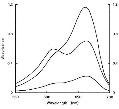
3.4 Absorption Spectra of Methylene Blue in O.IM HCl and in O.IM HCl/Ethanol... 73
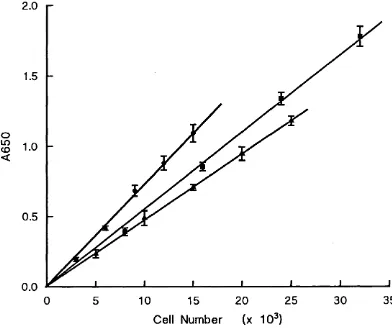
3.5 Linear Relationship between Cell Number and A650 in the Methylene Blue Assay for three Cell Lines . . . 74
Blue Assay on the Absorbance Obtained... 80
3.8 Effect of Serum Concentration on Growth of Rati
Cells in C u l t u r e ... 81
4.1 Production of Growth Factor by Alveolar Macrophages
in Response to Phagocytic Stimuli ... 96
4.2 Effect of the Concentration of Macrophages on
Growth Factor Secretion ... 98
5.1 Changes in Lung Collagen Content and Synthesis
Rates during Bleomycin-Induced Pulmonary Fibrosis in Rabbits... Ill
5.2 Mean Cell Yields from Bronchoalveolar Lavage . 120
5.3 Correlation between the Two Assays for Growth
Factor on Rati C e l l s ... 123
5.4 Growth Factor Secretion by Lavage Cells from Bleo
mycin and Control Animals Assayed on Rati Cells . .125 5.5 Growth Factor Secretion by Lavage Cells from Bleo
mycin and Control Animals Assayed on R9ab Cells . . 127
5.6 Correlation between Assays for Growth Factor
Secretion on Rati and R9ab C e l l s ... 128
5.7 Correlation between Assays for Growth Factor
Secretion on Dialysed and Undialysed Specimens, Using
Rati Fibroblasts... 129
Tables
2.1 Alveolar Macrophage Protein Synthesis in Vivo . 47
2.2 Relationship between Specific Radioactivity of
Proline in Plasma and Lavage Fluid ... 49
2.3 Alveolar Macrophage Protein Synthesis in Vitro 52
3.1 Reproducibility of Methylene Blue Assay . . . . 77
3.2 Reproducibility of Growth Factor Assay . . . . 83
4.1 Effect of Cycloheximide on Secretion of GF by
Alveolar Macrophages, and of Dialysis of Conditioned
Medium on Measured Levels of G F ... 99
4.2 Release of Growth Factor from Alveolar Macrophages
by Cell L y s i s ...101
5.1 Total Cell Yields and Viability at Bronchoalveolar
L a v a g e ... 117
5.2 Differential Cell Counts of the Lavage Cells from
the Bleomycin Treated Rabbits ... 119
5.3 Growth Factor Secretion by Lavage Cells from
Control and Bleomycin Treated Rabbits ... 122
Appendix 1 Lavage Cell Yields... 152
Appendix 2 Growth Factor Secretion by Lavage Cells 153
R e f e r e n c e s ... 154
Published Papers from this W o r k ... Back Cover
Comparison of protein-synthesis rates of alveolar
macrophages in vivo and in vitro. Oliver MH, Cole PJ and Laurent GJ. Biochem J (1984); 217i 761-5.
A rapid and convenient assay for counting cells cultured
in microwell plates: Application for assessment of growth
factors. Oliver MH, Harrison NK, Bishop JE, Cole PJ and
I would like to acknowledge my two supervisors. Professor
Peter Cole and Dr. Geoffrey Laurent. Without Professor
Cole's efforts this work would not have commenced, and his
support and advice were invaluable through to its conclusion. Dr. Laurent's expertise on the biochemical side of this work
was vital to its success, and his wise counsel combined with
a constant good humour guided and encouraged me through all
the stages of this research. I am very grateful to them
both.
A research project for a clinician is a voyage into uncharted waters. As well as my two principal pilots, many
others helped me to steer a relatively straight course. I
particularly wish to thank David Roberts, late of the Host Defence Unit, whose fund of stories and jokes was only
matched by his knowledge of where to find equipment, and who
to ask for advice if need be. Robin McAnulty of the
Biochemistry Uhit, was an inexhaustible source of practical help and never appeared to lose patience with the stream of
questions and queries that I directed to him. Without his
help this work would have taken much longer.
There are many others who gave me practical advice, tips
or ideas, over coffee in the Cardiothoracic Unit or the Chester Beatty Research Laboratory, and at meetings and
informal discussions there and in other laboratories. These
contacts were one of the great pleasures of my period in
research. I cannot acknowledge all of these individuals
here, so I must content myself with expressing my thanks to
A515 Absorbance measured at 515 nm
A650 Absorbance measured at 650 nm
AM Alveolar macrophage
DMEM Dulbecco's modification of Eagle's medium
GF Growth factor
IF Inhibitory factor
NCS Newborn calf serum
NSE Nonspecific esterase
PBS Phosphate buffered saline
PDGF Platelet derived growth factor
PGEg Prostaglandin E%
SD Standard deviation
SEM Standard error of the mean
SR Specific radioactivity
TCA Trichloroacetic acid
TNF Tumour necrosis factor
Statement of Personal Contribution to the Work
All of the work described in this thesis was carried out by
myself. The animals used were kept in the animal house of
the Cardiothoracic Unit and cared for by the Curator and
INTRODUCTION
The last two decades have seen an explosion of interest in
cell biology fuelled by improvements in cell culture techniques. These developments have illuminated many medical
problems. In particular the description of growth factors
has provided a tool for the study of local control of growth,
remodelling of tissues and repair mechanisms in damaged
tissue. It is to be hoped that this knowledge may finally
help in the treatment of many human diseases which have
remained resistant to our present day therapeutic
armamentarium.
One group of conditions greatly in need of such advances are the diffuse fibrotic disorders of the lung. These are a
heterogeneous group of diseases characterised by diffuse
pulmonary fibrosis which dominates the final clinical
picture. In most of these disorders therapy is either
unavailable, or uncertain of benefit and associated with
major side effects. The need for understanding of the
pathogenetic mechanisms in this situation is pressing.
1.1 HUMAN DISEASE
1.1.1 Diffuse Pulmonarv Fibrosis
The number of diseases in this category exceeds 100
(Crystal et al, 1981). Although not as common as disorders
such as asthma, chronic bronchitis or bronchial carcinoma,
part of every chest physician's weekly clinical load. They are all responsible for much morbidity in the form of
breathlessness which can be incapacitating, and in the later
stages damage to the pulmonary circulation causing pulmonary
hypertension and right ventricular failure. Many of these
diseases have a significant mortality rate. Cryptogenic
fibrosing alveolitis, for example, has a five year survival
from diagnosis of 50% (Turner-Warwick et al, 1980), no better
than many malignancies. In none of them is a completely
satisfactory form of therapy available, and in some
therapeutic decisions can be very difficult. These problems
are best illustrated by an outline sketch of three of these
diseases.
1.1.2 CrvDtoaenic Fibrosing Alveolitis
This disease can start at any time in adult life.
Examination of lung biopsies shows a pattern of alveolar wall damage and thickening together with the formation of
increasing amounts of fibrous tissue. Biochemical analysis
has confirmed the presence of increased amounts of collagen (Selman et al, 1986), in keeping with the histological
picture. In the end stages, the fine alveolar structure of
the lung is replaced by bands of thick fibrous tissue
separating enlarged airspaces. Varying degrees of cellul
ar ity of the lung biopsies are seen, with some suggestion
that the more cellular patterns are associated with potential
for improvement spontaneously or with treatment (Crystal et
al, 1976; Winterbauer et al, 1978).
The major symptoms are cough and breathlessness. The
course of the disease is very variable ranging from mild
symptoms with no change over many years of observation, to inexorable progression to death within months of diagnosis.
Spontaneous remissions and relapses are seen.
Treatment is unsatisfactory (Johnson et al, 1986). High
doses of corticosteroids benefit an uncertain proportion of
Cytotoxic drugs such as cyclophosphamide or azathioprine are
used. For none of these is there convincing evidence from
controlled trials of their benefit, although the general consensus is that they do benefit some 20% of patients, at a
cost of major side effects in some (Johnson et al, 1989) .
This marked variability in tempo, rate of progression, and
response to therapy of the disease makes decisions on therapy
for an individual patient very difficult. This is compounded
by the fact that response, if it occurs, is often slow so
that assessment of the success of therapy is difficult. So the clinician has desperate need of (1) a marker of disease
activity which would point to the need for therapy, (2) a
marker of response to treatment and (3) new forms of therapy
which are more effective and less toxic. The hope must be
that research into the pathogenesis of the disease might
satisfy some of these needs.
1.1.3 Sarcoidosis
Sarcoidosis is a multisystem disease of unknown aetiology characterised by the presence in affected tissues of
organised collections (granulomata) of chronic inflammatory
cells. It can involve almost any system in the body although
the brunt is most often borne by the lungs. Histological
examination of involved tissue generally shows non-caseating
granulomata consisting of collections of lymphocytes,
modified macrophages (epithelioid cells) and other chronic
inflammatory cells but without the central caseating necrosis
of the granulomata of tuberculosis. This abnormal pattern
may resolve with little or no permanent mark in the tissues,
but it may be replaced by bands of hyaline amorphous tissue
which becomes increasingly fibrotic with time.
For many patients pulmonary shadowing may be a chance finding on a chest X-ray or discovered on investigation of
minor respiratory or other symptoms. In some 50% of cases
this X-ray shadowing will clear without treatment leaving a
treatment with corticosteroids is necessary to achieve the
same result. In perhaps 25%, inspite of therapy, the
radiographic shadows evolve into those characteristic of
bands of pulmonary fibrosis. Often the process will arrest
after a time leaving some disability, but in a small
proportion of cases it is progressive, leading on to life
threatening respiratory insufficiency. The same problems in
assessment of likelihood of progression, response to therapy
and individual variations in tempo are seen with sarcoidosis
as with cryptogenic fibrosing alveolitis, accepting that the
overall prognosis is better. Here, where most patients will
not require therapy, the particular problem is to select at
an early stage those who will, before permanent lung damage
has occurred.
1.1.4 Mineral Dust Pneumoconiosis
This is a group of conditions characterised by pulmonary
fibrosis in response to the inhalation of mineral dusts such
as coal, silica, or asbestos. They vary as to the
radiographic pattern of the fibrosis and in the degree of
respiratory disability which results. Although the dose of
inhaled dust does correlate with severity, there are marked
individual variations in response. With a well defined and
controllable aetiological agent, reduction in dust exposure
offers the best hope of reducing future morbidity and
mortality. In patients with established pulmonary fibrosis
there is no therapy which can halt the progression of the
disease. Fortunately many patients see only minor
progression.
The pneumoconioses have become less prevalent because of
the adoption of dust control measures in the workplace.
Nonetheless they remain important as models for other forms
of pulmonary fibrosis because in their case the aetiological
1.2 THE MACROPHAGE. GROWTH FACTORS AND PULMONARY
FIBROGENESIS
Tissue destruction, derangement of tissue architecture and
fibrosis are all features of the pathology of the pulmonary
fibrotic diseases. An increase in macrophage numbers is a
consistent finding in all of these conditions. Several
historical threads have come together to identify the
macrophage as having a central role in one of these
pathological processes - fibrosis itself.
1.2.1 Lessons from Silicosis
This example of human pulmonary fibrosis has been well
documented clinically and pathologically for a number of
decades. The aetiological agent is readily available for
experimental study and so it is not surprising that the early
work in pathogenesis of pulmonary fibrosis focused on this condition.
Two themes have run through this literature:- the direct
cytotoxicity of silica and the role of the macrophage in the
development of fibrosis (reviewed by deShazo, 1982). It
would be expected that lung macrophages would ingest
particulate inhaled silica. The pathologists confirmed that
this process could be seen in lung tissue sections.
Experiments in vitro confirmed that macrophages did ingest
silica particles and that this process lead to the final
lysis of the cells with the release of lysosomal enzymes
(Davies and Allison, 1976) and free radicals (Gabor et al,
1980), which could cause lung damage. A more direct role for
the lung macrophage in fibrogenesis was suggested by
Hepplestone and Style's seminal paper (1967) where they
reported that macrophages exposed to silica in vitro released
substances which could stimulate fibroblasts to produce more
collagen. This observation lead to contributions from many
this early work is confused by the variations in experimental
conditions used by the different investigators, it did
establish that viable macrophages exposed to silica could
secrete factors which modified fibroblast replication and
collagen production. A study by Bateman et al (1980) of the
effects of fluid phase factors from mixtures of macrophages
and mineral dust contained in small diffusion chambers
provided evidence for the importance of continuing viability
of the macrophages in the development of fibrosis. The
chambers were implanted into the peritoneal cavities of mice
and the development of fibrosis observed. Asbestos, and
silica in low concentration permitted viable macrophages to
remain inside the chamber for two weeks, and the development
of fibrosis was noted. Silica in higher concentration
rapidly killed the macrophages and little fibrosis around
the chambers resulted. This evidence demonstrated the
importance in fibrogenesis of active secretion by macrophages
of mediators, rather than the passive release of substances
following macrophage cytotoxicity. This hypothesis is
attractive as it is of general application to fibrotic
disorders not linked to cytotoxic agents.
It seemed that the macrophage might have a central role in
the pathogenesis of all forms of diffuse pulmonary fibrosis.
Other developments in cell biology in the last decade
provided insights and tools for the study of macrophage-
fibroblast interaction.
1.2.2 Alveolitis
To the clinician studying the chest radiograph of patients with end-stage disease, or present at a post mortem
examination of such a patient, the fibrosis is obvious and
extensive. For years the morphologists have been pointing to
the presence of accumulations of inflammatory cells -
macrophages, lymphocytes, neutrophils etc - in the alveolar
structures of patients with pulmonary fibrosis. This
fibrosis although its nature may differ between different
conditions (Crystal, 1982).
There is now good evidence, provided by lung biopsy
techniques, that this alveolitis is the earliest
manifestation of these conditions, and may resolve completely
(Lacronique et al, 1981; Carrington et al, 1978). These
inflammatory cells have the potential to damage tissue by the
release of free radical species, and hydrolytic enzymes; to
secrete factors which activate nearby cells to contribute to the inflammatory process; and also to secrete chemotactic
factors which further increase the local numbers of
inflammatory cells (Shock and Laurent, 1990). Thus if the
alveolitis persists then increasing disorganisation and
fibrosis of the lungs can follow. This model of the
pathogenesis of pulmonary fibrosis has its parallels in other
organs e.g., the importance of the inflammatory infiltrate
seen in early cirrhosis of the liver to the subsequent tissue destruction, derangement and fibrosis seen in this condition.
Thus it is hoped that study of these inflammatory cells may illuminate pulmonary fibrogenesis, and treatment directed at
the alveolitis may forestall the development of the changes
apparent in end stage disease.
1.2.3 Growth Factors
It has long been known that diploid cells require serum to
replicate in culture. In 1971, Samuel Balk made a critical
observation about this effect of serum. He had designed a
culture medium which was more "physiological" in that serum
(derived from clotted whole blood) was replaced by plasma
where the platelets had been removed by centrifugation. In
this new medium the cells remained healthy but divided much
less readily. Balk suspected that a "wound hormone" was
released into serum during clot formation.
These observations were extended in 1974 by a number of
researchers (Ross et al, 1974; Kohler and Lipton, 1974; Scher
polypeptide which stimulates the growth of many cells in
culture. This is known as platelet derived growth factor
(PDGF). It is released when platelets are activated, for
example during coagulation of whole blood. Plasma also
contains growth promoting activity, different but
complementary in its action to PDGF. Both forms of growth
factor need to be present to sustain replication.
Mesenchymal cells in tissues are bathed in extracellular
fluid (effectively a platelet free ultrafiltrate of plasma)
which is not sufficient to cause growth. After tissue damage
with platelet activation and degranulation, the stimulus to
replication is complete. Thus PDGF provides control over
local variations in growth.
The partnership between PDGF and platelet poor plasma in
controlling the cell replication cycle has been analyzed
further (Scher et al, 1979) . The two stimuli can be applied sequentially but if so, the cells must be exposed to PDGF
before plasma to allow replication to proceed. PDGF induces cells to become "competent” to respond to the subsequent
stimulus of plasma, so that they "progress” from Gq into the
Gi and S phases of the cell cycle. Thus PDGF is known as a
"competence" growth factor and plasma contains a
"progression" factor or factors.
Since this pioneering work a large number of growth
factors have been identified and the list now exceeds thirty
(Deuel, 1987). The field is made particularly complex
because the detailed chemical structure of the majority is
not yet known. Some have, on stringent analysis, been shown
to be identical, so that as analysis of others is achieved,
the list may diminish and the picture become clearer. In
general each has been found to have either competence or
progression factor activity but not both. The target tissues
of these growth factors vary as to their sensitivity to each
GF, no doubt due to variable expression of the specific
receptors for each GF.
The discovery of this class of compounds and their
control of tissue growth locally, of differential growth of different cell types in a single tissue and of the
uncontrolled growth of tissue in malignant disease (Deuel,
1987; Druker et al, 1989).
1.2.4 Macrophages and the Fibroblast
In the cellular changes of wound healing, monocytes move from the capillaries into the granulation tissue of the wound
where they evolve into tissue macrophages, and this is
followed one to three days later by the appearance and
multiplication of fibroblasts. Collagen is laid down by
these fibroblasts to form scar tissue. Liebovich and Ross
(1975) studied the effects of monocyte depletion on this
progression of changes in healing skin wounds in guinea pigs.
They showed that fibroblast proliferation and collagen deposition were much reduced in the monocyte-depleted
animals. Their work suggested that tissue macrophages had a major role in controlling fibroblast proliferation and
possibily collagen secretion.
The increasing literature on serum and tissue growth
factors provided an impetus and new techniques to examine the
effects of monocytes and macrophages products on fibroblast
replication. The picture that has emerged is complex and
still evolving rapidly (Nathan, 1987). This complexity which
is partly due to the range of species and tissues studied and
partly due to the problems in characterising biochemically
the multiple activities secreted by macrophages, also
reflects the genuine intricacies of the networks of cell-cell
interaction being discovered.
Points of agreement are that
(a) Macrophages have the capability to produce a wide
range of mediators (cytokines) which can modulate the
function or replication of other cells in the local
environment (Nathan, 1987).
(b) The macrophage cytokines which have major effect on
in excess of 12,000 (Kelley, 1990).
(c) These polypeptide cytokines may have growth promoting
activity, or growth inhibiting activity or in the case of
interleukin-1, exhibit both activities depending upon the experimental conditions (Schmidt et al 1982; Rainer et al,
1989)
(d) Most of these cytokines can be secreted by many cell
types and are not unique to macrophages (Kelley, 1990).
Thus the macrophage has been found to have a central role
in wound healing and the formation of collagen in scars, is
present in increased numbers in the alveolitis which is the
forerunner of all forms of pulmonary fibrosis, and is capable
of secreting polypeptide cytokines which modulate fibroblast
replication. Fortunately this key cell can be obtained from
the lung for study both post mortem and in life by the technique of bronchoalveolar lavage.
1.2.5 Bronchoalveolar Lavaae
Standard clinical techniques such as respiratory function
tests, measurements of gas exchange and radiology shed little
light on the pathogenesis of pulmonary disease. Biopsy
samples allow histological study and biochemical analysis but
the risks involved in lung biopsy precludes its use as a
purely research tool. Samples will only be available if the
management of the patient indicates the need for biopsy. In
practice many patients with pulmonary fibrosis never undergo
lung biopsy and it would be rare for it to be performed more
than once in any patient. Sequential samples are not
available therefore.
The central role of an alveolitis in the development of
pulmonary fibrosis does offer a way out of this impasse.
Much may be learned from samples of inflammatory cells
obtained from the lung without sampling the parenchymal
cells. The technique of bronchoalveolar lavage, developed by
Inflammatory cells are to be found on both sides of the
alveolar wall i.e., in the interstitium and in the alveolar
fluid. The cells free in the alveolar fluid can be sampled
by the technique of bronchoalveolar lavage, whereby in life
a pulmonary segment, or post mortem the whole lung, can be
filled with physiological saline which is then aspirated. In
humans the procedure is carried out using a fibreoptic
bronchoscope wedged in a bronchus and is well tolerated with
a low morbidity rate. It can be repeated and therefore
permits sequential sampling.
Bronchoalveolar lavage has been accepted as both a
clinical and a research tool. In the former setting, the
interpretation of results finally rests on the pragmatic
question of whether they answer the questions posed by the
clinician i.e., questions of diagnosis or treatment. In a
research setting the value of bronchoalveolar lavage depends on the answer to the question "do lavage cells reflect events
in the lung interstitium"? A rapid inspection of a sample of lavage cells would show that the structural cells of the
interstitium - epithelial, endothelial, fibroblast etc - are
not represented. The cells seen are all "inflammatory" in
type. Within this group some differences between the
alveolar (lavage) and interstitial cell populations are
apparent. The epithelioid cells of granulomata are not seen
in lavage samples (Danel et al, 1983). Plasma cells and
lymphocytes predominate in the interstitium in cryptogenic
fibrosing alveolitis but eosinophils and neutrophils more
commonly accompany macrophages in lavage samples. However
Hunninghake and colleagues (1981) showed almost identical
percentage counts in lavage and lung extract samples in a
number of disease states. It is probable that the
distinction between interstitium and alveolar space becomes
less clearcut in many disease states (see Section 6.4.3 for
a fuller discussion of this). Thus, in disease,
bronchoalveolar lavage is likely to accurately reflect events
in the interstitium.
itself in research into the pathogenesis of pulmonary
fibrosis both in human disease and in animal models, and a
more limited role in the diagnosis of interstitial lung
disease in humans.
1.2.6 Macrophage Activation
The macrophage is sensitive to both specific and non
specific stimuli which can activate some or many of its
functions (Nathan, 1987). The investigator about to embark
on a study of cells in vitro must be concerned that the
techniques involved in isolating and culturing the cell may
of themselves stimulate or activate the cell, so that
measurements of basal level activity are falsely high. No
comparative study of a macrophage function measured both in
vitro and in vivo has been published, primarily because of
the problems of quantitating this in vivo. The difficulty in
addressing this question has been to identify an index of
activation which can be measured and compared for cells in
vivo and in vitro. Many secretion products of macrophages
are not unique to that cell e.g., arachidonic acid
metabolites, oxygen metabolites, complement components and
probably also growth factors. Therefore it would not be
possible to confidently assign a secretion rate of these
products measured in vivo to alveolar macrophages alone.
Even for those which are unique to the cell, calculation of
secretion in vivo is prevented by an unknown clearance rate.
Recently Laurent (1982) developed and validated a method of
measuring in vivo the protein synthesis rate of tissues such
as muscle, liver and heart. It seemed that this could be
applied to alveolar macrophages in vivo and thus provide a
parameter of alveolar macrophage activation which could be
1.3 ANIMAL MODELS OF PULMONARY FIBROSIS
1.3.1 The Need for Animal Models of Pulmonarv Fibrosis
It is a characteristic of most forms of human diffuse
pulmonary fibrosis that in the established case a large open
lung biopsy will reveal areas of early alveolitis and other
areas of heavy inflammatory infiltrate, and areas of mild and
marked fibrosis (Winterbauer et al, 1978). Thus all the
different "stages" in pulmonary fibrosis will be represented.
For the clinical investigator, this makes analysis of the component steps of the pathogenesis of the disease very
difficult. The analysis would be easier if the moment of
exposure to the aetiological agent was known so that a
temporal sequence of events could be charted. In the adult
respiratory distress syndrome (ARDS) the lung responds to an insult by developing widespread acute inflammatory changes
which then either resolve or progress to fibrosis. In many cases the insult is known, e.g., inhalation of toxic gas,
ingestion of a toxic chemical such as paraquat or a cytotoxic
drug, or widespread sepsis. Unfortunately ARDS has a high
mortality and these patients are a group in which research
investigation is constrained by ethical considerations and by
the presence of other pathological processes such as sepsis.
It has been possible to set up models of pulmonary fibrosis
in experimental animals often using the same agents seen to
cause ARDS in humans. Examples include paraquat
(Schoenberger et al, 1984), radiotherapy (Adamson and Bowden,
1983), oxygen (Rinaldo et al, 1982) and cytotoxic drugs such
as bleomycin (see next section). In these models the
potential exists for correlating the histological,
biochemical, inflammatory and physiological events, and
attempting "therapeutic" manipulation of these. Armed with
these insights it may be possible to return to our patients,
and develop diagnostic and therapeutic approaches of real
1.3.2 Bleomycin and the Luna
Bleomycin is an agent active against several solid
tumours. Its use is limited by the dose-related development
of pulmonary fibrosis (Blum et al, 1973). The histology of
the affected lung shows epithelial and endothelial cell
damage, interstitial oedema and fibrosis with derangement of
lung architecture i.e., similar to that of cryptogenic
fibrosing alveolitis.
The mechanism of action of bleomycin is dependent on the
presence of reduced iron i.e., Fe^^ (Sausville et al, 1978;
Burger et al, 1981) . Disruption of cellular DNA with the
production of toxic derivatives of nucleoside bases and free radical damage to cell membranes have been shown to be
important (Umezewa, 1974; Grollman, 1988).
Bleomycin also causes pulmonary fibrosis in a range of
experimental animals, including mice (Adamson and Bowden, 1977), hamsters (Snider et al, 1977), rats (Thrall et al,
1979) , baboons (McCullough et al, 1978) and rabbits (Laurent
et al, 1981) . In early experiments the drug was given
parenterally but Snider et al (1977) induced pulmonary
fibrosis in hamsters by endotracheal instillation. This
technique permits the use of lower doses of bleomycin with
less toxicity to other organs. This has proved the most
popular animal model of pulmonary fibrosis for study. The
cellular changes which follow bleomycin have been well
described (Snider et al, 1977; Thrall et al, 1979; Chandler
et al, 1983). Chandler and co-workers (1983) charted these
changes in detail using morphometric techniques in hamsters.
They studied animals 4, 7, 21, 28, 35 and 42 days after
instillation. They showed an early rise in neutrophil
numbers peaking at 7 days and falling to low levels by 21
days. Monocytes and macrophages also showed an early rise
but this was sustained throughout the study periçd with
macrophages being relatively more common at 21 and 28 days.
Fibroblasts on the other hand showed a steady rise in numbers
and only a minor fall thereafter. I These data fit in with the
more qualitative impressions gained in other animal models.
Thus the pattern seen is that observed in the wound healing
experiments referred to earlier. The same deductions may
well hold, i.e., macrophages play a central role in
increasing fibroblast numbers in bleomycin induced pulmonary
fibrosis.
1.3.3 The Rabbit Bleomvcin Model of Pulmonarv Fibrosis
Laurent and co-workers (1981, 1983) have described the
histological and biochemical changes seen in the lungs of
rabbits following the intratracheal instillation of bleomycin at a dose of lOmg/kg body weight. Their observations of the
pathological changes agree with reports from other animal
models. They commented that at four weeks there was still
only a little excess extracellular material staining for
reticulin, and for collagen with Masson's trichrome method.
At eight weeks "young fibrous tissue" was obvious and a
stronger trichrome reaction for collagen was seen. The slow
onset of histologically apparent fibrosis contrasts with the
biochemical evidence. In a first set of experiments (Laurent et al, 1981), examining animals at two, four and eight weeks
after bleomycin, the lung content of collagen, elastin, total
protein and DNA were all maximal by two weeks. Thereafter
the total protein content fell markedly although the other
components decreased only a little. This conflict between
the views of the histologist and the biochemist can be
explained by consideration of the nature of collagen and of
their respective techniques for detecting collagen.
Collagen exists in at least eleven different forms or
types, which differ in the detailed amino acid sequences of the component polypeptide chains. Types I and III collagens
are the major fibrous interstitial proteins. They consist of
three polypeptide chains (the a chains) covalently linked
(Laurent, 1986). This fibrillar protein is then assembled
and linked by covalent bonds to other matrix components such
as basement membrane collagens and proteoglycans. It is a
feature of all collagens that the amino acids glycine,
proline and hydroxyproline are a high proportion of the total
amino acid composition of this protein. Hydroxyproline is
found in a few other proteins (elastin, anticholinesterase
and the Clq component of complement) but collagens are by far
the largest source of hydroxyproline in all tissues.
Therefore the biochemist uses the tissue content of protein
bound hydroxyproline as a measure of the amount of collagen
present. This measure of collagen pays no regard to what
form the collagen is in. It is probably the complex chemical
environment of fully matured type I collagen which is detected by Masson's trichrome, and this maturation may lag
behind the initial collagen synthesis. Therefore in looking
to unravel the cellular control of collagen production we
must turn to the biochemical data to guide us in choosing time points to study.
This work confirmed that in the rabbit bleomycin model the deposition of excess collagen is an early and marked feature
of the development of pulmonary fibrosis. Subsequent work
provided more detail of the early time course of these
changes and of the relative importance of increased synthesis
and decreased degradation. This will be described in the
introduction to chapter 5.
1.3.4 Questions Raised bv the Biochemical Data
This work with the rabbit model has provided the most
detailed description published of the biochemical events
leading to net collagen deposition in an experimental animal
model of pulmonary fibrosis. The predominant collagens in
lung, as in most tissues, are type I and type III. It is
tissue fibroblasts that are the major source of these
collagen types. Therefore we must address ourselves to the
control of fibroblast numbers and function if we are to trace
deposition in this animal model.
An increase in tissue collagen might either be due to an
increase in fibroblast numbers or a change in collagen
metabolism (rise in synthesis, or fall in degradation) with
constant fibroblast numbers, or both mechanisms might
operate. The histological picture points to the first as an
important mechanism. Unfortunately we do not have
quantitative histological data in the rabbit bleomycin model.
In the hamster bleomycin model the increase in fibroblast
numbers - a peak increase of 273% over control values - was
more than sufficient to account for the increase in lung
collagen observed (Chandler et al, 1983; Chandler and Giri,
1982) . This is a rather naive calculation but it does
support the view that an increase in fibroblast numbers is
the main mechanism of increased collagen deposition.
Locally an increase in fibroblast numbers might be
achieved either by replication or migration of cells into the
area. Rather surprisingly fibroblasts are capable of
movement, within tissues, rather than being a fixed structural cell. This process is certainly important in the
control of the local distribution of fibroblasts (Rennard et al, 1981) but will not be relevant to their overall numbers.
The latter can only increase as a result of replication.
These arguments suggest that to understand what leads to the
deposition of collagen in damaged lung we need to look at the
1.4 AIMS OF THIS THESIS
The central objective of this work was to examine the role
of growth factor secretion by inflammatory cells obtained by
bronchoalveolar lavage in the pathogenesis of lung fibrosis.
This built upon the detailed picture described by Laurent and
coworkers (1981, 1983) of the biochemical events occurring in
bleomycin induced pulmonary fibrosis in rabbits. The
hypothesis tested was that the increase in collagen synthesis
rates seen might be mirrored by, and perhaps due to, a
proceeding increase in secretion of growth factors by
inflammatory cells in the pulmonary alveoli of bleomycin
treated rabbits.
The likely source of such growth factors was the
lung macrophage, and their secretion was to be studied in
vitro. Accordingly, three subsidiary problems were identified and also
investigated:-1. Culture of alveolar macrophages in vitro might
activate these cells, complicating interpretation of
secretion rates of growth factor. This was to be assessed by
measuring the macrophage protein synthesis rate in vivo and
in vitro, allowing a comparison of their states of activation.
2. There was a need for a rapid assay of fibroblast
numbers in microwell culture, as part of the measurement of
levels of growth factors secreted by macrophages. A novel
assay based on a colorimetric method was developed and
validated.
3. The ability of rabbit alveolar macrophages to produce
growth factor had not previously been described. This was
confirmed and some of the physicochemical properties of the
CHAPTER 2
MACROPHAGE ACTIVATION IN VIVO AND IN VITRO ASSESSED BY
MEASUREMENT OF PROTEIN SYNTHESIS RATE
2.1 INTRODUCTION
2.1.1 Macrophage Activation
Recently Laurent (1982) developed and validated a method
of measuring in vivo the protein synthesis rate of tissues
such as muscle, liver and heart. It seemed that this could
be applied to the small mass of tissue of alveolar
macrophages in vivo and thus achieve for the first time the
measurement of protein synthesis rate of a population of
macrophages in vivo.
It is technically simpler to estimate protein synthesis of
a cell population in culture. The quantitation of protein
synthesis rate provided a measure of alveolar macrophage
activation which could be compared in vivo and in vitro.
2.1.2 Principles of Measurement of Protein Svnthesis Rate
Measurement of the rate of protein synthesis of
intracellular protein requires a technique which labels the
newly synthesised protein. The use of a radioisotopically
labelled amino acid achieves this and has become the basis of
the standard biochemical methods in this field. After
exposure to radiolabelled amino acid of known specific
radioactivity (SR), the tissue is sampled and protein
isolated from free amino acids and other contaminants by
standard techniques. The proteins are hydrolysed and the SR
synthesis rate is calculated as follows
Fractional protein synthesis rate (%/day)
= SR of amino acid in protein____ x 100
SR of amino acid in precursor pool time (days)
This formula requires that the SR of the amino acid pool
is constant - the mathematics becomes more complicated if
this does not hold. A constant precursor SR is readily
achieved in vitro when the extracellular pool of amino acid
(i.e., in the culture medium) is large in relation to the
metabolic capacity of the tissue (e.g., cells in culture).
In vivo the same effect is achieved by the use of a large dose of injected proline (Laurent, 1982).
2.1.3 Choice of Experimental Conditions in Vitro
The present study is the first to examine macrophage
protein synthesis in vivo. However other workers have looked
at protein synthesis in vitro for AMs from guinea pigs
(Airhart et al, 1981) and from New Zealand White rabbits
(Hammer and Rannels, 1981) . It seemed wise to use New
Zealand White rabbits in this study of protein synthesis as
they were to be used in the subsequent bleomycin work. The
culture conditions described by Hammer and Rannels (1981)
were chosen to allow direct comparison with their work.
2.1.4 Choice of Assav
The measurement of specific radioactivity requires
knowledge of both the radioactivity and the molar amount of
amino acid present. For the in vivo experiments these were
provided by a chemical method (Peterkofsky and Prockop, 1962)
involving Chloramine-T oxidation of the proline. This method
has been modified and validated in this laboratory (Laurent
et al, 1982).
of proline to be obtained from the protein in AMs from a
rabbit was near it's lower limit of sensitivity. The effect
of different culture conditions on the protein synthesis rate
was to be studied and this required using smaller number of
cells in each individual culture. A different biochemical
technique for measuring proline (and phenylalanine - see
later) specific radioactivity was therefore employed. This
was the dansyl chloride double isotope method which is much
more sensitive than chemical assays for molar amounts of
proline but needs higher specific radioactivities to ensure
adequate radioactive counts from the smaller amounts of
proline. This could easily be achieved in vitro (but would
have been prohibitively profligate of radioisotope in vivo).
2.2 MATERIALS AND METHODS
2.2.1 MATERIALS
Laboratory stock chemicals were all of ANALAR reagent
grade.
Special chemicals were obtained from Sigma UK, unless
commented upon in the text. Where alternative Sigma
preparations are available, the catalogue number is quoted.
Radiochemicals were obtained from Amersham International.
PREPARATION OF ALVEOLAR LAVAGE CELLS
2.2.2 Animals
New Zealand White male rabbits were obtained from HOP
Laboratories, Cork Farm, Chilham, Kent, at a body weight of
1.7-2.0kg. These were specific pathogen free animals, and
the animals were screened regularly by animal house staff for
animal showing signs of disease was removed from the colony.
They were fed ad libitum.
Animals were weighed regularly and often showed an initial
fall after arrival related to the stress of transport. They
were not used until they were gaining weight. They had
usually been in the rabbit colony one to three weeks and were
between 1.7 and 2.3kg in weight at the time of experiment.
2.2.3 Luna Lavaae
Animals were killed with sodium pentabarbitone, 100 mg/kg
body weight (Euthatal, from May and Baker, Dagenham, England)
injected into an ear vein. The rabbit was then placed on its
back and the fur of the neck, thorax and abdomen soaked in
95% ethanol. The skin of the neck was opened by a vertical
incision and the trachea exposed by blunt dissection. A silk stay suture was placed behind the trachea. A tracheotomy was
formed with sterile scissors and the trachea intubated with a sterile 2.5mm polyvinylchloride tube. The silk suture was
tied to secure the tubing which was connected to a sterile
50ml polypropylene syringe via two 3-way taps connected in
series. Tubing led from the two side ports, so created, one to a 500ml bag of 0.9% saline for injection (Travenol, U.K.)
and the other to the collecting vessels held in crushed ice.
In the protein synthesis experiments sterile glass bottles whose interior surface had been treated with silicone
(Repelcote, BDH Chemicals) were used.
Saline (35ml) was drawn into the syringe and then slowly
injected into the lungs of the rabbit. The chest wall of the
animal was gently massaged, the lavage fluid aspirated back
into the syringe, and then transferred into the collecting
vessel. This process was repeated to a total of three times.
2.2.4 Lavaae Cell Preparation
Cells were separated from lavage fluid by centrifugation
phosphate buffered saline (PBS) and recentrifugation. Some
lavage returns were slightly contaminated with erythrocytes.
If so the cells were resuspended in PBS, layered onto
Ficoll/sodium metrizoate (d = 1.077, "Lymphoprep", Nyegaard
Ltd., Norway) and centrifuged at 400g for 10 minutes. The
nucleated cells were collected from the interface with a
silicone treated pasteur pipette, and washed twice in PBS.
An aliquot of the cell suspension was taken for cell differential count. The lavage macrophages were then either
placed in incubation vessels as detailed below, or mixed with
5% trichloroacetic acid as appropriate.
Throughout this preparation stage the cells were
maintained at 0-4°C. All manipulations were carried out in
a laminar air flow hood to maintain sterility.
2.2.5 Cell Viabilitv
The percentage of viable cells was determined by Trypan
Blue dye exclusion (Phillips, 1973). Equal volumes of the
cell suspension and 1% Trypan Blue in 0.15M NaCl were mixed
and allowed to stand at room temperature for 10 minutes, then
loaded into a haemocytometer for examination under a microscope. Two hundred cells were counted. The proportion
of viable cells, i.e., those whose cytoplasm did not stain
blue, was expressed as a percentage of the total.
2.2.6 Cell Counts
Cell counts were performed by mixing 100/xl aliquots of
cell suspension and stock 0.1% Crystal Violet in 0.15M
saline. A drop of the mixture was transferred into a
haemocytometer, and sufficient squares examined to count at
least 200 cells. From this the cell concentration was
2.2.7 Cell Differential Count
The Shandon Cytocentrifuge system was used. lOO/il
aliquots of a 1 0** cells/ml cell suspension were pipetted into
the cytocentrifuge cups, and the system operated at 300 rpm
for 10 minutes. The slides were then air dried before
staining. May-Grunewald-Giemsa staining was carried out by
the haematology department of the Brompton Hospital, Fulham
Rd, London. In addition slides were examined using the non
specific esterase method, which stained the cytoplasm of
macrophages (see below). Differential counts by microscopy
were performed by counting all the cells wholely within a
high power field before moving on to another part of the
slide until at least 400 cells had been examined.
2.2.8 Non Specific Esterase Method
This method depends on the presence in the cytoplasm of cells of the monocyte/macrophage series, of a non specific
esterase which converts a substrate a-napthyl butyrate into
a coloured product which is deposited in the cells. Non
specific esterase positive cells show an orange or rust brown
cytoplasmic staining. Lymphocytes and neutrophils show at
most a few granules of reaction product. Methyl Green is
used as a nuclear counter stain.
The following solutions were prepared.
1. Formol and acetone buffer.
20mg of Na2HP04 and lOOmg of KH2PO4 were dissolved in 30ml of
distilled water, to which was added 45ml acetone and 25ml
formalin. This could be stored for up to three weeks at
-40°C.
2. Non specific esterase stain.
A 4% pararosanaline/2M HCl solution was made by dissolving
This solution was mixed with an equal (50ml) volume of
freshly prepared 4% sodium nitrite solution. The pH of the
mixture was adjusted to 6.5 with 2M NaOH and filtered. The
filtrate is referred to as hexazotised pararosanaline.
Phosphate buffer was made by mixing, in approximately 5:1
proportions, 0.067M Na2HP04 and 0.15M KH2PO4, with final pH
adjustment to 7.6. 1,424ml of this phosphate buffer, 96ml of
hexazotised pararosanaline and 800ml of a 2% solution of a-
napthyl butyrate in ethylene glycol monomethyl ether, were
mixed. The pH was adjusted to 6.1 and the solution left for
fifteen minutes for the precipitate to form. After
filtration a yellow solution is obtained which was aliquotted
in 100ml amounts and stored at -70°C. This remains active
for six to eight weeks. Once thawed an aliquot was discarded
at the end of the day.
3. 1% Methyl Green in distilled water.
Slides were fixed using formol-acetone for thirty minutes
at 4°C, before washing three times in distilled water and air
drying. They were immersed in non specific esterase stain
for one hour at 37°C, and again washed three times in water.
Finally the counter stain was applied by immersion of the
slides for four minutes in 1% Methyl Green, and washing three
times in water. They were air dried and mounted. The
percentage of non specific esterase positive cells was
determined by a count of 400 cells per slide by light
microscopy.
IN VIVO EXPERIMENTS
2.2.9 Injection of Animals
Animals in groups of four to six, were injected with L-[5-
^H]proline mixed with unlabelled L-proline (7mmol/kg body
weight) dissolved in four ml sterile water at intervals of
solution was injected into an ear vein over one minute, and the animals returned to their cages. The dose of [^]proline
used (0.6-2.4mCi/kg) was highest for the 30 minute animals
and lowest for the animals killed at 120 minutes ensuring
that an adequate amount of radioactivity was incorporated
into macrophage protein at all time points. The time of
incorporation was taken from the start of the proline
injection until the instillation of the first bolus of lavage
saline. It was this measure which was used in calculation of
synthesis rate, although in practice it was never more than
six minutes adrift of the nominal time.
2.2.10 Separation of Samples for Assav of Specific
Radioactivity
Macrophage Protein.
At the end of the washing steps detailed in section 2.2.4
the pellet of lavage cells was mixed with 5% (w/v) tri
chloroacetic acid (TCA). The resulting precipitate was
washed three times in 5% TCA by centrifugation (600g, 5min) and then successively in acetone/12M HCl (400:1,v/v),
ethanol/diethyl ether (2:1) and diethyl ether alone. The
washed precipitate was dried and hydrolysed in one ml of 6M
HCl (110°C, 16 hours) in sealed glass tubes.
Lavage fluid.
The lavage fluid was freeze dried and then redissolved in
three ml of water before mixing with three ml 10% TCA. The
TCA soluble fraction (containing free proline) was obtained
by centrifugation (600g, 10 min) , discarding the precipitated
and sedimented protein.
Plasma.
Similarly the TCA soluble fraction of plasma was obtained
by mixing one ml volumes of plasma and 10% TCA, and
2.2.11 Chloramine-T Assav of Proline Specific Radioactivity
Oxidation of proline by Chloramine-T gives a product, A^-
pyrroline, which is soluble in toluene (Peterkofsky and
Prockop, 1962). Other amino acids and proline metabolites
are not converted to a toluene extractable product.
Therefore if the original sample contained 1 radiolabelled
proline, all the radioactive counts in a sample of the
toluene extract are derived from [^]proline. By measuring
chemically (using the ninhydrin assay) the quantity of A^-
pyrroline in another aliquot of the toluene extract the
specific radioactivity can be calculated (with a simple
correction for the volume of each aliquot).
2.2.12 Chloramine-T Reaction
The samples obtained were then assayed for proline
specific radioactivity as follows:
(a) Samples were placed into labelled 20ml glass tubes
fitted with screw tops.
(b) The pH was titrated with IM and O.IM potassium hydroxide
to 8.3-8.9 using phenolphthalein as pH indicator.
(c) The volume of solution in the tubes was equalised with
further water.
(d) Two ml of IM potassium borate/boric acid buffer pH 8.7
was added.
(e) The tubes were vortexed, and then two ml of a 0.25M
chloramine-T solution in 2-methoxyethanol was added to each
tube, revortexed and left for 20 minutes at room temperature.
(f) The oxidation was stopped by adding six ml of 3.6M
sodium thiosulphate solution.
(g) The mixture was saturated with KCl by adding
approximately two grams of crystalline KCl to each tube
(this improves the efficiency of extraction of the reaction
product into toluene).
capped tightly and then put on a horizontal shaker for 10
minutes.
(i) The toluene containing the proline oxidation product
pyrroline was aspirated and stored for subsequent assay.
2.2.13 Ninhvdrin Reaction
Both proline and A^-pyrroline react stoichiometrically
with ninhydrin to produce an identical coloured reaction
product, which could be measured photometrically. Aqueous
solutions of proline were used to produce a standard curve of absorbance of the reaction product against molar concen
tration. Aliquots of the toluene extract were reacted
similarly, the absorbance measured, and the concentration of
A^-pyrroline calculated.
(a) Standards of L-proline, 0.02-0.1/zmol in 0.02/xmol steps,
were set up in one ml of aqueous solution, together with a
tube containing one ml of water to act as a blank. The tubes
used were screw-topped tissue culture glass tubes of 12ml volume.
(b) Aliquots of the toluene extract from the chloramine-T
oxidation were pipetted into similar tubes. 0.2ml of the
plasma samples were used while one ml of the lavage and
macrophage protein samples were necessary because of the
lower proline concentration.
(c) A solution of ninhydrin 2.5g in 60ml glacial acetic
acid, 16ml of 15M phosphoric acid and 24ml HgO was prepared.
Three ml of this ninhydrin solution + three ml of glacial
acetic acid were added to each reaction tube.
(d) The tubes were placed in a boiling water bath with
gentle shaking for 60 minutes.
(e) The chromophore was extracted by shaking manually with
additional toluene to a final toluene volume of three ml, and
the tubes were allowed to cool, while the two liquid phases
separated under gravity.
transferred to an additional tube. Occasionally clouding of
the toluene solution was noted. This was attributed to
further cooling of the toluene causing the small amounts of
water which dissolved in the toluene layer at 100°C to
separate from the organic phase. This was initially remedied
by warming of the samples before placing them in the
photometer. It was subsequently found that adding 50^1 of
ethanol to each tube kept the water in solution, and this
modification was used thereafter.
(g) The absorbance (A515) of each toluene extract from the
ninhydrin reaction tubes was measured on a Perkin-Elmer spectrophotometer at 515nm. The standard sample containing
no proline constituted the blank needed to zero the
spectrophotometer.
(h) A515 was plotted against proline content of standards on
graph paper and the best straight line through these points
and zero drawn by eye. The slope of this line is the
conversion factor, C, from absorbance units measured to the quantity of proline present in sample.
2.2.14 Radioactive Counts in Toluene Extract
Aliquots, generally eight ml, of the toluene extract of
the chloramine-T oxidation product were placed in glass
scintillation vials, to which were added four ml of a 1.2%
solution of 2,5-diphenyloxazole in toluene. The radio
activity for each vial was then measured in a Packard liquid
scintillation counter (model 3380).
2.2.15 Calculation of Specific Radioactivity
Specific radioactivity was calculated from the following
formula:-Specific radioactivity of proline = R x Vxt
where R = radioactivity in a counting vial
Vr = volume of toluene extract counted
Vn = volume of toluene extract in ninhydrin assay
A515 = measure absorbance in ninhydrin assay
C = conversion factor from (h) above
2.2.16 Calculation of the Protein Svnthesis Rate
The information required for this calculation (see formula
in Section 2.1.2) was now available. The precursor pool
specific radioactivity was taken to be that of the free
proline in the lavage fluid. The validity of this assumption
will be discussed later.
MEASUREMENT OF PROTEIN SYNTHESIS IN VITRO
2.2.17 Cells
These were obtained as described previously using sterile
equipment throughout. Alveolar macrophages (AMs) from three
rabbits were pooled and a cell count performed and
cytocentrifuge slide preparations obtained. Cell viability
was assessed by Trypan Blue exclusion and was 97% prior to
incubation, and 96% of a sample of cells from the control
flasks at the end of the culture period.
2.2.18 Culture Conditions
AMs were incubated in five ml glass bijou bottles which
had been cleaned, treated with silicone (Repelcote, BDH
Chemicals) to inhibit adherence of the cells and then
sterilized in an autoclave. The bottles were fitted with
loose fitting caps to allow gas exchange, and maintained at
37°C in a water bath in an atmosphere of air/5% CO2. Each
culture contained 1.6 x 10® cells in 700/xl of Krebs-
additions:-glucose lOmM; benzylpenicillin lOOu/ml; normal rabbit plasma
concentrations of 18 amino acids (Block and Hubbard, 1962);
phenylalanine 345/xM (this is five times the physiological
concentration) with L-[ 4-^] phenylalanine to a specific
radioactivity of lOOCi/mol; L-[5-^H]proline to a specific
radioactivity of lOOCi/mol and various proline concentrations
(detailed below),
In order to explore the effect of varying concentrations
of proline and protein in the culture medium on the measured
protein synthesis rate, cultures were set up containing
lOOjuM, 1500/iM and 4000/iM proline, and in addition, cultures
containing 1500/xM proline with 6g/l and 20g/l bovine serum
albumen (Fraction V, Sigma, catalogue no. A3350). Each set of cultures was set up in quadruplicate flasks.
Prior to starting the incubation, the culture bottles were
placed in the water bath with the appropriate culture medium
in each. The AMs were centrifuged, resuspended in buffer and
aliquotted into the flasks to start the incubation. The
flasks were shaken at 80 strokes/min to keep them in
suspension.
After 126 minutes the flasks were removed and plunged into
crushed ice to stop cellular metabolism. The cell suspension
was transferred to centrifuge tubes in crushed ice, the
original culture bottles rinsed with one ml ice cold
phosphate buffered saline (PBS) into the centrifuge tubes,
and a further ten ml of cold PBS added. Thereafter the cell
processing was as described for in vivo experiments apart
from a reduction in scale of equipment appropriate to the
smaller numbers of cells involved in each sample.
2.2.19 Measurement of Specific Radioactivitv of Proline and
Phenvlalanine bv the Dansvl Chloride Double Isotope Method
The method of Airhart et al (1981) was followed,
dansyl chloride of known specific radioactivity is
reacted with the sample containing [^H]amino acids. The