Original Research Article.
195 |P a g e Int J Med Res Prof.2017 Nov; 3(6); 195-202. www.ijmrp.com
Prognostic Factors for Outcome of Childhood Lymphoblastic Leukemia
At Cancer Centers in Jeddah
Asheqah M. Al-Balwi
1*, Sleem Binmahfoz
2, Rajaa M. AL-Raddadi
3, Hassan A. Al-Trabolsi
4,
Abdullah A. Baothman
5, Mohamad Hasan Qari
61*Community Medicine Resident, King Faisal Hospital, Taif, Saudi Arabia.
2Family and Community Medicine Consultant, Trainer, Postgraduate Center for Family and Community Medicine, Jeddah, Saudi Arabia.
3Consultant Preventive Medicine, Head of MERS CoV Research Team Trainer, Postgraduate Center for Family and Community Medicine, Vice President Saudi Epidemiology Association Board Member, Saudi Society for Evidence Based Health Care, Saudi Arabia.
4Consultant, Pediatric Hematology / Oncology, Director, Pediatric Hematology / Oncology Fellowship Program, King Faisal Specialist Hospital and Research Center, Taif, Saudi Arabia.
5Consultant, Pediatric Hematology/Oncology, Princess Nourah Oncology Center, KAMC, WR.
6Consultant & Associate Professor of Hematology, King Abdul Aziz University Hospital, Jeddah, Saudi Arabia.
ABSTRACT
Background: Pediatric acute lymphoblastic leukemia (ALL) is disease of considerable interest, partly due to its high mortality rate if left untreated, and partly due to its high survival rates when properly treated. Early identification of the prognostic factors that will determine the long-term outcome of the disease is of crucial importance, as aggressive management of these factors may improve the outcome.
Objectives: To identify the prognostic indicators of ALL in Saudi Arabia and to determine outcome and the 5 and 10-year survival rates of the disease.
Subjects and Methods: 149 children diagnosed with ALL between 2004-2006 were recruited from 3 cancer-treating centers in Jeddah. Demographic, clinical, laboratory and outcome data were collected retrospectively from medical records at these centers.
Results: Using Kaplan Meier log-Rank test, females had higher survival probability at 5 and 10 years (0.865, 0.844) in comparison to males (0.631, 0.573) (p<0.0008). Splenomegaly, hepatomegaly, abnormal Cytogenetic and CNS involvement worsened survival probability at 5 and 10 years (p=0.002, 0.004, 0.028 and 0.048 respectively). Initial WBC<50000µ/L and LDH level<300U/L significantly enhanced survival (p = 0.003 and < 0.0001, respectively). Using Cox PH
model, Hazard ratios of CNS disease, Cytogenetic features, high WBC>50000µ/L, and LDH level>300U/L were 3.46, 5.64, 8.84 and 15.0 respectively. The mean 5 and 10-year survival probabilities were 0.736 and 0.688, respectively.
Conclusions: Certain factors had a negative impact on five and ten-year survival probability in children with ALL in Jeddah, namely male sex, abnormal cytogenetic, splenomegaly, hepatomegaly, CNS involvement, initial WBC>50000 µ/L, and initial LDH level >300 U/L.
Keywords: Pediatric, Acute Lymphoblastic Leukemia,
Survival, Prognosis.
*Correspondence to:
Dr. Asheqah M. Al-Balwi
Community Medicine Resident, King Faisal hospital, Taif, Saudi Arabia.
Article History:
Received: 12-10-2017, Revised: 05-11-2017, Accepted: 23-11-2017
Access this article online
Website:
www.ijmrp.com
Quick Response code
DOI:
10.21276/ijmrp.2017.3.6.039
INTRODUCTION
Lymphoblastic leukemia, especially acute lymphoblastic leukemia (ALL), is one of the most common malignancies diagnosed in children.1
Despite its relative rarity, pediatric ALL remains a disorder of considerable interest because of its high mortality rate when untreated.2 The overall survival rate of lymphoid leukemia is
approximately 80%. The vast majority of patients diagnosed with ALL responds well to chemotherapy, with certain subsets
experiencing cure rates greater than 98%. However, the likelihood of long-term cure depends on certain characteristics.3 Clinical
features; laboratory findings as well as treatment responses are the key characteristics in prognosis of the disease. They are used for classifying patients and using a risk group stratification approach to predict the outcome.4,5 In developed countries, such
during the years 2000-2005.6 However, the figures are less
promising in developing countries due to delayed diagnosis, absence of proper management, and toxicity due to suboptimal care. A survival rate of around 61% was reported in India7, and
that of over 78% was reported in Lebanon.8
In Saudi Arabia, the incidence of leukemia, in one of the studies conducted in Riyadh, was about 0.13%. Unlike Western countries, acute myeloid leukemia (AML) was more common than acute lymphocytic leukemia, with figures of 38% and 24% of all leukemias, respectively. Chronic myeloid leukemia, chronic lymphocytic leukemia (CLL), and lymph sarcoma cell leukemia were less common.9 To date, little is known about the actual
incidence and prevalence of pediatric lymphoblastic leukemia in the Middle East region. However, statistics from a large-scale study reported ALL as the most common type of childhood leukemias encountered in the Middle East population. B-cell type ALL was more prevalent, constituting around 85% of all leukemias. Came next the T-cell type ALL constituting 15%.10
Certain definitions are used to assess the status of leukemia e.g., remission, relapse and cure. A patient is considered to be in a morphological complete remission if all he meets the following criteria: blood cell counts, less than 5% blast cells in a bone marrow smear, no extramedullary evidence of disease (e.g., CNS,
soft tissue disease), absolute neutrophil count (ANC) ≥ 1,000/µL, platelets ≥ 100,000/µL, no signs or symptoms of the disease, and
being transfusion independent. A molecular complete remission is established when very sensitive tests, as polymerase chain reaction (PCR), are rendered unable to find any evidence of leukemic cells in bone marrow. Even with complete morphological and molecular remissions, patients are not considered in cure.11
Relapse is defined as the recurrence of disease after clinical remission. A relapse is diagnosed when at least one of the
following criteria are met: ≥ 5% blasts in the marrow or peripheral
blood, evidence of extramedullary disease, or clinical evidence of the disease through physical assessment.12 Patients who remain
in remission for ten years or more, with neither clinical events nor radiation therapy, are considered in cure. Those patients are expected to have a normal life span.13 This study aimed to trace
the survival trends for childhood lymphoblastic leukemia in patients diagnosed during the period 2004-2006 and completed treatment course at the cancer centres in Jeddah, through following them up in 2015 in order to estimate the 5-year and 10-year survival rates.
SUBJECTS AND METHODS
A record-based retrospective cohort study design was employed in Jeddah city. There are three centers in Jeddah to treat childhood lymphoblastic leukemia which are King Abdulaziz University Hospital, King Faisal Specialist Hospital and Research Center and King Abdulaziz Medical City. All children recruited to this study had the criteria of age less or equal to 14 years, diagnosis of ALL according to the WHO diagnostic criteria which require the presence of lymphoblasts in peripheral blood smear or bone marrow biopsy/aspirate with a diagnostic phenotype, and these blasts should account for more than 20% to 25% of the cells14, diagnosed between January 2004 and December 2006 in
one of the three study centers and had regular follow-up in one of the three centers. The records of the children who were transferred to other hospitals were excluded from this study
sample. The records of all the children eligible to be included in the study sample according to the inclusion and exclusion criteria were selected. Their total number from the three aforementioned cancer centers in Jeddah during the set time for the study was 149 records.
Data collection sheet was prepared to collect data from the selected records. The data collection sheet was divided into three main parts: demographic data ( thedate of birth, gender, and nationality), clinical data (the date of diagnosis, protocol of treatment, National Cancer Institute (NCI) risk, the presence of CNS or testicular disease at diagnosis, as well as the presence of hepatomegaly and/or splenomegaly. The laboratory data involved initial white blood cell (WBC) and platelet counts, initial levels of hemoglobin and lactate dehydrogenase (LDH). The genetic data consisted of immuno-phenotyping and cytogenetic type, and information about the outcome to estimate the 5-year and 10-year survival probabilities of the children. They included 1) the number of admissions to hospital; 2) the number and site of relapses after remission (defined as reemergence of the disease as indicated by
blast cells in the peripheral blood ≥5% of the blast cells in the
bone marrow, or extramedullary infiltration; 3) infection; 4) cure (defined as 10-year or more without a relapse of the disease or other complications); and 5) death.
A thorough review of the records and revision of the filled data collection sheet were done to ensure the validity of the obtained data.
Approval for research data collection was obtained from the health authority and ethical Committee in Jeddah.
Data were coded, entered and managed using Statistical Package for Social Sciences (SPSS) version 22. Categorical data were summarized using descriptive statistics such as frequencies and percentages with 95% confidence intervals (CI). For numeric quantitative data, mean and standard deviations (SD), median, and range were calculated. One of the objectives of this study was to evaluate the probability of survival from the date when the subject was diagnosed with Leukemia. Thus, 5-year and 10-year survival probabilities were estimated by the Kaplan–Meier method. The children who did not die at the point of study completion had their survival time censored at the date of being-known alive. The log-rank test was used to compare survival according to various prognostic factors. These were classified as follows: Demographic factors (Age; <2, 2-9 or >9 years old), Gender; Male or Female), Clinical/lab factors (Treatment protocol; Standard or High; Phenotype; B-Cell ALL or T-Cell ALL; Cytogenetic; normal or abnormal, extramedullary Invasion, Hepatomegaly (yes or no); Splenomegaly (yes or no), CNS Disease (yes or no), Testicular Disease (for males, yes or no), Level of lab Results at the diagnosis: Initial WBC Level at Diagnosis (Low: <50 or High: >=50); Initial LDH Level at Diagnosis (Low: <300, Middle: 300 –
disadvantages of log-rank test. However, its limitation is the assumption of proportionality, which requires that the hazard ratio between different levels of the factor of interest need to be constant. By presenting the hazard ratio as well as its
corresponding 95% confidence interval together with the p-value, the hazard of death can be compared and the extent of difference is quantified across different levels of a specific factor. In all tests, the level of statistical significance was considered at p<0.05.
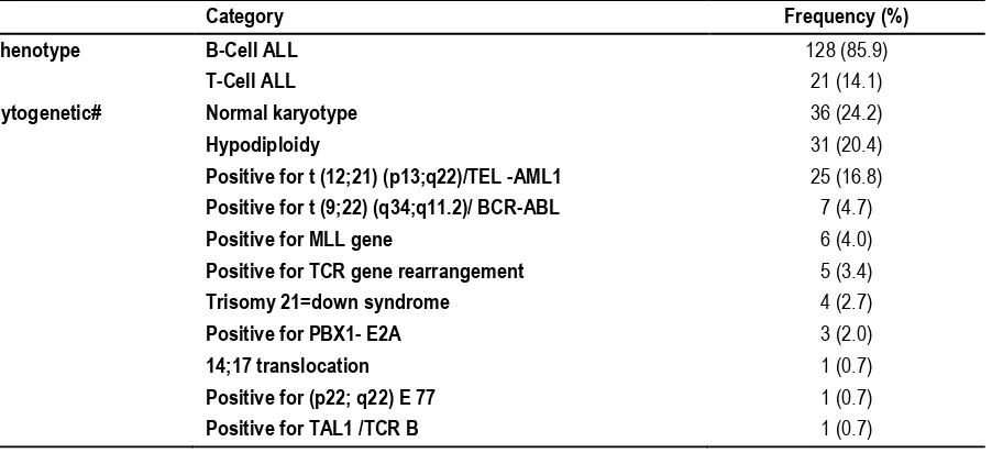
Table 1: Distribution of ALL children in Jeddah centers according to their phenotype & cytogenetic profile (N=149)
Category Frequency (%)
Phenotype B-Cell ALL 128 (85.9)
T-Cell ALL 21 (14.1)
Cytogenetic# Normal karyotype 36 (24.2)
Hypodiploidy 31 (20.4)
Positive for t (12;21) (p13;q22)/TEL -AML1 25 (16.8)
Positive for t (9;22) (q34;q11.2)/ BCR-ABL 7 (4.7)
Positive for MLL gene 6 (4.0)
Positive for TCR gene rearrangement 5 (3.4)
Trisomy 21=down syndrome 4 (2.7)
Positive for PBX1- E2A 3 (2.0)
14;17 translocation 1 (0.7)
Positive for (p22; q22) E 77 1 (0.7)
Positive for TAL1 /TCR B 1 (0.7)
(#) n=120
Table 2: Initial laboratory investigations of studied children with ALL (N=149)
Test at diagnosis Mean SD Median
WBC count 64.29 109.69 25.90
Hemoglobin (gm/dl) 11.37 40.778 8.10
Platelet count 141.48 498.84 51.85
Lactate dehydrogenase (IU) 685.80 1123.1 364.00
Table 3: Distribution of ALL Children according to the used treatment protocol (N=149)
Category Frequency (%)
Standard Risk Protocol 70 (47.0)
CCG 1991 protocol 36 (24.2)
CCG-1891 protocol 17 (11.4)
Standard risk protocol 12 (8.1)
LMP-96 protocol group C 5 (3.4)
High Risk Protocol 79 (53.0)
CCG 1961 protocol 38 (25.5)
High risk protocol 19 (12.8)
CCG-1882 regimen B 10 (6.7)
CCG-1882 regimen A 6 (4.0)
St. Jude Total 13 B protocol 5 (3.4)
Infant 99 ALL protocol 1 (0.7)
RESULTS
The study sample consisted of 149 medical records children diagnosed with ALL. It included 30 records from King Abdulaziz University Hospital, 38 from King Faisal Specialist Hospital, and 81 from King Abdulaziz Medical City. There were slightly more males (56.4%). Approximately two-thirds (63.8%) had their age between 2 and 9 years. As regards the nationality, 67.8% were Saudis.
The vast majority of children recruited to this study had B-cell ALL (85.9%) as shown in Table 1. As for the cytogenetics, done for 120 cases, the highest percentage had normal karyotype (24.2%),
followed by hypoploidy and “t(12:21)P13;q22/TEL-AML-1”
diagnosis was 64.29, with a high standard deviation (109.69), indicating wide variability. The median level of hemoglobin was low (8.10) indicating anemia in at least half of the sample. Similarly, the median platelet count was as low as 51.85. Finally, the mean (SD) level of Lactate Dehydrogenase (LDH) was high, 685.80 (1123.1). These laboratory values were categorized to examine their impact on the survival probability as prognostic factors as mentioned before. Ten different treatment protocols were adopted in the cancer-treating centers studied (Table 3).
The most commonly used two were CCG 1961 (25.5%) and CCG 1991 (24.2%) protocols. On the other hand, only 1 (0.7%) child was treated by Infant 99 ALL protocol. After re-grouping the
treatment groups into ‘High” and “Standard’ two different kinds of
protocols, 79 (53.0%) were under high level treatment group, and 70 (47.0%) were in the standard one.
Concerning outcomes, approximately two-thirds of the children had infections (61.7%). Meanwhile, the cure rate was only 10.7%, whereas the death rate was 28.2%.
Table 4: Five and ten-year survival probability in relation to different potential prognostic factors
Strata Strata Level Survival at Log-Rank
p-value
5 Years 10 Years
Overall 0.736 0.688
Age: 2 - <9 0.755 0.710 0.514
<2 0.647 0.575
>=9 0.733 0.692
Gender: Female 0.866 0.844 <0.001*
Male 0.636 0.573
Treatment Protocol: High Risk Protocol 0.723 0.673 0.689
Standard Risk Protocol 0.749 0.704
Phenotype: B-Cell ALL 0.756 0.699 0.326
T-Cell ALL 0.619 0.619
Cytogenetic: Abnormal 0.6547 0.6413 0.028*
Normal 0.8772 0.8772
Extramedullary Invasion:
Hepatomegaly: No 0.894 0.867 0.004*
Yes 0.657 0.601
Splenomegaly: No 0.901 0.836 0.002*
Yes 0.616 0.580
CNS Disease: No 0.766 0.710 0.048*
Yes 0.550 0.550
Initial WBC Level at Diagnosis High (>=50) 0.587 0.561 0.003*
Low (<50) 0.826 0.765
Initial LDH Level at Diagnosis High (>1000) 0.479 0.479 <.0001*
Middle (300-1000) 0.568 0.456
Low (<300) 0.952 0.952
Initial Hb Level at Diagnosis High (>=7) 0.762 0.723 0.115
Low (<7) 0.665 0.592
Initial Platelet Level at diagnosis: High (>100) 0.761 0.703 0.702
Middle (20-100) 0.724 0.704
Low (<20) 0.722 0.584
Testicular Disease (Male Only) No 0.649 0.584 0.087
Yes NE
NE: Not Estimable. (*) Statistically significant at p<0.05
Table 4 shows that the overall survival at 5 years and 10 years after ALL diagnosis was 0.736 and 0.688 respectively. The 5- and 10-year survival probabilities were significantly higher among female children compared to males based on log-rank test (p<0.001).
Similarly, the survival probabilities of children with abnormal cytogenicity, hepatomegaly, splenomegaly, CNS involvement, high initial WBCs count, and high initial LDH were significantly lower compared to those having no such complications or risk factors (p<0.05). As shown, the lowest 5-year survival probability
was among the children with high initial LDH levels (0.479), while the lowest 10-year survival probability was among those with middle initial LDH level (0.456). In the same table, the age at diagnosis, phenotype of ALL, treatment protocol used, initial hemoglobin (Hb) level, initial platelets count, and testicular involvement did not have a significant impact on the survival probabilities in the studied sample. Figures 1 displays the survival probability curves for the overall sample.
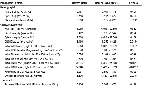
(p=0.001), the initial LDH levels (p<0.001), CNS disease (p=0.016), and cytogenicity (p=0.035). The hazard ratio for gender was 0.315, indicating that the hazard of death in female child is only 0.315 times that of male child.
As regards the clinical prognostic factors, the table indicates that the presence of CNS disease increased the hazard of death by 3.460 times. As for the laboratory prognostic factors, the table indicates that only the initial WBC count and LDH level were
statistically significant. Thus, the hazard for children with a high initial WBC count (>=50) is 8.838 times the hazard of those with low initial WBC count (<50). For the LDH initial level, the hazard of death among children with middle level was 20.193 times the hazard of those with low level, while the hazard of the high initial level compared to low initial level was 14.949. Lastly, the risk among children with abnormal cytogenicity is 5.636 with normal cytogenicity.
Table 5: Cox hazard ratios of different prognostic factors in children with ALL (without considering testicular disease as a factor)
Prognostic Factors Hazard Ratio Hazard Ratio (95% CI) p-value
Demographic:
Age Group (2 -<9 vs. <2) 0.481 0.164 - 1.415 0.184
Age Group (>=9 vs. <2) 0.515 0.146 - 1.823 0.304
Gender (Female vs. Male) 0.310 0.117 - 0.823 0.019*
Clinical/lab/genetic:
NCI Risk (High vs. Standard) 1.969 0.098 - 39.503 0.658
Hepatomegaly (Yes vs. No) 0.422 0.076 - 2.344 0.324
Splenomegaly (Yes vs. No) 2.800 0.612 - 12.804 0.184
CNS Disease (Yes vs. No) 3.460 1.256 - 9.528 0.016*
Initial WBC count (High: >=50 vs. Low: <50) 8.838 2.341 - 33.374 0.001* Initial HGB Level at Diagnosis (High: >=7 vs. Low: <7) 0.670 0.286 - 1.570 0.356 Initial Platelet count (Middle: 20 - 100 vs. Low: <20) 0.576 0.184 - 1.805 0.344 Initial Platelet count (High: >100 vs. Low: <20) 0.609 0.168 - 2.204 0.450 Initial LDH Level (Middle: 300 - 1000 vs. Low: <300) 20.193 5.373 - 75.885 <0.001* Initial LDH Level (High: >1000 vs. Low: <300) 14.949 3.421 - 65.329 <0.001* Phenotype (T-Cell ALL vs. B-Cell ALL) 2.597 0.856 - 7.885 0.092
Cytogenetic (Abnormal vs. Normal) 5.636 1.127 - 28.184 0.035*
Treatment:
Treatment Protocol (High Risk vs. Standard Risk) 0.106 0.007 - 1.670 0.111 (*) p-value of <0.05 is statistically significant
DISCUSSION
Despite the multiplicity of studies aimed at identifying the prognostic factors of pediatric lymphoblastic leukemia worldwide, data from the Middle East, including Saudi Arabia, are still deficient. The objectives of this research were to explore the profile of lymphoblastic leukemia is Saudi children, to identify the prognostic factors of the disease, and to determine the long-term outcomes and the survival probability in Saudi children.
There was a higher prevalence of pediatric lymphoblastic leukemia among males which goes with the figures reported from different studies in the literature.15,16 The highest percentage,
approximately two-thirds, of the children in our sample had an age ranging between 2 and 9 years, This age group is known to be the peak age for the incidence of ALL. In congruence with this, a study in Brazil reported that the incidence rate of pediatric ALL peaked at the age 1-4 years.17 The one-year difference with our
data could be explained by some delays in diagnosis in our data. On the same line, a study in the United States reported a high incidence of pediatric ALL before the age of 5 years.18
As regards the phenotypic characteristic of the recruited sample, B-cell ALL formed the vast majority of cases with a figure over 85%. This is similar to what is reported in the literature that B-cell ALL represents about 88% of all cases.19
As one of the overall outcomes of the studied sample, about one quarter of the children died, while only about one-tenth had total cure. The death rate is close to that reported in a study in Turkey involving 343 children with ALL followed-up for 15 years. The death rate was 20.1% among them.20 However, the total cure rate
is very low when compared to rates in the developed world, which reach up to 80% as mentioned by Abboud et al.21
According to the current study findings, over two-thirds of the ALL children in our sample developed infections. This is the most commonly encountered complication among ALL children either due to the disease itself or as a side effect of the treatment modalities. In congruence with this, a study on the infectious complications among ALL children in Taiwan it was found that 86.9% had fever, 39% had clinically documented and 44% microbiologically documented infections.22
The 5-year survival is lower than the figures reported in developed countries. A study in 2017mentioned that the 5-year survival rate of ALL children in the United States was as high as 90%.23
In the present study, child’s age at diagnosis does not seem to
have a significant impact on survival probability of the recruited patients. The finding is controversial with what is reported in literature. Thus, in disagreement with this, a retrospective record review study held in 2005 on 5181 children with ALL under the age of 18 years found reported that the best event-free survival rates were in the age group 1-5 years, and worst among in the age group <1 year, and the difference was statistically significant.24 As expected, child gender turned to be a significant
factor affecting the survival of ALL children in the present study. The results indicated that male children had lower 5- and 10-year survival probabilities, compared with females. The finding was confirmed after adjustment for confounders in the Cox PH analysis. In agreement with this present study finding, a multi-center retrospective study conducted in the U.S. to examine disparities in survival by gender in ALL children during 1973-2006 reported a higher 5-year survival rate among girls (70.1%) compared with 66.3% among boys.25
The results of the current study do not indicate any statistically significant impact of phenotype. However, the abnormal cytogenetic feature had a significant impact on the survival of ALL children, and their death risk increased by more than fivefold. The finding regarding phenotype is in agreement with a study which demonstrated almost equal survival rates among ALL children with B-cell and T-cell phenotypes.26
The presence of hepatomegaly has a significant impact on the survival of ALL children. Thus, the 5- and 10-year survival probabilities among the ALL children having hepatomegaly turned to be significantly lower in comparison with those having no hepatomegaly. In congruence with this result, a retrospective study conducted in Pakistan during 1989-2006 to explore the prognostic factors in pediatric ALL reported that the 5-year survival rate in patients with hepatomegaly was 56% compared with 76.1% among patients without hepatomegaly.27 In fact, the
presence of hepatomegaly indicates that the disease has extended to involve extra-medullary structures; hence, it is more likely to be associated with a worse outcome.
Similarly, splenomegaly had statistically significant effects on the 5- and 10-year survival probabilities among the ALL children in the present study. This is in agreement with the results reported by a retrospective record review study exploring the prognostic factors in childhood T-cell ALL in California in 1990, which indicated that absence of splenomegaly contributed to improving the survival rate to 66%. Meanwhile, the patients who had splenomegaly, along with other factors, had average survival rates of 43% and 19%.28 The negative impact of organomegaly on the survival and
outcomes of ALL in children was also documented in a recent study in Mexico.29
The 5- and 10-year survival probabilities of the children with CNS involvement were significantly lower compared with those having no CNS affection. Moreover, in Cox HR analysis, the involvement of the CNS had a statistically significant effect on the hazard of
child’s death, which was more than threefold the hazard among
children with no CNS involvement. In agreement with our log-rank results, a retrospective cohort study on 48 children with ALL in 2005 in Taiwan aimed at exploring the clinical significance of CNC involvement at diagnosis of childhood T-cell acute lymphoblastic leukemia, reported that initial CNS involvement was an unfavorable prognostic factor. The overall survival rate in patients with CNS involvement was 15%, in comparison to 88% in those without CNS affection.30 However, a more recent study in
Denmark, involving 1877 ALL patients could not reveal any significant effect of CNS involvement at diagnosis and survival.31
The present study findings indicate negative impact of an initial high WBC count (>50000/ µL). Moreover, the Cox HR analysis showed that the children who had high initial WBC count at time of diagnosis had more than eightfold risk of death compared with those with low initial WBC count. The findings are in agreement with those of a retrospective record review study exploring the prognostic factors in childhood T-cell ALL in California in 1990.28
Among study limitations, being a record-based study, the researcher collected data from medical records, and could not meet all the patients to interview and examine them at the time of the study. Some records were deficient so that some data were missed. Due to the paucity of cases with certain important characteristics (e.g. patients with acute lymphoblastic leukemia <1 year), the prognostic value of these characteristics could not be properly calculated. Lastly, the low number of cases treated with certain protocols might have impaired the results of analyses, as the effect of different treatment protocol could not be appropriately evaluated. Further multi-center prospective studies are suggested for better evaluation of all prognostic factors, and assessment of long-term outcome on a larger number of patients recruited from different cities in Saudi Arabia as well as Middle East countries. In conclusion, children with ALL in Saudi Arabia have 5- and 10-survival probabilities that are lower compared with the figures reported in more developed countries. Their survival is affected by certain prognostic factors similar to these reported in literature.
REFERENCES
1. Childhood cancer. In: Howlader N, Noone AM, Krapcho M, et al., eds.: SEER Cancer Statistics Review, 1975-2010. Bethesda, Md: National Cancer Institute, 2013, Section 28. available online at:https://seer.cancer.gov/archive/csr/1975_2010/results_merged/ sect_28_childhood_cancer.pdf. Last accessed August 02, 2017. 2. Tion SO, Richard AL. Current management of Acute Lymphoblastic Leukemia in adults | Cancer Network | The Oncology Journal Oncol. J. 2015;9.
3. Cooper SL, Brown PA.Treatment of pediatric acute lymphoblastic leukemia.Pediatr. Clin. North Am.2015;62(1):61–73. 4. Pui CH, Carroll WL, Meshinchi S, Arceci RJ. Biology, Risk Stratification, and Therapy of Pediatric Acute Leukemias: An Update. J. Clin. Oncol. 2011;29(5):551–565.
5. Ribeiro KB, Buffle PA, Metayer C. Socioeconomic status and childhood acute lymphocytic leukemia incidence in São Paulo, Brazil. Int. J. Cancer 2008; 123(8):1907–1912.
6. Hunger SP, Lu X, Devidas M, Camitta BM, Gaynon PS, Winick NJ, Get al. Improved survival for children and adolescents with acute lymphoblastic leukemia between 1990 and 2005: A report
from the children’s oncology group J. Clin. Oncol.
2012;30(14):1663–1669.
7. Magrath I, Shanta V, Advani S, Adde M, Arya LS, Banavali S, et al. Treatment of acute lymphoblastic leukaemia in countries with limited resources; lessons from use of a single protocol in India over a twenty year peroid. European Journal of Cancer 2005;41(11): 1570–1583.
8. Muwakkit S, Al-Aridi C, Samra A, Saab R, Mahfouz RA, Farra C, et al. Implementation of an intensive risk-stratified treatment protocol for children and adolescents with acute lymphoblastic leukemia in Lebanon. Am. J. Hematol. 2012;87(7):678–683. 9. Khan MQ, Shivarudrappa AS, el-Bialy S, Khawagi MZ, al-Mofarreh M. Leukaemia cases in Central Hospital, Riyadh (Saudi Arabia). J. Indian Med. Assoc. 1991;89(2):38–42.
10. Al-Mulla NA, Chandra P, Khatta M, Madanat F, Vossough P, Torfa E, et al. Childhood acute lymphoblastic leukemia in the Middle East and neighboring countries: A prospective multi-institutional international collaborative study (CALLME1) by the Middle East Childhood Cancer Alliance (MECCA). Pediatr. Blood Cancer 2014;61(8):1403–1410.
11. American Cancer society. How Is Acute Lymphocytic Leukemia Classified?, 2016. [Online]. Available: www.cancer.org/cancer/acute-lymphocytic-leukemia/detection-diagnosis-staging/how-classified.html. [Accessed: 07-Sep-2017]. 12. De Greef GE, Van Putten WLJ, Boogaerts M, Huijgens PC, Verdonck LF, Vellenga E, et al. Criteria for defining a complete remission in acute myeloid leukaemia revisited. An analysis of patients treated in HOVON-SAKK co-operative group studies. Br. J. Haematol. 2005;128(2):184–19.
13. Pui CH, Pei D, Campana D, Cheng C, Sandlund JT, Bowman WP, et al. A revised definition for cure of childhood acute lymphoblastic leukemia. Leukemia 2014;28(12): 2336–2343. 14. Swerdlow J, Campo SH, Harris E, Jaffe NL, Pileri ES, Stein SA, et al. WHO Classification of Tumours of Haematopoietic and Lymphoid Tissues, Fourth Edition - WHO - OMS -,” in WHO
Classification of Tumours, Volume 2, 2008, p. 439.
15. Hjalgrim LL, Rostgaard K, Schmiegelow K, Söderhäll S, Kolmannskog S,. Vettenranta K, et al. Age- and sex-specific incidence of childhood leukemia by immunophenotype in the Nordic countries. J. Natl. Cancer Inst. 2003; 92(20): 1539–1544 16. Rivera-Luna R, Velasco-Hidalgo L, Zapata-Tarres M, Cardenas-Cardos R, Aguilar-Ortiz MR. Current outlook of childhood cancer epidemiology in a middle-income country under a public health insurance program. Pediatric hematology and oncology. 2017;34(1):43-50.
17 .Lins MM, Santos MdO, de Albuquerque MdFPM, de Castro CCL, Mello MJG, de Camargo B. Incidence and survival of childhood leukemia in Recife, Brazil: A population-based analysis. Pediatric Blood & Cancer. 2017;64(8):e26391-n/a.
18. Whitehead TP, Metayer C, Wiemels JL, Singer AW, Miller MD. Childhood Leukemia and Primary Prevention. Current problems in pediatric and adolescent health care. 2016;46(10):317-52. 19. Wartenberg D, Groves FD, Adelman AS. Acute Lymphoblastic Leukemia: epidemiology and etiology. In: Acute Leukemias, Berlin, Heidelberg: Springer Berlin Heidelberg, 2008; 77–93. 20. Gunes AM, Oren H, Baytan B, Bengoa SY, Evim MS, Gozmen S, et al. The long-term results of childhood acute lymphoblastic leukemia at two centers from Turkey: 15 years of experience with the ALL-BFM 95 protocol. Annals of hematology. 2014;93(10):1677-84.
21. Abboud MR, Ghanem K, Muwakkit S. Acute lymphoblastic leukemia in low and middle-income countries: disease characteristics and treatment results. Current opinion in oncology. 2014;26(6):650-5.
22. Li MJ, Chang HH, Yang YL, Lu MY, Shao PL, Fu CM, et al. Infectious complications in children with acute lymphoblastic leukemia treated with the Taiwan Pediatric Oncology Group protocol: A 16-year tertiary single-institution experience. Pediatr Blood Cancer. 2017;64(10).
23. Kansagra A, Dahiya S, Litzow M. Continuing challenges and current issues in acute lymphoblastic leukemia. Leukemia & lymphoma. 2017:1-16.
26. Jabbour E, O'Brien S, Konopleva M, Kantarjian H. New insights into the pathophysiology and therapy of adult acute lymphoblastic leukemia. Cancer. 2015;121(15):2517-28.
27. Khalid S, B. Moiz B, Adil SN, Khurshid M. Retrospective review of pediatric patients with acute lymphoblastic leukemia: a single center experience. Indian J. Pathol. Microbiol. 2010;53(4): 704–10.
28. Shuster JJ, Falletta JM, Pullen DJ, Crist WM, Humphrey GB, Dowell BL, et al. Prognostic factors in childhood T-cell acute lymphoblastic leukemia: a Pediatric Oncology Group study. Blood 1990; 75(1):166–173.
29. Jaime-Perez JC, Pinzon-Uresti MA, Jimenez-Castillo RA, Colunga-Pedraza JE, Gonzalez-Llano O, Gomez-Almaguer D. Relapse of childhood acute lymphoblastic leukemia and outcomes at a reference center in Latin America: organomegaly at diagnosis is a significant clinical predictor. Hematology (Amsterdam, Netherlands). 2017:1-9.
30. Jaing TH, Yang CP, Hung IJ, Tsay PK, Tseng CK, Chen SH. Clinical significance of central nervous system involvement at diagnosis of childhood T-cell acute lymphoblastic leukemia. Pediatr. Blood Cancer 2005;45(2): 135–8.
31. Ranta S, Palomaki M, Levinsen M, Taskinen M, Abrahamsson J, Mellgren K, et al. Role of neuroimaging in children with acute lymphoblastic leukemia and central nervous system involvement at diagnosis. Pediatr Blood Cancer. 2017;64(1):64-70.
32. Zeidler L, Zimmermann M, Möricke A, Meissner B, Bartels D, Tschan C, et al. Low platelet counts after induction therapy for childhood acute lymphoblastic leukemia are strongly associated with poor early response to treatment as measured by minimal residual disease and are prognostic for treatment outcome. Haematologica 2012 Mar;97(3): 402–9.
[
Source of Support: Nil.
Conflict of Interest: None Declared.
Copyright: © the author(s) and publisher. IJMRP is an official publication of Ibn Sina Academy of Medieval Medicine & Sciences, registered in 2001 under Indian Trusts Act, 1882. This is an open access article distributed under the terms of the Creative Commons Attribution Non-commercial License, which permits unrestricted non-commercial use, distribution, and reproduction in any medium, provided the original work is properly cited.
Cite this article as: Asheqah M. Al-Balwi, Sleem Binmahfoz, Rajaa M. AL-Raddadi, Hassan A. Al-Trabolsi, Abdullah A. Baothman, Mohamad Hasan Qari. Prognostic Factors for Outcome of Childhood Lymphoblastic Leukemia At Cancer Centers in Jeddah. Int J Med Res Prof. 2017 Nov; 3(6):195-202.