OncoTargets and Therapy
Dove
press
O r i g i n a l r e s e a r c h
open access to scientific and medical research
Open access Full Text article
cXcr3 is a prognostic marker and a potential
target for patients with solid tumors: a
meta-analysis
Yang Zhang1
linjuan Xu2
Minggang Peng2
1Department of clinical laboratory,
Union hospital, Tongji Medical college, huazhong University of science and Technology, Wuhan, china; 2Department of Obstetrics and
gynecology, Union hospital, Tongji Medical college, huazhong University of science and Technology, Wuhan, china
Objective: To deeply verify the clinical significance of CXCR3 in prediction of cancer patients’ prognosis.
Data sources: We performed a meta-analysis including 12 studies searched from PubMed, Web of Science, Embase, and Cochrane databases. A total of 1,751 patients were used to ana-lyze the association between CXCR3 and patients’ prognosis, based on either overall survival or time to tumor progression.
Study selection: Studies evaluating CXCR3 expression for predicting prognosis in human solid tumors were included.
Results: It showed that patients with higher expression of CXCR3 had significantly shorter OS (pooled hazard ratio =2.315, 95% CI: 1.162–4.611, P=0.017). In addition, higher CXCR3 expression was associated with distant metastasis (yes vs no: pooled relative ratio [RR] =1.828,
95% CI: 1.140–2.931, P=0.012) in solid tumors and indicated advanced tumor stage (III/IV
vs I/II, RR =2.656, 95% CI: 1.809–3.900, P0.001) and lymph node metastasis (yes vs no:
RR =2.28, 95% CI: 1.61–3.25, P0.001) in colorectal cancer.
Conclusion: Our study highlights the role of CXCR3 as a potential prognostic marker and a promising therapeutic target in solid tumors.
Keywords: CXCR3, meta-analysis, solid tumor, prognostic marker, overall survival
Introduction
Chemokines comprise a family of chemotactic cytokines with low molecular weight which participate in multiple biologic processes such as angiogenesis, migration of leukocytes, tumor growth, and metastasis.1–3 There are currently four major families
of chemokines based on the position of conserved cysteines of these small inducible proteins: CXC, CC, CX3C, and C.4 The CXC subgroup can be further classified into
two groups, ELR+ and ELR− chemokines, according to the presence or absence of the “glu-leu-arg (ELR)” motif.5 Most, if not all, chemokines function through binding
to and activating a family of G-protein–coupled receptors, namely, CXCR or CCR.6
Recent studies have highlighted the clinical importance of chemokines and their receptors in tumor initiation, progression, and metastasis.7 Among them, we noticed
CXCR3 and analyzed several roles of it that seem to be of particular importance to us, especially its effects on tumor prognosis.
As the main receptor of the ELR− chemokines, CXCR3 is activated by specific binding of the ligands, CXCL4/PF4, CXCL9/MIG, CXCL10/IP10, CXCL11/IP9, and thus results in diverse cellular responses such as chemotactic migration, cell proliferation, or inhibition of migration according to different cell types and distinct correspondence: Minggang Peng
Department of Obstetrics and gynecology, Union hospital, Tongji Medical college, huazhong University of science and Technology, 1277 JieFang avenue, Wuhan 430022, china Tel +86 1 355 465 1079 email mgpengwh@163.com
Journal name: OncoTargets and Therapy Article Designation: Original Research Year: 2018
Volume: 11
Running head verso: Zhang et al
Running head recto: CXCR3 in solid tumors DOI: 157421
OncoTargets and Therapy downloaded from https://www.dovepress.com/ by 118.70.13.36 on 25-Aug-2020
For personal use only.
Dovepress
Zhang et al
microenvironment.8,9 It is reported that CXCR3 was
upregu-lated in many human tumors; furthermore, the increased levels were correlated with poor prognosis in breast cancer, melanoma, renal cancer, prostate cancer, and colorectal cancer patients.9–13 In colon cancer specimens, high
expres-sion of CXCR3 was correlated with the metastatic frequencies to lymph nodes and distant organs; meanwhile, CXCR3-positive colon cancer patients exhibited shorter survival than CXCR3-negative patients.7 Importantly, the systematic
administration of a CXCR3 inhibitor, AMG487, was recently reported to inhibit lung metastasis of colon cancer and breast cancer in a mouse model.14,15 Another group demonstrated
that CXCR3 could be a molecular target in breast cancer metastasis.16 However, a study involving 96 gastric
can-cer patients reported opposite results.17 Overexpression of
CXCR3 was found to be inversely associated with invasion depth and metastasis in gastric cancer, and further analysis showed that high CXCR3 expression was an independent prognostic factor and associated with favorable prognosis.
As for the multiple roles of CXCR3 in various biophysical processes, especially its divergent performance in different types of cancers, we herein carried out this meta-analysis to address the overall roles of CXCR3 in cancer patients’ prognosis. This study aimed at defining the clinical value of CXCR3 molecule, thereby supporting the use of specific genetic diagnosis for cancer patients and development of targeted strategies against CXCR3.
Materials and methods
literature search
For this study, we searched papers published before 1 April 2017 from PubMed, Embase, Web of Science, and Cochrane databases, using the following search strategy: (“CXCR3” or “CMKBR3” or “CD183” or “Chemokine C-C Motif Recep-tor 3” or “CXC Chemokine RecepRecep-tor 3”) and (“cancer” or “tumor” or “carcinoma” or “neoplasm”) and (“prognosis” or “survival” or “mortality” “death”). Furthermore, we manually searched the reference lists of relevant literature. When multiple studies of the same patient population were identified, we included the published study with the largest sample size.
selection criteria
Articles were selected if they met the following criteria: 1) evaluation of CXCR3 expression for predicting prog-nosis in human cancer; 2) studies reporting survival data; 3) studies that detected CXCR3 protein expression by
immunohistochemistry; and 4) studies with adequate data of pooled HRs and 95% CIs to be extracted or calculated. The exclusion criteria were as follows: 1) duplicate publication; 2) non-English papers; 3) author’s view, commentary, confer-ence abstract, or review articles; 4) sample number fewer than 40 patients; 5) study only focused on animal models or cancer cell lines; and 6) study lacking sufficient data for individual HRs and 95% CIs to be extracted or calculated.18 All
evalu-ations were independently performed by three individual researchers to ensure the accurate inclusion of studies.
Data collection
Three investigators independently extracted the data from eligible studies using a predefined form. The collected data included the name of first author, the publication year, patients’ country of origin, tumor type, number of patients, sex, cancer stage or grade, percentage showing high CXCR3 expression and the corresponding cutoff value, and outcome. Multivariate HRs and 95% CIs were chosen if both univari-ate and multivariunivari-ate results were reported in an individual study. For studies that presented only Kaplan–Meier curves, Engauge Digitizer version 4.1 was used to extract the survival data.19 The estimated HRs and 95% CIs were calculated by
Tierney’s method.18 By checking among the three
investiga-tors, the final data collection was performed.
statistical analysis
Using the data collected from each eligible study, we per-formed the meta-analysis to evaluate the relationship between solid tumor’s CXCR3 expression and patients’ prognosis. Stata version 14.0 (Stata Corporation, College Station, TX, USA) was used to carry out the statistical analysis. As the outcome endpoints disease-free survival, progression-free survival, and recurrence-free survival are similar in mean-ing, they were combined and a unified prognostic parameter, TTP, was used for the meta-analysis. The meta-analysis was, therefore, based on two outcome endpoints: OS and TTP. Pooled HRs and 95% CIs for the two outcome endpoints (OS, TTP) were used to evaluate the association of CXCR3 expression with solid tumor prognosis. Pooled RRs and 95% CIs were used to assess the correlations between CXCR3 expression and the clinicopathologic features of solid tumor, including TNM stage, T stage, lymph node metastasis, distant metastasis, and tumor differentiation. Heterogeneity assump-tion was checked using I2 statistic. I2 values 25% may be
considered “low”, values around 50% may be considered “moderate”, and values 75% may be considered “high”.20
OncoTargets and Therapy downloaded from https://www.dovepress.com/ by 118.70.13.36 on 25-Aug-2020
Dovepress cXcr3 in solid tumors
When the I2 values were 50%, a random-effect model was
used to calculate the pooled HRs or RRs; otherwise, a fixed-effect model was used.21 An observed HR 1 and P0.05
implied worse prognosis for patients with high CXCR3 expression. An observed RR 1 and P0.05 implied more advanced clinicopathologic characteristics for the group of high CXCR3 expression.
Publication bias and sensitivity analysis
Publication bias was tested using Begg’s funnel plot and Egger’s test.22 If the funnel plot is asymmetric and theEgger’s test reports a P-value of 0.05, publication bias is deemed to be probably existent. Meanwhile, we performed the sensitivity analysis by omitting each study or specific studies to assess the influence of individual studies to the entire meta-analysis.
Data availability
All data generated or analyzed during this study are included in this published article.
Results
search results
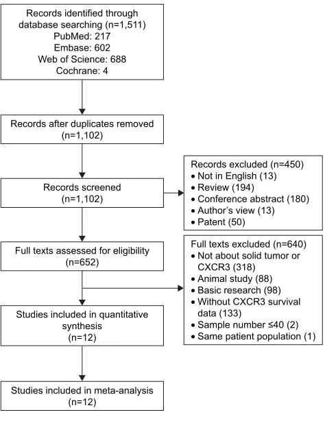
A total of 1,511 articles were retrieved by a comprehen-sive search from PubMed, Embase, Web of Science, and Cochrane databases. A total of 409 duplicate reports were excluded. After screening the titles and abstracts, 450 articles were excluded for reasons such as commentary, review, author’s view, conference abstracts, and non-English paper. The remaining articles were viewed in full text for further selection. Six hundred and forty articles were excluded for reasons such as animal study, basic research, not about solid tumor or CXCR3, without CXCR3 survival data, sample number 40, and duplicated patient population. Finally, 12 studies that reported at least one outcome endpoint were included in this meta-analysis.10–12,23–31 A flowchart of the
study selection process is shown in Figure 1.
characteristics of studies
Detailed information of these eligible studies is summarized in Table 1. In total, the 12 studies provided a sample of 1,751 patients to assess the relationship between CXCR3 expression and solid tumor prognosis. The median sample size was 146, with a range from 45 to 364. Among all the cohorts, China (n=6) became the major source region of literature, followed by USA (n=2), Ireland (n=1), Sweden (n=1), Iran (n=1), and Japan (n=1). As for the cancer type,
four studies evaluated colorectal cancer, three studies evalu-ated breast cancer, two studies evaluevalu-ated gastric cancer, two studies evaluated renal cancer, and one study evaluated glioblastoma. Nine studies out of 12 focused on overall sur-vival (OS), 3 focused on disease-free sursur-vival, 1 focused on progression-free survival, and 1 focused on recurrence-free survival. Various clinicopathologic data were reported in five studies (TNM stage in three studies, pathologic T stage in five studies, lymph node metastasis in three studies, distant metastasis in three studies, and tumor differentiation in five studies). All studies applied immunohistochemical staining to investigate CXCR3 expression. The cutoff values of posi-tive CXCR3 expression varied among different studies, so we classified all the cases according to their original studies (negative or positive staining).
Meta-analysis
The meta-analysis of CXCR3 expression was based on two outcome endpoints: OS and time to tumor progression (TTP). Nine studies were included in the meta-analysis of OS. A random-effect model was applied to calculate the pooled hazard ratio (HR) and 95% CI because the heterogeneity
5HFRUGVDIWHUGXSOLFDWHVUHPRYHG Q
5HFRUGVH[FOXGHGQ
• 1RWLQ(QJOLVK
• 5HYLHZ
• &RQIHUHQFHDEVWUDFW
• $XWKRU¶VYLHZ
• 3DWHQW
)XOOWH[WVH[FOXGHGQ
• 1RWDERXWVROLGWXPRURU &;&5
• $QLPDOVWXG\
• %DVLFUHVHDUFK
• :LWKRXW&;&5VXUYLYDO GDWD
• 6DPSOHQXPEHU
• 6DPHSDWLHQWSRSXODWLRQ 5HFRUGVLGHQWLILHGWKURXJK
GDWDEDVHVHDUFKLQJQ 3XE0HG (PEDVH :HERI6FLHQFH
&RFKUDQH
5HFRUGVVFUHHQHG Q
)XOOWH[WVDVVHVVHGIRUHOLJLELOLW\ Q
6WXGLHVLQFOXGHGLQTXDQWLWDWLYH V\QWKHVLV
Q
6WXGLHVLQFOXGHGLQPHWDDQDO\VLV Q
Figure 1 Flow diagram of study selection.
Abbreviation: cXcr3, c–X–c motif chemokine receptor 3.
OncoTargets and Therapy downloaded from https://www.dovepress.com/ by 118.70.13.36 on 25-Aug-2020
Dovepress
Zhang et al
Table 1 characteristics of studies included in the meta-analysis
Reference Year Country Cancer type Case Male/
female Stage/ grade
Percentage of high CXCR3 expression, cutoff value
Outcome
Wu et al30 2012 china colorectal carcinoma 112 59/53 na 60/112 (53.6%), 25% of cells Os
Kawada et al11 2007 Japan colon cancer 92 32/60 TnM: i–iV 31/92 (33.7%), 10% of cells Os
Bai et al23 2016 china colorectal cancer 143 na TnM: ii 14/143 (9.8%), na Os, DFs
Du et al12 2014 china colorectal cancer 145 84/61 TnM: i/ii 26/145 (17.9%), 50% of cells DFs
Zhou et al31 2016 china gastric cancer 103 72/31 grade: 1–3 75/103 (72.8%), na Os
li et al26 2015 china gastric cancer 192 138/54 TnM: i–iV 112/192 (58.33%), 25% of cells Os
Ma et al10 2009 Usa Breast cancer 75 0/75 na 18/75 (24.0%), 75% of cell Os
Mulligan et al27 2013 ireland Breast cancer 261 na grade: 1–3 52/263 (19.8%), 33% of cells Os
hilborn et al24 2014 sweden Breast cancer 364 na na 290/364 (79.7%), na css, rFs
rezakhaniha et al29 2016 iran renal cell carcinoma 45 28/17 TnM: i–iV 34/45 (75.55%), 30% of cells Os
Klatte et al25 2008 Usa clear cell renal cell carcinoma 154 100/54 grade: 1–4 134/154 (87.0%), 30% of cells DFs
Pu et al28 2015 china glioblastoma 65 24/41 na 27/65 (41.54%), staining score 4 Os, PFs
Abbreviations: cXcr3, c–X–c motif chemokine receptor 3; DFs, disease-free survival; na, not available; Os, overall survival; PFs, progression-free survival; rFs,
recurrence-free survival; CSS, cancer specific survival.
6WXG\,'
&RORUHFWDOFDQFHU
%UHDVWFDQFHU
*DVWULFFDQFHU
*OLREODVWRPD
5HQDOFHOOFDUFLQRPD
ZHLJKW +5&,
± ± ±
±
± ±
±
± ±
±
±
±
±
±
±
%DLHWDO
:XHWDO
.DZDGDHWDO
6XEWRWDO, 3
6XEWRWDO, 3
6XEWRWDO, 3
2YHUDOO, 3
6XEWRWDO
6XEWRWDO
0XOOLJDQHWDO
0DHWDO
=KRXHWDO
/LHWDO
3XHWDO
5H]DNKDQLKDHWDO
Figure 2 Forest plots of studies evaluating cXcr3 expression level and patients’ overall survival with regard to cancer type. Note: Weights are from random-effects analysis.
Abbreviations: cXcr3, c–X–c motif chemokine receptor 3; hr, hazard ratio.
test reported a P0.001 and an I2 value of 84.9%. The
results showed that CXCR3 overexpression was associ-ated with poorer OS of solid tumors (pooled HR =2.315, 95% CI: 1.162–4.611, P=0.017; Figures 2 and 3). Five studies included in the meta-analysis reported TTP. A ran-dom-effect model was again used to calculate the pooled
HR and 95% CI because the heterogeneity test reported a P0.001 and an I2 value of 88.0%. The results
demon-strated that there was no significant association between CXCR3 overexpression and TTP (pooled HR =2.273, 95% CI: 0.910–5.676, P=0.079; Figure 4). Furthermore, subgroup analysis was stratified by cancer type and races.
OncoTargets and Therapy downloaded from https://www.dovepress.com/ by 118.70.13.36 on 25-Aug-2020
Dovepress cXcr3 in solid tumors
Study ID HR (95% CI)
0.28 1 36.7
% weight
10.47 12.30 9.00 11.90 12.82 11.97
68.47
11.79 10.55 9.19
31.53
100
1.56 (0.82–2.89) 13.04 (4.64–36.69)
5.72 (1.49–21.98) 1.31 (0.63–2.71) 0.46 (0.28–0.76) 3.82 (1.88–7.77)
2.37 (0.91–6.14)
3.04 (1.43–6.43) 2.07 (0.75–5.75) 1.85 (0.50–6.83)
2.49 (1.44–4.31)
2.32 (1.16–4.61) Subtotal (I2=89.9%, P=0.000)
Overall (I2=84.9%, P=0.000)
Subtotal (I2=0.0%, P=0.745) Bai et al23 (2016)
Asia
Caucasian
Wu et al30 (2012)
Kawada et al11 (2007)
Zhou et al31 (2016)
Li et al26 (2015)
Pu et al28 (2015)
Mulligan et al27 (2013)
Ma et al10 (2009)
Rezakhaniha et al29 (2016)
Figure 3 Forest plots of studies evaluating cXcr3 expression level and patients’ overall survival with regard to ethnicity. Note: Weights are from random-effects analysis.
Abbreviations: cXcr3, c–X–c motif chemokine receptor 3; hr, hazard ratio.
With regard to race, high CXCR3 expression was associated with poorer OS (a fixed-effect model, HR =2.490, 95% CI: 1.439–4.311, P=0.001) in Caucasian patients while no dif-ference on TTP (Figures 3 and 5; Table 2). For the cancer type, high CXCR3 expression was associated with poorer OS in colorectal cancer (a random-effect model, HR =4.635, 95% CI: 1.114–19.293, P=0.035; I2=84.3%; P=0.002),
breast cancer (a fixed-effect model, HR =2.654, 95% CI: 1.449–4.858, P=0.002; I2=0.00%; P=0.555), and
glioblas-toma (HR =3.823, 95% CI: 1.880–7.773, P0.001). Mean-while, high CXCR3 expression was associated with poorer TTP in glioblastoma (HR =2.180, 95% CI: 1.090–4.390,
P=0.028) and clear cell renal cell carcinoma (HR =2.460, 95% CI: 1.040–5.800, P=0.040; Figure 4; Table 2).
In the comprehensive analysis of the role of CXCR3 expression in solid tumors as a biomarker, we investigated the association of high CXCR3 expression and clinicopathologic characteristics in each cancer type. As shown in Table 3, high CXCR3 expression was significantly associated with distant metastasis (yes vs no: relative ratio [RR] =1.828, 95% CI: 1.140–2.931, P=0.012), lymph node metastasis in colorectal cancer (yes vs no: RR =2.28, 95% CI: 1.61–3.25,
P0.001), TNM stage in colorectal cancer (III/IV vs I/II:
RR =2.656, 95% CI: 1.809–3.900, P0.001), and lymph node metastasis in gastric cancer (yes vs no: RR =0.714, 95% CI: 0.610–0.837, P0.001). However, no significant relationship was observed between CXCR3 overexpression and other clinical characteristics such as T stage and tumor differentiation in solid tumors, which may be due to insuf-ficient data available.
Publication bias
Begg’s funnel plot and Egger’s test were used to estimate the publication bias of the included literature. The shapes of the funnel plots for TTP showed no evidence of obvious asymmetry (Figure 6), and Egger’s tests revealed nonsig-nificant values (P=0.619). However, publication bias may exist for OS (P=0.035) in the analysis of high vs low CXCR3 expression.
sensitivity analysis
Moreover, sensitivity analysis was carried out to assess the influence of individual studies on the overall results of OS and TTP. No individual study dominated this meta-analysis, and the removal of any single study had no significant effect on the overall conclusion (Figure 7).
OncoTargets and Therapy downloaded from https://www.dovepress.com/ by 118.70.13.36 on 25-Aug-2020
Dovepress
Zhang et al
± ± ±
±
± ±
±
± +5&,
ZHLJKW
$VLD 6WXG\,'
%DLHWDO
3XHWDO
'XHWDO
6XEWRWDO, 3
&DXFDVLDQ
+LOERUQHWDO
.ODWWHHWDO
6XEWRWDO, 3
2YHUDOO, 3
Figure 5 Forest plots of studies evaluating cXcr3 expression level and patients’ TTP. Notes: Patients’ TTP with regard to race. Weights are from random-effects analysis.
Abbreviations: cXcr3, c–X–c motif chemokine receptor 3; hr, hazard ratio; TTP, time to tumor progression.
6WXG\,'
&RORUHFWDOFDQFHU
6XEWRWDO, 3
2YHUDOO, 3
6XEWRWDO
6XEWRWDO
6XEWRWDO %DLHWDO
'XHWDO
+LOERUQHWDO
.ODWWHHWDO
3XHWDO
*OLREODVWRPD
%UHDVWFDQFHU
&OHDUFHOOUHQDOFHOOFDUFLQRPD
± ± ±
± ±
± ±
± ±
± +5&O
ZHLJKW
Figure 4 Forest plots of studies evaluating cXcr3 expression level and patients’ TTP. Notes: Patients’ TTP with regard to cancer type. Weights are from random-effects analysis.
Abbreviations: cXcr3, c–X–c motif chemokine receptor 3; hr, hazard ratio; TTP, time to tumor progression.
OncoTargets and Therapy downloaded from https://www.dovepress.com/ by 118.70.13.36 on 25-Aug-2020
Dovepress cXcr3 in solid tumors
Table 3 Meta-analysis of the association between high cXcr3 expression and clinicopathologic features of each cancer type
Variables Cancer type Studies Pooled RR 95% CI P-value Model Heterogeneity
I2 (%)
P-value
TnM stage Overall 3 1.654 0.641–4.270 0.298 random 93.4 0.001
renal cell carcinoma 1 2.206 0.604–8.062 0.232 Fixed – –
gastric cancer 1 0.893 0.761–1.048 0.166 Fixed – –
colorectal cancer 1 2.656 1.809–3.900 0.001 Fixed – –
T stage Overall 5 1.242 0.870–1.772 0.232 random 88.5 0.001
colorectal cancer 3 1.245 0.879–1.764 0.217 random 82.1 0.004
gastric cancer 2 1.448 0.294–7.130 0.649 random 94.4 0.001
lymph node metastasis Overall 3 1.329 0.678–2.605 0.407 random 94.3 0.001
colorectal cancer 2 2.605 1.604–4.230 0.001 random 52.2 0.148
gastric cancer 1 0.714 0.610–0.837 0.001 Fixed – –
Distant metastasis Overall 3 1.828 1.140–2.931 0.012 random 51.2 0.129
colorectal cancer 2 1.770 0.955–3.283 0.07 random 73.1 0.054
renal cell carcinoma 1 2.265 0.833–6.160 0.109 Fixed – –
Tumor differentiation Overall 5 1.111 0.856–1.442 0.43 random 76.8 0.001
colorectal cancer 3 1.199 0.799–1.799 0.381 random 87.0 0.001
gastric cancer 1 1.160 0.897–1.500 0.258 Fixed – –
renal cell carcinoma 1 0.768 0.481–1.228 0.271 Fixed – –
Note: “–” indicates no data.
Abbreviations: CXCR3, C–X–C motif chemokine receptor 3; RR, relative ratio; Random, random-effect model; Fixed, fixed-effect model. Table 2 subgroup analysis of pooled hr for solid tumor patients with cXcr3 overexpression
Outcome Subgroup Studies Pooled HR 95% CI P-value Model Heterogeneity
I2 (%)
P-value
Os Cancer type
colorectal cancer 3 4.635 1.114–19.293 0.035 random 84.3 0.002
Breast cancer 2 2.654 1.449–4.858 0.002 Fixed 0.0 0.555
gastric cancer 2 0.752 0.273–2.072 0.581 random 81.4 0.020
glioblastoma 1 3.823 1.880–7.773 0.001
renal cell carcinoma 1 1.854 0.503–6.830 0.354
Race
asian 6 2.365 0.912–6.136 0.077 random 89.9 0.001
caucasian 3 2.490 1.439–4.311 0.001 Fixed 0.0 0.745
TTP Cancer type
colorectal cancer 2 3.511 0.380–32.434 0.268 random 93.0 0.001
glioblastoma 1 2.180 1.090–4.390 0.028
Breast cancer 1 0.950 0.580–1.560 0.839
clear cell renal cell carcinoma 1 2.460 1.040–5.800 0.040
Race
asian 3 3.028 0.814–11.263 0.098 random 88.5 0.001
caucasian 2 1.429 0.568–3.596 0.449 random 71.7 0.060
Abbreviations: cXcr3, c–X–c motif chemokine receptor 3; hr, hazard ratio; Os, overall survival; TTP, time to tumor progression; random, random-effect model;
Fixed, fixed-effect model.
Discussion
Our current meta-analysis is so far the only one that has made a comprehensive assessment of the published studies regarding CXCR3 expression and patients’ survival. We systematically analyzed survival data for 1,751 patients with various solid tumors. Among tumors in which CXCR3 overexpression is commonly observed (colorectal cancer, breast cancer, glio-blastoma), there was a strong association between CXCR3 overexpression and unfavorable outcome compared with normal CXCR3 expression. Besides, we also evaluated the association between CXCR3 expression and clinicopathologic
features, which showed a significant correlation between CXCR3 overexpression and tumor distant metastasis. Espe-cially in colorectal cancer, high CXCR3 expression was related to unfavorable performances such as advanced tumor stage and lymph node metastasis. All these results indicated that CXCR3 probably participates in tumor progression and finally affects tumor prognosis. However, there seemed to be a negative association between high CXCR3 expression and lymph node metastasis in gastric cancer, and one of the two studies regarding gastric cancer, indeed, showed a protective role for CXCR3 in patients’ OS, while the other one showed
OncoTargets and Therapy downloaded from https://www.dovepress.com/ by 118.70.13.36 on 25-Aug-2020
Dovepress
Zhang et al
6(RIORJK %HJJ¶VIXQQHOSORWZLWK SVHXGRFRQILGHQFHOLPLWV
$
/RJK
±
6(RIORJK %HJJ¶VIXQQHOSORWZLWK SVHXGRFRQILGHQFHOLPLWV
%
/RJK
Figure 6 Begg’s funnel plot estimating the publication bias of the included literature. Note: (A) For cXcr3 and Os and (B) for cXcr3 and TTP.
Abbreviations: cXcr3, c–X–c motif chemokine receptor 3; Os, overall survival; se, standard error; TTP, time to tumor progression.
Figure 7 sensitivity analysis of this meta-analysis.
Note: (A) For cXcr3 and Os and (B) for cXcr3 and TTP.
Abbreviations: cXcr3, c–X–c motif chemokine receptor 3; Os, overall survival; TTP, time to tumor progression.
(VWLPDWH 8SSHU&,OLPLW /RZHU&,OLPLW
=KRXHWDO
/LHWDO
3XHWDO
%DLHWDO
:XHWDO
.DZDGDHWDO
5H]DNKDQLKDHWDO
0XOOLJDQHWDO
0DHWDO
%DLHWDO
3XHWDO
'XHWDO
+LOERUQHWDO
.ODWWHHWDO
$
%
0HWDDQDO\VLVHVWLPDWHVJLYHQQDPHGVWXG\LVRPLWWHG 0HWDDQDO\VLVHVWLPDWHV
JLYHQQDPHGVWXG\LVRPLWWHG
no significant correlation. We noticed that CXCR3 appeared to act differently in gastric cancer than the other cancer types. Actually, in Li et al’s study, CXCR3 was highly expressed in advanced gastric cancer tissues compared with corresponding paracancerous tissues, which is the same as in other cancer types. However, in advanced gastric cancer tissues, higher CXCR3 correlated with better prognosis.26 To explain this,
they suggested a possible pathway that the CXCR3 played a role in recruiting tumor-infiltrating lymphocytes to promote antitumor function resulting in a better prognosis. Due to the limited studies evaluating gastric cancer and the scale of the study, it is hard to make a conclusion here, but we think it is worthy to investigate the underlying mechanism for the divergent performance.
Just like other chemokine receptors, CXCR3 is a seven transmembrane G-protein–coupled receptor which is
reported to trigger several downstream pathways such as MAPK, SRC, and PI3K signaling that affect several cellular responses (calcium influx, proliferation, actin rearrange-ment, migration).32 In tumor organs, CXCR3 is expressed
on the tumor cells, stroma cells, and recruited leukocytes, with various ligands also expressed on most of these cells. Consequently, CXCR3 mediated tumor progression directly or indirectly by regulating tumor growth, migration, invasion, angiogenesis, and immunity.32 It has been generally verified
that the expression profiles of CXCR3 exhibited a significant increase in metastatic organs compared to corresponding primary cancer tissues in colorectal cancer, breast cancer, and melanoma,10,33,34 further confirming the important role
of CXCR3 in tumor progression.
It is notable that CXCR3 is a risk factor for cancer distant metastasis combining all the included cancer types, since
OncoTargets and Therapy downloaded from https://www.dovepress.com/ by 118.70.13.36 on 25-Aug-2020
Dovepress cXcr3 in solid tumors
metastatic cancers are responsible for about 90% of cancer-related mortality.35 Disseminated cancers are usually more
refractory to prior cancer treatments; thus, an ideal strategy is to prevent metastasis by limiting initial dissemination and preventing secondary spread.36 Consequently, CXCR3 may
be a promising therapeutic target as increasing evidence has shown its overexpression in many primary and metastatic tumors. Moreover, targeting CXCR3 using its inhibitor AMG487 significantly suppressed metastasis and improved host antitumor immunity in metastatic breast cancer; while CXCR3-knockout mice showed decreased metastasis.16
Though there are no clinical trials regarding CXCR3-targeted therapy currently, it is, indeed, a potential target and more studies are required to verify this. In most cases, chemokines are released locally and their effects are usually confined to local tissues, which are different from those of other cytok-ines that may cause systemic effects. Thus, CXCR3 could be better targets with less off-target side effects.
In conclusion, this meta-analysis demonstrates that high level of CXCR3 expression is associated shorter OS and tumor metastasis for patients, which suggests that CXCR3 is a valuable prognostic marker and a promising therapeutic target for solid tumors. We are looking forward to see more supportive data regarding CXCR3 in various tumors in the near future.
Author contributions
YZ and MP: concept and design, data collection, data analysis, and drafting the paper. LX: concept and critical revision of the paper. MP: study supervision. All authors contributed toward data analysis, drafting and revising the paper and agree to be accountable for all aspects of the work.
Disclosure
The authors report no conflicts of interest in this work.
References
1. Struyf S, Proost P, Van Damme J. Regulation of the immune response by the interaction of chemokines and proteases. Adv Immunol. 2003;81:1–44. 2. Billottet C, Quemener C, Bikfalvi A. CXCR3, a double-edged sword
in tumor progression and angiogenesis. Biochim Biophys Acta. 2013; 1836(2):287–295.
3. Vandercappellen J, Van Damme J, Struyf S. The role of CXC chemokines and their receptors in cancer. Cancer Lett. 2008;267(2):226–244. 4. Zlotnik A, Yoshie O. Chemokines: a new classification system and their
role in immunity. Immunity. 2000;12(2):121–127.
5. Hu M, Li K, Maskey N, et al. Corrigendum to “Overexpression of the chemokine receptor CXCR3 and its correlation with favorable prognosis in gastric cancer” (Hum Pathol. 2015;46:1872–1880). Hum Pathol. 2016;54:203–204.
6. Rot A, von Andrian UH. Chemokines in innate and adaptive host defense: basic chemokinese grammar for immune cells. Annu Rev Immunol. 2004; 22:891–928.
7. Murakami T, Kawada K, Iwamoto M, et al. The role of CXCR3 and CXCR4 in colorectal cancer metastasis. Int J Cancer. 2013;132(2):276–287. 8. Dagan-Berger M, Feniger-Barish R, Avniel S, et al. Role of CXCR3
car-boxyl terminus and third intracellular loop in receptor-mediated migra-tion, adhesion and internalization in response to CXCL11. Blood. 2006; 107(10):3821–3831.
9. Wu Q, Dhir R, Wells A. Altered CXCR3 isoform expression regu-lates prostate cancer cell migration and invasion. Mol Cancer. 2012; 11:3.
10. Ma X, Norsworthy K, Kundu N, et al. CXCR3 expression is associated with poor survival in breast cancer and promotes metastasis in a murine model. Mol Cancer Ther. 2009;8(3):490–498.
11. Kawada K, Hosogi H, Sonoshita M, et al. Chemokine receptor CXCR3 promotes colon cancer metastasis to lymph nodes. Oncogene. 2007;26(32):4679–4688.
12. Du C, Yao Y, Xue W, Zhu WG, Peng Y, Gu J. The expression of chemokine receptors CXCR3 and CXCR4 in predicting postoperative tumour progression in stages I–II colon cancer: a retrospective study. BMJ Open. 2014;4(8):e005012.
13. Monteagudo C, Martin JM, Jorda E, Llombart-Bosch A. CXCR3 chemokine receptor immunoreactivity in primary cutaneous malignant melanoma: correlation with clinicopathological prognostic factors. J Clin Pathol. 2007;60(6):596–599.
14. Cambien B, Karimdjee BF, Richard-Fiardo P, et al. Organ-specific inhibition of metastatic colon carcinoma by CXCR3 antagonism. Br J Cancer. 2009;100(11):1755–1764.
15. Walser TC, Rifat S, Ma X, et al. Antagonism of CXCR3 inhibits lung metastasis in a murine model of metastatic breast cancer. Cancer Res. 2006;66(15):7701–7707.
16. Zhu G, Yan HH, Pang Y, et al. CXCR3 as a molecular target in breast cancer metastasis: inhibition of tumor cell migration and promotion of host anti-tumor immunity. Oncotarget. 2015;6(41):43408–43419. 17. Hu M, Li K, Maskey N, et al. Overexpression of the chemokine receptor
CXCR3 and its correlation with favorable prognosis in gastric cancer. Hum Pathol. 2015;46(12):1872–1880.
18. Tierney JF, Stewart LA, Ghersi D, Burdett S, Sydes MR. Practical methods for incorporating summary time-to-event data into meta-analysis. Trials. 2007;8:16.
19. Zhang Y, Cai P, Liang T, Wang L, Hu L. TIM-3 is a potential prog-nostic marker for patients with solid tumors: a systematic review and meta-analysis. Oncotarget. 2017;8(19):31705–31713.
20. DerSimonian R, Laird N. Meta-analysis in clinical trials. Control Clin Trials. 1986;7(3):177–188.
21. Higgins JP, Thompson SG, Deeks JJ, Altman DG. Measuring incon-sistency in meta-analyses. BMJ. 2003;327(7414):557–560.
22. Egger M, Davey Smith G, Schneider M, Minder C. Bias in meta-analysis detected by a simple, graphical test. BMJ. 1997;315(7109):629–634. 23. Bai M, Chen X, Ba YI. CXCL10/CXCR3 overexpression as a biomarker
of poor prognosis in patients with stage II colorectal cancer. Mol Clin Oncol. 2016;4(1):23–30.
24. Hilborn E, Sivik T, Fornander T, Stal O, Nordenskjold B, Jansson A. C-X-C ligand 10 and C-X-C receptor 3 status can predict tamoxifen treatment response in breast cancer patients. Breast Cancer Res Treat. 2014;145(1):73–82.
25. Klatte T, Seligson DB, Leppert JT, et al. The chemokine receptor CXCR3 is an independent prognostic factor in patients with localized clear cell renal cell carcinoma. J Urol. 2008;179(1):61–66.
26. Li K, Zhu Z, Luo J, et al. Impact of chemokine receptor CXCR3 on tumor-infiltrating lymphocyte recruitment associated with favorable prognosis in advanced gastric cancer. Int J Clin Exp Pathol. 2015; 8(11):14725–14732.
27. Mulligan AM, Raitman I, Feeley L, et al. Tumoral lymphocytic infiltra-tion and expression of the chemokine CXCL10 in breast cancers from the Ontario Familial Breast Cancer Registry. Clin Cancer Res. 2013;19(2): 336–346.
28. Pu Y, Li S, Zhang C, Bao Z, Yang Z, Sun L. High expression of CXCR3 is an independent prognostic factor in glioblastoma patients that pro-motes an invasive phenotype. J Neurooncol. 2015;122(1):43–51.
OncoTargets and Therapy downloaded from https://www.dovepress.com/ by 118.70.13.36 on 25-Aug-2020
OncoTargets and Therapy
Publish your work in this journal
Submit your manuscript here: http://www.dovepress.com/oncotargets-and-therapy-journal
OncoTargets and Therapy is an international, peer-reviewed, open access journal focusing on the pathological basis of all cancers, potential targets for therapy and treatment protocols employed to improve the management of cancer patients. The journal also focuses on the impact of management programs and new therapeutic agents and protocols on
patient perspectives such as quality of life, adherence and satisfaction. The manuscript management system is completely online and includes a very quick and fair peer-review system, which is all easy to use. Visit http://www.dovepress.com/testimonials.php to read real quotes from published authors.
Dovepress
Dove
press
Zhang et al
29. Rezakhaniha B, Dormanesh B, Pirasteh H, et al. Immunohistochemi-cal distinction of metastases of renal cell carcinoma with molecular analysis of overexpression of the chemokines CXCR2 and CXCR3 as independent positive prognostic factors for the tumorigenesis. IUBMB Life. 2016;68(8):629–633.
30. Wu Z, Han X, Yan J, et al. The prognostic significance of chemokine receptor CXCR3 expression in colorectal carcinoma. Biomed Pharma-cother. 2012;66(5):373–377.
31. Zhou H, Wu J, Wang T, Zhang X, Liu D. CXCL10/CXCR3 axis pro-motes the invasion of gastric cancer via PI3K/AKT pathway-dependent MMPs production. Biomed Pharmacother. 2016;82:479–488.
32. Ma B, Khazali A, Wells A. CXCR3 in carcinoma progression. Histol Histopathol. 2015;30(7):781–792.
33. Rubie C, Kollmar O, Frick VO, et al. Differential CXC receptor expres-sion in colorectal carcinomas. Scand J Immunol. 2008;68(6):635–644. 34. Kawada K, Taketo MM. Significance and mechanism of lymph node
metas-tasis in cancer progression. Cancer Res. 2011;71(4):1214–1218. 35. Mehlen P, Puisieux A. Metastasis: a question of life or death. Nat Rev
Cancer. 2006;6(6):449–458.
36. Wells A, Grahovac J, Wheeler S, Ma B, Lauffenburger D. Targeting tumor cell motility as a strategy against invasion and metastasis. Trends Pharmacol Sci. 2013;34(5):283–289.
OncoTargets and Therapy downloaded from https://www.dovepress.com/ by 118.70.13.36 on 25-Aug-2020