SPECIAL ARTICLE
The International Liaison Committee on
Resuscitation (ILCOR) Consensus on Science With
Treatment Recommendations for Pediatric and
Neonatal Patients: Neonatal Resuscitation
The International Liaison Committee on Resuscitation
The authors have indicated they have no financial relationships relevant to this article to disclose.
A
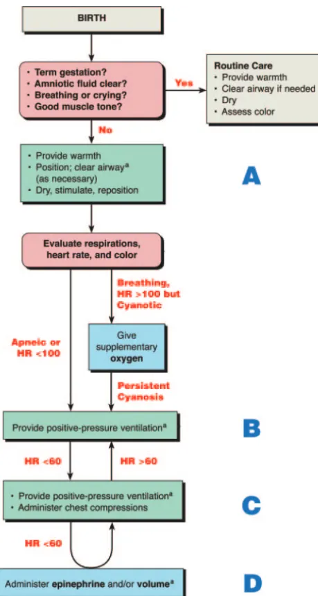
PPROXIMATELY 10% OFnewborns require some assistance to begin breathing at birth, and about 1% require extensive resuscitation. Although the vast majority of newborn infants do not require intervention to make the transition from intrauterine to extrauterine life, the large number of births worldwide means that many infants require some resuscitation. Newborn infants who are born at term, had clear amniotic fluid, and are breathing or crying and have good tone must be dried and kept warm but do not require resuscitation.All others need to be assessed for the need to receive 1 or more of the following actions in sequence:
A. Initial steps in stabilization (clearing the airway, positioning, stimulating) B. Ventilation
C. Chest compressions
D. Medications or volume expansion
Progression to the next step is based on simultaneous assessment of 3 vital signs: respirations, heart rate, and color. Progression occurs only after successful com-pletion of the preceding step. Approximately 30 seconds is allotted to complete 1 step successfully, reevaluate, and decide whether to progress to the next (Fig 1). Since publication of the last International Liaison Committee on Resuscitation
(ILCOR) document,1several controversial neonatal resuscitation issues have been
identified. The literature was researched and a consensus was reached on the role of supplementary oxygen, peripartum management of meconium, ventilation strategies, devices to confirm placement of an advanced airway (eg, tracheal tube or laryngeal mask airway [LMA]), medications, maintenance of body temperature, postresuscitation management, and considerations for withholding and discon-tinuing resuscitation.
INITIAL RESUSCITATION
Supplementary OxygenW202A,W202B
There is growing evidence from both animal and human studies that air is as effective as 100% oxygen for the resuscitation of most infants at birth. There are concerns about potential adverse effects of 100% oxygen on breathing physiology, cerebral circulation, and potential tissue damage from oxygen free radicals.
www.pediatrics.org/cgi/doi/10.1542/ peds.2006-0350
doi:10.1542/peds.2006-0350
This work was presented at the 2005 International Consensus Conference on Cardiopulmonary Resuscitation and Emergency Cardiovascular Care Science With Treatment Recommendations, hosted by the American Heart Association; January 23–30, 2005; Dallas, TX
This report was copublished inCirculation. 2005;112:III-91–III-99; andResuscitation. 2005;67:293–303.
Key Words
resuscitation
Abbreviations
ILCOR—International Liaison Committee on Resuscitation
LOE—level of evidence
PEEP—positive end-expiratory pressure CPAP— continuous positive air pressure FRC—functional residual capacity IV—intravenous
Consensus on Science
Studies examining blood pressure, cerebral perfusion, and biochemical indicators of cell damage in asphyxiated animals resuscitated with 100% vs 21% oxygen show
conflicting results (level of evidence [LOE] 6).2–6 One
study of preterm infants (⬍33 weeks of gestation)
ex-posed to 80% oxygen found lower cerebral blood flow when compared with those stabilized with 21% oxygen (LOE 2).7Some animal data indicate the opposite effect, that is, reduced blood pressure and cerebral perfusion
with air versus 100% oxygen (LOE 6).2
Meta-analysis of 4 human studies showed a reduction in mortality and no evidence of harm in infants resus-citated with air compared with those resusresus-citated with
100% oxygen (LOE 1).8,9The 2 largest newborn human
studies of room air versus oxygen resuscitation were not blinded. In those studies, if there was no response after
90 seconds, those resuscitated with air were switched to supplementary oxygen; a similar proportion who failed to respond while receiving oxygen were not crossed over to room air.10,11These results require careful interpreta-tion because of significant methodologic concerns (re-garding patient selection, lack of blinding, randomiza-tion methods, and follow-up).
Trials have not examined in sufficient detail infants
with a birth weight of ⬍1000 g, those with known
congenital pulmonary or cyanotic heart disease, and those without discernible signs of life at birth.10–13 Con-tinuous oximetry studies show that term healthy
new-borns may take ⬎10 minutes to achieve a preductal
oxygen saturation⬎95% and nearly 1 hour to achieve
this postductally (LOE 5).14–16
Treatment Recommendation
There is currently insufficient evidence to specify the concentration of oxygen to be used at initiation of re-suscitation. After initial steps at birth, if respiratory ef-forts are absent or inadequate, lung inflation/ventilation should be the priority. Once adequate ventilation is es-tablished, if the heart rate remains low, there is no evidence to support or refute a change in the oxygen concentration that was initiated. Rather, the priority should be to support cardiac output with chest compres-sions and coordinated ventilations. Supplementary oxy-gen should be considered for infants with persistent central cyanosis. Some have advocated adjusting the oxygen supply according to pulse oximetry measure-ments to avoid hyperoxia, but there is insufficient evi-dence to determine the appropriate oximetry goal be-cause observations are confounded by the gradual increase in oxyhemoglobin saturation that normally oc-curs following birth. Excessive tissue oxygen may cause oxidant injury and should be avoided, especially in the premature infant.
Peripartum Management of Meconium
Management of meconium was examined from 2 per-spectives: (1) suctioning of the meconium from the in-fant’s airway after delivery of the head but before deliv-ery of the shoulders (intrapartum suctioning) and (2) suctioning of the infant’s trachea immediately after birth (tracheal suctioning).
Intrapartum SuctioningW206
Consensus on Science
Previous studies have yielded conflicting results about the value of intrapartum oropharyngeal and nasopha-ryngeal suctioning of infants born with meconium-stained fluid (LOE 317; LOE 418,19). A recent large multi-center randomized trial found that intrapartum suctioning of meconium does not reduce the incidence of meconium aspiration syndrome (LOE 1).20
FIGURE 1
Treatment Recommendation
Routine intrapartum oropharyngeal and nasopharyn-geal suctioning for infants born with meconium-stained amniotic fluid is no longer recommended.
Tracheal SuctioningW206
Consensus on Science
A randomized, controlled trial showed that tracheal intubation and suctioning of meconium-stained but
vig-orous infants at birth offers no benefit (LOE 1).17 The
benefit of tracheal suctioning in meconium-stained, de-pressed infants has not been systematically studied (LOE 5).21–23
Treatment Recommendation
Meconium-stained, depressed infants should receive tracheal suctioning immediately after birth and before stimulation, presuming the equipment and expertise is available. Tracheal suctioning is not necessary for infants with meconium-stained fluid who are vigorous.
VENTILATION STRATEGIES
Ventilation strategy was examined from 4 perspectives: (1) the characteristics of the initial assisted breaths, (2) devices to assist ventilation, (3) special considerations for infants born preterm, and (4) the role of positive end-expiratory pressure (PEEP) or continuous positive air pressure (CPAP) during or following resuscitation.
Initial BreathsW203A,W203C
Consensus on Science
When performed properly, positive-pressure ventila-tion alone is effective for resuscitating almost all apneic
or bradycardic newborn infants (LOE 5).24The primary
measure of adequate initial ventilation is prompt
im-provement in heart rate (LOE 6).25–27 The presence or
absence of chest wall movement has been described but
not assessed adequately (LOE 5).28
In term infants, initial inflations, either spontaneous or assisted, create a functional residual capacity (FRC)
(LOE 5).28–33 The optimum pressure, inflation time, and
flow required to establish an effective FRC have not been determined. In case series reporting the physiologic changes associated with initial ventilation of term hu-man neonates, peak pressures used to initiate ventilation
varied widely (18 – 60 cm H2O). Average initial peak
inflating pressures of 30 to 40 cm H2O were used to
successfully ventilate unresponsive term infants (LOE 5).31–35In a single small series, a sustained inflation pres-sure of 30 cm H2O for 5 seconds for the first breath was effective in establishing lung volume in term infants requiring resuscitation (LOE 5)31; the risk and benefits of this practice have not been evaluated. Ventilation rates of 30 to 60 breaths per minute are commonly used, but
the relative efficacy of various rates has not been inves-tigated (LOE 8).
Treatment Recommendation
Establishing effective ventilation is the primary objec-tive in the management of the apneic or bradycardic newborn infant in the delivery room. In the bradycardic infant, prompt improvement in heart rate is the primary measure of adequate initial ventilation; chest wall move-ment should be assessed if heart rate does not improve. Initial peak inflating pressures necessary to achieve an increase in heart rate or movement of the chest are variable and unpredictable and should be individualized with each breath. If pressure is being monitored, an initial inflation pressure of 20 cm H2O may be effective,
but a pressureⱖ30 to 40 cm H2O may be necessary in
some term infants. If pressure is not being monitored, the minimal inflation required to achieve an increase in heart rate should be used. There is insufficient evidence to recommend optimal initial or subsequent inflation times.
Assisted Ventilation DevicesW203B
Consensus on Science
Studies on humans and manikins suggest that effec-tive ventilation can be achieved with either a flow-inflating or self-flow-inflating bag or with a T-piece mechan-ical device designed to regulate pressure (LOE 436,37; LOE 538). The pop-off valves of self-inflating bags are flow-dependent, and pressures generated during resuscitation
may exceed the target values (LOE 6).39Target inflation
pressures and long inspiratory times are achieved more consistently in mechanical models when using T-piece
devices than when using bags (LOE 6),40although the
clinical implications are not clear. To provide the desired pressure, health care providers need more training to use flow-inflating bags than they need to use self-inflat-ing bags (LOE 6).41
Treatment Recommendation
A self-inflating bag, a flow-inflating bag, or a T-piece mechanical device designed to regulate pressure as needed can be used to provide bag-mask ventilation to a newborn.
Laryngeal Mask AirwayW215A,W215B
Consensus on Science
Masks that fit over the laryngeal inlet are effective for
ventilating newborn term infants (LOE 242; LOE 543).
(LOE 5).43,46A single randomized, controlled trial found no significant difference between the laryngeal mask airway and tracheal intubation during resuscitation of infants by experienced providers after cesarean section (LOE 2).42Case reports suggest that when ventilation via a face mask has been unsuccessful and tracheal intuba-tion is unsuccessful or not feasible, the laryngeal mask airway may provide effective ventilation (LOE 5).47,48
Treatment Recommendation
The laryngeal mask airway may enable effective ven-tilation during neonatal resuscitation if bag-mask venti-lation is unsuccessful and tracheal intubation is unsuc-cessful or not feasible. There is insufficient evidence to recommend use of the laryngeal mask airway as the primary airway device during neonatal resuscitation or in the settings of meconium-stained amniotic fluid, when chest compressions are required, or for the deliv-ery of drugs into the trachea.
VENTILATION STRATEGIES FOR PRETERM INFANTSW203A,W203C
Consensus on Science
There has been little research evaluating initial ven-tilation strategies in the resuscitation of preterm infants. Animal studies indicate that preterm lungs are more easily injured by large-volume inflations immediately after birth (LOE 6).49Additional studies in animals indi-cate that when positive-pressure ventilation is applied immediately after birth, the application of end-expira-tory pressure protects against lung injury and improves
lung compliance and gas exchange (LOE 6).50,51 Case
series in infants indicate that most apneic preterm in-fants can be ventilated with an initial inflation pressure
of 20 to 25 cm H2O, although some infants who do not
respond require a higher pressure (LOE 5).52,53
Treatment Recommendation
Providers should avoid creation of excessive chest wall movement during ventilation of preterm infants immediately after birth. Although measured peak infla-tion pressure does not correlate well with volume deliv-ered in the context of changing respiratory mechanics, monitoring of inflation pressure may help provide con-sistent inflations and avoid unnecessarily high pressures. If positive-pressure ventilation is required, an initial in-flation pressure of 20 to 25 cm H2O is adequate for most preterm infants. If prompt improvement in heart rate or chest movement is not obtained, then higher pressures may be needed.
Use of CPAP or PEEPW204A,W204B
Consensus on Science
Spontaneously breathing newborns establish FRC more quickly and with lower transpulmonary pressures
than sick neonates (LOE 5).32In the sick neonate, CPAP
helps stabilize and improve lung function (LOE 4).54
Excessive CPAP, however, can overdistend the lung, increase the work of breathing, and reduce cardiac
out-put and regional blood flow (LOE 6).55,56 There are no
prospective, randomized, controlled clinical trials of suf-ficient power to compare CPAP and positive-pressure ventilation (via bag-mask or bag-tracheal tube) during resuscitation of either the preterm or term neonate. When compared with historical controls, use of CPAP for extremely premature infants in the delivery room was associated with a decrease in requirement for intubation, days on mechanical ventilation, and use of postnatal steroids (LOE 4).53A small underpowered feasibility trial of delivery room CPAP/PEEP versus no CPAP/PEEP did not show a significant difference in immediate outcomes (LOE 2).57
Treatment Recommendation
There are insufficient data to support or refute the routine use of CPAP during or immediately after resus-citation in the delivery room.
Exhaled CO2Detectors to Confirm Tracheal Tube
PlacementW212A,W212B
Consensus on Science
After tracheal intubation, adequate ventilation is
as-sociated with a prompt increase in heart rate (LOE 5).35
Exhaled CO2detection is a reliable indicator of tracheal
tube placement in infants (LOE 5).58–61 A positive test
(detection of exhaled CO2) confirms tracheal placement
of the tube, whereas a negative test strongly suggests esophageal intubation (LOE 5).58,60,61Poor or absent pul-monary blood flow may give false-negative results, but tracheal tube placement is identified correctly in nearly all patients who are not in cardiac arrest (LOE 7).62In critically ill infants with poor cardiac output, a false-negative result may lead to unnecessary extubation.
Exhaled CO2 detectors identify esophageal
intuba-tions faster than clinical assessments (LOE 5).58,61Clinical techniques for confirmation of correct tracheal tube placement (eg, evaluation of condensed humidified gas during exhalation, chest movement) have not been evaluated systematically in neonates.
Treatment Recommendation
Tracheal tube placement must be confirmed after in-tubation, especially in infants with a low heart rate that is not rising. Exhaled CO2detection is useful to confirm tracheal tube placement. During cardiac arrest, if
ex-haled CO2 is not detected, tube placement should be
confirmed with direct laryngoscopy.
MEDICATIONS
neonatal resuscitation. Those that may be used include epinephrine and fluids. Very rarely, a narcotic
antago-nist, sodium bicarbonate,W200 or vasopressors may be
useful after resuscitation.
Route and Dose of EpinephrineW213A,W213B,W217,W220
Consensus on Science
Despite the widespread use of epinephrine/adrena-line during resuscitation, no placebo-controlled studies have evaluated either the tracheal or intravenous (IV) administration of epinephrine at any stage during car-diac arrest in human neonates. A pediatric study (LOE
7)63and studies in newborn animals (LOE 6)64,65showed
no benefit and a trend toward reduced survival rates and worse neurologic status after administration of
high-dose IV epinephrine (100 g/kg) during resuscitation.
Animal and adult human studies show that when given tracheally, considerably higher doses of epinephrine than currently recommended are required to show a positive effect (LOE 6).66–68
One neonatal animal study using the currently
rec-ommended dose of tracheal epinephrine (10 g/kg)
showed no benefit (LOE 6).69One neonatal cohort study
of 9 preterm infants requiring resuscitation showed that tracheal epinephrine was absorbed, but the study used 7
to 25 times the dose recommended currently (LOE 5).70
Treatment Recommendation
Despite the lack of human data, it is reasonable to continue to use epinephrine when adequate ventilation and chest compressions have failed to increase the heart
rate to ⬎60 beats per minute. Use the IV route for
epinephrine as soon as venous access is established. The recommended IV dose is 0.01 to 0.03 mg/kg. If the tracheal route is used, give a higher dose (up to 0.1 mg/kg). The safety of these higher tracheal doses has not been studied. Do not give high doses of IV epinephrine.
Volume Expansion: Crystalloids and ColloidsW208
Consensus on Science
Three randomized, controlled trials in neonates showed that isotonic crystalloid is as effective as albumin for the treatment of hypotension (LOE 7).71–73No studies have compared the relative effectiveness of crystalloid during resuscitation.
Treatment Recommendation
In consideration of cost and theoretical risks, an iso-tonic crystalloid solution rather than albumin should be the fluid of choice for volume expansion in neonatal resuscitation.
Other Drugs: NaloxoneW214A,W214B
Consensus on Science
There are no studies examining the use of naloxone in infants with severe respiratory depression from
ma-ternal opioids. Vigorous newborns whose mothers re-ceived opioids had brief improvement in alveolar venti-lation with naloxone without affecting Apgar score, pH,
PaCO2, or respiratory rate (LOE 7).74 Compared with
intramuscular naloxone, IV naloxone produces higher plasma concentrations but has a shorter half-life (LOE
5).75 Tracheal or subcutaneous administration has not
been examined in neonates, nor has the current recom-mended dose of 0.1 mg/kg been studied.
Naloxone may interfere with critical functions of en-dogenous opioids and exacerbate long-term neurohisto-logic injury of cerebral white matter in asphyxiated
an-imals (LOE 6).76,77 Cardiac arrhythmias, hypertension,
and noncardiogenic pulmonary edema have been re-ported in adolescents and adults, especially when high
doses have been used (LOE 7).78Naloxone given to an
infant born to an opioid-addicted mother was associated with seizures.79
Treatment Recommendation
Naloxone is not recommended as part of the initial resuscitation of newborns with respiratory depression in the delivery room. Before naloxone is given, providers should restore heart rate and color by supporting venti-lation. The preferred route should be IV or intramuscu-lar. Tracheal administration is not recommended. There is no evidence to support or refute the current dose of 0.1 mg/kg.
SUPPORTIVE THERAPY
Temperature Control: Maintenance of Body TemperatureW210A,W210B
Consensus on Science
Numerous observational studies showed an associa-tion between hypothermia and increased mortality in premature newborns. Premature infants continue to be at risk for hypothermia when treated according to cur-rent recommendations (dry the infant, remove wet
lin-ens, place the infant on a radiant warmer) (LOE 5).80
Two randomized, controlled trials (LOE 2)81,82 and 3
observational studies (LOE 483,84; LOE 585) confirm the efficacy of plastic bags or plastic wrapping (food-grade, heat-resistant plastic) in addition to the customary radi-ant heat in significradi-antly improving the admission
tem-perature of premature infants of ⬍28 weeks’ gestation
when compared with standard care (LOE 281,82; LOE
483,84; LOE 585). There is no direct evidence that this improves mortality or long-term outcomes. Temperature must be monitored closely because there is a small risk that this technique may produce hyperthermia (LOE 2).82
have not been compared with the plastic wrap technique for premature infants (LOE 8).86,87
Treatment Recommendation
Very low birth weight preterm infants remain at risk for hypothermia. Consider the use of plastic bags or plastic wrapping under radiant heat as well as standard techniques to maintain temperature. All initial resusci-tation steps, including intubation, chest compression, and insertion of lines, can be performed with these temperature-controlling interventions in place.
POSTRESUSCITATION MANAGEMENT
Temperature
HyperthermiaW201
Consensus on Science
Infants born to febrile mothers (temperature⬎38°C)
have an increased risk of death, perinatal respiratory depression, neonatal seizures, and cerebral palsy (LOE 4).88,89During the first 24 hours after adult stroke, fever is associated with a marked increase in neurologic
mor-bidity and mortality (LOE 7).90,91 Adult animal studies
indicate that hyperthermia during or after ischemia is associated with a progression of cerebral injury (LOE 6).92,93
Treatment Recommendation
The goal is to achieve normothermia and to avoid iatrogenic hyperthermia in infants who require resusci-tation.
Therapeutic HypothermiaW211A,W211B
Consensus on Science
A reduction of body temperature by 2 to 3°C (modest hypothermia) following cerebral hypoxia-ischemia re-duces cerebral metabolic and biochemical abnormalities and cerebral injury and improves function in
experi-mental neonatal models (LOE 6).94–96In adults, induced
hypothermia (temperature of 32–34°C) for 12 to 24 hours improves neurologic outcome after cardiac arrest due to ventricular arrhythmias but not after trauma or
stroke (LOE 7).97 In a multicenter trial involving
new-borns with suspected asphyxia (indicated by need for resuscitation at birth, metabolic acidosis, and early en-cephalopathy), selective head cooling to achieve a rectal temperature of 34 to 35°C was associated with a non-significant reduction in the overall number of survivors with severe disability at 18 months but a significant benefit in the subgroup with moderate encephalopathy (LOE 2).98
Infants with severe electroencephalographic (EEG) suppression and seizures did not benefit from treatment
with modest hypothermia (LOE 2).98 A second small
controlled pilot study in asphyxiated infants with early induced systemic hypothermia that achieved a rectal
temperature of 33°C resulted in fewer deaths and dis-ability at 12 months (LOE 2).99
Modest hypothermia is associated with bradycardia and elevated blood pressure that do not usually require treatment, but a rapid increase in body temperature may
cause hypotension (LOE 5).100 Profound hypothermia
(core temperature⬍33°C) may cause arrhythmia,
bleed-ing, thrombosis, and sepsis, but these complications have not been reported in infants treated with modest hypo-thermia (LOE 2).98,99,101,102
Treatment Recommendation
There are insufficient data to recommend the routine use of systemic or selective cerebral hypothermia after resuscitation of infants with suspected asphyxia. Further clinical trials are needed to confirm that treatment with cooling is beneficial, to identify infants who will benefit most, and to determine the most effective method and timing of cooling.
GENERAL SUPPORTIVE CARE
GlucoseW218A,W218B,W219A,W219B
Consensus on Science
Low blood glucose is associated with adverse neuro-logic outcomes in a neonatal animal model of asphyxia
and resuscitation (LOE 6).103Hypoglycemia in animals at
the time of an anoxic or hypoxic-ischemic insult resulted in larger areas of cerebral infarction and/or decreased survival rates when compared with controls (LOE 6).104,105 One clinical study showed an association
be-tween hypoglycemia (blood glucose ⬍40 mg/dL)
mea-sured shortly after resuscitation and poor neurologic
outcome following perinatal asphyxia (LOE 4).106
Hyperglycemia induced in neonatal animal models of hypoxia-ischemia had conflicting effects on the extent of brain injury (LOE 6).107,108 No clinical neonatal studies have investigated this topic.
Treatment Recommendation
Based on available evidence, the optimal range of blood glucose concentration to minimize brain injury following asphyxia and resuscitation cannot be defined. Infants requiring resuscitation should be monitored and treated to maintain glucose in the normal range.
Timing of Cord ClampingW216A,W216B
Consensus on Science
Treatment Recommendation
No recommendation can be made about the timing of cord clamping when resuscitation is required.
WITHHOLDING OR DISCONTINUING RESUSCITATIVE EFFORTSW209A,W209B
Consensus on Science
Mortality and morbidity for newborns varies
accord-ing to region and availability of resources (LOE 5).113
Social science studies indicate that parents would like a larger role in decisions to start resuscitation and con-tinue life support of severely compromised newborns. Opinions among neonatal providers vary widely regard-ing the benefits and disadvantages of aggressive thera-pies in such newborns (LOE 5).114,115
Some data are available to help identify conditions associated with high mortality and poor outcome (LOE 5).80,116Such conditions may include extreme prematu-rity and infants with anomalies that predict extreme morbidity or early death. Data from infants without signs of life lasting at least 10 minutes or longer from birth despite continuous and adequate resuscitative ef-forts document either high mortality or severe neurode-velopmental disability (LOE 5).117,118
Treatment Recommendation
A consistent and coordinated approach to individual cases by obstetric and neonatal teams and parents is an important goal. Not starting resuscitation and discontin-uation of life-sustaining treatment during or after resus-citation are ethically equivalent, and clinicians should not be hesitant to withdraw support when functional survival is highly unlikely. The following guidelines must be interpreted according to current regional out-comes and societal principles:
● When gestation, birth weight, or congenital anomalies
are associated with almost certain early death and an unacceptably high morbidity is likely among the rare survivors, resuscitation is not indicated. Examples from the published literature from developed coun-tries include:
● Extreme prematurity (gestational age⬍23 weeks or
birth weight⬍400 g)
● Anomalies such as anencephaly and confirmed
tri-somy 13 or 18
● In conditions associated with a high rate of survival
and acceptable morbidity, resuscitation is nearly al-ways indicated.
● In conditions associated with uncertain prognosis,
when there is borderline survival and a relatively high rate of morbidity, and where the burden to the child is high, the parents’ views on starting resuscitation should be supported.
If there are no signs of life after 10 minutes of con-tinuous and adequate resuscitative efforts, it may be justifiable to stop resuscitation.
ACKNOWLEDGMENTS
The Neonatal ILCOR Task Force would like to acknowl-edge the seminal contribution of Jeff Perlman, MB, ChB, to this document. Additional contributions were made by the following Task Force members, writers, work-sheet authors and conference participants:
Jeffrey M. Perlman, MB, ChB John Kattwinkel, MD
Sam Richmond, MD David Boyle, MD Steve Byrne, MD Waldemar Carlo, MD William A. Engle, MD Marliyn Escobedo, MD Jay P. Goldsmith, MD Ruth Guinsburg, MD Louis P. Halamek, MD Jane E. McGowan, MD Colin Morley, MD Susan Niermeyer, MD Nalini Singhal, MD Michael Speer, MD Ben J. Stenson, MD Edgardo Szyld, MD Enrique Udaeta, MD Sithembiso Velaphi, MD Dharmapuri Vidyasagar, MD Michael Watkinson, MD Gary M. Weiner, MD Myra H Wyckoff, MD Jonathan Wyllie, MD
Wendy Marie Simon, MA, CAE
REFERENCES
1. American Heart Association in collaboration with Interna-tional Liaison Committee on Resuscitation. Guidelines 2000 for Cardiopulmonary Resuscitation and Emergency Cardio-vascular Care: International Concensus on Science, Part 11: Neonatal Resuscitation. Circulation. 2000;102(suppl I):I-343–I-358
2. Solas AB, Kutzsche S, Vinje M, Saugstad OD. Cerebral hypox-emia-ischemia and reoxygenation with 21% or 100% oxygen in newborn piglets: effects on extracellular levels of excitatory amino acids and microcirculation.Pediatr Crit Care Med.2001; 2:340 –345
3. Solas AB, Munkeby BH, Saugstad OD. Comparison of short-and long-duration oxygen treatment after cerebral asphyxia in newborn piglets.Pediatr Res.2004;56:125–131
4. Solas AB, Kalous P, Saugstad OD. Reoxygenation with 100 or 21% oxygen after cerebral hypoxemia-ischemia-hypercapnia in newborn piglets.Biol Neonate.2004;85:105–111
pressure and striatal dopamine metabolism in newborn pig-lets.J Neurochem.1995;64:292–298
6. Kutzsche S, Ilves P, Kirkeby OJ, Saugstad OD. Hydrogen peroxide production in leukocytes during cerebral hypoxia and reoxygenation with 100% or 21% oxygen in newborn piglets.Pediatr Res.2001;49:834 – 842
7. Lundstrom KE, Pryds O, Greisen G. Oxygen at birth and prolonged cerebral vasoconstriction in preterm infants.Arch Dis Child Fetal Neonatal Ed.1995;73:F81–F86
8. Tan A, Schulze A, O’Donnell CP, Davis PG. Air versus oxygen for resuscitation of infants at birth.Cochrane Database Syst Rev. 2004;(3):CD002273
9. Davis PG, Tan A, O’Donnell CP, Schulze A. Resuscitation of newborn infants with 100% oxygen or air: a systematic re-view and meta-analysis.Lancet.2004;364:1329 –1333 10. Saugstad OD, Rootwelt T, Aalen O. Resuscitation of
asphyx-iated newborn infants with room air or oxygen: an interna-tional controlled trial: the Resair 2 study. Pediatrics. 1998; 102(1). Available at: www.pediatrics.org/cgi/content/full/ 102/1/e1
11. Ramji S, Rasaily R, Mishra PK, et al. Resuscitation of asphyx-iated newborns with room air or 100% oxygen at birth: a multicentric clinical trial.Indian Pediatr.2003;40:510 –517 12. Ramji S, Ahuja S, Thirupuram S, Rootwelt T, Rooth G,
Saug-stad OD. Resuscitation of asphyxic newborn infants with room air or 100% oxygen.Pediatr Res.1993;34:809 – 812 13. Vento M, Asensi M, Sastre J, Garcia-Sala F, Pallardo FV, Vina
J. Resuscitation with room air instead of 100% oxygen pre-vents oxidative stress in moderately asphyxiated term neo-nates.Pediatrics.2001;107:642– 647
14. Harris AP, Sendak MJ, Donham RT. Changes in arterial oxy-gen saturation immediately after birth in the human neonate. J Pediatr.1986;109:117–119
15. Reddy VK, Holzman IR, Wedgwood JF. Pulse oximetry satu-rations in the first 6 hours of life in normal term infants.Clin Pediatr (Phila).1999;38:87–92
16. Toth B, Becker A, Seelbach-Gobel B. Oxygen saturation in healthy newborn infants immediately after birth measured by pulse oximetry.Arch Gynecol Obstet.2002;266:105–107 17. Wiswell TE, Gannon CM, Jacob J, et al. Delivery room
man-agement of the apparently vigorous meconium-stained neonate: results of the multicenter, international collabora-tive trial.Pediatrics.2000;105:1–7
18. Carson BS, Losey RW, Bowes WA Jr, Simmons MA. Com-bined obstetric and pediatric approach to prevent meconium aspiration syndrome.Am J Obstet Gynecol.1976;126:712–715 19. Falciglia HS. Failure to prevent meconium aspiration
syn-drome.Obstet Gynecol.1988;71:349 –353
20. Vain NE, Szyld EG, Prudent LM, Wiswell TE, Aguilar AM, Vivas NI. Oropharyngeal and nasopharyngeal suctioning of meconium-stained neonates before delivery of their shoulders: multicentre, randomised controlled trial. Lancet. 2004;364:597– 602
21. Gregory GA, Gooding CA, Phibbs RH, Tooley WH. Meconium aspiration in infants: a prospective study.J Pediatr.1974;85: 848 – 852
22. Rossi EM, Philipson EH, Williams TG, Kalhan SC. Meconium aspiration syndrome: intrapartum and neonatal attributes. Am J Obstet Gynecol.1989;161:1106 –1110
23. Davis RO, Philips JB 3rd, Harris BA Jr, Wilson ER, Huddleston JF. Fatal meconium aspiration syndrome occurring despite airway management considered appropriate.Am J Obstet Gy-necol.1985;151:731–736
24. Perlman JM, Risser R. Cardiopulmonary resuscitation in the delivery room: associated clinical events.Arch Pediatr Adolesc Med.1995;149:20 –25
25. Adamsons K Jr, Behrman R, Dawes GS, James LS, Koford C.
Resuscitation by positive pressure ventilation and tris-hydroxymethylaminomethane of rhesus monkeys asphyxi-ated at birth.J Pediatr.1964;65:807– 818
26. Campbell AM. A comparison of air and O2in a hyperbaric chamber or by positive pressure ventilation, in the resuscita-tion of newborn rabbits.J Pediatr.1966;68:153–163 27. Dawes GS, Jacobson HN, Mott JC, Shelley HJ, Stafford A. The
treatment of asphyxiated, mature foetal lambs and rhesus monkeys with intravenous glucose and sodium carbonate. J Physiol.1963;169:167–184
28. Upton CJ, Milner AD. Endotracheal resuscitation of neonates using a rebreathing bag.Arch Dis Child.1991;66:39 – 42 29. Karlberg P, Koch G. Respiratory studies in newborn infants.
III. Development of mechanics of breathing during the first week of life: a longitudinal study.Acta Paediatr.1962;(suppl 135):121–129
30. Mortola JP, Fisher JT, Smith JB, Fox GS, Weeks S, Willis D. Onset of respiration in infants delivered by cesarean section. J Appl Physiol.1982;52:716 –724
31. Vyas H, Milner AD, Hopkin IE, Boon AW. Physiologic re-sponses to prolonged and slow-rise inflation in the resuscita-tion of the asphyxiated newborn infant.J Pediatr.1981;99: 635– 639
32. Boon AW, Milner AD, Hopkin IE. Lung expansion, tidal ex-change, and formation of the functional residual capacity during resuscitation of asphyxiated neonates.J Pediatr.1979; 95:1031–1036
33. Hull D. Lung expansion and ventilation during resuscitation of asphyxiated newborn infants.J Pediatr.1969;75:47–58 34. Milner AD, Vyas H, Hopkin IE. Efficacy of facemask
resusci-tation at birth.Br Med J (Clin Res Ed).1984;289:1563–1565 35. Palme-Kilander C, Tunell R. Pulmonary gas exchange during
facemask ventilation immediately after birth.Arch Dis Child. 1993;68:11–16
36. Allwood AC, Madar RJ, Baumer JH, Readdy L, Wright D. Changes in resuscitation practice at birth.Arch Dis Child Fetal Neonatal Ed.2003;88:F375–F379
37. Hoskyns EW, Milner AD, Hopkin IE. A simple method of face mask resuscitation at birth.Arch Dis Child.1987;62:376 –378 38. Cole AF, Rolbin SH, Hew EM, Pynn S. An improved ventilator
system for delivery-room management of the newborn. An-esthesiology.1979;51:356 –358
39. Ganga-Zandzou PS, Diependaele JF, Storme L, et al. Is Ambu ventilation of newborn infants a simple question of finger-touch [in French]?Arch Pediatr.1996;3:1270 –1272
40. Finer NN, Rich W, Craft A, Henderson C. Comparison of methods of bag and mask ventilation for neonatal resuscita-tion.Resuscitation.2001;49:299 –305
41. Kanter RK. Evaluation of mask-bag ventilation in resuscita-tion of infants.Am J Dis Child.1987;141:761–763
42. Esmail N, Saleh M, Ali A. Laryngeal mask airway versus endotracheal intubation for Apgar score improvement in neo-natal resuscitation.Egypt J Anaesth.2002;18:115–121 43. Gandini D, Brimacombe JR. Neonatal resuscitation with the
laryngeal mask airway in normal and low birth weight in-fants.Anesth Analg.1999;89:642– 643
44. Lonnqvist PA. Successful use of laryngeal mask airway in low-weight expremature infants with bronchopulmonary dysplasia undergoing cryotherapy for retinopathy of the pre-mature.Anesthesiology.1995;83:422– 424
45. Brimacombe J, Gandini D. Airway rescue and drug delivery in an 800 g neonate with the laryngeal mask airway.Paediatr Anaesth.1999;9:178
46. Paterson SJ, Byrne PJ, Molesky MG, Seal RF, Finucane BT. Neonatal resuscitation using the laryngeal mask airway. An-esthesiology.1994;80:1248 –1253
mask airway guided tracheal intubation in a neonate with the Pierre Robin syndrome. Acta Anaesthesiol Scand. 1995;39: 129 –131
48. Osses H, Poblete M, Asenjo F. Laryngeal mask for difficult intubation in children.Paediatr Anaesth.1999;9:399 – 401 49. Ingimarsson J, Bjorklund LJ, Curstedt T, et al. Incomplete
protection by prophylactic surfactant against the adverse ef-fects of large lung inflations at birth in immature lambs. Intensive Care Med.2004;30:1446 –1453
50. Nilsson R, Grossmann G, Robertson B. Bronchiolar epithelial lesions induced in the premature rabbit neonate by short periods of artificial ventilation.Acta Pathol Microbiol Scand [A]. 1980;88:359 –367
51. Probyn ME, Hooper SB, Dargaville PA, et al. Positive end expiratory pressure during resuscitation of premature lambs rapidly improves blood gases without adversely affecting ar-terial pressure.Pediatr Res.2004;56:198 –204
52. Hird MF, Greenough A, Gamsu HR. Inflating pressures for effective resuscitation of preterm infants. Early Hum Dev. 1991;26:69 –72
53. Lindner W, Vossbeck S, Hummler H, Pohlandt F. Delivery room management of extremely low birth weight infants: spontaneous breathing or intubation? Pediatrics. 1999;103: 961–967
54. Morley C. Continuous distending pressure.Arch Dis Child Fetal Neonatal Ed.1999;81:F152–F156
55. Heulitt MJ, Holt SJ, Wilson S, Hall RA. Effects of continuous positive airway pressure/positive end-expiratory pressure and pressure-support ventilation on work of breathing, using an animal model.Respir Care.2003;48:689 – 696
56. Furzan JA, Gabriele G, Wheeler JM, Fixler DE, Rosenfeld CR. Regional blood flows in newborn lambs during endotracheal continuous airway pressure and continuous negative pressure breathing.Pediatr Res.1981;15:874 – 878
57. Finer NN, Carlo WA, Duara S, Fet al. Delivery room contin-uous positive airway pressure/positive end-expiratory pres-sure in extremely low birth weight infants: a feasibility trial. Pediatrics.2004;114:651– 657
58. Aziz HF, Martin JB, Moore JJ. The pediatric disposable end-tidal carbon dioxide detector role in endotracheal intubation in newborns.J Perinatol.1999;19:110 –113
59. Bhende MS, Thompson AE. Evaluation of an end-tidal CO2 detector during pediatric cardiopulmonary resuscitation. Pe-diatrics.1995;95:395–399
60. Repetto JE, Donohue P-CP, Baker SF, Kelly L, Nogee LM. Use of capnography in the delivery room for assessment of endo-tracheal tube placement.J Perinatol.2001;21:284 –287 61. Roberts WA, Maniscalco WM, Cohen AR, Litman RS,
Chhib-ber A. The use of capnography for recognition of esophageal intubation in the neonatal intensive care unit.Pediatr Pulmo-nol.1995;19:262–268
62. Bhende MS, Karasic DG, Karasic RB. End-tidal carbon dioxide changes during cardiopulmonary resuscitation after experi-mental asphyxial cardiac arrest.Am J Emerg Med. 1996;14: 349 –350
63. Perondi MB, Reis AG, Paiva EF, Nadkarni VM, Berg RA. A comparison of high-dose and standard-dose epinephrine in children with cardiac arrest. N Engl J Med. 2004;350: 1722–1730
64. Berg RA, Otto CW, Kern KB, et al. A randomized, blinded trial of high-dose epinephrine versus standard-dose epinephrine in a swine model of pediatric asphyxial cardiac arrest.Crit Care Med.1996;24:1695–1700
65. Burchfield DJ, Preziosi MP, Lucas VW, Fan J. Effects of graded doses of epinephrine during asphxia-induced bradycardia in newborn lambs.Resuscitation.1993;25:235–244
66. Ralston SH, Voorhees WD, Babbs CF. Intrapulmonary
epi-nephrine during prolonged cardiopulmonary resuscitation: improved regional blood flow and resuscitation in dogs.Ann Emerg Med.1984;13:79 – 86
67. Ralston SH, Tacker WA, Showen L, Carter A, Babbs CF. Endotracheal versus intravenous epinephrine during electro-mechanical dissociation with CPR in dogs.Ann Emerg Med. 1985;14:1044 –1048
68. Redding JS, Asuncion JS, Pearson JW. Effective routes of drug administration during cardiac arrest.Anesth Analg.1967;46: 253–258
69. Kleinman ME, Oh W, Stonestreet BS. Comparison of intra-venous and endotracheal epinephrine during cardiopulmo-nary resuscitation in newborn piglets.Crit Care Med.1999;27: 2748 –2754
70. Schwab KO, von Stockhausen HB. Plasma catecholamines after endotracheal administration of adrenaline during post-natal resuscitation.Arch Dis Child Fetal Neonatal Ed.1994;70: F213–F217
71. So KW, Fok TF, Ng PC, Wong WW, Cheung KL. Randomised controlled trial of colloid or crystalloid in hypotensive preterm infants.Arch Dis Child Fetal Neonatal Ed.1997;76:F43–F46 72. Oca MJ, Nelson M, Donn SM. Randomized trial of normal
saline versus 5% albumin for the treatment of neonatal hy-potension.J Perinatol.2003;23:473– 476
73. Emery EF, Greenough A, Gamsu HR. Randomised controlled trial of colloid infusions in hypotensive preterm infants.Arch Dis Child.1992;67:1185–1188
74. McGuire W, Fowlie PW. Naloxone for narcotic-exposed new-born infants.Cochrane Database Syst Rev.2002;(4):CD003483 75. Moreland TA, Brice JE, Walker CH, Parija AC. Naloxone
pharmacokinetics in the newborn.Br J Clin Pharmacol.1980; 9:609 – 612
76. Laudenbach V, Calo G, Guerrini R, Lamboley G, Benoist JF, Evrard P, Gressens P. Nociceptin/orphanin FQ exacerbates excitotoxic white-matter lesions in the murine neonatal brain.J Clin Invest.2001;107:457– 466
77. de-Castro RM, Cabral-Filho JE, Costa JA, Costa FB, Gallindo MA, Hecksher CA. Neonatal treatment with naloxone causes permanent hyperalgesia in rats.Braz J Med Biol Res.1993;26: 747–751
78. Hasan RA, Benko AS, Nolan BM, Campe J, Duff J, Zureikat GY. Cardiorespiratory effects of naloxone in children.Ann Pharmacother.2003;37:1587–1592
79. Gibbs J, Newson T, Williams J, Davidson DC. Naloxone haz-ard in infant of opioid abuser.Lancet.1989;2(8655):159 –160 80. Costeloe K, Hennessy E, Gibson AT, Marlow N, Wilkinson AR. The EPICure study: outcomes to discharge from hospital for infants born at the threshold of viability.Pediatrics.2000; 106:659 – 671
81. Vohra S, Frent G, Campbell V, Abbott M, Whyte R. Effect of polyethylene occlusive skin wrapping on heat loss in very low birth weight infants at delivery: a randomized trial.J Pediatr. 1999;134:547–551
82. Vohra S, Roberts RS, Zhang B, Janes M, Schmidt B. Heat Loss Prevention (HeLP) in the delivery room: a randomized con-trolled trial of polyethylene occlusive skin wrapping in very preterm infants.J Pediatr.2004;145:750 –753
83. Lyon AJ, Stenson B. Cold comfort for babies.Arch Dis Child Fetal Neonatal Ed.2004;89:F93–F94
84. Lenclen R, Mazraani M, Jugie M, et al. Use of a polyethylene bag: a way to improve the thermal environment of the pre-mature newborn at the delivery room [in French].Arch Pedi-atr.2002;9:238 –244
prevent-ing hypothermia in the newborn. Lancet. 1968;1(7544): 672– 673
87. Besch NJ, Perlstein PH, Edwards NK, Keenan WJ, Sutherland JM. The transparent baby bag: a shield against heat loss. N Engl J Med.1971;284:121–124
88. Petrova A, Demissie K, Rhoads GG, Smulian JC, Marcella S, Ananth CV. Association of maternal fever during labor with neonatal and infant morbidity and mortality.Obstet Gynecol. 2001;98:20 –27
89. Lieberman E, Eichenwald E, Mathur G, Richardson D, Heffner L, Cohen A. Intrapartum fever and unexplained seizures in term infants.Pediatrics.2000;106:983–988
90. Hajat C, Hajat S, Sharma P. Effects of poststroke pyrexia on stroke outcome: a meta-analysis of studies in patients.Stroke. 2000;31:410 – 414
91. Azzimondi G, Bassein L, Nonino F, et al. Fever in acute stroke worsens prognosis: a prospective study. Stroke. 1995;26: 2040 –2043
92. Coimbra C, Boris-Moller F, Drake M, Wieloch T. Diminished neuronal damage in the rat brain by late treatment with the antipyretic drug dipyrone or cooling following cerebral isch-emia.Acta Neuropathol (Berl).1996;92:447– 453
93. Dietrich WD, Busto R, Halley M, Valdes I. The importance of brain temperature in alterations of the blood-brain barrier following cerebral ischemia.J Neuropathol Exp Neurol.1990; 49:486 – 497
94. Gunn AJ, Gunn TR, de Haan HH, Williams CE, Gluckman PD. Dramatic neuronal rescue with prolonged selective head cool-ing after ischemia in fetal lambs. J Clin Invest. 1997;99: 248 –256
95. Laptook AR, Corbett RJ, Sterett R, Burns DK, Garcia D, Tollefsbol G. Modest hypothermia provides partial neuropro-tection when used for immediate resuscitation after brain ischemia.Pediatr Res.1997;42:17–23
96. Thoresen M, Bagenholm R, Loberg EM, Apricena F, Kjellmer I. Posthypoxic cooling of neonatal rats provides protection against brain injury.Arch Dis Child Fetal Neonatal Ed.1996;74: F3–F9
97. Bernard SA, Gray TW, Buist MD, et al. Treatment of comatose survivors of out-of-hospital cardiac arrest with induced hypo-thermia.N Engl J Med.2002;346:557–563
98. Gluckman PD, Wyatt JS, Azzopardi D, et al. Selective head cooling with mild systemic hypothermia after neonatal encephalopathy: multicentre randomised trial.Lancet. 2005; 365:663– 670
99. Eicher DJ, Wagner CL, Katikaneni LP, et al. Moderate hypo-thermia in neonatal encephalopathy: efficacy outcomes. Pedi-atr Neurol.2005;32:11–17
100. Thoresen M, Whitelaw A. Cardiovascular changes during mild therapeutic hypothermia and rewarming in infants with hypoxic-ischemic encephalopathy.Pediatrics.2000;106:92–99 101. Shankaran S, Laptook A, Wright LL, et al. Whole-body hy-pothermia for neonatal encephalopathy: animal observations as a basis for a randomized, controlled pilot study in term infants.Pediatrics.2002;110:377–385
102. Eicher DJ, Wagner CL, Katikaneni LP, et al. Moderate hypo-thermia in neonatal encephalopathy: safety outcomes.Pediatr Neurol.2005;32:18 –24
103. Brambrink AM, Ichord RN, Martin LJ, Koehler RC, Trayst-man RJ. Poor outcome after hypoxia-ischemia in newborns is associated with physiological abnormalities during early recovery: possible relevance to secondary brain injury after head trauma in infants.Exp Toxicol Pathol.1999;51:151–162 104. Vannucci RC, Vannucci SJ. Cerebral carbohydrate
metabo-lism during hypoglycemia and anoxia in newborn rats.Ann Neurol.1978;4:73–79
105. Yager JY, Heitjan DF, Towfighi J, Vannucci RC. Effect of
insulin-induced and fasting hypoglycemia on perinatal hy-poxic-ischemic brain damage.Pediatr Res.1992;31:138 –142 106. Salhab WA, Wyckoff MH, Laptook AR, Perlman JM. Initial
hypoglycemia and neonatal brain injury in term infants with severe fetal acidemia.Pediatrics.2004;114:361–366
107. Hattori H, Wasterlain CG. Posthypoxic glucose supplement reduces hypoxic-ischemic brain damage in the neonatal rat. Ann Neurol.1990;28:122–128
108. LeBlanc MH, Huang M, Patel D, Smith EE, Devidas M. Glu-cose given after hypoxic ischemia does not affect brain injury in piglets.Stroke.1994;25:1443–1447; discussion 1448 109. Rabe H, Reynolds G, Diaz-Rossello J. Early versus delayed
umbilical cord clamping in preterm infants.Cochrane Database Syst Rev.2004;(4):CD003248
110. Ibrahim HM, Krouskop RW, Lewis DF, Dhanireddy R. Pla-cental transfusion: umbilical cord clamping and preterm in-fants.J Perinatol.2000;20:351–354
111. Linderkamp O, Nelle M, Kraus M, Zilow EP. The effect of early and late cord-clamping on blood viscosity and other hemorheological parameters in full-term neonates.Acta Pae-diatr.1992;81:745–750
112. Nelle M, Zilow EP, Bastert G, Linderkamp O. Effect of Leboyer childbirth on cardiac output, cerebral and gastrointestinal blood flow velocities in full-term neonates.Am J Perinatol. 1995;12:212–216
113. De Leeuw R, Cuttini M, Nadai M, et al. Treatment choices for extremely preterm infants: an international perspective.J Pe-diatr.2000;137:608 – 616
114. Sanders MR, Donohue PK, Oberdorf MA, Rosenkrantz TS, Allen MC. Perceptions of the limit of viability: neonatologists’ attitudes toward extremely preterm infants.J Perinatol.1995; 15:494 –502
115. Kopelman LM, Irons TG, Kopelman AE. Neonatologists judge the “Baby Doe” regulations.N Engl J Med.1988;318:677– 683 116. Draper ES, Manktelow B, Field DJ, James D. Tables for pre-dicting survival for preterm births are updated.BMJ.2003; 327:872
117. Jain L, Ferre C, Vidyasagar D, Nath S, Sheftel D. Cardiopul-monary resuscitation of apparently stillborn infants: survival and long-term outcome.J Pediatr.1991;118:778 –782 118. Haddad B, Mercer BM, Livingston JC, Talati A, Sibai BM.
Outcome after successful resuscitation of babies born with Apgar scores of 0 at both 1 and 5 minutes.Am J Obstet Gynecol. 2000;182:1210 –1214
WORKSHEETS CITED
W200.
http://circ.ahajournals.org/cgi/content/full/CIRCULATIONAHA. 105.170522/DC344
W201.
http://circ.ahajournals.org/cgi/content/full/CIRCULATIONAHA. 105.170522/DC345
W202A.
http://circ.ahajournals.org/cgi/content/full/CIRCULATIONAHA. 105.170522/DC346
W202B.
http://circ.ahajournals.org/cgi/content/full/CIRCULATIONAHA. 105.170522/DC347
W203A.
http://circ.ahajournals.org/cgi/content/full/CIRCULATIONAHA. 105.170522/DC348
W203B.
http://circ.ahajournals.org/cgi/content/full/CIRCULATIONAHA. 105.170522/DC349
W203C.
W204A.
http://circ.ahajournals.org/cgi/content/full/CIRCULATIONAHA. 105.170522/DC351
W204B.
http://circ.ahajournals.org/cgi/content/full/CIRCULATIONAHA. 105.170522/DC352
W206.
http://circ.ahajournals.org/cgi/content/full/CIRCULATIONAHA. 105.170522/DC354
W208.
http://circ.ahajournals.org/cgi/content/full/CIRCULATIONAHA. 105.170522/DC357
W209A.
http://circ.ahajournals.org/cgi/content/full/CIRCULATIONAHA. 105.170522/DC358
W209B.
http://circ.ahajournals.org/cgi/content/full/CIRCULATIONAHA. 105.170522/DC359
W210A.
http://circ.ahajournals.org/cgi/content/full/CIRCULATIONAHA. 105.170522/DC360
W210B.
http://circ.ahajournals.org/cgi/content/full/CIRCULATIONAHA. 105.170522/DC361
W211A.
http://circ.ahajournals.org/cgi/content/full/CIRCULATIONAHA. 105.170522/DC362
W211B.
http://circ.ahajournals.org/cgi/content/full/CIRCULATIONAHA. 105.170522/DC363
W212A.
http://circ.ahajournals.org/cgi/content/full/CIRCULATIONAHA. 105.170522/DC364
W212B.
http://circ.ahajournals.org/cgi/content/full/CIRCULATIONAHA. 105.170522/DC365
W213A.
http://circ.ahajournals.org/cgi/content/full/CIRCULATIONAHA. 105.170522/DC366
W213B.
http://circ.ahajournals.org/cgi/content/full/CIRCULATIONAHA. 105.170522/DC367
W214A.
http://circ.ahajournals.org/cgi/content/full/CIRCULATIONAHA. 105.170522/DC368
W214B.
http://circ.ahajournals.org/cgi/content/full/CIRCULATIONAHA. 105.170522/DC369
W215A.
http://circ.ahajournals.org/cgi/content/full/CIRCULATIONAHA. 105.170522/DC370
W215B.
http://circ.ahajournals.org/cgi/content/full/CIRCULATIONAHA. 105.170522/DC371
W216A.
http://circ.ahajournals.org/cgi/content/full/CIRCULATIONAHA. 105.170522/DC372
W216B.
http://circ.ahajournals.org/cgi/content/full/CIRCULATIONAHA. 105.170522/DC373
W217.
http://circ.ahajournals.org/cgi/content/full/CIRCULATIONAHA. 105.170522/DC374
W218A.
http://circ.ahajournals.org/cgi/content/full/CIRCULATIONAHA. 105.170522/DC375
W218B.
http://circ.ahajournals.org/cgi/content/full/CIRCULATIONAHA. 105.170522/DC376
W219A.
http://circ.ahajournals.org/cgi/content/full/CIRCULATIONAHA. 105.170522/DC377
W219B.
http://circ.ahajournals.org/cgi/content/full/CIRCULATIONAHA. 105.170522/DC378
W220.
DOI: 10.1542/peds.2006-0350 originally published online April 17, 2006;
2006;117;e978
Pediatrics
Neonatal Resuscitation
Science With Treatment Recommendations for Pediatric and Neonatal Patients:
The International Liaison Committee on Resuscitation (ILCOR) Consensus on
Services
Updated Information &
http://pediatrics.aappublications.org/content/117/5/e978
including high resolution figures, can be found at:
References
http://pediatrics.aappublications.org/content/117/5/e978#BIBL
This article cites 113 articles, 26 of which you can access for free at:
Subspecialty Collections
sub
http://www.aappublications.org/cgi/collection/emergency_medicine_ Emergency Medicine
following collection(s):
This article, along with others on similar topics, appears in the
Permissions & Licensing
http://www.aappublications.org/site/misc/Permissions.xhtml
in its entirety can be found online at:
Information about reproducing this article in parts (figures, tables) or
Reprints
http://www.aappublications.org/site/misc/reprints.xhtml
DOI: 10.1542/peds.2006-0350 originally published online April 17, 2006;
2006;117;e978
Pediatrics
Neonatal Resuscitation
Science With Treatment Recommendations for Pediatric and Neonatal Patients:
The International Liaison Committee on Resuscitation (ILCOR) Consensus on
http://pediatrics.aappublications.org/content/117/5/e978
located on the World Wide Web at:
The online version of this article, along with updated information and services, is
by the American Academy of Pediatrics. All rights reserved. Print ISSN: 1073-0397.