University of Pennsylvania
ScholarlyCommons
Publicly Accessible Penn Dissertations
1-1-2015
Homologous Recombination-Directed
Mechanisms of Alternative Lengthening of
Telomeres
Nam Woo Cho
University of Pennsylvania, namwcho@mail.med.upenn.edu
Follow this and additional works at:http://repository.upenn.edu/edissertations Part of theCell Biology Commons, and theMolecular Biology Commons
This paper is posted at ScholarlyCommons.http://repository.upenn.edu/edissertations/1655 For more information, please contactlibraryrepository@pobox.upenn.edu.
Recommended Citation
Cho, Nam Woo, "Homologous Recombination-Directed Mechanisms of Alternative Lengthening of Telomeres" (2015).Publicly Accessible Penn Dissertations. 1655.
Homologous Recombination-Directed Mechanisms of Alternative
Lengthening of Telomeres
Abstract
Telomere length maintenance is a requisite feature of cellular immortalization and a hallmark of human cancer. While most human cancers express telomerase activity, ∼10%-15% employ a recombination-dependent telomere maintenance pathway known as alternative lengthening of telomeres (ALT) that is characterized by multi-telomere clusters and associated promyelocytic leukemia protein bodies. However, the mechanisms that govern the lengthening process are poorly understood. Here, we show that a DNA double-strand break (DSB) response at ALT telomeres triggers long-range movement and clustering between chromosome termini, resulting in homology-directed telomere synthesis. Damaged telomeres initiate increased random surveillance of nuclear space before displaying rapid directional movement and association with recipient telomeres over micron-range distances. This phenomenon required Rad51 and the
Hop2-Mnd1 heterodimer, which are essential for homologous chromosome synapsis during meiosis. Recruitment of Rad51 and Hop2 to damaged telomeres was dependent on ATR and Chk1 signaling. These findings implicate a specialized homology searching mechanism in ALT-dependent telomere maintenance and provide a molecular basis underlying the preference for recombination between nonsister telomeres during ALT.
Degree Type
Dissertation
Degree Name
Doctor of Philosophy (PhD)
Graduate Group
Cell & Molecular Biology
First Advisor
Roger A. Greenberg
Second Advisor
Michael A. Lampson
Keywords
Alternative Lengthening of Telomeres, Homologous recombination
Subject Categories
Cell Biology | Molecular Biology
HOMOLOGOUS RECOMBINATION-DIRECTED MECHANISMS OF
ALTERNATIVE LENGTHENING OF TELOMERES
Nam Woo Cho
A DISSERTATION
in
Cell and Molecular Biology
Presented to the Faculties of the University of Pennsylvania
in
Partial Fulfillment of the Requirements for the Degree of Doctor of Philosophy
2015
Supervisor of Dissertation
__________________
Roger A. Greenberg, M.D. Ph.D., Associate Professor of Cancer Biology
Graduate Group Chairperson
____________________
Daniel S. Kessler, Ph.D., Associate Professor of Cell and Developmental Biology
Dissertation Committee:
Michael Lampson, Ph.D., Associate Professor of Biology
F. Bradley Johnson, M.D. Ph.D., Associate Professor of Pathology and Laboratory Medicine
HOMOLOGOUS RECOMBINATION-DIRECTED MECHANISMS OF
ALTERNATIVE LENGTHENING OF TELOMERES
COPYRIGHT
2015
Nam Woo Cho
This work is licensed under the Creative Commons Attribution- NonCommercial-ShareAlike 3.0 License
To view a copy of this license, visit
iii
Acknowledgement
I would like to extend my sincerest gratitude for those who made the work contained
within this thesis possible. Roger has been the best mentor and a source of constant
learning that I hoped for, walking with me through the entire journey. I also thank Robert,
Niraj, Qinqin, Shane and Karl, the lab members on whom I constantly relied until the
finish line. I thank Mike, who has provided instrumental help and guidance in
visualization and analysis of telomeres. Last but not least, I thank my family and friends.
The very last words of acknowledgement, I saved for my wife Eun Ji for her
iv
ABSTRACT
HOMOLOGOUS RECOMBINATION-DIRECTED MECHANISMS OF
ALTERNATIVE LENGTHENING OF TELOMERES
Nam Woo Cho
Roger A. Greenberg
Telomere length maintenance is a requisite feature of cellular immortalization and
a hallmark of human cancer. While most human cancers express telomerase activity,
∼10%-15% employ a recombination-dependent telomere maintenance pathway known as
alternative lengthening of telomeres (ALT) that is characterized by multi-telomere
clusters and associated promyelocytic leukemia protein bodies. However, the
mechanisms that govern the lengthening process are poorly understood. Here, we show
that a DNA double-strand break (DSB) response at ALT telomeres triggers long-range
movement and clustering between chromosome termini, resulting in homology-directed
telomere synthesis. Damaged telomeres initiate increased random surveillance of nuclear
space before displaying rapid directional movement and association with recipient
telomeres over micron-range distances. This phenomenon required Rad51 and the
Hop2-Mnd1 heterodimer, which are essential for homologous chromosome synapsis during
meiosis. Recruitment of Rad51 and Hop2 to damaged telomeres was dependent on ATR
v
mechanism in ALT-dependent telomere maintenance and provide a molecular basis
vi
Table of Contents
ACKNOWLEDGEMENT ... III
ABSTRACT ... IV
LIST OF FIGURES ... VIII
CHAPTER 1. INTRODUCTION ... 1
I. Homologous recombination safeguards genome integrity ... 1
II. DNA damage responses at the telomere ... 2
III. Mechanisms of telomere length maintenance ... 3
IV. Hallmarks of ALT ... 4
V. Substrates of recombination of ALT telomeres ... 6
VI. Current gap in knowledge and potential mechanisms of ALT recombination ... 7
CHAPTER 2. INTERCHROMOSOMAL HOMOLOGY SEARCHES DRIVE DIRECTIONAL ALT TELOMERE MOVEMENT AND SYNAPSIS ... 10
I. Introduction ... 10
II. Telomere Double-Strand Breaks Increase the Hallmarks of ALT Recombination ... 11
III. Double-Strand Breaks Initiate Directional ALT Telomere Movement and Clustering ... 14
IV. Homologous Recombination Predominates at ALT Telomere DSBs ... 18
V. Rad51 and the HR Machinery Control Directional ALT Telomere Movement and Clustering ... 19
V. Hop2-Mnd1 Regulate ALT Telomere Movement and Recombination ... 21
VII. Discussion ... 23
vii
CHAPTER 3. REGULATION OF ALT TELOMERE RECOMBINATION BY
THE ATR-CHK1 KINASE PATHWAY ... 35
I. Introduction ... 35
II. Inhibition of ATR and Chk1 activity disrupts HR factor localization at ALT telomeres ... 36
III. Inhibition of ATR and Chk1 results in genome-wide DNA fragmentation ... 37
IV. Limiting replication-dependent fork breakage partially rescues the loss of Rad51 and Hop2 from ALT telomeres ... 39
V. Abrogation of Hop2 expression by CRISPR/Cas9 disrupts ALT activity ... 40
VI. Discussion ... 41
VII. Experimental Procedures ... 43
CHAPTER 4. CONCLUSIONS AND FUTURE DIRECTIONS ... 46
FIGURES ... 54
viii
List of Figures
Figure 1. TRF1-FokI DSBs Promote Telomeric Clustering in ALT Cells
Figure 2. TRF1-FokI Induced Clustering Occurs Specifically in ALT Cells
Figure 3. TRF1-FokI DSBs Promote ALT Activity
Figure 4. TRF1-FokI DSBs Promote ALT Activity
Figure 5. ALT Telomere DSBs Rapidly Associate by Long Range, Directional
Movement
Figure 6. Damage-dependent Chromatin Movement in ALT is Telomere Specific
Figure 7. Homologous Recombination Predominates at ALT Telomere DSBs
Figure 8. Increased Single-stranded Telomeric Signal is Largely from Telomeric
Overhangs
Figure 9. Rad51 Promotes Diffusive and Directed ALT Telomere Movement
Figure 10. Rad51 Guides ALT Telomere Movement and Clustering
Figure 11. Hop2-Mnd1 Regulate Telomere Clustering and Recombination in ALT
Figure 12. Hop2-Mnd1 Regulates Telomere Clustering and Recombination in ALT
Figure 13. Model for a Specialized Homology Search Mechanism that drives ALT
Telomere Recombination
ix
Figure 15. Hop2 localization to telomeres is disrupted upon ATR or Chk1 inhibition
Figure 16. ATR and Chk1 inhibition reduces Rad51, but not RPA, localization to ALT
telomeres
Figure 17. Genome fragmentation occurs following ATR or Chk1 inhibition
Figure 18. γH2Ax is increased following ATR or Chk1 inhibition in LM216ALT and
LM216TEL cells
Figure 19. Roscovitine and Mus81 knockdown rescues the effects of ATRi and Chk1i
Figure 20. CRISPR-Cas9 mediated deletion of Hop2 suppress ALT phenotypes
Movie 1. Live cell imaging of telomeres after DSB induction demonstrates movement
and clustering.
Movie 2. Live cell imaging of telomeres in the absence of DSBs.
Movie 3. Example of spontaneous telomere clustering.
Movie 4. Live cell imaging of mCherry-TRF1FokI foci and interconnecting GFP-Rad51
filaments in a VA13 cell.
Movie 5. Live cell imaging of telomeres in U2OS transfected with control siRNA.
Movie 6. Live cell imaging of telomeres in U2OS transfected with siRNA targeted to
x
Movie 7. Live cell imaging of telomeres in U2OS transfected with siRNA targeted to
1
CHAPTER 1. Introduction
I. Homologous recombination safeguards genome integrity
Homologous recombination (HR) is an evolutionarily conserved mechanism of
DNA repair that is essential to genome integrity in meiotic and mitotic cells (Mazon et
al., 2010; Moynahan and Jasin, 2010). In particular, this form of DNA repair as a
response to double-strand breaks (DSBs) requires a highly coordinated process that
culminates in an accurate search for and copying of a template DNA with sequence
homology to the broken DNA. One of the critical early steps in this pathway is
nucleolytic resection of DNA ends to generate 3’ single-stranded DNA (ssDNA)
overhangs—this precludes ligation repair by the potentially more mutagenic
non-homologous end-joining (NHEJ) pathway (Symington, 2014). Instead, the ssDNA is
rapidly bound by replication protein A (RPA), which is subsequently replaced by the
Rad51 recombinase. Rad51-coated ssDNA nucleoprotein filament then initiates a search
for homologous sequences (Qi et al., 2015). Successful capture of homology entails base
pairing between invading single stranded DNA with the complementary strand of duplex
DNA, forming a displacement loop (D-loop). Subsequent close association of the
homologous strands (synapsis) and extension by DNA polymerases enables
template-directed DNA repair.
HR-mediated DNA repair mechanisms are largely cell-cycle restricted to S and
G2 phases in mitotic cells in eukaryotes when a sister chromosome is present and
2
Ira et al., 2004). HR between sister chromatids, rather than homologous chromosomes, is
thought to be the vastly preferred mechanism of HR in mitotic cells (Johnson and Jasin,
2000). Conversely, meiotic HR is not limited to sister chromatid recombination, but
occurs extensively between sequences on homologous chromosomes (Neale and Keeney,
2006). Meiotic recombination involves a lineage-restricted, programmatic form of HR
that is initiated by Spo11 induced DSBs and culminates in synapsis of distant
homologous loci. This process requires Rad51 and Dmc1 as well as the heterodimeric
Hop2-Mnd1 proteins, which promote Rad51- and Dmc1-dependent D-loop formation in
vitro and are epistatic to these RecA homologs during meiosis in yeast and in mammalian
organisms (Bishop, 1994; Chi et al., 2007; Petukhova et al., 2003; Pezza et al., 2007).
Thus, 3-dimensional genome organization during physiologic meiotic recombination is
intimately linked to the repair mechanisms that execute homology searches between
non-sister, homologous chromosomes.
II. DNA damage responses at the telomere
A region of the genome that is particularly susceptible to DSB repair mechanisms
is the telomere, a stretch of repetitive homologous DNA sequences (TTAGGG in
vertebrates) that encase each eukaryotic chromosome termini. By virtue of their position
at the end of linear chromosomes, telomeres are potentially recognized as DSBs in the
absence of protective mechanisms such as T-loop formation and presence a multi-protein
complex termed the Shelterin complex (de Lange, 2005; Griffith et al., 1999). Depletion
3
specific pathways of DNA repair—NHEJ dependent telomere fusions occur in the
absence of TRF2, for instance, and HR can proceed in the setting of POT1 and Ku70/80
depletion (Sfeir and de Lange, 2012). These Shelterin-deficient or -free telomeres elicit a
robust DNA damage response, and are referred to as “dysfunctional” or “deprotected”
telomeres.
In addition, telomeres can shorten through a process known as the end replication
problem. Due to the inability of the replication machinery to duplicate the region
occupied by the RNA primer on the lagging strand, each cell cycle results in telomere
shortening of about 50-150 base pairs (Martens et al., 2000). Cells that have gone through
a sufficient number of divisions to reach a telomere length of 1-2 kilobases will undergo
senescence. Those that can bypass senescence will most likely die from excessive
damage responses at the telomere, especially during mitosis, in a phase known as crisis
(Hayashi et al., 2015). Thus, telomeres represent a critical location in the genome at
which DNA damage responses are normally appropriately suppressed in order to
safeguard genome integrity and cellular viability.
III. Mechanisms of telomere length maintenance
Cells that continuously divide must avert cell death from telomere shortening and
deprotection. The first mechanism is activation of a reverse transcriptase enzyme
complex known as telomerase, and this occurs in 80-85% of all cancers as well as in
4
complex uses a non-coding RNA template to processively add telomeric repeats to the
ends of chromosomes. The second mechanism of telomere elongation, occurring in
10-15% of human cancers, is termed the Alternative Lengthening of Telomeres (ALT). ALT
is defined as telomere maintenance occurring in the absence of telomerase activity. In
spite of the potential sequelae of aberrant DNA repair reactions at the telomere, cells that
utilize ALT rely on template directed DNA recombination to maintain or lengthen their
telomeres (Dunham et al., 2000). In cancers, telomerase reactivation and ALT each occur
exclusively, even though each mechanism does not suppress the other given that ectopic
expression of hTERT in ALT cells causes both length maintenance mechanisms to
function concurrently (Cerone et al., 2001; Perrem et al., 2001). While anti-telomerase
therapy has been explored, blocking telomerase may result in resistance by upregulation
of ALT activity (Hu et al., 2012), highlighting the importance of understanding
mechanisms in ALT to devise therapeutic approaches.
IV. Hallmarks of ALT
ALT is defined as a mechanism of telomere length maintenance in the absence of
telomerase activity. However, while this definition is one of exclusion, ALT cells
typically demonstrate several hallmarks that positively identify them. The first is the
presence of ALT-associated Promyelocytic leukemia Bodies (APBs), which contain
telomeric DNA within the PML nuclear bodies, along with telomere associated proteins,
DNA repair proteins and chromatin modifying proteins (Yeager et al., 1999). Since DNA
5
of telomeric DNA is an exclusive finding in ALT-utilizing cells (Dellaire and
Bazett-Jones, 2004).
The presence of APBs correlates with telomere maintenance by ALT. Disruption
of APBs by depletion of PML itself or components of the Mre11/Rad50/Nbs1 (MRN)
complex suppresses telomere length maintenance in ALT cells (Jiang et al., 2005; Zhong
et al., 2007). Furthermore, bromodeoxyuridine (BrdU) accumulation can occur within
APBs in non-S phase cells, indicating that active DNA synthesis is occurring likely as a
part of a DNA repair process as opposed to normal DNA replication (Nabetani et al.,
2004). Importantly, APBs are often strikingly large in size—they can reach sizes up to
several microns—and can contain multiple telomeric material from different
chromosomes as well as extrachromosomal telomeric fragments (Draskovic et al., 2009;
Fasching et al., 2007; Komosa et al., 2015). This is critical for ALT-sustaining
recombination reactions that require a donor and a recipient molecule for the formation of
the D-loop. A high local concentration of DNA repair factors such as RPA32 and Rad51
among a host of other factors likely facilitates recombination.
ALT has also been found to occur in cells lacking APBs (Cerone et al., 2005;
Fasching et al., 2005; Marciniak et al., 2005). Interestingly, some of these cell lines
display arrays of non-telomeric and telomeric amplificons arranged in tandem. This is
analogous to ALT in Saccharomyces cerevisiae, where type I survivors show
ampifications of both telomeric G-rich DNA and subtelomeric Y’ repeat elements, and
6
Mechanisms governing these potential distinct pathways within ALT have not been
investigated in detail.
The other hallmarks of ALT include long and heterogeneous telomere lengths,
telomere sister chromatid exchanges (T-SCEs), and the presence of extrachromosomal
telomeric repeats (ECTRs) such as double-stranded T-circles, largely single stranded
c-cicrles and linear DNA (Cesare and Griffith, 2004; Henson et al., 2009). These hallmarks
are likely generated from the possible recombination reactions in ALT, which will be
discussed next.
V. Substrates of recombination of ALT telomeres
Due to the repetitive nature of telomeres, recombination at telomeres can
theoretically occur with a variety of substrates and not necessitate a sister strand, which is
normally the vastly preferred substrate for HR (Johnson and Jasin, 2000). These
substrates, which are available even outside of S/G2 phases of the cell cycle, include
telomeres from another chromosome, and ECTRs such as T-circles or C-circles.
Interchromosomal telomeric recombination is evidenced in the initial study which
characterized ALT, demonstrating interchromosomal copying of a telomere-integrated
neomycin resistance tag (Dunham et al., 2000). Furthermore, interchromosomal telomeric
bridges composed of telomeric sequences can be found in metaphase chromosome
7
mechanism such as those demonstrated in yeast is a possibility (Natarajan and
McEachern, 2002), this has not yet been shown in human ALT cells.
Another possible substrate is the sister telomere in the S/G2 phases of the cell
cycle. This activity is suggested by the presence of T-SCEs, which are post-replicative
exchanges in G- or C-rich telomeric sequence resulting in a double signal at a particular
chromosome end (Bailey et al., 2004; Londono-Vallejo et al., 2004). T-SCEs are
generally elevated in ALT cells, although whether these exchange events actually lead to
telomere elongation or merely represent increased recombination activity at ALT
telomeres remains to be sorted out. Furthermore, exchange events can often occur at only
one of the two sister chromatids, whereas a normal exchange between sister chromatids
would result in double signals at both sister chromatids (Conomos et al., 2014). It is
likely that these one-sided exchanges represent recombination with a non-sister substrate.
VI. Current gap in knowledge and potential mechanisms of ALT recombination
A mechanistic understanding of how ALT telomeres recombine is needed. A
plausible model follows a classical model for HR. In this model, the initial step of
recombination involves generation of a 3’ overhang, which if at the end of the
chromosome, is composed of the G-rich sequence. This overhang could be potentially
generated by various nucleases such as Mre11, Exo1, CTIP and Dna2. Alternatively, they
may be unmasked during the normal progression of replication through the telomere.
8
telomere and form a D-loop. It is unclear, however, how this search for homology occurs.
What are the factors that regulate the initial resection and homology search process?
Once the D-loop is formed, several reactions can proceed depending on the nature
of the invading molecule. If both sides of the break contain homology to the intact
substrate, repair could occur by formation of a double Holliday Junction leading to gene
conversion (GC), or by synthesis-dependent strand annealing (SDSA). Gene conversion
is the predominant outcome for breaks at the vast majority of DSBs in somatic cells.
However, if the homology is one-sided or if the break is one-ended, repair can proceed by
a process that may be similar to break-induced replication (BIR) as described in yeast
(McEachern and Haber, 2006). This mechanism is particularly attractive to explain ALT
recombination since the repair would proceed by continuous leading and lagging strand
synthesis for greater than 100 kilobases, resulting in net elongation of the invading strand
(Donnianni and Symington, 2013).
While Rad51 is predicted to be important for the critical strand invasion process,
it is notable that BIR in yeast can occur in a Rad51-dependent and –independent manner
(Malkova et al., 1996). Furthermore, the two types of yeast survivors lacking telomerase
can arise in the setting of Rad51 deletion (type II, telomeric amplification only) and in the
setting of Rad50 deletion (type I, telomeric and subtelomeric amplification). It is unclear
whether these types of pathways exist in human ALT cells and to what extent these
9
Lastly, it is unknown what roles the major DNA polymerases play in telomeric
DNA synthesis during ALT recombination. Pol32, a subunit of the delta polymerase
complex, is important for yeast BIR (Lydeard et al., 2007) and its human ortholog PolD3
has been reported to be important for BIR in mammalian cells (Costantino et al., 2014).
Given these findings, it would be instructive to test the role of PolD3 in ALT telomere
synthesis.
10
CHAPTER 2. Interchromosomal Homology Searches Drive Directional
ALT Telomere Movement and Synapsis
I. Introduction
A subset of ALT telomeres coalesces into characteristic ALT-associated PML
Body (APB) structures that display multiple telomeres from different chromosomes in
association with PML (Draskovic et al., 2009; Jegou et al., 2009; Molenaar et al., 2003;
Yeager et al., 1999). These multi-telomere bodies are thought to be sites of
homology-directed telomere synthesis (Nabetani et al., 2004). While the nature of the initiating
stimulus for ALT recombination is unclear, one plausible mechanism is that DSB
responses at a subset of ALT telomeres would represent a seminal event that initiates the
search and capture of distant homologous DNA. Pairing and recombination between
telomeres from different chromosomes during ALT would necessitate long range
telomere movement.
Damage dependent increases in local DNA mobility have been documented in
both prokaryotes and eukaryotes, which may suggest that increased movement of broken
chromosomes assists in repair of these loci (Aten et al., 2004; Chen et al., 2013;
Dimitrova et al., 2008; Dion et al., 2012; Krawczyk et al., 2012; Mine-Hattab and
Rothstein, 2012; Roukos et al., 2013). Interestingly, DNA damaging agents increase the
prevalence of APBs in ALT cells, and a subset of ALT telomeres accumulates DNA
11
majority of telomeres in ALT-positive osteosarcoma U2OS cells display relatively slow
mobility confined to a radius of less than 0.5µm, up to 15% of telomeres show unusually
high mobility (Jegou et al., 2009; Molenaar et al., 2003). Yet, how increased mobility
would facilitate efficient associations between damaged DNA and homologous genomic
regions remains enigmatic, as are molecular events underlying such migration of DNA
across the nucleoplasm lacking canonical structures of cellular transport such as
microtubules.
This section provides direct evidence that telomeric DSB responses drive
inter-telomere associations in the context of ALT telomeric chromatin. Strikingly, increased
ALT telomere mobility culminated in rapid and directional movement over micron
distances toward a recipient telomere, providing a real-time cellular visualization of
homology search and synapsis in a mammalian cell nucleus. This process required the
HR machinery including Rad51, which could be directly visualized in between
recombining telomeres, representing a putative recombination intermediate. Moreover,
these studies reveal that ALT cells commandeer proteins critical for meiotic
recombination searching mechanisms, providing insights into this specialized form of
HR-driven telomere maintenance.
II. Telomere Double-Strand Breaks Increase the Hallmarks of ALT Recombination
Telomeric chromatin is bound by a set of proteins that recognize double and
12
Karlseder, 2012; Palm and de Lange, 2008). Fusion of the telomere repeat binding factor,
TRF1, to the FokI nuclease catalytic domain targets DSBs specifically at telomeres in
both telomerase positive and ALT cells, leading to a robust induction of DSB responses
that extend hundreds of kilobases into subtelomeric chromatin (Tang et al., 2013).
Further characterization of TRF1-FokI expression revealed a DSB response equivalent to
approximately 1-2 Gy ionizing radiation in U2OS cells as assessed by western blot using
antibodies to γH2AX, and phosphorylated-ATM (Figures 1A and B). Notably, Chk2
phosphorylation was not increased to similar levels as phospho-ATM, consistent with
prior reports that telomere damage signals are not efficiently transmitted to some ATM
substrates (Cesare et al., 2013). Despite reduced transmission of ATM phosphorylation to
Chk2, TRF1-FokI expression resulted in a nearly 2-fold increase in cells in the G2 phase
of the cell cycle, consistent with the induction of a G2/M checkpoint (Figure 2A).
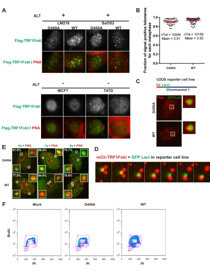
Interestingly, TRF1-FokI expression resulted in up to 4-fold increases in average
telomere foci size and reduced numbers of telomeres in each of 4 different ALT positive
cell lines in comparison to cells expressing the nuclease inactive TRF1-FokI D450A
mutant (Figures 1C, 1D and 2B). Telomere foci size increases did not occur in telomerase
negative primary human IMR90 fibroblasts or 4 different telomerase positive cell lines.
Telomere length difference between ALT and telomerase positive cells was not sufficient
to explain foci size increases, as TRF1-FokI expression did not significantly increase
telomere foci size in the telomerase positive HeLa 1.3 cells (Figure 1D), which have a
13
Telomeres within these larger foci in ALT cells contain chromosomally attached
telomeres. This is supported by the observation that metaphase chromosome spreads from
D450A and WT TRF1-FokI were not appreciably different with respect to the percentage
of chromosome ends displaying telomeric signal, and by the presence of subtelomeric
fluorescence in situ hybridization (FISH) signals or subtelomeric lac operator transgene
repeats juxtaposing telomeres in interphase U2OS cells (Shanbhag et al., 2010) (Figures
2C-E). Furthermore, expression of TRF1-FokI increased the percentage of multiple
subtelomeric FISH signals accumulating at a telomere cluster (Figure 1E). These data are
in agreement with previous reports that APB bodies contain chromosomally attached
telomeres (Draskovic et al., 2009). However, they do not exclude the possibility that
extra-chromosomal telomeric repeats (ECTRs), which increase in response to DNA
damage, are also present in these large telomere bodies (Cesare and Griffith, 2004;
Fasching et al., 2007).
These findings suggest that DSB responses at ALT telomeric chromatin provide
the initiating stimulus for telomere clustering. Consistent with this expectation,
TRF1-FokI expression induced multiple hallmarks of ALT recombination, including significant
increases in telomeres associated with promyelocytic leukemia bodies (APBs) and
telomere associated DNA synthesis as evidenced by incorporation of thymidine analog
5-ethynyl-2’-deoxyuridine (edU) in non S-phase cells (Figures 3A-D and Figure 4A).
Similar findings were not detectable in telomerase positive cells. Expression of
TRF1-FokI also increased c-circle formation, a specific indicator of ALT activity (Figures
14
heterogeneity by Terminal Restriction Fragment Analysis in 3 different ALT cell lines
(Figure 3E). The increased heterogeneity could result from a combination of factors,
including telomere cutting by TRF1-FokI, as well as ALT recombination associated
length changes and ECTR generation. These telomeres were sensitive to digestion by
Bal-31, an exonuclease that degrades duplex DNA from both 3’ and 5’ ends, indicating
that the longer telomere fragments observable following TRF1-FokI WT expression were
not a consequence of telomere-telomere end joining (Figure 3F).
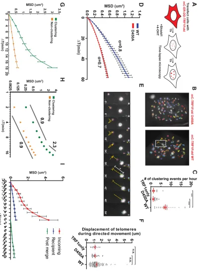
III. Double-Strand Breaks Initiate Directional ALT Telomere Movement and
Clustering
The presence of intense ALT-like telomere clusters suggests that DSB responses
initiate a homology search process, followed by synapsis and recombination between
distant telomeres. To directly test this hypothesis, we visualized telomere movement
using an inducible mCherryTRF1-FokI fused to a modified estradiol receptor and
destabilization domain, which allowed small molecule induction by administration of
4-hydroxytamoxifen and Shield1 ligand (Figure 5A). Following a 1-hour induction period,
TRF1-FokI expressing cells were monitored over the following hour by capturing
confocal z stacks of the entire nucleus every 2 minutes. Telomere foci were tracked in the
z-projected plane, and a registration process (Thevenaz et al., 1998) assisted
15
Strikingly, telomeres in TRF1-FokI WT expressing cells demonstrated increased
mobility and an average of 7 telomere-telomere clustering events per hour between foci
separated by up to 5µm (Figures 5B, C, E, F and Movie 1). Telomere clustering and
movement were greatly diminished in the nuclease inactive D450A mutant or in cells
expressing mCherry-TRF1 (Figures 5B-D and Movies 2 and 3). Importantly, less
frequent instances of clustering were observed at mCherry-D450A and TRF1 containing
telomeres, consistent with a previous report of an association of two telomeres in an
unperturbed U2OS cell (Molenaar et al., 2003). TRF1-FokI induced DSBs at ALT
telomeres greatly increase the frequency of telomere associations that normally occur in
these cells.
To determine if DSBs at other regions of the genome in ALT cells would
demonstrate similar movement and clustering as those observed at telomeres, we
monitored targeted and random DSB positions at non-telomeric locations. Fusion of FokI
to the Lac repressor (mCherryLacIFokI) enables efficient visualization of DSB responses
at lac operator repeat sequences integrated into chromosome 1p36 (Shanbhag et al.,
2010). mCherryLacIFokI DSBs did not display large increases in mobility at this locus in
U2OS cells (Figures 6A and B). GFP-53BP1 movement and clustering was also minimal
at most ionizing radiation induced foci during time lapse imaging in U2OS cells (Figure
6C). Conversely, telomeric DSBs moved coordinately with the sub-telomeric LacO locus
in cells expressing both GFP-LacI and mCherryTRF1-FokI (Figure 6D). The lack of
16
reports that ALT cells display elevated recombination at telomeres, but not elsewhere in
the genome (Bechter et al., 2003; Dunham et al., 2000).
Importantly, the robust increase in ALT telomere movement allowed a
quantitative analysis of this type of chromatin movement. Telomere tracks were subjected
to a mean squared displacement (MSD) analysis, which plots the average squared
displacements at each time interval, given by equation MSD = <(x(t+Δt)-x(t))2>, where x
is the position of the focus and t is time. The MSD trajectories were then fitted to a single
exponential time dependence diffusion model described by 𝑀𝑆𝐷 = Γ𝑡! where Γ is a
generalized coefficient, and α is a time dependence coefficient which can be used to
determine the type of motion. For α ~ 1, the particle is undergoing normal diffusion, and
α < 1 represents sub-diffusion, also known as anomalous diffusion. Subdiffusive target
searches in cells can result from molecular crowding of the nucleus and cytoplasm
(Guigas and Weiss, 2008). Finally, α ≥ 2 represents an exponential dependence on time
that indicates that the particle is moving in a directed manner, an example of which is
active cellular transport.
A comparison of averaged MSD trajectories for all telomeres in TRF1-FokI WT
or D450A expressing U2OS cells revealed that α = 0.8 for WT and α = 0.7 for D450A,
both characteristic of subdiffusive motion (Figure 5D). The Γ coefficient, which describes
the magnitude of the behavior characterized by α, was greater for WT than for D450A,
with values of 4.7x10⁻²µm2s-α and 3.3x10⁻²µm2s-α respectively. Calculation of
time-dependent diffusion coefficient 𝐷 𝑡 = 𝑀𝑆𝐷/𝑡 =Γ𝑡!!! showed that the diffusion
17
scale, consistent with subdiffusive motion (Saxton, 2007). For D450A, D(t) at 15 minutes
was 1.4x10⁻²µm2min-1, consistent with values for normal U2OS telomeres (Molenaar et
al., 2003), and decreased to 0.9x10⁻²µm2min-1 at the end of the observation period (Figure
6A). For WT, however, D(t) was consistently elevated at 2.6x10⁻²µm2min-1 and 1.9x10
-2µm2min-1 at 15 minutes and 60 minutes respectively (Figure 6E). These results indicate
that damaged telomeres move faster and roam a larger nuclear territory.
While all telomeres considered in sum demonstrated diffusive movement, it was
readily apparent from imaging experiments that faster, “incoming” telomeres displayed a
striking long-range directional movement prior to association with a comparatively
slow-moving “recipient” telomere (Figure 5B, E and Movie 1). For a quantitative analysis of
this observation, mobility data from telomeres that merged into a recipient telomere was
isolated. The terminal behavior of such telomeres was characterized by MSD analysis of
the last 10 timepoints of each track, with the ultimate timepoint representing the merge
event. The shape of the resulting MSD trajectory suggested an initial, increased diffusive
movement for Δt of up to 10 minutes, followed by a transition to directed movement at
large Δt (Figure 5G) This change in behavior was clearly visualized on a log-log plot.
The α coefficient for the initial portion of the clustering telomere trajectory was 0.9
suggestive of diffusive motion, but between Δt of 12-18 minutes, there was a clear
transition of α to ~ 2.3, indicative of directed movement (Figure 5H).
The average displacement of telomeres during this directed phase was ~1.3µm
with up to 4-5 µm observed for some tracks (Figure 5E, F). Following the clustering
18
the searching process that underlies directional movement had concluded. Interestingly,
the less mobile, “recipient” telomere was associated with PML in 85% of clustering
events (Figure 6F). This supports a model in which PML promotes clustering and
recombination of telomeres within APBs (Chung et al., 2011; Draskovic et al., 2009).
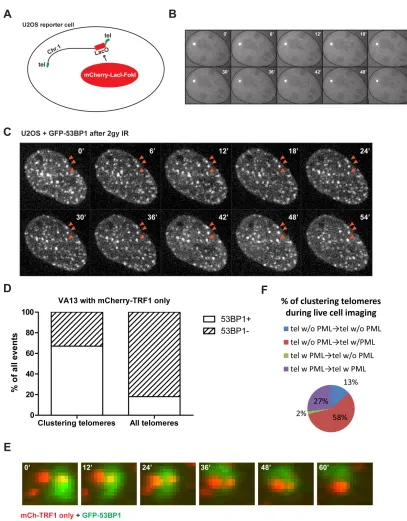
To further address whether the driving force behind ALT telomere movement is a
DSB response, spontaneous telomere clustering events in mcherryTRF1 expressing
VA13 cells were quantified with respect to colocalization of GFP-53BP1 as a marker of
DSBs. Greater than 60% of clustering telomeres accumulated GFP-53BP1 prior to
association, while 15% of all telomeres were associated with GFP-53BP1. This indicates
that telomere movement and clustering is closely correlated with a local DNA damage
response (Figure 6G, H).
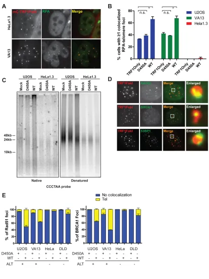
IV. Homologous Recombination Predominates at ALT Telomere DSBs
The presence of random surveillance followed by directional DSB induced
telomere movement could be a consequence of a homology search and capture between
distant telomeres. Resection of telomeric ends would be a critical determinant of this
pathway choice. RPA localization was assessed at telomeres in cells expressing
TRF1-FokI in ALT and telomerase positive cells. HeLa 1.3 did not significantly accumulate
RPA at telomeres. Conversely, telomeres in both U2OS and VA13 cells were associated
with RPA at baseline, which further increased in the presence of TRF1-FokI (Figures 7A,
19
single-stranded telomeres as assessed by electrophoresis and hybridization of telomeric
probes under native conditions (Figure 7C). The increased ssDNA was largely derived
from telomeric overhangs, since the native single stranded telomeric signal was reduced
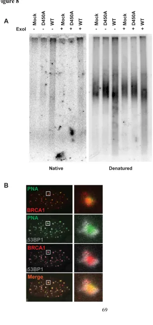
following treatment with ExoI ssDNA exonuclease (Figure 8A).
Consistent with the observed increases in resection, homologous recombination
proteins BRCA1 and Rad51 were present directly overlying 20-60% of telomeres in ALT
cells and only 5-15% of telomeres in telomerase positive cells after TRF1-FokI DSB
induction (Figures 7D and E). 53BP1 immunofluoresence juxtaposed BRCA1 in both
ALT and telomerase positive cells, consistent with known differences of BRCA1 and
53BP1 chromatin localization adjacent to DSBs (Figures 7D and 8B) (Chapman et al.,
2012; Tang et al., 2013).
V. Rad51 and the HR Machinery Control Directional ALT Telomere Movement and
Clustering
These observations raise the possibility of homology directed telomere
movement, analogous to the reported Rad51 dependency for DSB movement that occurs
during homology searches in yeast (Dion et al., 2012; Kalocsay et al., 2009; Mine-Hattab
and Rothstein, 2012; Oza et al., 2009). To test this hypothesis, TRF1-FokI induced
telomere clustering was quantified following siRNA-targeted depletion of factors
involved in either HR or NHEJ (Figure 9A and 10A). Knockdown of NBS1 and SMC5
20
Yu, 2007; Wu et al., 2003; Zhong et al., 2007). Similar reductions were observed in cells
following knockdown of either BRCA2 or Rad51, but not 53BP1 (Figure 9A) (Jiang et
al., 2007). Interestingly, ALT telomere clustering was independent of BRCA1, consistent
with the HR competency of cells that exhibit extensive resection as a consequence of
53BP1 deficiency (Bouwman et al., 2010; Bunting et al., 2010).
Rad51 molecules nucleate onto RPA-coated ssDNA forming a dynamic
nucleoprotein filament which mediates the presynaptic search for homology (Renkawitz
et al., 2014). Remarkably, expression of GFP-tagged Rad51 in VA13 cells allowed
visualization of GFP-Rad51 filaments that originate specifically at telomeres and
extended to distant telomeres (Figure 9B). Live cell imaging revealed that clustering
could proceed by rapid shortening of the GFP-Rad51 filament with synchronous
directional movement of the incoming telomere (Figures 9C and Movie 4). Of 35
clustering events in cells in which a bridging filament formation was evident, 86%
showed Rad51 localization. Rad51 filament could be directly visualized between
recombining telomeres in approximately 46% of cases in which Rad51 was observable at
telomeres (Figure 10B).
MSD analysis revealed that Rad51 knockdown restricted telomere mobility as
well as telomere clustering events that occur as a result of directed movement (Figures
9D, E and Movies 5 and 6). Interestingly, telomere clustering was decreased by
expression of an ATPase defective dominant negative mutant of Rad51, K133R, which
21
extended conformation that cannot transition to a compressed filament (Figure 10C)
(Robertson et al., 2009; Stark et al., 2002).
V. Hop2-Mnd1 Regulate ALT Telomere Movement and Recombination
TRF1-FokI DSB-induced telomere recombination resembles certain aspects of
recombination between homologous chromosomes during meiosis, which is also initiated
by programmed DSBs and requires RecA homologs Rad51 and Dmc1. The Hop2-Mnd1
heterodimer is necessary for Dmc1 and Rad51 dependent inter-homolog recombination in
vivo during gametogenesis in yeast and in mice (Leu et al., 1998; Petukhova et al., 2003),
and strongly stimulates Rad51 or Dmc1 dependent D-loop formation in vitro (Bugreev et
al., 2014; Chi et al., 2007; Petukhova et al., 2005; Pezza et al., 2007). Moreover,
Hop2-Mnd1 or Dmc1 mutant yeast and mice display epistasis with respect to meiotic
chromosome inter-homolog synapsis. Hop2-Mnd1 binds double stranded DNA and
induces rapid condensation of large stretches of DNA in vitro, consistent with its
requirement for homolog synapsis (Pezza et al., 2010).
Hop2 protein was broadly expressed in all 16 different ALT cell lines and in
telomerase positive cancer cell lines tested, with lower levels detected in primary human
fibroblasts (Figures 11A and 12A-C). Endogenous Hop2 localized to approximately
10-20% of TRF1-FokI damaged telomeres in VA13 cells and at lower levels in the absence
of TRF1-FokI (Figure 12E, F). GFP-Hop2 foci localized adjacent to telomeres in a subset
22
within the Hop2 Leucine Zipper domain (Figures 12G, H). This domain is required for
homolog pairing and recombination, with the Leucine Zipper also being necessary for
Hop2 dependent D-loop formation in vitro (Pezza et al., 2006). Hop2 or Mnd1
knockdown strongly reduced telomere clustering, mobility and directional movement to
levels observed in D450A control cells (Figures 11B-D, 12D and Movie 7). Hop2-Mnd1
depletion did not affect Rad51 localization to damaged telomeres (Figure 12I), in
agreement with established roles for the heterodimer in meiotic inter-homolog pairing but
not Rad51 or Dmc1 recruitment to Spo11 dependent DSBs (Petukhova et al., 2003).
To determine if these results would be recapitulated with respect to telomere
clustering and recombination in ALT cell lines that did not express TRF1-FokI, several
different ALT lines were quantified for spontaneous APB formation following
knockdown of Hop2 or Mnd1 with 5 different targeting siRNAs (Figures 11E, F and
14A, B). Knockdown of either Hop2 or Mnd1 significantly reduced APB formation in
each of these lines. The reduction in APBs could be fully rescued by stable expression of
full length Hop2, which is resistant to a siRNA targeted to the 3’UTR (Figure 11G). To
assess the impact of Hop2-Mnd1 on ALT telomere recombination, telomere chromatid
exchanges were assessed by chromosome orientation-FISH (CO-FISH). Knockdown of
Hop2 or Mnd1 reduced telomere chromatid exchanges by 50% or greater in ALT cells
(Figures 11H, I and 14C). Collectively, these data reveal that the forces driving
directional telomere movement are intimately connected to the mechanism of ALT
23
VII. Discussion
The phenomenon of DSB movement has been described in prokaryotes, yeast and
also in mammalian cells within distinct experimental contexts (Aten et al., 2004;
Dimitrova et al., 2008; Dion et al., 2012; Kalocsay et al., 2009; Lesterlin et al., 2013;
Mine-Hattab and Rothstein, 2012; Oza et al., 2009; Roukos et al., 2013). Telomeres
appear to be a particularly predisposed genomic location to DNA damage induced
mobility increases. Diffusive movement of damaged telomeres in telomerase positive
cells has been reported in several independent studies (Chen et al., 2013; Dimitrova et al.,
2008). Notably, the NHEJ promoting factor 53BP1 was required for movement of
deprotected mouse telomeres. However, TRF1-FokI induced directional ALT telomere
mobility required HR factors and was independent of 53BP1, indicative of distinct
mechanisms underlying telomere mobility in each case. Extensive end resection and more
prominent accumulation of HR factors at damaged ALT telomeric chromatin likely
contribute to these differences (Figure 13).
Expression of TRF1-FokI enabled quantitative characterization of an
unanticipated type of chromatin movement. Damaged ALT telomeres initially roamed a
larger nuclear territory at greater velocities than D450A controls, but notably, these
movements culminated in rapid and directional movements of up to 5µm to synapse with
a more stationary recipient telomere. These displacements were also much larger in
magnitude and occurred over a longer time period than those of stochastic unidirectional
“jumps” that could be seen in interphase chromatin (Levi et al., 2005). We note, however,
24
highly mobile particles. The analysis of directionality was limited to clustering telomeres,
and this analysis does not preclude the possibility that a proportion of non-clustering
telomeres could move directionally.
To our knowledge, directional ALT telomere movement provides the first
example of real time visualization of homology searches and synapsis in mammalian
cells. Given our data, we favor a model in which Rad51 nucleoprotein filaments
interrogate surrounding nuclear space, leading to homology capture of a non-sister
telomere and subsequent directional movement during synapsis (Figure 13). Interestingly,
dynamic formation of long stretches of prokaryotic RecA coated filaments mediated
rapid associations between DSBs and homologous genomic regions that are separated by
1.3µm (Lesterlin et al., 2013), which are similar to the distances of directional phase
movement we describe for ALT telomeres. The reported structure of ssDNA filaments in
association with RecA reveals an extended conformation that is stretched to ~1.5 fold
longer B-form DNA (Chen et al., 2008). Thus, it is predicted that 1.3µm of nuclear space
connecting non-sister telomeres could theoretically require only ~2.5kb of Rad51 ssDNA
filament for directional movement, which is well within the length possible for ALT
telomeres. Furthermore, as vertebrate telomeres contain extensive regions of homology
consisting of TTAGGG repeats, in effect every chromosome is a “homolog” with respect
to telomere recombination. This feature of primary telomere sequence would be predicted
to increase the probability of recombination between different chromosomes, enabling
successful capturing of distant homology on timescales similar to those observed in much
25
Rad51 foci, consistent with the presence of Rad51 independent mechanisms of ALT in
type II survivors of telomerase deficiency in yeast (Chen et al., 2001).
Several obvious parallels exist between meiotic recombination and ALT. Both
processes involve DSB responses to initiate recombination between homologous DNA
sequences on non-sister chromatids. Hop2-Mnd1 uniquely contributes to chromosome
pairing in meiotic recombination and ALT, but is not known to be important for sister
chromatid recombination. Both constituents of this heterodimer are broadly expressed in
ALT and telomerase positive cancers, yet appear to promote telomere recombination only
in cells that use ALT. This may be a consequence of the known interaction of
Hop2-Mnd1 with Rad51, which did not efficiently nucleate damaged telomeres in telomerase
positive cells. It is also plausible that other factors related to the specific chromatin
environment in ALT cells, such as the absence of ATRX and the association between
ALT and defective histone chaperone activity (Heaphy et al., 2011; Lovejoy et al., 2012;
O'Sullivan et al., 2014; Schwartzentruber et al., 2012), may promote inter-telomere
recombination. Mechanistic studies into this process are warranted, as is the extent to
which ALT recapitulates known mechanisms of meiotic recombination.
VIII. Experimental Procedures
Cell culture
Saos2 cells were grown in McCoy’s 5A medium (Invitrogen) with 15% fetal
26
calf serum and 1% penn/strep. All other cell lines were grown in DMEM (Invitrogen)
with 10% calf serum and 1% pen/strep. VA13 cell line refers to WI-38 VA-13 subline
2RA.
Plasmids, primers and siRNAs
Flag-TRF1-FokI fusion protein was cloned as previously described (Tang et al.,
2013) into the HFUW lentiviral vector. The coding sequence for mCherry, the modified
estrogen receptor (ER) and destabilization domains (DD) were PCR amplified and cloned
in frame into the N terminus of TRF1-FokI. Rad51 cDNA was cloned into pEGFP-C1.
The N terminal GFP tagged Hop2 expression vector was generated by PCR amplification
and ligation of Hop2 cDNA corresponding to isoform 2 (RefSeq NM_016556.3) from
ProQuest HeLa cDNA Library (Invitrogen). Hop2 M110P mutant was generated by
site-directed mutagenesis.
The following primers were used for qRT-PCR: GAPDH (IDT, PrimeTime assay
Hs.PT.39a.22214836), Hop2/PSMC3IP (IDT, PrimeTime assay Hs.PT.56a.40246325.g),
Mnd1 (IDT, PrimeTime assay Hs.PT.56a.1133039).
The following siRNA sequences were used:
Luciferase, 5’-GCCAUUCUAUCCUCUAGAGGAUG-3’
Control, (Qiagen Allstars)
27 Rad51 #2, 5’-CCAGAUCUGUCAUACGCUA-3’
Rad51 #3, 5’-UGGAGGGCUGACAGCUUCC-3’
NBS1 (Santa Cruz)
SMC5, 5’-GAAGCAAGAUGUUAUAGAA-3’
53BP1, 5’-UAUUACCGUCUCCUCGUUC-3’
CTIP, 5’-ACACACUCAUGGUGAUAAA-3’
BRCA1, 5’-AGAUAGUUCUACCAGUAAA-3’
BRCA2, 5’-GAAGAAUGCAGGUUUAAU-3’
Hop2 #1, 5’-GCAGCUACCAAUCAUGUGA-3’
Hop2 #2, 5’-AAGAGAAGAUGUACGGCAA-3’
Hop2 #3, 5’-UCUGCUUAAAGGUGAAAGUAGCAGG-3’
Hop2 #4, 5’-UAAAUGUUAACCUCAAGCUACUGCA-3’
Mnd1 #1, 5’-GCUAACAGAUGGACUGAUA-3’
All siRNAs were transfected at 20nM, except for double transfections where each siRNA
concentration was at 10nM, using Lipofectamine RNAiMAX (Invitrogen).
For Hop2 Mnd1 knockdown for APB quantification, knockdown was performed in series
on day 1 and day 4 and cells were fixed on day 7.
Antibodies
The following antibodies were used: anti-53BP1 (rabbit, Novus), anti-BRCA1 D9
28
Bethyl), anti-phosphoATM Ser-1981 (mouse, ECM Biosciences), anti-phosphoChk2
Thr-68 (Rabbit, Cell Signaling), anti-Flag M2 (mouse, Sigma), anti-PML (mouse, Santa
Cruz), anti-Rad51 H-92 (mouse, Santa Cruz), anti-Hop2/PSMC3IP (rabbit, ProteinTech
11339-1-AP; rabbit, Novus NBP1-92301), anti-RPA2 (9H8, mouse, Novus).
Transfections and lentiviral transductions
Transient plasmid transfections were carried out with LipoD293 (Signagen), and siRNA
transfections with Lipofectamine RNAiMax (Invitrogen) according to manufacturer’s
instructions. Concentrated TRF1-FokI lentivirus with polybrene (8ug/ml) diluted in
media was added to cells at a minimum titer resulting in greater than 90% expression at
24 hours by immunofluorescence. Analyses were performed 16 hours after transfection of
plasmids, and 48-72 hours after siRNA transfection. Analyses were performed 24 hours
after transduction of cells with Flag-TRF1-FokI lentivirus.
Western blot
Cells were collected and lysed in RIPA buffer or for histone fractions, acid extracted.
Proteins were resolved on a 4-12% bis-tris gel (Invitrogen). Transferred membranes were
blocked in 5% milk and incubated with primary antibody overnight. For western blot
following TRF1-FokI expression, cells were collected 24 hours after addition of
29
Immunofluorescence, IF-PNA FISH and subtelomeric FISH
For anti-Hop2 immunofluorescence, permeabilized coverslips were blocked with 10%
goat serum for 30 minutes at 37°C, followed by incubation with primary antibody for 16h
at 37°C. Coverslips were washed and incubated with appropriate secondary antibody for
20 minutes at 37°C, then mounted onto glass slides using Vectashield mounting medium
with DAPI (Vector Labs).
edU fluorescence assay
16 hours after cells were transfected with mCherry-ER-DD-TRF1FokI, media was
changed to contain 10µM (final) edU. Cells were pre-incubated for 2 hours then Shield1
and 4-OHT were added to the media to induce TRF1FokI for 2 hours. Cells were fixed
and edU was labeled with Alexa488 using Click-iT chemistry (Invitrogen).
Pulsed field gel electrophoresis and in-gel hybridization
Genomic DNA was purified using the Masterpure DNA Purification Kit (Epicentre) per
manufacturer’s instructions. 15ug of purified DNA was digested using AluI and MboI,
and resolved on a 1% PFGE agarose gel (Biorad) in 0.5X TBE buffer using the
30
Following electrophoresis, the gel was dried at 50ºC for 3h and processed for native or
denaturing probe hybridization. For native hybridization, the dried gel was incubated
with p32 end labeled (TTAGGG)6 oligo probe at 42º for 16h in Church buffer, washed 4
times in 4X SSC and exposed onto a storage phosphor screen (GE Healthcare) and
scanned using STORM 860 with ImageQuant (Molecular Dynamics). For denaturing
hybridization, the gel was denatured in 0.5N NaOH/1.5M NaCl, neutralized and
incubated with p32 end labeled (TTAGGG)6 oligo probe as above.
Nuclease digestion of genomic DNA
For Bal31 digestion, purified genomic DNA (5ug) was incubated with Bal31 (2
units per reaction, TaKaRa/Clontech) in buffer provided by manufacturer for various
timepoints at 30ºC, followed by inactivation in 20mM EDTA at 65ºC for 10 minutes. The
Bal31 digested DNA was then purified by phenol-chloroform extraction and resolved by
PFGE as above. For Exo1, purified genomic DNA (10ug) was incubated with Exo1 for
16h at 37ºC, purified with phenol-chloroform extraction and resolved by PFGE.
C-circle Assay
Purified genomic DNA was digested using AluI and MboI (New England
Biolabs). 15ng or 30ng of DNA was diluted to 10ul, then combined with 10ul of 0.2
31
7.5U φ29 DNA polymerase (NEB). Samples were incubated for 8 hours at 30°C then the
polymerase was inactivated for 20 minutes at 65°C. Samples were diluted with 2x SSC to
60ul, then dot-blotted onto Hybond N+ membrane (GE Healthcare). After UV
crosslinking, the membrane was hybridized with p32 labeled (CCCTAA)6 oligo at 37°C
in Church buffer for 16h. Membrane was exposed onto a storage phosphor screen (GE
Healthcare) and scanned using STORM 860 with ImageQuant (Molecular Dynamics).
Chromosome orientation FISH (CO-FISH)
Telomeric CO-FISH can be used to assess the level of postreplicative exchanges
involving a telomere and another TTAGGG repeat sequence, and has been described in
detail previously(Bailey et al., 2004; Londono-Vallejo et al., 2004). Briefly, cells were
incubated with BrdU/BrdC (7.5µM, 2.5µM respectively) for 21h (less than one cell
cycle). Mitotic cells were collected after treatment with 0.1ug/ml colcemid for 90
minutes. Subsequently, cells were incubated in 75 mM KCl and fixed on ice with fresh
fixative (3:1 methanol / acetic acid). Fixed cells were dropped onto slides at 42ºC, and
allowed to dry o/n. The slides were treated with 0.5mg/ml RNaseA for 15 minutes at
37ºC, then with 0.5mg/ml Hoechst 33258 for 15 minutes at RT. Slides were exposed to
365nm UV light for 30 minutes to introduce nicks in the BrdU/C incorporated replicated
strands. The nicked strands were digested twice with 10U/ul ExonucleaseIII (Promega) in
buffer supplied by manufacturer at RT for 10 minutes. Ethanol dried slides were stained
32
Live cell imaging
Cells were transfected with mCherry-ER-DD-TRF1-FokI 16 hours prior to
induction with 4-OHT and Shield1 ligand for 60 minutes. Confocal images were acquired
under temperature controlled conditions calibrated to 37ºC, using a 100x 1.4 NA
objective on an inverted fluorescence microscope (DM6000, Leica Microsystems)
equipped with an automated XYZ stage (Ludl Electronic Products), a charge-coupled
device camera (QuantEM 512SC, Photometrics), an X-LIGHT Confocal Imager (Crisel
Electrooptical Systems), and a SPECTRA X Light Engine (Lumencor), controlled by
Metamorph Software (MDS Analytical Technologies). Images were collected as z stacks
at 0.6 µm intervals that covered the entire nucleus, at 2-minute intervals for a total of 60
minutes. Images were processed using ImageJ (NIH). Each z stack was projected onto a
single z plane for each timepoint, and then the t stack was registered using StackReg
plugin in ImageJ to normalize for cell movement. Tracking of individual foci was
performed using the TrackMate plugin for ImageJ (Perry N, Tinevez JY and Schindelin
J). The Mean Square Displacement (MSD) values were calculated using MSD =
<(x(t+Δt)-x(t))2>, where x is the position of the telomere and t is time in minutes.
Resulting data were analyzed and visualized in MATLAB (MathWorks) using the class
@msdanalyzer (written by JY Tinevez, Institute Pasteur). The error bars for each data
point represent the weighted s.e.m. where weights are set to be the number of points
33
The MSD trajectories were fitted to a single exponential diffusion model
described by 𝑀𝑆𝐷 = Γ𝑡! where Γ is a generalized coefficient, and α is a time
dependence coefficient. The fitting was performed using least-squares fitting in
GraphPad Prism.
Calculation of the time dependent diffusion coefficient was performed by
calculating 𝐷 𝑡 = 𝑀𝑆𝐷/𝑡= Γ𝑡!!! (Saxton, 2007). For particles undergoing
anomalous/subdiffusion, D(t) decreases linearly with time with a slope of α-1when
plotted on a log-log scale.
For analysis of clustering telomeres as in Figure 3G, H, telomere tracks were
pre-selected for those that displayed clustering during the course of imaging. Telomeres that
clustered during the final 2/3 of the movie were selected to ensure enough datapoints for
movement preceding the association event. Then, the last 10 time points for each of these
tracks were used for analysis (last 10 points prior to the joining event). For the analysis of
incoming and recipient telomeres vs. post merge as in Figure 3I, clustering events that
occurred during the middle 1/3 of the movie were analyzed.
Analysis of telomere foci size and clustering
For measurements of telomeric foci size, ImageJ (NIH) was used to apply a
constant threshold to images and subsequent binarization. Foci sizes were measured as
square pixels for each telomeric focus within a nucleus and the average size was
34
transfection, a cluster was defined as a telomeric focus equal to or greater than four-fold
the area as based on the radius, compared to the average size of undamaged telomeres.
Statistics
35
CHAPTER 3. Regulation of ALT telomere recombination by the
ATR-Chk1 kinase pathway
I. Introduction
A network of signaling pathways ensure proper coordination of the complex
sequence of molecular reactions that are required for completion of DNA repair.
Double-strand breaks will activate ataxia-telangiectasia mutated (ATM) kinase. If the DSB is
further resected to reveal single-stranded DNA onto which RPA rapidly binds, this
structure recruits and activates ataxia-telangiectasia mutated and Rad3-related (ATR)
kinase (Ball et al., 2005). Importantly, ATR is also activated during normal replication
when ssDNA is exposed and ensures proper progression of the replication fork especially
during periods of replication stress (Flynn and Zou, 2011). Downstream of ATM and
ATR kinase activation, Chk2 and Chk1 kinases are phosphorylated respectively (Falck et
al., 2001; Liu et al., 2000). Subsequent phosphorylation-mediated inhibition of
Cdc25A/B/C phosphatases results in inhibition of cyclin-dependent kinases (CDKs),
leading to cell cycle arrest. This temporary block allows sufficient time for completion of
DNA repair.
The ATR-Chk1 pathway is critical for protection during replication stress both
locally and globally. At the stalled replication fork, ATR stabilizes the structure of the
fork and prevents collapse of the fork (Cobb et al., 2003; Lucca et al., 2004). In the
absence of ATR, collapsed forks could degenerate into DSBs by structure-specific
36
ATR also phosphorylates Chk1, which diffuses globally throughout the nucleus to
suppress new origin firing (Shechter et al., 2004; Sorensen and Syljuasen, 2012).
Inhibition of ATR or Chk1 results in deregulated origin firing and generation of DSBs
globally from collapsed forks, leading to irreversible catastrophe and cell death (Buisson
et al., 2015; Toledo et al., 2013).
It was recently shown that ALT activity is disrupted upon ATR inhibition and that
ALT positive cells are selectively killed by ATR inhibition (Flynn et al., 2015). RPA
coated ssDNA telomeres, characteristic of ALT cells, likely serves as a substrate to
activate ATR. However, it is unclear how ATR would regulate ALT activity. One
possibility is a direct effect of ATR-mediated phosphorylation on proteins mediating
ALT recombination. Alternatively, ATR inhibition may exert its effects through
disruption of replication at telomeres as well as the rest of the genome. In this section, I
show that inhibition of ATR or Chk1 activities disrupt Rad51 and Hop2 localization to
ALT telomeres, and that prevention of excessive replication origin firing and DSB
generation partially alleviates this stress.
II. Inhibition of ATR and Chk1 activity disrupts HR factor localization at ALT
telomeres
To test if ATR or Chk1 activity contributes to ALT telomere recombination by
altering recruitment of HR factors, ALT cells were treated with small molecule inhibitors
37
and LY-2603618, which inhibits Chk1 with an IC50 of 7nM, strongly reduced Hop2
localization to telomeres in VA13 cells (Figure 15A, C). Hop2 localization was reduced
to less than 50% by 4 hours of treatment, and continued to decline until 24 hours of
treatment (Figure 15 B, D). CHIR-124, another potent Chk1 inhibitor with an IC50 of
0.3nM, reduced Hop2 colocalization with telomeres similarly to LY-2603618 in VA13
cells (Figure 15F). Reduction of Hop2 at telomeres by ATRi was also apparent in
GM847, another ALT cell line (Figure 15 E). Knockdown of ATR recapitulated the loss
of Hop2 in VA13 cells, while knockdown of ATM only marginally affected Hop2 and
knockdown of RNF168 significantly increased Hop2 localization to telomeres (Figure
15G).
In order to test if localization of other HR factors is affected by ATR and Chk1
inhibition, RPA and Rad51 were visualized at ALT telomeres by immunofluorescence.
Interestingly, RPA32 foci maintained colocalization with telomeres even after treatment
with VE-821 in VA13 cells (Figure 16A, B). U2OS cells expressing TRF1-FokI also
largely retained RPA32 foci after treatment with VE-821 or LY-2603618 (Figure 16E).
On the other hand, inhibition of ATR and Chk1 significantly reduced Rad51 recruitment
to telomeres similarly to Hop2 (Figure 16C, D, F). These results show that ATR or Chk1
inhibition, but not ATM or RNF168 inhibition, specifically perturb Rad51 and Hop2
localization to ALT telomeres while RPA recruitment is unaffected.