INTRODUCTION
A burn injury represents one of the most severe forms of trauma and occurs in over two million people in the United States of America per year (1). The liver, with its metabolic, inflammatory, im-mune and acute-phase functions, plays a pivotal role in patient outcome and re-covery by modulating multiple path-ways (2–7). It has been suggested that because the liver modulates metabolic pathways, inflammatory processes and acute-phase responses, limiting hepatic damage after traumatic injury may im-prove patient outcomes significantly (7). In a recent study, Price and colleagues (8)
looked at outcomes of 290 burned pa-tients who suffered from liver disease prior to burn injury. They showed that preexisting liver disease increased mor-tality risk from 6% (total population) to 27%. The authors concluded that liver impairment worsens the prognosis in pa-tients with thermal injury (8). This sug-gests that therapeutic agents, which pro-tect against liver dysfunction after traumatic injury, may improve patient morbidity and mortality significantly.
Hepatic dysfunction after burn injury persists over a prolonged time period (3,7,9,10). At the molecular level, we re-cently have shown that burn injury leads
to gross alterations in hepatocyte calcium homeostasis (7,11). Burn injury causes depletion of endoplasmic reticulum (ER) calcium stores and increased cytosolic calcium, which subsequently induces mi-tochondrial damage and cytochrome c release (11). Cytochrome c binds to the ER-resident inositol 1,4,5-trisphosphate receptor (IP3R) calcium channel, aug-menting the depletion of ER calcium stores leading to ER stress and induction of the unfolded protein response (UPR) (7,11). ER stress/UPR due to calcium store depletion leads to activation of the JNK signaling pathway and ultimately hepatocyte apoptosis. Burn injury also induces the hepatic acute phase response and a concomitant decrease in the pro-duction of constitutive serum proteins such as albumin (3,7,9,12).
The introduction of insulin and the concept of tight euglycemic control rep-resent a cornerstone of modern critical care medicine (13,14). Studies in severely burned patients conducted by our group
Marc G Jeschke,
1,2,3,5Robert Kraft,
1,2Juquan Song,
1,2Gerd G Gauglitz,
1,6Robert A Cox,
1Natasha C Brooks,
3Celeste C Finnerty,
1,2Gabriela A Kulp,
1David N Herndon,
1,2and Darren Boehning
41Shriners Hospitals for Children and Departments of 2Surgery, 3Biochemistry and Molecular Biology, 4Neuroscience and Cell Biology, The University of Texas Medical Branch, Galveston, Texas, United States of America; 5Sunnybrook Health Sciences Centre and University of Toronto Department of Surgery, Division of Plastic Surgery, Toronto, Canada; and 6Department of Dermatology, Ludwig-Maximilians University of Munich, Germany
Burn injury causes hepatic dysfunction associated with endoplasmic reticulum (ER) stress and induction of the unfolded protein response (UPR). ER stress/UPR leads to hepatic apoptosis and activation of the Jun-N-terminal kinase (JNK) signaling pathway, leading to vast metabolic alterations. Insulin has been shown to attenuate hepatic damage and to improve liver function. We therefore hypothesized that insulin administration exerts its effects by attenuating postburn hepatic ER stress and subsequent apo-ptosis. Male Sprague Dawley rats received a 60% total body surface area (TBSA) burn injury. Animals were randomized to receive saline (controls) or insulin (2.5 IU/kg q. 24 h) and euthanized at 24 and 48 h postburn. Burn injury induced dramatic changes in liver structure and function, including induction of the ER stress response, mitochondrial dysfunction, hepatocyte apoptosis, and up-regulation of inflammatory mediators. Insulin decreased hepatocyte caspase-3 activation and apoptosis significantly at 24 and 48 h postburn. Furthermore, insulin administration decreased ER stress significantly and reversed structural and functional changes in hepatocyte mitochondria. Finally, insulin attenuated the expression of inflammatory mediators IL-6, MCP-1, and CINC-1. Insulin alleviates burn-induced ER stress, hepatocyte apoptosis, mitochondrial abnormalities, and inflammation leading to improved he-patic structure and function significantly. These results support the use of insulin therapy after traumatic injury to improve patient outcomes.
© 2011 The Feinstein Institute for Medical Research, www.feinsteininstitute.org Online address: http://www.molmed.org
doi: 10.2119/molmed.2010.00166
Address correspondence and reprint requests toMarc Jeschke, Director Ross Tilley Burn Centre, Sunnybrook Health Sciences Centre, Department of Surgery, Division of Plastic Sur-gery, University of Toronto Senior Scientist Sunnybrook Research Institute, Room D7 04, 2075 Bayview Avenue, Toronto, ON CANADA M4N 3M5. Phone: 416-480-6703; Fax: 416-480-6763; E-mail: [email protected].
indicated that insulin improves hyper-metabolism by decreasing proinflamma-tory cytokines and hepatic acute-phase protein production (10,15,16). While in-sulin administration postburn is known to attenuate hepatic damage and im-prove liver function (5,17), the underly-ing mechanisms by which insulin exerts its effects on liver structure and function are not understood. The aim of this study was to determine the molecular mechanisms by which insulin adminis-tration improves hepatic structure and function postburn. We hypothesized that postburn insulin administration would improve hepatic function by decreasing ER stress and downstream mitochondrial dysfunction and apoptosis, leading to a marked reduction in acute phase protein production and activation of associated inflammatory pathways.
MATERIAL AND METHODS
Animals
All animal manipulations were ap-proved by the Institutional Animal Care and Use Committee of the University of Texas Medical Branch at Galveston. The National Academy Press Guide for the Care and Use of Laboratory Animals(1996) were met. Sprague Dawley rats, 325 to 350 g, were used in these studies and were allowed to acclimate for 1 wk be-fore experiments. Rats were housed in an institutional animal care facility and re-ceived a high-protein diet (Ensure, Ab-bott Laboratories, Waukegan, IL, USA) and water ad libitumthroughout the study. Ensure (Abbott) was administered 7 d before the study to adjust the animals to the liquid diet.
Burn Injury
A well-established method was used to induce a full-thickness scald burn in our model (18). Animals were anes-thetized with general anesthesia (keta-mine 40 mg/kg body weight and xylazine 5 mg/kg body weight, both injected intraperitoneally [i.p.]) and re-ceived analgesia (buprenorphine
0.05 mg/kg body weight, injected
subcu-taneously). The dorsum of the trunk and the abdomen were shaved, and a 60% total body surface area (TBSA) burn was administered by placing the animals in a mold exposing defined areas of the skin of the back and abdomen under general anesthesia and analgesia. The mold was placed in 96–98°C water, scalding the back for 10 sec and the abdomen for 2 sec. Lactated Ringer’s solution (40 mL/kg body weight) was adminis-tered i.p. immediately after the burn for resuscitation. After burn and resuscita-tion, animals were observed, received oxygen and were then placed into cages. Unburned (sham) animals received the same treatment except for the scald burn.
Animals were pair-fed with a liquid high-protein high-amino acid nutrition (Ensure, Abbott Laboratories) and water
ad libitum. Ensure has a caloric distribution of 24% protein, 21% fat and 55% carbohy-drate. All rats were pair-fed according to the caloric intake. The feeding protocol was as follows: 25 calories on the day of burn or sham, 51 calories on the first day postburn, 76 calories on the second, and 101 calories from the third day postburn on, to the end of the study. Nutritional in-take was the same in all groups.
Treatment Groups
Animals received a 60% total body surface area (TBSA) burn and animals were injected once daily with either 2.5 IU/kg body weight of insulin glargine (Insulin Lantus, Sanofi Aventis, Kansas City, MO, USA; insulin group), or an equal amount of saline subcutaneous (control group, n = 10). Animals were killed by decapitation 24 and 48 h after burn injury and blood and liver was col-lected immediately thereafter (n = 10 for each group and time point). No animals were excluded from the study, and there were no significant differences in mortal-ity rate between the groups.
Serum Markers
Serum was separated by centrifugation at 3,000gfor 3 min at 4°C and stored at –80°C until analyzed. The levels of IL-6, cytokine-induced neutrophil
chemoat-tractant (CINC)-1 and monocyte chemo-tactic protein (MCP)-1 as markers of sys-temic inflammation were determined by enzyme-linked immunosorbent assay (ELISA) (R&D Systems Inc., Minneapolis, MN, USA) according to the protocol of the manufacturer. Absolute cytokine con-centrations were determined by compari-son to a standard curve.
SDS-Polyacrylamide Gel
Electrophoresis and Immunoblotting Liver tissue was homogenized and sol-ubilized in 150 mmol/L NaCl, 50 mmol/L Tris-HCl, pH 7.8, 1% (w/v) Triton X-100, 1 mmol/L EDTA, 0.5 mmol/L phenyl -methane sulfonyl fluoride and 1 ×protease inhibitor cocktail (Roche Molecular Bio-chemicals, Basel, Switzerland) for 5 min on ice and spun at 12,000g, and 20 µg of the resultant supernatant was run on a 4% to 20% gradient SDS-polyacrylamide gel and subsequently electrotransferred to nitrocellulose membranes (Bio-Rad Laboratories, Hercules, CA, USA). Nitro-cellulose sheets then were probed with primary antibody against phosphorylated inositol requiring enzyme-1 (IRE1), c-Jun N-terminal Kinase (JNK), Thr183/185
- phosphorylated-JNK, Thr980 phosphorylated PKR-like endoplasmic reticulum kinase (PERK), ATF6, Grp78/ Bip, and actin. Blots were developed with chemiluminescent substrates (Pierce Biotechnology, Rockford, IL, USA) and were quantified by densitometry (GENE GENIUS, Bio Imaging System, Syngene, Frederick, MD, USA). After quantifica-tion, the intensities of the phosphory-lated protein bands were divided by the total form of the respective protein or by the intensity of the respective actin band. In those cases where a single blot was probed sequentially with more than one antibody, the nitrocellulose was stripped at 60°C for 30 min in stripping buffer (2% SDS, 100 mmol/L β-mercaptoethanol, 62.5 mmol/L Tris-HCl) before probing with the next antibody.
Liver Apoptosis
Balti-more, MD, USA) staining to identify apoptotic hepatocytes in situwas per-formed as suggested by the manufac-turer. Six sections of each liver block were obtained at 40- to 50-µm intervals. Within each section, a blinded observer selected five fields for counting TUNEL-positive cells. Three blinded observers counted TUNEL-positive cells and the data pooled. The data were quantified as the percentage of apoptotic cells per hun-dred hepatocytes.
Mitochondrial Isolation and Respiration
Liver tissue (400 mg) was minced on ice and transferred (10% w/v) to isolation buffer (250 mmol/L sucrose, 10 mmol/L HEPES, 0.5 mmol/L EGTA, 0.1% bovine serum albumin [BSA], pH 7.4). The sam-ple was homogenized gently by 3 to 4 strokes with a Dounce homogenizer with a loose fitting pestle. The homogenate
was centrifuged at 500gfor 5 min at 4°C. The supernatant fraction was retained, whereas the pellet was washed with iso-lation buffer and centrifuged again (500g
for 5 min at 4°C). The combined super-natant fractions were centrifuged at 7800g
for 10 min at 4°C to obtain a crude mito-chondria pellet. The mitomito-chondria pellet was resuspended in isolation buffer with-out EGTA and BSA and centrifuged again at 7800gfor 10 min. Oxygen consumption of isolated mitochondria was measured at 25°C by using a model 782 oxygen meter system and model 1302 Microcath-ode oxygen electrMicrocath-ode (Strathkelvin, Glas-gow, UK).
Statistical Analysis
All data is presented as the mean ± the standard error of the mean. Statistical significance between two groups was ex-amined with a Student ttest. A Pvalue < 0.05 was determined to be significant. All data were analyzed by using Graph-Pad Prism software (GraphGraph-Pad Software, Inc, San Diego, CA, USA).
RESULTS
We first determined whether insulin at a dose of 2.5 units/kg body weight alter serum glucose levels postburn. We found that insulin significantly decreased glu-cose levels at 24 h and 48 h postburn
when compared with burn, P< 0.05 (Table 1).
We next examined whether insulin ad-ministration would improve hepatic apo-ptosis postburn. As shown in Figure 1A, we found that burn induces a 5- to 7-fold increase in hepatic caspase-3 activation. Insulin significantly attenuated caspase-3 activation compared with burn (Fig-ure 1A). Consistent with burn-induced activation of hepatocyte apoptosis, we found a 5- to 6-fold increase in the num-ber of TUNEL-positive hepatocytes after burn injury, which was inhibited signifi-cantly by insulin administration (Fig-ure 1B). Burn-induced hepatic apoptosis was associated with activation of the acute phase response as indicated by a drop in production of the constitutive protein albumin (Figure 1C). Impor-tantly, insulin administration increased serum albumin levels significantly post-burn (see Figure 1C).
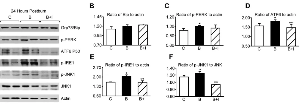
We next asked whether insulin pro-tects against burn-induced induction of the hepatic ER stress response. Confirm-ing previous studies (11,19), we found that burn injury is a potent inducer of the hepatic ER stress response 24 h after injury (Figure 2A). Burn increased levels of phospho-PERK significantly, cleaved ATF-6, phospho-IRE-1 and phospho-JNK (Figure 2C–F). These effects were signifi-Table 1.Blood glucose levels postburn
in mg/dL.a
Normal Burn Burn + insulin
24 h 95 ± 5 145 ± 11 105 ± 6b
48 h 98 ± 6 138 ± 12 103 ± 7b
aData presented as mean ± SD. bSignificant difference between burn
versus burn + insulin, P < 0.05.
cantly inhibited in all cases by insulin administration. At 48 h postburn, similar changes persisted, and we also found significant upregulation of the resident chaperone Grp78/Bip, which is a transcriptional target of ATF6 (Figure 3A–F).
Burn has been shown to cause pro-found calcium alterations in the ER by depleting ER stores and increasing cy-tosolic calcium (11). Increased cycy-tosolic calcium has been shown to induce mito-chondrial swelling, decrease respiratory
capacity and release of mitochondrial apoptotic factors such as cytochrome c (20–23). Previously we have shown that burn injury causes mitochondrial swelling and decreased state-3 respira-tion (11). Consistent with our previous studies, by using transmission electron microscopy we found marked alterations in mitochondrial morphology after burn injury (Figure 4A–C). Mitochondrial swelling, loss of cristae (double asterisks; Figure 4B) and loss of matrix electron density (single asterisk, Figure 4B) were
prominent in hepatocytes from burned animals. Furthermore, burn injury was associated with fragmentation of the tu-bular ER membrane structure (arrows; Figure 4B). Importantly, insulin adminis-tration reversed these ultrastructural changes in the mitochondria (Figure 4C). To examine mitochondrial function, we measured state-3 respiration in isolated hepatic mitochondria. We found that burn injury caused a marked impairment in hepatic mitochondria state-3 respira-tion, and this could be reversed signifi-Figure 2.Insulin decreases hepatic ER stress 24 h after burn injury. (A) Representative Western blots from control (C), burn (B), and burn + insulin (B + I) liver lysates 24 h after burn injury. Two separate animals from each group are shown. (B) Densitometric ratio of Grp78/Bip to actin. (C) Ratio of phospho (active) PERK to actin. (D) Ratio of cleaved (active) ATF6 to actin. (E) Ratio of phospho (active) IRE1 to actin. (F) Ratio of phospho (active) JNK to total JNK. *P < 0.05 relative to control. **P < 0.05 relative to burn.
cantly by insulin administration (Fig-ure 4D).
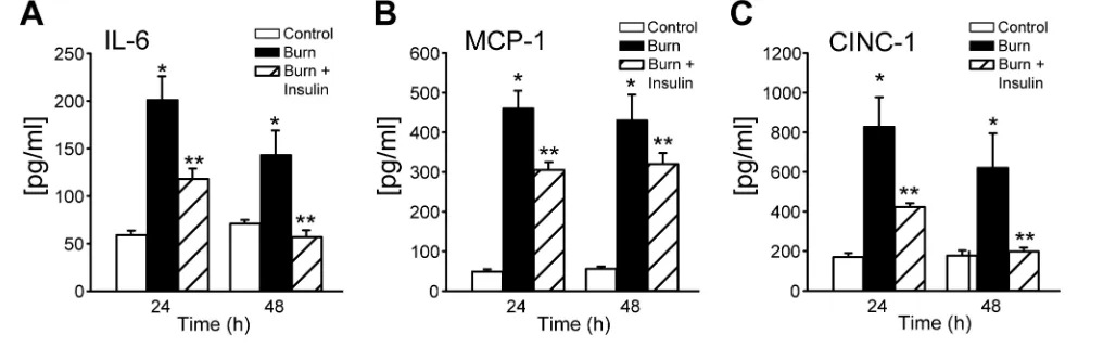
Burn injury induces the acute phase response and subsequent production of inflammatory mediators such as IL-6, CINC-1 and MCP-1. We found that burn increases the production of these cy-tokines 5- to 20-fold compared with control 24 and 48 h after burn injury (Figure 5). Importantly, insulin adminis-tration reduces IL-6, MCP-1, and CINC-1 significantly to almost normal levels, dicating that insulin suppresses the in-flammatory response potently postburn.
DISCUSSION
The liver with a wide range of central physiologic, metabolic and immune
functions is central for survival in criti-cally ill patients or after a severe injury such as a burn injury (7,9,12). We have shown previously that a burn leads to marked alterations in liver integrity and function associated with gross alterations in ER calcium, with increased cytosolic calcium concentrations and hepatic apo-ptosis and ER stress/UPR that impact postburn morbidity and mortality (7,9,11,19,24). We now hypothesize that attenuation of hepatic apoptosis and ER stress will be associated with alleviated hypermetabolism and subsequent im-proved postburn outcome. To test this hypothesis we used an agent that is safe and widely used in the clinical setting, insulin (13,14,16). We found that insulin
administration postburn attenuated he-patic ER stress, which was associated with an improved mitochondrial func-tion, decreased apoptosis and decreased inflammation.
Intensive insulin therapy is beneficial in terms of postburn morbidity (16,25). In a recent prospective randomized trial, we found that intensive insulin therapy decreased the incidence of infections and sepsis significantly, along with damp-ened acute phase and inflammatory responses (15,16). We also showed that intensive insulin improved hepatic and renal function (15–17). The mechanisms by which insulin causes these effects are not determined, but based on this study and several other studies (5,25,26), all of Figure 4.Insulin reverses mitochondrial damage and associated respiratory dysfunction after burn injury. (A) Transmission electron microscopy of hepatocytes from normal liver, (B) hepatocytes from burned animals and (C) hepatocytes from burn-plus-insulin rats. A single asterisk highlights a mitochondrion with significant loss of matrix electron density. Two mitochondria with loss of cristae are highlighted with double asterisks. Arrows indicate fragmented ER. (D) State-3 respiration in isolated mitochondria from control, burn and burn + insulin rats. *P < 0.05 relative to control. **P < 0.05 relative to burn.
which showed that insulin had antiin-flammatory effects, improved organ function, and decreased incidence of fection and sepsis, we suggest that in-sulin exerts antiinflammatory effects and improves organ function by alleviating ER stress/UPR and exerting mitogenic effects in various organs, such as liver, heart and kidney (5,15,16). In the present study, we did not test the hy-pothesis that an alleviated ER stress/ UPR leads to an improved survival after a severe injury, but we speculate, based on a recent study in a burn-sepsis model in which it was demonstrated clearly that insulin improves survival, that a reduced ER stress response is associated with im-proved morbidity and mortality (26).
The ER, a membranous organelle that functions in the synthesis and processing of secretory and membrane proteins, is critical in the cellular stress response (27). Certain pathological stress condi-tions disrupt ER homeostasis and lead to accumulation of unfolded or misfolded proteins in the ER lumen (27–29). The ER stress response limits the unfolded pro-tein burden in the ER lumen by inhibit-ing translation and inducinhibit-ing the nuclear transcription of additional chaperone proteins. If the unfolded protein burden cannot be reversed, apoptotic cell death ensues. To cope with this stress, cells ac-tivate a signal transduction system link-ing the ER lumen with the cytoplasm and nucleus, called the unfolded protein response (UPR) (28,29). ER stress is de-tected by transmembrane proteins that monitor the load of unfolded proteins in the ER lumen, and transmit this signal to the cytosol (27). Two of these proteins, inositol-requiring enzyme-1 and PKR-like ER kinase, undergo oligomerization and phosphorylation in response to in-creased ER stress (27). Work in our labo-ratory recently has demonstrated in-creased phosphorylation of IRE-1 and PERK in rodents and humans after burn injury, indicating postburn activation of ER-stress signaling pathways.
ER stress and UPR not only have been shown to affect inflammatory and stress responses, but also various metabolic
re-sponses (27,30,31). Specifically, several recent studies linked ER stress/UPR and JNK signaling to insulin resistance and hyperglycemia (11,12,19,32–35). Upon binding to the α-subunit on the extracel-lular portion of its receptor, insulin in-duces autophosphorylation of the β-subunit, leading to conformational changes and phosphorylation of insulin receptor substrate (IRS)-1 at a critical ty-rosine residue, which in turn leads to ac-tivation of the phosphatidylinositol-3 ki-nase (PI3K)/Akt pathway (33,34). Recent work now suggests that stress-induced insulin resistance may in part be due to phosphorylation-based negative feed-back, which may uncouple the insulin receptor or insulin receptor-associated proteins from their downstream signal-ing pathways, altersignal-ing insulin action (34). Specifically, phosphorylation of IRS-1 at serine residues by various kinases may preclude its tyrosine phosphoryla-tion by the insulin receptor tyrosine ki-nase, thus inhibiting insulin receptor trafficking (36).
We did not evaluate the effect of in-sulin administration on inin-sulin resistance in the present study. The reason being that the study approach would have been to give the animals a peritoneal glu-cose tolerance test (PGTT) and therefore have the variable of PGTT stress altering our primary outcomes. Therefore, a PGTT was not within our outcome mea-sures. However, in a recent clinical trial, we found that insulin administration im-proves insulin resistance (16) thus indi-cating that insulin not only alters glucose levels but also improves insulin resist-ance. We found in the present study that insulin decreased JNK activation. Acti-vated JNK phosphorylates the IRS-1 ki-nase, which blocks IRS-1 tyrosine phos-phorylation by the insulin receptor, leading to impaired insulin receptor sig-naling (32–34). We propose that this is one of the underlying mechanisms by which burn injury induces insulin resist-ance and hyperglycemia. Insulin de-creased JNK activation, and it is there-fore possible that insulin administration improves insulin resistance through
in-hibiting the JNK pathway (and thus al-lowing IRS-1 activation by the insulin re-ceptor). That a burn in fact induces major insulin resistance not only has been shown clinically, but also in a rodent burn study which was very similar to the present study (19). The authors found that burn induces marked hepatic insulin resistance 1 to 3 d postburn associated with IR, PI3K, Akt and JNK signaling (19). Therefore, we suggest that insulin has a dual effect, decreasing glucose lev-els by increasing cellular uptake and by restoring insulin receptor signaling.
To examine mitochondrial and ER structure in situ, we performed transmis-sion electron microscopy of liver sections from control, burn and burn-plus-insulin treated animals. We observed a signifi-cant loss of mitochondrial electron den-sity and cristae in liver sections of burned animals. In addition, our electron microscopy analysis revealed fragmenta-tion of the rough ER after burn injury. Thus, physiological changes observed in isolated mitochondria in vitro(that is, de-creased state-3 respiration) are associated with consistent morphological changes in mitochondrial and ER structures in situ. Furthermore, we found that insulin treatment reversed these changes and improved hepatic mitochondrial and ER structure. These results clearly indicate that insulin is beneficial by improving not only hepatic function, but also by al-leviating hepatocyte damage.
antiinflammatory effects and alleviates the hepatic stress response.
In the present study, we found that in-sulin decreased hepatocyte apoptosis after a burn injury. Insulin administra-tion further decreased ER stress/UPR, and improved hepatic mitochondrial res-piration. The underlying mechanisms by which insulin improves hepatocyte apo-ptosis are not known. We hypothesize that insulin activates PI3K/Akt leading to decreased IP3R activation, which will restore intracellular calcium signaling and storage. Restored calcium signaling will reduce ER stress/UPR, which, in turn, will decrease JNK expression, which will decrease insulin resistance. Therefore, we conclude that insulin ad-ministration is a beneficial adjunct in critical care medicine to improve hepatic function and structure.
ACKNOWLEDGMENTS
The authors wish to thank Hal K Hawkins for assistance with the trans-mission electron microscopy. We also like to thank Eileen Figueroa and Steven Schuenke for their assistance in editing the manuscript. This work was sup-ported by the Shriners Hospitals for Children grants 8660 (MG Jeschke), 8640 (MG Jeschke), 8760 (DN Herndon), 9145 (DN Herndon), and National Institutes of Health grants GM081685 (DB Boehn-ing), GM008256 (DN Herndon), R01 GM087285 (MG Jeschke) and GM60338 (DN Herndon).
DISCLOSURE
The authors declare that they have no competing interests as defined by Molec-ular Medicine, or other interests that might be perceived to influence the re-sults and discussion reported in this paper.
REFERENCES
1. Bringham PA, McLoughlin E. (1996) Burn inci-dence and medical care use in the United States: estimates, trends and data sources. J. Burn Care Rehabil. 17:95–107.
2. Jeschke MG, et al. (2001) Cell proliferation, apo-ptosis, NF-kappaB expression, enzyme, protein, and weight changes in livers of burned rats. Am.
J. Physiol. Gastrointest. Liver Physiol. 280:G1314–20.
3. Jeschke MG, Mlcak RP, Finnerty CC, Herndon DN. (2007) Changes in liver function and size after a severe thermal injury. Shock. 28:172–7. 4. Jeschke MG, et al. (2005) Insulin prevents liver
damage and preserves liver function in lipopolysaccharide-induced endotoxemic rats.
J.Hepatol. 42:870–9.
5. Klein D, Schubert T, Horch RE, Jauch KW, Jeschke MG. (2004) Insulin treatment improves hepatic morphology and function through mod-ulation of hepatic signals after severe trauma.
Ann. Surg. 240:340–9.
6. Moshage H. (1997) Cytokines and the hepatic acute phase response. J. Pathol. 181:257–66. 7. Jeschke MG. (2009) The hepatic response to
ther-mal injury: is the liver important for postburn outcomes? Mol. Med. 15:337–51.
8. Price LA, Thombs B, Chen CL, Milner SM. (2007) Liver disease in burn injury: evidence from a na-tional sample of 31,338 adult patients. J. Burns Wounds. 7:e1.
9. Jeschke MG, Barrow RE, Herndon DN. (2004) Ex-tended hypermetabolic response of the liver in severely burned pediatric patients. Arch. Surg. 139:641–7.
10. Jeschke MG, Boehning DF, Finnerty CC, Hern-don DN. (2007) Effect of insulin on the inflam-matory and acute phase response after burn in-jury. Crit. Care Med. 35:S519–23.
11. Jeschke MG, et al. (2009) Calcium and ER stress mediate hepatic apoptosis after burn injury.
J.Cell Mol. Med. 13:1857–65.
12. Jeschke MG, et al. (2008) Pathophysiologic re-sponse to severe burn injury. Ann. Surg. 248:387–401.
13. Van den Berghe G, et al. (2006) Intensive insulin therapy in the medical ICU. N. Engl. J. Med. 354:449–61.
14. van den Berghe G, et al. (2001) Intensive insulin therapy in the critically ill patients. N. Engl. J.
Med. 345:1359–67.
15. Jeschke MG, Klein D, Herndon DN. (2004) In-sulin treatment improves the systemic inflamma-tory reaction to severe trauma. Ann. Surg. 239:553–60.
16. Jeschke MG, et al. (2010) Intensive insulin ther-apy in severely burned pediatric patients: a pro-spective randomized trial. Am. J. Respir. Crit. Care Med. 182:351–9.
17. Jeschke MG, Einspanier R, Klein D, Jauch KW. (2002) Insulin attenuates the systemic inflammatory response to thermal trauma. Mol. Med. 8:443–50. 18. Herndon DN, Wilmore DW, Mason AD Jr. (1978)
Development and analysis of a small animal model simulating the human postburn hyperme-tabolic response. J. Surg. Res. 25:394–403. 19. Gauglitz GG, et al. (2009) Post-burn hepatic
in-sulin resistance is associated with ER stress.
Shock. 2009, June 18 [Epub ahead of print]. 20. Boehning D, et al. (2003) Cytochrome c binds to
inositol (1,4,5) trisphosphate receptors, amplify-ing calcium-dependent apoptosis. Nat. Cell Biol. 5:1051–61.
21. Boehning D, Patterson RL, Snyder SH. (2004) Apoptosis and calcium: new roles for cy-tochrome c and inositol 1,4,5-trisphosphate. Cell Cycle. 3:252–254.
22. Boehning D, van Rossum DB, Patterson RL, Snyder SH. (2005) A peptide inhibitor of cy-tochrome c/inositol 1,4,5-trisphosphate receptor binding blocks intrinsic and extrinsic cell death pathways. Proc. Natl. Acad. Sci. U. S. A. 102:1466–71.
23. Wozniak AL, et al. (2006) Requirement of bipha-sic calcium release from the endoplasmic reticu-lum for Fas-mediated apoptosis. J. Cell Biol. 175:709–14.
24. Song J, Finnerty CC, Herndon DN, Boehning D, Jeschke MG. (2009) Severe burn-induced endo-plasmic reticulum stress and hepatic damage in mice. Mol. Med. 15:316–20.
25. Hemmila MR, Taddonio MA, Arbabi S, Maggio PM, Wahl WL. (2008) Intensive insulin therapy is associated with reduced infectious complications in burn patients. Surgery. 144:629–37.
26. Gauglitz GG, et al. (2009) Insulin increases resist-ance to burn wound infection-associated sepsis.
Crit. Care Med. 38:202–8.
27. Ron D, Walter P. (2007) Signal integration in the endoplasmic reticulum unfolded protein re-sponse. Nat Rev Mol. Cell. Biol. 8:519–29. 28. Hampton RY. (2000) ER stress response: getting
the UPR hand on misfolded proteins. Curr. Biol. 10:R518–21.
29. Mori K. (2000) Tripartite management of un-folded proteins in the endoplasmic reticulum.
Cell. 101:451–4.
30. Ozcan U, et al. (2004) Endoplasmic reticulum stress links obesity, insulin action, and type 2 dia-betes. Science. 306:457–61.
31. Ozcan U, et al. (2006) Chemical chaperones re-duce ER stress and restore glucose homeostasis in a mouse model of type 2 diabetes. Science. 313:1137–40.
32. White MF. (1997) The insulin signalling system and the IRS proteins. Diabetologia. 40 Suppl 2:S2–17. 33. Kahn CR, et al. (1993) The insulin receptor and
its substrate: molecular determinants of early events in insulin action. Recent Prog. Horm. Res. 48:291–339.
34. Le Roith D, Zick Y. (2001) Recent advances in our understanding of insulin action and insulin re-sistance. Diabetes Care. 24:588–97.
35. Gauglitz GG, Herndon DN, Jeschke MG. (2008) Insulin resistance postburn: underlying mecha-nisms and current therapeutic strategies. J. Burn Care Res. 29:683–94.