R E S E A R C H
Open Access
Comparative modeling and docking studies of
p16ink4/Cyclin D1/Rb pathway genes in lung
cancer revealed functionally interactive residue of
RB1 and its functional partner E2F1
Syeda Naqsh e Zahra
1, Naureen Aslam Khattak
2and Asif Mir
1** Correspondence:[email protected] 1
Department of Bioinformatics and Biotechnology, International Islamic University, Islamabad, Pakistan Full list of author information is available at the end of the article
Abstract
Background:Lung cancer is the major cause of mortality worldwide. Major
signalling pathways that could play significant role in lung cancer therapy include (1) Growth promoting pathways (Epidermal Growth Factor Receptor/Ras/
PhosphatidylInositol 3-Kinase) (2) Growth inhibitory pathways (p53/Rb/P14ARF, STK11) (3) Apoptotic pathways (Bcl-2/Bax/Fas/FasL).Insilicostrategy was implemented to solve the mystery behind selected lung cancer pathway by applying comparative modeling and molecular docking studies.
Results:YASARA [v 12.4.1] was utilized to predict structural models ofP16-INK4and
RB1genes using template 4ELJ-A and 1MX6-B respectively. WHAT CHECK evaluation tool demonstrated overall quality of predicted P16-INK4 and RB1 with Z-score of −0.132 and−0.007 respectively which showed a strong indication of reliable structure prediction. Protein-protein interactions were explored by utilizing STRING server, illustrated thatCDK4andE2F1showed strong interaction withP16-INK4and
RB1based on confidence score of 0.999 and 0.999 respectively. In order to facilitate a comprehensive understanding of the complex interactions between candidate genes with their functional interactors, GRAMM-X server was used. Protein-protein docking investigation ofP16-INK4revealed four ionic bonds illustrating Arg47, Arg80,Cys72 and Met1 residues as actively participating in interactions withCDK4while docking results ofRB1showed four hydrogen bonds involving Glu864, Ser567, Asp36 and Arg861 residues which interact strongly with its respective functional interactorE2F1.
Conclusion:This research may provide a basis for understanding biological insights ofP16-INK4andRB1proteins which will be helpful in future to design a suitable drug to inhibit the disease pathogenesis as we have determined the interacting amino acids which can be targeted in order to design a ligandin-vitroto propose a drug for clinical trials. Protein -protein docking of candidate genes and their important interacting residues likely to be provide a gateway for developing computer aided drug designing.
Background
Lung cancer is the most prevalent type of cancer which causes greater than millions worldwide cancer-related death [1,2]. About 85−90% of lung cancer is caused due to tobacco smoking resulting in bronchogenic carcinoma [3,4].
It has been classified into four distinct histological types, namely, small cell lung carcin-oma (SCLC) and three non-small cell lung carcincarcin-oma (NSCLC) types; adenocarcincarcin-oma (ADC), squamous cell carcinoma (SQC), and large cell carcinoma (LCC) [5]. This type of cancer develops its proliferation through alterations in oncogenes, such as EGFR and tumor suppressor genes, such as TP53, RB1, CDKN2A/p16 [1,6]. Smoking is the most important root of all lung cancer types but small-cell lung cancer and squamous-cell carcinoma are more strongly caused by tobacco smoke. However, in patients who have never smoked in their life, adenocarcinoma is the most frequent type.
Epigenetic changes have also a profound impact in development of lung cancer. In the DNA promoter sequence of protein-coding genes, hypermethylation of cytosine in clusters of CpG dinucleotides can cause loss of gene expression. Research indicated that more than 80 genes are hypermethylated including tumour suppressor genes, e.g. p16INK4a in this type of cancer. Early detection of methylated DNA in sputum or blood of a patient can be an effective biomarker for diagnosis of lung cancer at initial stages. DNA promotor methylation and histone deacetylation are reversible processes; therefore, pharmacological inhibition can be used as therapeutic strategy to cure this disorder as this strategy may reverse gene silencing which will be beneficial in curing lung cancer [7].
Table 1 Templates sorted by their overall quality Z-scores and E-values
Protein Model ID Z-Score Alignment BLAST E-value
RB1 4ELJ-A −0.007 Good 0
P16-INK4A 1MX6-B −0.289 Optimal 7e-032
1BLX-B −0.528 Good 1e-034
1BD8-A −0.620 Good 2e-036
1BI7-B −1.084 Good 6e-042
2A5E-A −2.110 Good 9e-050
Several different signalling pathways play significant roles in lung cancer therapy, for example, Growth promoting pathways (Epidermal Growth Factor Receptor/Ras/ Phospha-tidylInositol 3-Kinase),Growth inhibitory pathways (p53/Rb/P14ARF, STK11), Apoptotic pathways (Bcl-2/Bax/Fas/FasL),DNA repair and immortalisation genes. Among these pathways, we have selectedp16INK4/cyclin D1/Rbpathway for this particular study.
Expression profiling of eleven genes involved in this pathway was done by utilizing several databases like BioGPS, HPRD and GeneCards. Two candidate genes were short listed based on (i) Molecular Function, (ii) Biological process and (iii) Cellular location. Furthermore, common functional partners of selected pathway genes through STRING database were evaluated and it was found that three dimensional structures of these short listed proteinsP16-INK4AandRB1are not reported to have been resolved yet. Therefore, in current study, 3-D structures are predicted using a computational methodology i.e., homology modeling. Furthermore, Protein-protein docking was performed for proteins encoded by these genes.
Results
Templates selected for all proteins with optimal alignment of fist template and good alignment for remaining templates sorted by their overall quality Z-scores and E-values are listed in Table 1.
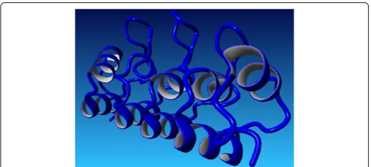
Hybrid structure of RB1 protein was generated using best aligned parts of tem-plates (Figure 1). Among the selected temtem-plates for RB1, 4ELJ was best scoring template used for modeling. Plot of its overall quality Z-score, shown per residue is displayed in Figure 2. ForP16-INK4Aprotein, hybrid structure was generated using
Figure 2Overall quality of predicted RB1 model.
Figure 4Overall quality of predicted P16-INK4A model.
Figure 5Functional partners of P16-INK4A protein through STRING database.
best aligned parts of all the five templates (Figure 3). The best scoring template used for modeling was 1MX6. Plot of its overall quality Z-score, shown per residue is displayed in Figure 4.
Protein-protein docking
GRAMM-X was utilized for protein-protein docking of two proteinsRB1andP16-INK4A for which no ligand was reported in literature/databases. Figure 5 and 6 shows the functional partners for these proteins obtained through STRING database. Table 2 shows the functional proteins which are found to be common between RB1 and P16-INK4A. Table 3 displays the protein and their functional interactors considered for docking. Table 4 shows the GRAMM-X docking results and Figure 7, 8, 9 and 10 displays the docked complexes and their interactions.
Discussion
In current study, 3D structures of the prioritized genes are predicted.RB1andP16-INK4A are found to have expressions in lung tissue. Best docking complex of RB1 and E2F1 analysis suggested that , hydrogen bond interactions are found between O of Glu 864, Ser 567, Asp 36, Arg 861(ofRB1) and H of Gln 290, Arg 22,Lys 89 and Thr 285 (ofE2F1) with bond distances of 2.5,2.7,2.2 and 1.9 respectively. P16-INK4AandCDK4 protein-protein complex showed ionic bond interactions between Arg47, Arg80, Met 1, Cys72, Thr 104, Trp 106 and Val9 with 3.6, 2.2, 2.9 and 3.4 bond distances indicating the potential role of Table 2 Common functional partners between RB1 and P16-INK4A
Proteins Common functional partners
RB1andP16-INK4A CDK4
E2F1 MDM2 CCND1
Table 3 Proteins and interactors for protein-protein docking
Receptor protein Functional interactors
P16-INK4A CDK4
Rb1 E2F1
Table 4 Binding interactions forRb1andP16-INK4A
Receptor protein Interacting protein Interactions (Receptor residue→ Interacting protein residue)
Bond distance
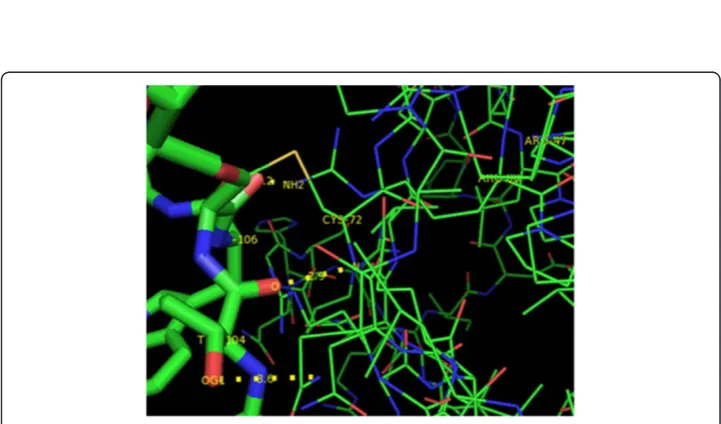
P16-INK4A CDK4 Arg47:NH2→Thr 104:OG1 3.6
Arg 80:NH2→Trp 106: O 2.2
Met 1: N→Val 9: O 3.4
Cys72:N→Thr104:O 2.9
RB1 E2F1 Glu 864:O→Gln 290:2HEZ 2.5
Ser 567:OG→Arg 22:1HH1 2.7
Asp 36: OD2→Lys 89:1HZ 2.2
Figure 7Docked complex of P16-INK4A and CDK4 showing P16-INK4A in blue and CDK4 in green.
Figure 8Interactions between P16-INK4A and CDK4.P16-INK4A is shown in lines and CDK4 in sticks format.
these residues in protein-protein interaction. Interactions ofRB1andE2F1complexes will help in cell cycle arrest in G1 phase asRB1acts as a transcription repressor of E2F1 target genes. The underphosphorylated, active form of RB1 interacts with E2F1 and represses its transcription activity, leading to cell cycle arrest.P16-INK4AandCDK4interactions help to inhibit the proliferation of the cells . Results revealed through Protein-protein binding may provide a basis for designing a suitable drug for preventing this widely spreading disease by using the information retrieved about the amino acids involved in interactions with the respective proteins.
Conclusion
3-dimensional structure prediction of most plausible candidate genes proposed that it may be used further to understand the potential mechanism of lung cancer develop-ment and role of these proteins in causing abnormalities. By exploring protein- protein docking interaction with in wild type and mutant protein can open the new gate for computer aided drug designing for the better identification of potential drug inhibitor.
Materials & methods
Sequence retrieval and 3d model building
Sequences in FASTA format ofP16-INK4andRB1were retrieved from NCBI (National Centre of Biotechnology Information) having accession numbers of P42771, P06400 and OMIM id’s of 614041 and 600160 respectively. Since the target sequence was the
Figure 10Interactions between RB1 and E2F1 showing RB1 in lines and E2F1 in sticks format.
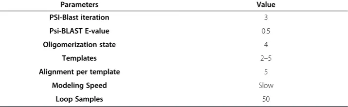
Table 5 Parameters selected for YASARA comparative modeling
Parameters Value
PSI-Blast iteration 3
Psi-BLAST E-value 0.5
Oligomerization state 4
Templates 2–5
Alignment per template 5
Modeling Speed Slow
only available information, possible templates were identified by running 3 PSI-BLAST iterations to search the PDB for match (i.e. hits with an E-value below the homology modeling cutoff 0.5).
Comparative modeling approach was implemented to generate 3D structures of genes using YASARA software. YASARA generated a hybrid structure using 2–5 templates which are ranked on the basis of alignment score (PSI_BLAST) and structural quality (Z_Score) according to WHAT CHECK [8] obtained from the PDBFinder2 [8] database for all six candidate genes. Selected Parameters used by YASARA for structure predic-tion are menpredic-tioned in Table 5.
Model validation
YASARA softwares uses WHAT CHECK [8] obtained from the PDBFinder2 [8] data-base for generating plot of overall quality Z-score.
Molecular docking
Protein-protein docking of P16-INK4 and RB1 was carried out through GRAMM-X docking web server.
Protein-protein docking
Protein to be used as a ligand in protein-protein docking was retrieved from STRING database, an online database for physical (direct) and functional (indirect) protein– -protein interactions [9] and its 3D structure was predicted using ab-initio approach through I-TASSER server. GRAMM-X docking server [10] was used for Protein-protein docking which generated a docked complex. Post docking analysis was carried out using Pymol software which is a molecular visualization system for use in structural biology which provides a user with high quality 3D images of small molecules and biological macromolecules, such as proteins.
Competing interests
The authors declare that they have no competing interests.
Authors’contributions
The work presented here was carried out in collaboration between all authors. AM and NAK defined the research theme and designed methods analyzed the data, interpreted the results and wrote the paper. NZ carried out all the work and analysis of results under the guidance of NAK. AM also provided suggestions to the interpretation of results. All authors have contributed to, seen, read and approved the manuscript.
Acknowledgements
We are thankful to university institute of biochemistry and biotechnology (UIBB), PMAS- Arid Agriculture university for providing molecular modeling software, YASARA forinsilicoanalysis.
Author details 1
Department of Bioinformatics and Biotechnology, International Islamic University, Islamabad, Pakistan.2Institute of Biochemistry and Biotechnology, Department of Biochemistry, Arid Agriculture University Rawalpindi, Rawalpindi, Pakistan.
Received: 23 October 2012 Accepted: 11 December 2012 Published: 1 January 2013
References
1. Herbst RS, Heymach JV, Lippman SM:Lung cancer.N Engl J Med2008,359:1367–1380.
2. Brenner DR, McLaughlin JR JR, Rayjean J, Hung RJ:Previous lung diseases and lung cancer risk: a systematic review and meta-analysis.PLoS One2011,3:e17479.
4. Blot WJ, McLaughlin JK:Passive smoking and lung cancer risk: what is the story now.J Natl Cancer Inst1998, 90:1416–1417.
5. Kohno T, Otsuka A, Girard L, Sato M, Iwakawa R, Ogiwara H, Cespedes MS, Minna JD, Yokota J:A Catalog of Genes Homozygously Deleted in Human Lung Cancer and the Candidacy of PTPRD as a Tumor Suppressor Gene.Genes Chromosomes Cancer2010,4:342–352.
6. Minna JD, Roth JA, Gazdar AF:Focus on lung cancer.Cancer Cell2002,1:49–52.
7. Belinsky AS, Liechty CK, Gentry DF, Wolf JH, Rogers J, Vu K, Haney J, Kennedy CT, Hirsch RF, Miller Y, Franklin AW, Herman GJ, Baylin BS, Bunn AP, Byers T:Promoter Hypermethylation of Multiple Genes in Sputum Precedes Lung Cancer Incidence in a High-Risk Cohort.Cancer Res2006,66:3338–3344.
8. Hooft RW, Vriend G, Sander C, Abola EE:Errors in protein structures.Nature1996,6580:272.
9. Szklarczyk D, Franceschini A, Kuhn M, Simonovic M, Roth A, Minguez P, Doerks T, Stark M, Muller J, Bork P, Jensen JL, Mering VCL:The STRING database in 2011: functional interaction networks of proteins, globally integrated and scored.Nucleic Acids Research2011,39:D561–D568.
10. Tovchigrechko A, Vakser AI:GRAMM-X public web server for protein–protein docking.Nucleic Acids Res2006, 34:W310–W314.
doi:10.1186/1742-4682-10-1
Cite this article as:e Zahraet al.:Comparative modeling and docking studies of p16ink4/Cyclin D1/Rb pathway genes in lung cancer revealed functionally interactive residue of RB1 and its functional partner E2F1.Theoretical Biology and Medical Modelling201310:1.
Submit your next manuscript to BioMed Central and take full advantage of:
• Convenient online submission
• Thorough peer review
• No space constraints or color figure charges
• Immediate publication on acceptance
• Inclusion in PubMed, CAS, Scopus and Google Scholar
• Research which is freely available for redistribution