Open Access
Research
Quantification of gamma-secretase modulation differentiates
inhibitor compound selectivity between two substrates Notch and
amyloid precursor protein
Ting Yang, Dilyara Arslanova, Yongli Gu, Corinne Augelli-Szafran and
Weiming Xia*
Address: Center for Neurologic Diseases, Department of Neurology, Brigham and Women's Hospital, Harvard Medical School, Harvard University, Boston, MA, USA
Email: Ting Yang - [email protected]; Dilyara Arslanova - [email protected]; Yongli Gu - [email protected]; Corinne Augelli-Szafran - [email protected]; Weiming Xia* - [email protected]
* Corresponding author
Abstract
Background: Deposition of amyloid-β protein (Aβ) is a major pathological hallmark of Alzheimer's disease (AD). Aβ is generated from γ-secretase cleavage of amyloid precursor protein (APP). In addition to APP, γ-secretase also cleaves other type I integral membrane proteins, including the Notch receptor, a key molecule involved in embryonic development.
Results: To explore selective γ-secretase inhibitors, a combination of five methods was used to systematically determine these inhibitors' profiles on the γ-secretase cleavage of APP and Notch. When two potent γ-secretase inhibitors, compound E (cpd E) and DAPT, were used in a conventional in vitro γ-secretase activity assay, cpd E completely blocked Aβ generation from the cleavage of substrate APP C100, but only had a minor effect on Notch cleavage and NICD generation. Next, cpd E and DAPT were applied to HEK293 cells expressing a truncated Notch substrate NotchΔE. Both cpd E and DAPT were more potent in blocking Aβ generation than NICD generation. Third, a reporter construct was created that carried the NICD targeting promoter with three Su(H) binding sequences followed by the luciferase gene. We found that the inhibition of NICD generation by cpd E and DAPT was consistent with the reduced expression of luciferase gene driven by this Notch targeting promoter. Fourth, levels of "Notch-Aβ-like" (Nβ*) peptide derived from two previously reported chimeric APP with its transmembrane domain or the juxtamembrane portion replaced by the Notch sequence were quantified. Measurement of Nβ* peptides by ELISA confirmed that EC50's of cpd E were much higher for Nβ* than Aβ. Finally, the
expression levels of Notch target gene her6 in cpd E or DAPT-treated zebrafish were correlated with the degree of tail curvature due to defective somitogenesis, a well characterized Notch phenotype in zebrafish.
Conclusion: Our ELISA-based quantification of Aβ and Nβ* in combination with the test in zebrafish provides a novel approach for efficient cell-based screening and in vivo validation of APP selective γ-secretase inhibitors.
Published: 4 November 2008
Molecular Brain 2008, 1:15 doi:10.1186/1756-6606-1-15
Received: 29 September 2008 Accepted: 4 November 2008
This article is available from: http://www.molecularbrain.com/content/1/1/15
© 2008 Yang et al; licensee BioMed Central Ltd.
Background
Genetic and neuropathologic evidence suggests that Alzheimer's disease (AD) is caused partly by the
overpro-duction and lack of clearance of the amyloid β peptide
(Aβ) [1]. This Aβ peptide is generated by sequential
cleav-ages of the amyloid precursor protein (APP) by β
-secre-tase, which generates a 12 kDa C-terminal stub of APP
(C99), and by γ-secretase to yield two major species of Aβ
that end at residue 40 (Aβ40) or 42 (Aβ42) [2,3]. In
addi-tion to cleaving APP, γ-secretase also mediates the final
proteolytic cleavage of the Notch receptor [4,5]. Notch signaling is critical to a wide variety of cell fate determina-tions during embryonic development as well as through-out adulthood. After ectodomain shedding, the remaining
membrane-bound C-terminal stub is cleaved by γ
-secre-tase to release the Notch-1-β peptide (Nβ, similar to
amy-loid β peptide from APP) and the Notch IntraCellular
Domain (NICD). NICD is subsequently translocated to the nucleus where it regulates gene expression [5-7].
There are about 50 γ-secretase substrates in addition to
APP and Notch that include DCC [8], ErbB-4 [9,10], E-and N-cadherin [11,12], CD44 [13,14], LRP [15],
Nectin1α [16], Delta and Jagged [17], Glutamate Receptor
Subunit 3 [18], APLP1 and APLP2 [19-21], p75 Neuro-trophin Receptor [22], Syndecan3 [23], Colony Stimulat-ing factor-1 [24] and Interleukin-1 Receptor II [25]. All of these substrates are type I membrane proteins and have diverse functions, including transcriptional regulation, cell-cell adhesion, regulation of ion conductance, and neurotrophin signaling. The cleavage of these proteins can
be blocked by reported γ-secretase inhibitors and are fully
dependent on each γ-secretase component [26].
γ-Secretase is composed of presenilin 1 (PS1), anterior
pharynx defective-1 (Aph-1), presenilin enhancer-2
(Pen-2), and nicastrin (Nct). PS1 carries the catalytic site of γ
-secretase, as we have demonstrated that a mutation of two critical aspartate (Asp) residues abrogates enzymatic
activ-ity [27]. Nicastrin is required for γ-secretase activity
[28-35] and is an important component in the complex, pos-sibly functioning as the receptor for different substrates [36]. Genetic screens further revealed the aph-1 gene and the pen-2 gene that encodes two essential components of
the γ-secretase complex [37,30,38]; overexpression of all
four components results in increased γ-secretase activity,
both in mammalian cells [39-44] and in yeast [45].
Among all reported γ-secretase inhibitors, transition-state
analogues prevent Aβ generation and bind directly to PS1
and PS2 [46,47]. Most reported γ-secretase inhibitors
spe-cifically block the cleavage at both sites in APP and Notch without differentiating between the two substrates. It has been reported that a subset of NSAIDS (nonsteroidal anti-inflammatory drugs) that include ibuprofen,
indometh-acin and sulindac sulphide, specifically block the cleavage
of the γ-secretase substrates at the middle of
transmem-brane domain (TMD) without affecting the generation of the intracellular domains (ICDs) of several type I trans-membrane proteins that include APP, ErbB-4, and Notch
[48]. These NSAIDs directly modulate γ-secretase complex
and become a part of a new class of γ-secretase modulators
[49-54]. Another γ-secretase modulator is Gleevec that has
been approved for the treatment of chronic myeloid leukemia and gastrointestinal stromal tumors. In addition to Gleevec binding to Abelson leukemia (Abl) tyrosine kinase, it also was shown to selectively inhibit APP
cleav-age and Aβ production without affecting Notch cleavage
at the concentration of 10 μM [55].
Two potent γ-secretase inhibitors, DAPT and compound E
(cpd E), show a range of IC50 values in blocking γ-secretase
activity in both in vitro and cell-based assays. For cpd E,
the IC50 for NICD and Aβ generation in cultured cells was
found as low as 1.7 nM [56] and 0.3 nM [57], respectively.
De novo Aβ and AICD generation in vitro was inhibited by
DAPT with IC50 values ranging from 10–100 nM [58,59].
A direct comparison of NICD and AICD levels in an in vitro γ-secretase activity assay showed a partial inhibition of NICD generation by DAPT at 50 nM, and AICD at 100 nM [60]. Different assay systems were implemented in
these various studies to measure the IC50 values of the γ
-secretase inhibitors. Since there were a variety of assays used, it was difficult to compare the potency toward the cleavage of APP and Notch among different systems.
The current study combined five assay methods and sys-tematically determined the pharmacological profile of
cpd E and DAPT on γ-secretase cleavage of APP and Notch.
This approach includes the measurements of the potency
of γ-secretase inhibitors and their effect on the inhibition
of the γ-secretase activity in vitro, NICD generation, NICD
downstream transcription activation, cleavage of APP/ Notch chimeric substrates, and Notch downstream target gene expression in zebrafish. Previous studies showed that treating zebrafish with DAPT at the late blastula stage caused defects in somitogenesis and neurogenesis [61]. Similarities have been observed between DAPT-treated embryos and previously reported zebrafish Notch
path-way mutants like bea, des, aei, and wit [62,63]. The
increased neurogenesis in DAPT-treated embryos can be reduced by microinjecting NICD mRNA. Interestingly, defective somitogenesis was not observed in zebrafish
embryos that were treated with the Aβ-lowering JLK
non-peptidic isocoumarin inhibitors [64]. In this study, the expression levels of Notch target gene her 6 were corre-lated to the phenotypes that were observed in the embryos treated with DAPT and cpd E. This provided an in vivo
sys-tem to test the effect of γ-secretase inhibitors on Notch
Results
Low concentration of compound E selectively blocks Aβ
production with minimum effect on NICD generation in
vitro
To characterize the direct effect of two γ-secretase
inhibi-tors cpd E and DAPT on APP/Notch cleavage, a
conven-tional in vitro γ-secretase assay to quantify their inhibitory
potency was used [58,65,66]. The incubation of γ
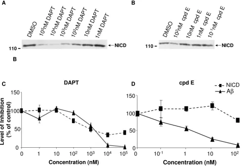
-secre-tase complex with purified substrates at 37°C for 4 hr was followed by Western Blot (WB) to determine the quantity of newly generated NICD. A newly generated band that corresponds to the predicted size of the NICD-Flag was detected (Fig. 1A and 1B). A clear reduction of NICD
gen-eration in samples containing DAPT (Fig. 1A) or cpd E (Fig. 1B) was found, and the reduction was dose depend-ent.
The same preparation of γ-secretase complex was mixed
with C100Flag followed by ELISA to quantify the levels of
newly generated Aβ. As expected, both DAPT (Fig. 1C)
and cpd E (Fig. 1D) blocked γ-secretase cleavage of APP
C100Flag and caused a dose-dependent reduction of Aβ
production.
Close comparison of the inhibition profile of cpd E and
DAPT on Aβ and NICD generation revealed a divergence
Generation of NICD and Aβ from purified APP and Notch substrate C100 and N100 in an in vitro γ-secretase activity assay
Figure 1
Generation of NICD and Aβ from purified APP and Notch substrate C100 and N100 in an in vitro γ-secretase activity assay. The E. coli generated APP- and Notch-based, 100-residue γ-secretase substrates C100-Flag and N100-Flag were mixed with the membrane vesicles solubilized in CHAPSO after DMSO or compounds were added. The mixture was incubated at 37°C for 4 hours. A. A dose-dependent inhibition of NICD generation by DAPT. Generation of NICD was detected by Western blot (WB) with antibody 1744 specifically recognizing N-terminus of NICD. B. Generation of NICD was inhibited in the presence of 100 nM of cpd E. C. Levels of NICD determined by WB were quantified by densitometry (dotted line). Levels of Aβ generated from the γ-secretase cleavage of C100 in the presence of DAPT were determined by ELISA (solid line). Comparison of NICD and Aβ generation in the presence of DAPT suggests that high concentrations of DAPT were more potent in blocking Aβ than NICD generation. D. NICD (dotted line) and Aβ (solid line) generation in the presence of cpd E were compared. Cpd E inhibited Aβ generation with an IC50 of 1 nM and is more potent in inhibiting Aβ than NICD.
1
0
5
n
M
D
A
P
T
D
M
S
O
1
0
n
M
D
A
P
T
1
0
4
n
M
D
A
P
T
1
0
3
n
M
D
A
P
T
1
0
2
n
M
D
A
P
T
1
n
M
D
A
P
T
1
0
2
n
M
c
p
d
E
1
0
n
M
c
p
d
E
1
n
M
c
p
d
E
D
M
S
O
1
0
-1
n
M
c
p
d
E
A
110
B
cpd E
0
50
100
150
10
-11
10
10
2Concentration (nM)
0
NICD
A
β
1
10
10
210
310
410
5Concentration (nM)
0
DAPT
0
50
100
150
Lev
e
l of Inhi
bition
(%
of
control)
110
NICD NICD
B
in their potencies. Low concentrations of DAPT did not
show much difference in inhibiting NICD and Aβ
genera-tion, but 10 and 100 μM of DAPT blocked ~60% of NICD
generation compared to a complete depletion of Aβ
pro-duction (Fig. 1C). While 100 nM of cpd E could almost
deplete any Aβ generation from substrate APP C100, its
effect on NICD was much less obvious (Fig. 1D). There was only minor reduction of NICD levels compared to
DMSO controls. This led to the speculation that certain γ
-secretase inhibitors may specifically inhibit APP at a par-ticular range of doses that have minimum effect on Notch signaling.
Compound E and DAPT differentially inhibit Aβ and NICD
generation in cultured cells
Since many compounds could behave differently in vitro versus in culture cells, cpd E and DAPT were tested in cul-tured cells (Fig. 2). HEK293 cells stably expressing Swed-ish mutant APP were transiently transfected with
NotchΔE, a truncated Notch construct that is readily
cleaved by the γ-secretase to generate NICD for
down-stream signaling transduction [67]. NotchΔE expressing
cells were treated with increasing concentrations of DAPT or cpd E. Cell lysates were subjected to WB for measuring the generation of NICD (Fig. 2A), and conditioned media
were collected for Aβ measurement by ELISA (Fig. 2B,C).
Semi-quantification of NICD levels was detected by WB, and the inhibition profile of DAPT (Fig. 2B) and cpd E
(Fig. 2C) were compared on NICD and Aβ generation in
cultured cells. It was found that high doses of DAPT and cpd E could not completely eliminate NICD generation in
cultured cells. This was in contrast to Aβ levels that were
efficiently reduced to almost undetectable levels. Since Notch signaling and levels of NICD can be examined by quantifying the expression of the Notch target gene, a Hes-1 reporter construct (Hes-Luc) was generated by insertion of three Su(H) binding sequences in the pGL3-pro
luci-ferase reporter vector. Hes-Luc and NotchΔE were
tran-siently transfected into HEK293 cells, and transfected cells were treated with different concentrations of cpd E or DAPT. Consistent with the levels of NICD that was freshly generated in cultured cells, luciferase activities were inhib-ited by relatively high doses of cpd E and DAPT. At the concentrations of cpd E and DAPT that completely
blocked Aβ generation (Fig. 2B and 2C), about 50%
luci-ferase activities remained, i.e., inhibition of NICD
genera-tion was less efficient compared to Aβ blockage (Fig. 2D).
A chimeric APP-Notch ELISA differentiates cpd E in inhibiting APP versus chimeric APP-Notch
Two cDNA constructs expressing chimeric APP and Notch
were previously reported to generate chimeric "Notch-Aβ
-like" (Nβ*) peptide [68]. One construct has its
transmem-brane domain (TMD) replaced by the Notch TMD
(APP-m-NOTCH) and the other with the juxtamembrane
por-tion of the APP ectodomain (the α-secretase cleavage
region) replaced by the corresponding sequence in Notch
(APP-α-NOTCH) [68]. Taking advantage of different
com-binations of ELISA antibodies (see Methods), effects of
cpd E on the generation of Aβ and Nβ* peptides from
these chimeric APP-Notch substrates were quantified by ELISA.
Individual construct APP, APP-α-Notch or APP-m-Notch
was transiently transfected into HEK293 cells. These chi-meric protein expressing cells were treated with cpd E, and
the levels of Aβ and Nβ* were measured by ELISA. Again,
it was found that the effective concentration (EC) for
inhibiting 50% of Aβ production by cpd E was less than
0.1 nM, but the EC50 for Nβ* from α-Notch was at ~8 nM
(Fig. 3A). Similar results were obtained when m-Notch was expressed in HEK293 cells. At least two magnitude of
difference was observed, with EC50 for cpd E was ~0.03
nM for APP, compared to EC50 for Nβ* at ~1 nM (Fig. 3B).
Defective zebrafish phenotypes illustrated inhibition of Notch signaling
Measurements of in vitro γ-secretase activity and cell-based
Aβ/NICD generation have shown different inhibition
potencies. To examine the inhibitory effect in vivo, zebrafish embryos were treated with DAPT or cpd E.
Because different γ-secretase inhibitors may impact
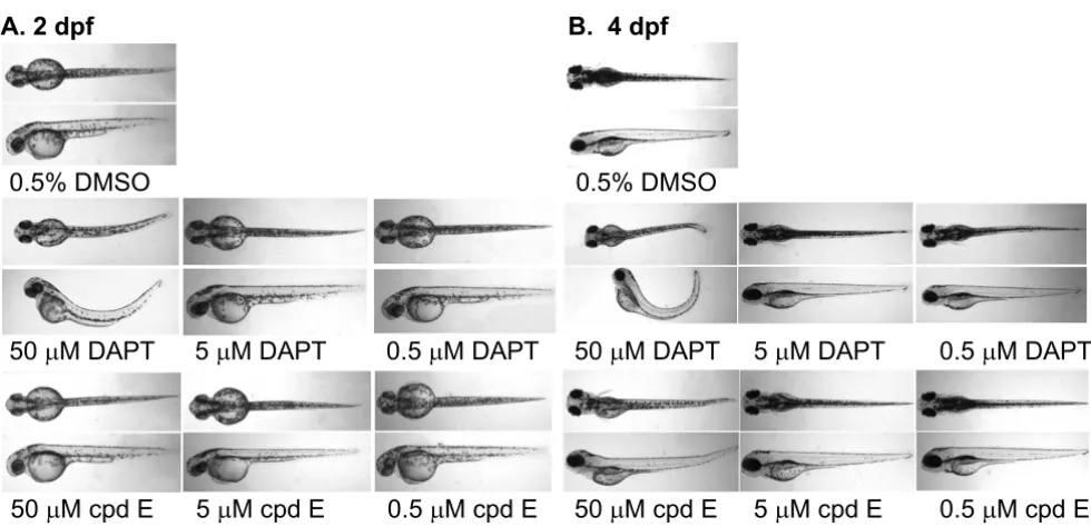
vari-ous metabolic pathways in zebrafish embryos, especially during development, the phenotypes of zebrafish embryos treated with high concentrations of DAPT and cpd E were compared. The major phenotype we examined was curved tail caused by defective somitogenesis.
Morphological alteration in DAPT- or cpd E-treated embryos was compared and correlated to the somitogen-esis associated with the inhibition of Notch signaling. The treated embryos were examined using a stereomicroscope
and it was found that embryos treated with 50 μM DAPT
had a much shorter and curved tail, compared to control DMSO-treated embryos (Fig. 4A). The curvature was obvi-ous when a lateral view of zebrafish was obtained. Cpd E, on the other hand, did not show any curvature when
treated at 50 μM. Because the EC50 values for DAPT and
cpd E to reduce NICD generation in cultured cells were ~1000 nM and 10 nM, respectively (Fig. 2B and 2C), 50
μM of DAPT and cpd E were chosen as the highest
concen-trations for the treatment.
When embryos were kept for four days, embryos treated
with 50 μM DAPT continued to show the curvature of the
Expression of Notch target gene her6 correlates with the
phenotypes of zebrafish treated with γ-secretase inhibitors
To examine the effect of DAPT and cpd E on Notch sign-aling, embryos treated with different concentrations of DAPT or cpd E were stained by whole mount in situ hybridization using a her6 probe (Fig 5). The expression level of Notch downstream target gene her6 correlates to
the levels of Notch signaling, i.e., a loss of her6 staining
corresponds to an inhibition of γ-secretase mediated
Notch signaling. We have focused on the specific effect of
γ-secretase inhibitors on Notch signaling in brain region.
In DMSO-treated embryos, her6 expression was mainly
clustered in the ventral midbrain and ventral hindbrain
Generation of NICD and Aβ from NotchΔE and APP expressing cells
Figure 2
Generation of NICD and Aβ from NotchΔE and APP expressing cells. A. Twenty four hrs after the construct carrying NotchΔE was transfected into APP expressing HEK293 cells, cells were treated with DAPT or cpd E for 8 hr and lysed for WB with antibody 1744 to specifically detect the N-terminus of NICD. Bottom panel, the antibody against α-tubulin was applied to normalize the amounts of lysates used for WB. B. Levels of NICD determined by WB were quantified by densitometry (dotted line). Levels of Aβ generated from the γ-secretase cleavage of APP in the presence of DAPT were determined by ELISA. Com-parison of NICD and Aβ generation in the presence of DAPT suggests that high concentrations of DAPT had a greater inhibi-tion of Aβ than NICD. C. NICD (dotted line) and Aβ (solid line) production from cpd E-treated cells were compared. Cpd E inhibited Aβ generation with an IC50 of ~8 nM, and it shows a greater inhibition of Aβ than NICD. D. A luciferase reporter construct driven by HES1 promoter was transfected into HEK293 cells followed by treatment with cpd E or DAPT. Both γ -secretase inhibitors blocked transcriptional activation of NICD dependent luciferase activity.
B
DAPT
0
50
100
150
1
10
10
210
310
410
5Concentration (nM)
Lev
e
l of Inhi
bition
(%
of
control)
0
100
150
cpd E
0
50
Concentration (nM)
0
10
-210
-11
10
10
210
3C
A
β
NICD
NICD
1
0
3
n
M
c
p
d
E
1
0
2
n
M
c
p
d
E
1
0
n
M
c
p
d
E
1
n
M
c
p
d
E
1
0
-1
n
M
c
p
d
E
110
A
D
M
S
O
1
0
n
M
D
A
P
T
1
0
4
n
M
D
A
P
T
1
0
3
n
M
D
A
P
T
1
0
2
n
M
D
A
P
T
1
n
M
D
A
P
T
D
M
S
O
50
αααα
-tubulin
0
50
100
concentration (nM)
HE
S-Luc A
c
tivit
y
(%
of
control)
DAPT
cpd E
0
1
10
10
210
310
410
510
6(Fig. 5). In the presence of 50 μM DAPT, the her6 expres-sion was significantly reduced or disappeared in most
areas, reflecting a strong inhibition of γ-secretase activity.
When embryos were treated with a lower concentration of
DAPT at 5 μM, staining of her6 started to appear in those
areas found in DMSO-treated embryos. Embryos treated
with 0.5 μM DAPT showed a very similar staining pattern
to the control embryos. Interestingly, cpd E showed a weaker effect on the expression levels of her6. There was a
reduction of her6 staining in those embryos that were
treated with highest testing doses of cpd E. When the her6
staining is linked to morphological alterations (Fig. 5), the level of reduction in Notch signaling is closely linked with the severity of phenotypes that was observed in these zebrafish, i.e., the curvature of the tails.
Discussion
Our knowledge of γ-secretase components distinguishing
different substrates provides a molecular basis for the
modulation of γ-secretase complex. Nicastrin has been
found to interact with both APP and Notch and is involved in substrate recognition and interaction [36]. An
Generation of Aβ and Nβ* from chimeric APP-Notch expressing cells
Figure 3
Generation of Aβ and Nβ* from chimeric APP-Notch expressing cells. A. The juxtamembrane portion of the APP ectodomain (the α-secretase cleavage region) was replaced by the corresponding sequence in Notch (α-NOTCH). Levels of Nβ* in the media from cells expressing APP-α-Notch (dotted line) and levels of Aβ from APP expressing cells (solid line) were determined by ELISA. B. A chimeric cDNA constructs express APP with its transmembrane domain (TMD) replaced by the Notch TMD (APP-m-NOTCH). Levels of Aβ (solid line) and Nβ* (dotted line) were determined by ELISA.
A
0
50
100
150
Concentration (nM)
Relat
iv
e
A
β β β β
and N
ββββ∗∗∗∗
le
v
e
ls
(%
of
c
o
nt
rol)
APP
APP-
α
-Notch
Concentration (nM)
B
0
50
100
150
APP
APP-m-Notch
0
10
-310
-210
-11
10
0
10
-310
-210
-11
10
Relat
iv
e
A
β β β β
and N
ββββ∗∗∗∗
le
v
e
ls
(%
of
c
o
nt
artificial elongation of the Pen-2 N-terminus leads to an
increased Aβ42 production [69], indicating that Pen-2
might function as a modulator to influence the γ-secretase
cleavage of APP. Identification of a key regulator of γ
-secretase complex TMP21 further suggests that cleavage of APP and Notch could be distinguished and modulated
[70]. While the development of γ-secretase inhibitors is
one of the major directions for AD therapeutics,
com-pletely blocking the γ-secretase-mediated proteolytic
proc-ess of about 50 substrates interferes with fundamental steps in many biological functions. Therefore, identifying
γ-secretase modulators that only block the cleavage of
APP, but not other substrates is ideal. Different from ear-lier studies that have identified NSAIDs and Gleevec for
specific blockage of Aβ production without affecting the γ
-secretase cleavage of Notch, the current study has
pro-vided a systematic approach to identify γ-secretase
inhibi-tors to modulate the γ-secretase cleavage of APP and
Notch separately.
We have analyzed two potent γ-secretase inhibitors DAPT
and cpd E using different quantification methods to deter-mine the pharmacological profile of blocking the cleavage of APP and Notch. The range of inhibition concentrations vary among these methods. However, the effective
inhib-itory concentrations for Notch cleavage were always found to be higher than those concentrations for APP
cleavage. In a conventional in vitro γ-secretase activity
assay, 0.1 μM of cpd E completely blocked Aβ generation
from the cleavage of substrate APP C100, and only had minor effect on Notch cleavage and NICD generation.
Cpd E selectively inhibited the γ-secretase cleavage of APP
at low concentrations, i.e., from 0.1 nM to 10 nM. How-ever, at the same concentrations, we found that DAPT did
not inhibit the γ-secretase cleavage of APP and Notch (Fig.
1C). When higher concentration of DAPT was used in our in vitro γ-secretase activity assay, a partial inhibition of Notch cleavage was observed, in contrast to an almost complete inhibition of APP cleavage. Therefore, DAPT
selectively blocked the γ-secretase cleavage of APP at
higher concentration compared to compound E. When cpd E or DAPT were applied to HEK293 cells that
expressed the substrate NotchΔE, we found that both
compounds were more potent in blocking Aβ generation
than NICD production. DAPT at concentrations of 1 μM
or higher reduced Notch cleavage to about 50% in both in vitro γ-secretase activity assay (Fig. 1B) and cell culture
based assay (Fig. 2B). Cpd E at 0.1 μM reduced Notch
cleavage to ~50% in both systems. For the γ-secretase
cleavage of APP, DAPT was able to inhibit the levels of Aβ
Treatment of zebrafish embryos with DAPT causes curved tails
Figure 4
Treatment of zebrafish embryos with DAPT causes curved tails. A. A stock of DAPT or cpd E in DMSO was diluted in embryo medium, and increasing concentrations of DAPT or cpd E were applied to de-chorionated zebrafish embryos incu-bated at 28°C from 24 hpf to 48 hpf. Control embryos were mock-treated with embryo medium containing the same concen-tration of DMSO. Treatment of zebrafish embryos with 50 μM DAPT caused curved trunk and tails. B. DAPT- or cpd E-treated embryos were kept until 4 dpf, and images were acquired at 40 × magnification.
P
P
P
P
P
P
P
P
P
P
to 50% at the concentration of 1 μM in vitro and ~0.5 μM in cultured cells, respectively. Compound E, on the other
hand, was able to reduce the levels of Aβ to 50% at the
concentrations of 1 nM and 5 nM in two systems. There-fore, DAPT and cpd E showed similar potencies in
cul-tured cells and in vitro γ-secretase activity assay. The level
of NICD inhibition was consistent with the reduced expression of Luciferase gene driven by a Notch target gene promoter containing three Su(H) binding sequences. Using two previously reported chimeric cDNA
constructs expressing APP-m-NOTCH or APP-α-NOTCH,
cpd E showed much higher EC50's for lowering the levels
of Nβ* derived from the cleavage of APP-m-NOTCH and
APP-α-NOTCH. Finally, the expression levels of Notch
target gene her6 in a whole animal zebrafish, as measured by in situ hybridization, were correlated with the dose-dependent phenotypic effect of DAPT. The effect of cpd E was less obvious and hence, consistent with a less reduc-tion of her6 expression.
Previous studies have applied similar compounds to
dif-ferentiate their effect on the γ-secretase cleavage of Notch
and APP, and some showed selective inhibition of Aβ
pro-duction without Notch phenotypes in animals [71]. Lewis et al. have used a set of compounds for the test, and some of these compounds (like compound 1) have similar structures to DAPT [72]. Using cultured cells to test the potencies of different compounds, they found that Notch and APP cleavages cannot be easily dissected apart [72]. We have used additional methods to determine the inhi-bition profile of DAPT and cpd E, including in vivo
ani-mal based assays. In cultured cells expressing NotchΔE or
chimeric APP-Notch proteins, cpd E was more effective in inhibiting APP than Notch substrate. DAPT showed
simi-lar effect in cultured cells and in an in vitro γ-secretase
activity assay. Both γ-secretase inhibitors DAPT and cpd E
are believed to interact with the core component of the γ
-secretase complex, PS. Mutation of two aspartate residues
in PS1 leads to a complete loss of function for γ-secretase
Expression levels of Notch target gene her6 are consistent with the curvature phenotype
Figure 5
Expression levels of Notch target gene her6 are consistent with the curvature phenotype. Increasing concentra-tions of DAPT or cpd E were applied to de-chorionated zebrafish embryos from 24 hpf until 48 hpf, and in situ hybridization of compound-treated embryos was carried out at 48 hpf using the her6 probe. At least 10 to 20 embryos were examined for each experiment. Images were taken at the 64× magnification for stained embryos.
0.5% DMSO
50
μ
M DAPT
50
μ
M cpd E
5
μ
M DAPT
0.5
μ
M DAPT
which suggests that these two aspartates may constitute
the active site of γ-secretase [27]. Both aspartyl protease
transition state mimic and non-transition-state γ-secretase
inhibitor could specifically bind the N- and C-terminal
fragments of PS1 [73,46,57]. The binding of the γ
-secre-tase inhibitor to PS1 NTF/CTF could be then competi-tively suppressed by the presence of cpd E [57]. DAPT was found to specifically interact with the C-terminal region of PS1 [59]. Studies that use helical peptide inhibitors to
block the γ-secretase complex suggest that a docking and
an active site exist for the γ-secretase complex, and that the
docking site might be located at the PS subunit interface, a site very close to the active site [74]. It is not clear whether different concentrations of DAPT and cpd E may affect the docking site in a way that differentiate the
bind-ing of APP and Notch to the γ-secretase complex.
Both DAPT and cpd E have been used to treat animals. DAPT was specifically tested in zebrafish [61]. Zebrafish
have a highly conserved γ-secretase complex. Both
zebrafish PS1 (Psen1) and the PS2 homolog (Psen2) are expressed during the segmentation and later stages [75-77]. Nicastrin has been identified in the zebrafish genome, and only one copy of Psen1, Psen2, Pen-2, and Aph-1 gene has been found [30]. Once the highly similar
zebrafish γ-secretase complex is inhibited by DAPT,
somi-togenesis is severely affected leading to curved tails, a phe-notype well-characterized for altered Notch signaling [61]. In this study, a dose dependent effect of DAPT on zebrafish phenotypes was observed, and a curvature of
zebrafish tail was found in embryos treated with 50 μM of
DAPT. Although the EC50 of DAPT for inhibiting Notch
signaling was much lower in cultured cells (1–10 μM, Fig.
2), it is not surprising that a high concentration of DAPT was necessary to induce a phenotype in a whole animal
(50 μM, Fig. 4). For cpd E, the highest concentration used
to treat embryos was 50 μM compared to an EC50 that was
below 0.1 μM for the inhibition of NICD generation in
cultured cells (Fig. 2). For both DAPT and cpd E, there is no data on pharmacokinetics, pharmacodynamics and ADME of these two compounds in zebrafish. While both cpd E and DAPT are cell permeable, a lack of dramatic
phenotypic alteration in embryos treated with 50 μM cpd
E could be best explained by a slightly reduced expression level of her6 gene. This indicates that Notch signaling was not significantly perturbed at this concentration of cpd E in a whole animal.
Administration of cpd E into guinea pig resulted in a
dose-dependent inhibition of brain cortical γ-secretase activity
and correspondingly, decreases in plasma, CSF and brain
Aβ levels [78]. Treatment of a transgenic mouse expressing
human familial AD linked V717F APP with DAPT also
leads to a dose-dependent, acute decrease in brain Aβ
[79]. Treatment of AD patients with another γ-secretase
inhibitor, LY450139 dihydrate, reduces plasma Aβ40.
This compound was well tolerated in these patients
[80-82]. Therefore, modulated inhibition of γ-secretase is
fea-sible in human subjects, and potent inhibitors used at appropriate doses appear to be promising in preventing
the progression of Aβ pathology.
Conclusion
Our measurement of Aβ and Notch-Aβ-like peptides from
chimeric APP proteins could be used for efficient
cell-based screening of γ-secretase modulators. These
modula-tors could be tested by in vitro γ-secretase activity assay.
The in vivo test results presented here of these compounds in a vertebrate zebrafish further validate our quantitative methods to differentiate their selectivity for APP, Notch
and potentially other γ-secretase substrates.
Methods
In Vitro γ-Secretase Activity Assay
The E. coli generated APP- and Notch-based, 100-residue
γ-secretase substrates C100-Flag and N100-Flag were
puri-fied as previously described [65]. C100-Flag substrate con-tains an initiating methionine, the 99 C-terminal residues
of APP that start at the α-secretase site, and a Flag tag.
N100-Flag substrate contains a similar initiating methio-nine, 99 amino acids that start at the TACE cleavage site, and a Flag tag. The membrane vesicles were solubilized in 1% CHAPSO-HEPES and diluted in a final concentration of 0.2% CHAPSO-HEPES. Phosphatidylethanolamine (PE) and phosphatidylcholine (PC) were added to the final concentration of 0.02% PE and 0.08% PC. After
add-ing DMSO or test compounds to the solubilized γ
-secre-tase complex, substrate C100-Flag or N100-Flag was added to the mixture, then followed by incubation at 37°C for 4 hours [65,83]. Two compounds have been used in this study, compound E (cpd E), {(S,S)-2-[2-(3,5- Difluorophenyl)-acetylamino]-N-(1-methyl-2-oxo-5-phenyl-2,3-dihydro-1H-benzo [e] [1,4] diazepin-3-yl)-propionamide} [57] and DAPT
{N-[N-(3,5-Difluoroph-enacetyl-L-alanyl)]-S-phenylglycine t-Butyl Ester} [79].
Cpd E was provided by Dr. Michael Wolfe.
ELISAs and Antibodies
Sandwich ELISAs for Aβ were performed as described [84].
The capture antibodies, 2G3 (to Aβ residues 33–40) and
4G8 (to Aβ residues 17–24), were used for Aβ40 and Aβ
total species, respectively. The detecting antibodies were
biotinylated 82E1 (to Aβ residues 1–16) for Aβ1–40/total
or biotinylated 266 for Aβx-40 species. The use of
mid-region and C-terminal capturing antibodies and
N-termi-nal detecting antibody for chimeric "Notch-Aβ-like"
pep-tide (Nβ*) has been documented [68]. The combination
of several capture/detecting antibodies are use to measure
Aβ and Nβ* derived from different precursors. The
for measuring Nβ* from APP-α-Notch expressing cells.
Since the APP-α-NOTCH is the fusion protein with its
jux-tamembrane portion of the APP ectodomain (the α
-secre-tase cleavage region) replaced by the corresponding sequence in Notch, the epitopes in APP sequence could still be recognized by 2G3 (C-terminus) and 82E1 (N-ter-minus). The capture antibody 4G8 and detecting antibody
82E1 were used for measuring Nβ* from APP-m-Notch
expressing cells. Since the APP-m-NOTCH is the fusion protein with its transmembrane domain (TMD) replaced by the Notch TMD, the epitopes in APP sequence could be recognized by 4G8 (mid-region before TMD) and 82E1 (N-terminus). Antibody 82E1 was purchased from Immuno-Biological Laboratories, Inc., Minneapolis, MN. Antibody 4G8 was purchased from Signet Laboratories, Inc., Dedham, MA. Antibody 1744 that specifically detect the N-terminus of NICD was purchased from Cell Signal-ing Technology, Danvers, MA.
cDNA constructs for cell based γ-secretase activity assay
The cDNA construct NotchΔE has a c-myc tag and is a
truncated Notch molecule that is an immediate substrate
for γ-secretase cleavage to generate Notch intracellular
domain (NICD) [85]. Two chimeric cDNA constructs express APP with (APP-m-NOTCH), or else the
juxtamem-brane portion of the APP ectodomain (the α-secretase
cleavage region) replaced by the corresponding sequence
in Notch (APP-α-NOTCH). These cDNA constructs were
provided by Dr. Dennis Selkoe [68]. Hes-1 reporter con-struct (Hes-Luc) was generated by insertion three of Su(H) binding sequence
5'-AGGTTCTCACTGTGGGGTAAGAAGGTTCTCACAGT-GGGGTAAGAGGTTCTCACAGTC in the pGL3-pro luci-ferase reporter vector (Promega, Madison, WI). The final assemble is similar to a previously reported Notch reporter construct [86].
Human embryonic kidney (HEK) 293 cells stably express-ing Swedish mutant human APP695 were transfected with
different cDNA constructs and maintained in 200 μg/ml
G418 (Invitrogen, Carlsbad, CA). Transfected cells were
treated with two γ-secretase inhibitors cpd E or DAPT for
8 hr. Conditioned media were collected for ELISA, and cell lysates were analyzed by Western blot as described
[87]. Cells co-transfected with Hes-Luc and NotchΔE were
treated with compounds followed by the measurement of luciferase activity (Luciferase Assay System, Promega, Madison, WI).
Zebrafish Embryo Treatment
Zebrafish embryos were raised and staged according to Kimmel, et al. [88]. Compounds were dissolved in egg water at various final concentrations, and 0.5% DMSO was used as a negative control. Prior to the treatment at 24
hour post fertilization, embryos were de-chorionated manually. Embryos were placed in a 24-well plate (5–6 embryos/well) and treated with the compound contain-ing egg water. Embryos were incubated at 28°C, and pho-tographic images were taken at 2 days and 4 days post fertilization (dpf).
Microscope Imaging
Compound-treated embryos were observed under an OLYMPUS SZX12 microscope. For examination, embryos were removed from the compound containing medium and placed into 0.4% tricane (3-amino benzoic acidethyl-ester, Sigma, St. Louis, MO) solution. Upon anesthetizing, embryos were placed in 3% methylcellulose for position-ing and images were recorded with OLYMPUS Q-COLOR3 camera. Images were taken at the 40× magnifi-cation for embryos at 2 and 4 dpf.
In situ Hybridization
In situ hybridization of compound-treated embryos was carried out at 2 dpf according to standard protocols [89] using the her6 probe. Single-stranded RNA probes against her6 were synthesized from a cDNA clone (provided by Dr. P Raymond, University of Michigan, Ann Arbor, MI) using T7 RNA polymerase after linearization by restriction digest. The probe was then labeled with digoxigenin-UTP (Roche, Basel, Switzerland). At least 10 to 20 embryos were examined for each experiment. Images were taken at 64× magnification for stained embryos.
Abbreviations
AD: Alzheimer's disease; Aβ: amyloid β protein; APP:
amyloid precursor protein; Abl: Abelson leukemia; cpd E: compound E; dpf: days post fertilization; EC: effective concentration; HEK: human embryonic kidney; hpf:
hours post fertilization; Nβ*: Notch-Aβ-like; NICD:
Notch intracellular domain; PS: Presenilin; TMD: trans-membrane domain; WB: Western blot.
Competing interests
The authors declare that they have no competing interests.
Authors' contributions
TY participated in the design of the study and carried out biochemical studies, DA carried out the zebrafish assays, YG and CAS provided reagents and intellectual
contribu-tion to the in vitro γ-secretase activity assay, WX conceived
of the study, participated in its design, and drafted the manuscript. All authors read and approved the final man-uscript.
Acknowledgements
her6, Dr. Pamela Osenkowski and Wenjuan Ye for helpful discussions. This work was supported by NIH grant AG015379 (WX).
References
1. Selkoe DJ: Alzheimer disease: mechanistic understanding pre-dicts novel therapies. Ann Intern Med 2004, 140(8):627-638. 2. Haass C, Schlossmacher M, Hung AY, Vigo-Pelfrey C, Mellon A,
Ostaszewski B, Lieberburg I, Koo EH, Schenk D, Teplow D, et al.: Amyloid b-peptide is produced by cultured cells during nor-mal metabolism. Nature 1992, 359:322-325.
3. Shoji M, Golde TE, Ghiso J, Cheung TT, Estus S, Shaffer LM, Cai X, McKay DM, Tintner R, Frangione B, et al.: Production of the Alzhe-imer amyloid b protein by normal proteolytic processing. Science 1992, 258:126-129.
4. De Strooper B, Annaert W, Cupers P, Saftig P, Craessaerts K, Mumm JS, Schroeter EH, Schrijvers V, Wolfe MS, Ray WJ, et al.: A preseni-lin-1-dependent gamma-secretase-like protease mediates release of Notch intracellular domain. Nature 1999, 398(6727):518-522.
5. Okochi M, Steiner H, Fukumori A, Tanii H, Tomita T, Tanaka T, Iwat-subo T, Kudo T, Takeda M, Haass C: Presenilins mediate a dual intramembranous gamma-secretase cleavage of Notch-1. Embo J 2002, 21(20):5408-5416.
6. Fortini ME: Gamma-secretase-mediated proteolysis in cell-surface-receptor signalling. Nat Rev Mol Cell Biol 2002, 3(9):673-684.
7. Kopan R, Goate A: Aph-2/Nicastrin: an essential component of gamma-secretase and regulator of Notch signaling and Presenilin localization. Neuron 2002, 33(3):321-324.
8. Taniguchi Y, Kim SH, Sisodia SS: Presenilin-dependent "gamma-secretase" processing of deleted in colorectal cancer (DCC). J Biol Chem 2003, 278(33):30425-30428.
9. Ni CY, Murphy MP, Golde TE, Carpenter G: gamma-Secretase Cleavage and Nuclear Localization of ErbB-4 Receptor Tyrosine Kinase. Science 2001, 294(5549):2179-2181.
10. Lee HJ, Jung KM, Huang YZ, Bennett LB, Lee JS, Mei L, Kim TW: Presenilin-dependent gamma-secretase-like intramem-brane cleavage of ErbB4. J Biol Chem 2002, 277(8):6318-6323. 11. Marambaud P, Shioi J, Serban G, Georgakopoulos A, Sarner S, Nagy
V, Baki L, Wen P, Efthimiopoulos S, Shao Z, et al.: A presenilin-1/ gamma-secretase cleavage releases the E-cadherin intracel-lular domain and regulates disassembly of adherens junc-tions. Embo J 2002, 21(8):1948-1956.
12. Marambaud P, Wen PH, Dutt A, Shioi J, Takashima A, Siman R, Robakis NK: A CBP binding transcriptional repressor pro-duced by the PS1/epsilon-cleavage of N-cadherin is inhibited by PS1 FAD mutations. Cell 2003, 114(5):635-645.
13. Lammich S, Okochi M, Takeda M, Kaether C, Capell A, Zimmer A-K, Edbauer D, Walter J, Steiner H, Haass C: Presenilin dependent intramembrane proteolysis of CD44 leads to the liberation of its intracellular domain and the secretion of an Abeta-like peptide. J Biol Chem 2002, 277:44754-44759.
14. Murakami D, Okamoto I, Nagano OKY, Tomita T, Iwatsubo T, De Strooper B, Yumoto E, Saya H: Presenilin-dependent gamma-secretase activity mediates the intramembranous cleavage of CD44. Oncogene 2003, 22:1511-1516.
15. May P, Reddy YK, Herz J: Proteolytic processing of low density lipoprotein receptor-related protein mediates regulated release of its intracellular domain. J Biol Chem 2002, 277(21):18736-18743.
16. Kim D, Ingano L, Kovacs D: Nectin-1a, an immunoglobulin-like receptor involved in the formation of synapses, is a substrate for presenilin/g-secretase-like cleavage. J Biol Chem 2002, 277:49976-49981.
17. Ikeuchi T, Sisodia S: The Notch ligands, Delta1 and Jagged2, are substrates for presenilin-dependent "gamma-secretase" cleavage. J Biol Chem 2003, 278(10):7751-7754.
18. Meyer E, Strutz N, Gahring LCRS: Glutamate Receptor Subunit 3 Is Modified by Site-specific Limited Proteolysis Including Cleavage by {gamma}-Secretase. J Biol Chem 2003, 278(26):23786-23796.
19. Scheinfeld MH, Ghersi E, Laky K, Fowlkes BJ, D'Adamio L, Roncarati R, Sestan N, Berechid BE, Lopez PA, Meucci O, et al.: Processing of beta-amyloid precursor-like protein-1 and -2 by
gamma-secretase regulates transcription. J Biol Chem 2002, 277(46):44195-44201.
20. Walsh DM, Fadeeva JV, LaVoie MJ, Paliga K, Eggert S, Kimberly WT, Wasco W, Selkoe DJ: gamma-Secretase cleavage and binding to FE65 regulate the nuclear translocation of the intracellu-lar C-terminal domain (ICD) of the APP family of proteins. Biochemistry 2003, 42(22):6664-6673.
21. Eggert S, Paliga K, Soba P, Evin G, Masters CL, Weidemann A, Beyreu-ther K: The proteolytic processing of the amyloid precursor protein gene family members APLP-1 and APLP-2 involves alpha -, beta -, gamma -, and epsilon -like cleavages. Modula-tion of APLP-1 processing by N-glycosylaModula-tion. J Biol Chem 2004, 279:18146-18156.
22. Kanning KC, Hudson M, Amieux PS, Wiley JC, Bothwell M, Schecter-son LC: Proteolytic processing of the p75 neurotrophin receptor and two homologs generates C-terminal fragments with signaling capability. J Neurosci 2003, 23(13):5425-5436. 23. Schulz JG, Annaert W, Vandekerckhove J, Zimmermann P, De
Strooper B, David G: Syndecan 3 intramembrane proteolysis is presenilin/gamma-secretase-dependent and modulates cytosolic signaling. J Biol Chem 2003, 278(49):48651-48657. 24. Wilhelmsen K, Geer P van der: Phorbol 12-myristate
13-acetate-induced release of the colony-stimulating factor 1 receptor cytoplasmic domain into the cytosol involves two separate cleavage events. Mol Cell Biol 2004, 24(1):454-464.
25. Kuhn PH, Marjaux E, Imhof A, De Strooper B, Haass C, Lichtenthaler SF: Regulated intramembrane proteolysis of the interleukin-1 receptor II by alpha-, beta-, and gamma-secretase. J Biol Chem 2007, 282(16):11982-11995.
26. Xia W, Wolfe M: Intramembrane proteolysis by presenilin and presenilin-like proteases. J Cell Sci 2003, 116(Pt 14):2839-2844. 27. Wolfe MS, Xia W, Ostaszewski BL, Diehl TS, Kimberly WT, Selkoe DJ: Two transmembrane aspartates in presenilin-1 required for presenilin endoproteolysis and g-secretase activity. Nature 1999, 398:513-517.
28. Yu G, Nishimura M, Arawaka S, Levitan D, Zhang L, Tandon A, Song YQ, Rogaeva E, Chen F, Kawarai T, et al.: Nicastrin modulates presenilin-mediated notch/glp-1 signal transduction and betaAPP processing. Nature 2000, 407(6800):48-54.
29. Chen F, Yu G, Arawaka S, Nishimura M, Kawarai T, Yu H, Tandon A, Supala A, Song Y, Rogaeva E, et al.: Nicastrin binds to membrane-tethered Notch. Nat Cell Biol 2001, 3:751-754.
30. Francis R, McGrath G, Zhang J, Ruddy D, Sym M, Apfeld J, Nicoll M, Maxwell M, Hai B, Ellis MC, et al.: aph-1 and pen-2 are required for Notch pathway signaling, g-secretase cleavage of bAPP and presenilin protein accumulation. Dev Cell 2002, 3:85-97. 31. Lee S, Shah S, Li H, Yu C, Han W, Yu G: Mammalian APH-1
Inter-acts with Presenilin and Nicastrin, and is Required for Intramembrane Proteolysis of APP and Notch. J Biol Chem 2002, 277:45013-45019.
32. Siman R, Velji J: Localization of presenilin-nicastrin complexes and gamma-secretase activity to the trans-Golgi network. J Neurochem 2003, 84(5):1143-1153.
33. Shirotani K, Edbauer D, Capell ASJ, Steiner H, Haass C: gamma-Secretase Activity Is Associated with a Conformational Change of Nicastrin. J Biol Chem 2003, 278(19):16474-16477. 34. Alves da Costa C, Mattson MP, Ancolio K, Checler F: The
C-termi-nal fragment of presenilin 2 triggers p53-mediated stau-rosporine-induced apoptosis, a function independent of the presenilinase-derived N-terminal counterpart. J Biol Chem 2003, 278(14):12064-12069.
35. Li T, Ma G, Cai H, Price DL, Wong PC: Nicastrin Is Required for Assembly of Presenilin/gamma-Secretase Complexes to Mediate Notch Signaling and for Processing and Trafficking of beta-Amyloid Precursor Protein in Mammals. J Neurosci 2003, 23(8):3272-3277.
36. Shah S, Lee SF, Tabuchi K, Hao YH, Yu C, LaPlant Q, Ball H, Dann CE 3rd, Sudhof T, Yu G: Nicastrin functions as a gamma-secretase-substrate receptor. Cell 2005, 122(3):435-447.
37. Goutte C, Tsunozaki M, Hale VA, Priess JR: APH-1 is a multipass membrane protein essential for the Notch signaling pathway in Caenorhabditis elegans embryos. Proc Natl Acad Sci 2002, 99(2):775-779.
39. Baulac S, LaVoie MJ, Kimberly WT, Strahle J, Wolfe MS, Selkoe DJ, Xia W: Functional gamma-secretase complex assembly in Golgi/ trans-Golgi network: interactions among presenilin, nicas-trin, Aph1, Pen-2, and gamma-secretase substrates. Neurobiol Dis 2003, 14(2):194-204.
40. De Strooper B: Aph-1, Pen-2, and Nicastrin with Presenilin generate an active gamma-Secretase complex. Neuron 2003, 38(1):9-12.
41. Hu Y, Fortini M: Different cofactor activities in gamma-secre-tase assembly: evidence for a nicastrin-Aph-1 subcomplex. J Cell Biol 2003, 161(4):685-690.
42. Kimberly W, LaVoie M, Ostaszewski BL, Ye W, Wolfe MS, Selkoe DJ: Gamma-secretase is a membrane protein complex com-prised of presenilin, nicastrin, Aph-1, and Pen-2. Proc Natl Acad Sci USA 2003, 100(11):6382-6387.
43. Luo WJ, Wang H, Li H, Kim BS, Shah S, Lee HJ, Thinakaran G, Kim TW, Yu G, Xu H: PEN-2 and APH-1 coordinately regulate pro-teolytic processing of presenilin 1. J Biol Chem 2003, 278:7850-7854.
44. Takasugi N, Tomita T, Hayashi I, Tsuruoka M, Niimura M, Takahashi Y, Thinakaran G, Iwatsubo T: The role of presenilin cofactors in
the gamma-secretase complex. Nature 2003,
422(6930):438-441.
45. Edbauer D, Winkler E, Regula JT, Pesold B, Steiner H, Haass C: Reconstitution of gamma-secretase activity. Nat Cell Biol 2003, 5:486-488.
46. Esler WP, Kimberly WT, Ostaszewski BL, Diehl TS, Moore CL, Tsai JY, Rahmati T, Xia W, Selkoe DJ, Wolfe MS: Transition-state ana-logue inhibitors of gamma-secretase bind directly to prese-nilin-1. Nat Cell Biol 2000, 2(7):428-434.
47. Li Y-M, Xu M, Lai M-T, Huang Q, Castro JL, DiMuzio-Mower J, Har-rison T, Lellis C, Nadin A, Neduvelli JG, et al.: Photoactivated g-secretase inhibitors directed to the active site covalently label presenilin 1. Nature 2000, 405:689-694.
48. Weggen S, Eriksen JL, Sagi SA, Pietrzik CU, Golde TE, Koo EH: Abeta42-lowering nonsteroidal anti-inflammatory drugs preserve intramembrane cleavage of the amyloid precursor protein (APP) and ErbB-4 receptor and signaling through the APP intracellular domain. J Biol Chem 2003, 278(33):30748-30754.
49. Weggen S, Eriksen JL, Das P, Sagi SA, Wang R, Pietrzik CU, Findlay KA, Smith TE, Murphy MP, Bulter T, et al.: A subset of NSAIDs lower amyloidogenic Abeta42 independently of cyclooxyge-nase activity. Nature 2001, 414(6860):212-216.
50. Gasparini L, Rusconi L, Xu H, del Soldato P, Ongini E: Modulation of beta-amyloid metabolism by non-steroidal anti-inflamma-tory drugs in neuronal cell cultures. J Neurochem 2004, 88(2):337-348.
51. Qin W, Ho L, Pompl PN, Peng Y, Zhao Z, Xiang Z, Robakis NK, Shioi J, Suh J, Pasinetti GM: Cyclooxygenase (COX)-2 and COX-1 potentiate beta-amyloid peptide generation through mech-anisms that involve gamma-secretase activity. J Biol Chem 2003, 278(51):50970-50977.
52. Yan Q, Zhang J, Liu H, Babu-Khan S, Vassar R, Biere AL, Citron M, Landreth G: Anti-inflammatory drug therapy alters beta-amy-loid processing and deposition in an animal model of Alzhe-imer's disease. J Neurosci 2003, 23(20):7504-7509.
53. Eriksen JL, Sagi SA, Smith TE, Weggen S, Das P, McLendon DC, Ozols VV, Jessing KW, Zavitz KH, Koo EH, et al.: NSAIDs and enantiom-ers of flurbiprofen target gamma-secretase and lower Abeta 42 in vivo. J Clin Invest 2003, 112(3):440-449.
54. Weggen S, Eriksen JL, Sagi SA, Pietrzik CU, Ozols V, Fauq A, Golde TE, Koo EH: Evidence that nonsteroidal anti-inflammatory drugs decrease amyloid beta 42 production by direct modu-lation of gamma-secretase activity. J Biol Chem 2003, 278(34):31831-31837.
55. Netzer WJ, Dou F, Cai D, Veach D, Jean S, Li Y, Bornmann WG, Clarkson B, Xu H, Greengard P: Gleevec inhibits beta-amyloid production but not Notch cleavage. Proc Natl Acad Sci USA 2003, 100(21):12444-12449.
56. Milano J, McKay J, Dagenais C, Foster-Brown L, Pognan F, Gadient R, Jacobs RT, Zacco A, Greenberg B, Ciaccio PJ: Modulation of notch processing by gamma-secretase inhibitors causes intestinal goblet cell metaplasia and induction of genes known to spec-ify gut secretory lineage differentiation. Toxicol Sci 2004, 82(1):341-358.
57. Seiffert D, Bradley JD, Rominger CM, Rominger DH, Yang F, Meredith JE Jr, Wang Q, Roach AH, Thompson LA, Spitz SM, et al.: Presenilin-1 and -2 are molecular targets for gamma-secretase inhibi-tors. J Biol Chem 2000, 275(44):34086-34091.
58. Fraering PC, LaVoie MJ, Ye W, Ostaszewski BL, Kimberly WT, Selkoe DJ, Wolfe MS: Detergent-dependent dissociation of active gamma-secretase reveals an interaction between Pen-2 and PS1-NTF and offers a model for subunit organization within the complex. Biochemistry 2004, 43(2):323-333.
59. Morohashi Y, Kan T, Tominari Y, Fuwa H, Okamura Y, Watanabe N, Sato C, Natsugari H, Fukuyama T, Iwatsubo T, et al.: C-terminal fragment of presenilin is the molecular target of a dipeptidic gamma-secretase-specific inhibitor DAPT (N-[N-(3,5-dif-luorophenacetyl)-L-alanyl]-S-phenylglycine t-butyl ester). J Biol Chem 2006, 281(21):14670-14676.
60. Fraering PC, Ye W, LaVoie MJ, Ostaszewski BL, Selkoe DJ, Wolfe MS: gamma-Secretase substrate selectivity can be modulated directly via interaction with a nucleotide-binding site. J Biol Chem 2005, 280(51):41987-41996.
61. Geling A, Steiner H, Willem M, Bally-Cuif L, Haass C: A gamma-secretase inhibitor blocks Notch signaling in vivo and causes a severe neurogenic phenotype in zebrafish. EMBO Rep 2002, 3(7):688-694.
62. Jiang YJ, Brand M, Heisenberg CP, Beuchle D, Furutani-Seiki M, Kelsh RN, Warga RM, Granato M, Haffter P, Hammerschmidt M, et al.: Mutations affecting neurogenesis and brain morphology in the zebrafish, Danio rerio. Development 1996, 123:205-216. 63. van Eeden FJ, Granato M, Schach U, Brand M, Furutani-Seiki M,
Haffter P, Hammerschmidt M, Heisenberg CP, Jiang YJ, Kane DA, et al.: Mutations affecting somite formation and patterning in the zebrafish, Danio rerio. Development 1996, 123:153-164. 64. Petit A, Pasini A, Alves Da Costa C, Ayral E, Hernandez JF,
Duman-chin-Njock C, Phiel CJ, Marambaud P, Wilk S, Farzan M, et al.: JLK isocoumarin inhibitors: selective gamma-secretase inhibi-tors that do not interfere with notch pathway in vitro or in vivo. J Neurosci Res 2003, 74(3):370-377.
65. Esler WP, Kimberly W, Ostaszewski B, Ye W, Diehl T, Selkoe D, Wolfe MS: Activity dependent isolation of the presenilin-g-secretase complex reveals nicastrin and a g substrate. Proc Natl Acad Sci 2002, 99(5):2720-2725.
66. Campbell W, Wolfe M, Xia W: Assays for Amyloid Precursor Protein g-Secretase Activity. In Amyloid Precursor Protein, A Prac-tical Approach Edited by: Xia W, Xu H. Boca Raton: CRC Press; 2005:51-68.
67. Ray WJ, Yao M, Nowotny P, Mumm J, Zhang W, Wu JY, Kopan R, Goate AM: Evidence for a physical interaction between prese-nilin and Notch. Proc Natl Acad Sci USA 1999, 96(6):3263-3268. 68. Zhang J, Ye W, Wang R, Wolfe MS, Greenberg BD, Selkoe DJ:
Pro-teolysis of chimeric beta-amyloid precursor proteins con-taining the Notch transmembrane domain yields amyloid beta-like peptides. J Biol Chem 2002, 277(17):15069-15075. 69. Isoo N, Sato C, Miyashita H, Shinohara M, Takasugi N, Morohashi Y,
Tsuji S, Tomita T, Iwatsubo T: Abeta42 overproduction associ-ated with structural changes in the catalytic pore of gamma-secretase: common effects of Pen-2 N-terminal elongation and fenofibrate. J Biol Chem 2007, 282(17):12388-12396. 70. Chen F, Hasegawa H, Schmitt-Ulms G, Kawarai T, Bohm C, Katayama
T, Gu Y, Sanjo N, Glista M, Rogaeva E, et al.: TMP21 is a presenilin complex component that modulates gamma-secretase but
not epsilon-secretase activity. Nature 2006,
440(7088):1208-1212.
71. Best JD, Smith DW, Reilly MA, O'Donnell R, Lewis HD, Ellis S, Wilkie N, Rosahl TW, Laroque PA, Boussiquet-Leroux C, et al.: The novel gamma secretase inhibitor N-[cis-4-[(4-chlorophenyl)sulfo-nyl]-4-(2,5-difluorophenyl)cyclohexyl]-1,1,1-trifl uorometh-anesulfonamide (MRK-560) reduces amyloid plaque deposition without evidence of notch-related pathology in the Tg2576 mouse. J Pharmacol Exp Ther 2007, 320(2):552-558. 72. Lewis HD, Perez Revuelta BI, Nadin A, Neduvelil JG, Harrison T,
Pol-lack SJ, Shearman MS: Catalytic site-directed gamma-secretase complex inhibitors do not discriminate pharmacologically between Notch S3 and beta-APP cleavages. Biochemistry 2003, 42(24):7580-7586.
Publish with BioMed Central and every scientist can read your work free of charge "BioMed Central will be the most significant development for disseminating the results of biomedical researc h in our lifetime."
Sir Paul Nurse, Cancer Research UK
Your research papers will be:
available free of charge to the entire biomedical community
peer reviewed and published immediately upon acceptance
cited in PubMed and archived on PubMed Central
yours — you keep the copyright
Submit your manuscript here:
http://www.biomedcentral.com/info/publishing_adv.asp
BioMedcentral
gamma-secretase inhibitors directed to the active site cova-lently label presenilin 1. Nature 2000, 405(6787):689-694. 74. Kornilova AY, Bihel F, Das C, Wolfe MS: The initial
substrate-binding site of gamma-secretase is located on presenilin near the active site. Proc Natl Acad Sci USA 2005, 102(9):3230-3235. 75. Leimer U, Lun K, Romig H, Walter J, Grunberg J, Brand M, Haass C:
Zebrafish (Danio rerio) Presenilin Promotes Aberrant Amy-loid b-Peptide Production and Requires a Critical Aspartate Residue for Its Function in Amyloidogenesis. Biochem 1999, 38:13602-13609.
76. Groth C, Nornes S, McCarty R, Tamme R, Lardelli M: Identification of a second presenilin gene in zebrafish with similarity to the human Alzheimer's disease gene presenilin2. Dev Genes Evol 2002, 212:486-490.
77. Nornes S, Groth C, Camp E, Ey P, Lardelli M: Developmental con-trol of Presenilin1 expression, endoproteolysis, and interac-tion in zebrafish embryos. Exp Cell Res 2003, 289(1):124-132. 78. Grimwood S, Hogg J, Jay MT, Lad AM, Lee V, Murray F, Peachey J,
Townend T, Vithlani M, Beher D, et al.: Determination of guinea-pig cortical gamma-secretase activity ex vivo following the systemic administration of a gamma-secretase inhibitor. Neuropharmacology 2005, 48(7):1002-1011.
79. Dovey HF, John V, Anderson JP, Chen LZ, de Saint Andrieu P, Fang LY, Freedman SB, Folmer B, Goldbach E, Holsztynska EJ, et al.: Func-tional gamma-secretase inhibitors reduce beta-amyloid pep-tide levels in brain. J Neurochem 2001, 76(1):173-181.
80. Siemers ER, Dean RA, Friedrich S, Ferguson-Sells L, Gonzales C, Far-low MR, May PC: Safety, tolerability, and effects on plasma and cerebrospinal fluid amyloid-beta after inhibition of gamma-secretase. Clin Neuropharmacol 2007, 30(6):317-325.
81. Siemers ER, Quinn JF, Kaye J, Farlow MR, Porsteinsson A, Tariot P, Zoulnouni P, Galvin JE, Holtzman DM, Knopman DS, et al.: Effects of a gamma-secretase inhibitor in a randomized study of
patients with Alzheimer disease. Neurology 2006,
66(4):602-604.
82. Fleisher AS, Raman R, Siemers ER, Becerra L, Clark CM, Dean RA, Farlow MR, Galvin JE, Peskind ER, Quinn JF, et al.: Phase 2 safety trial targeting amyloid beta production with a gamma-secre-tase inhibitor in Alzheimer disease. Arch Neurol 2008, 65(8):1031-1038.
83. Xia W, Xu H: Amyloid Precursor Protein, A Practical Approach. Boca Raton, FL.: CRC Press; 2004.
84. Johnson-Wood K, Lee M, Motter R, Hu K, Gordon G, Barbour R, Khan K, Gordon M, Tan H, Games D, et al.: Amyloid precursor protein processing and A beta42 deposition in a transgenic mouse model of Alzheimer disease. Proc Natl Acad Sci USA 1997, 94(4):1550-1555.
85. Ray WJ, Yao M, Mumm J, Schroeter E, Saftig P, Wolfe M, Selkoe D, Kopan R, Goate AM: Cell surface presenilin-1 participates in the g-secretase-like cleavage of Notch. J Biol Chem 1999, 274:36801-36807.
86. Berechid BE, Kitzmann M, Foltz DR, Roach AH, Seiffert D, Thompson LA, Olson RE, Bernstein A, Donoviel DB, Nye JS: Identification and characterization of presenilin-independent Notch signaling. J Biol Chem 2002, 277(10):8154-8165.
87. Xia W, Zhang J, Perez R, Koo EH, Selkoe DJ: Interaction between amyloid precursor protein and presenilins in mammalian cells: implications for the pathogenesis of Alzheimer disease. Proc Natl Acad Sci USA 1997, 94(15):8208-8213.
88. Kimmel CB, Ballard WW, Kimmel SR, Ullmann B, Schilling TF: Stages of embryonic development of the zebrafish. Dev Dyn 1995, 203(3):253-310.