C A S E R E P O R T
Open Access
A novel unbalanced
de novo
translocation
der(5)t(4;5)(q26;q21.1) in adult T-cell
precursor lymphoblastic leukemia
Eigil Kjeldsen
1*and Anne Stidsholt Roug
2Abstract
We here describe a novel unbalancedde novotranslocation der(5)t(4;5)(q26;q21.1) in a 39-year-old male diagnosed with acute T-cell lymphoblastic leukemia. Bone marrow (BM) was massively infiltrated with 85 % highly proliferative polymorphic T-cell precursors. Immunologically, the malignant cells stained positive for CD7, CD34, intracytoplasmic CD3+, TdT + and negative for CD3 and CD5. G-banded chromosome analysis of BM cells showed the normal karyotype 46,XY[25] whereas BAC-based aCGH analysis revealed partial gain of 4q and partial loss of 5q. Multicolor karyotyping confirmed the presence of an unbalanced der(5)t(4;5) as the sole structural abnormality. Subsequent high-resolution oligonucleotide-based aCGH analysis showed that the der(5)t(4;5)(q26;q21.1) resulted in partial trisomy of 4q26qter (117,719,015-190,613,014) and partial monosomy of 5q21.1qter (100,425,442-180,857,866) and that there was no indication of any gene disruptions resulting from the breakages. Interphase FISH analysis using BAC-based specific probes for 4q26 and 5q21.1 confirmed the breakpoints and revealed approximately 80 % abnormal cells accordingly. At 4q26 the MIR1973 gene is located centromeric to the breakpoint in the copy number neutral region and the TRAM1L1 gene is located within the gained region. At 5q21.1 the genes ST8SIA4 and MIR548p are located centromeric to the breakpoint and no known genes up to approximately 1 Mb telomeric to the breakpoint in the copy number loss region. Interestingly, only the gene ST8SIA4 at 5q21.1 have been implicated in T-cell regulation as it encodes one of the key enzymes for polysialysation of surface proteins on dendritic cells which are important regulators for T-cell proliferation. The der(5)t(4;5) is thought to play a crucial role in the pathogenesis of acute T-ALL due to either gain of 4q, the loss of 5q, or deregulation of genes in proximity to the breakpoints.
Keywords:T-ALL, Unbalanced translocation, Oligonucleotide array CGH, der(5)t(4;5)
Background
Precursor T-lymphoblastic leukemia (T-ALL) accounts for approximately 25 % of patients with adult acute lymphoblastic leukemia and is a high-risk malignancy of lymphocytes committed to the T-cell lineage [1]. It is a heterogeneous disease and is diagnosed according to the expression of specific cytoplasmic or surface markers. The lymphoblasts are TdT positive and most often ex-press CD3 and CD7. Moreover, variable exex-pression of CD1a, CD2, CD4, CD5 CD7 and CD8 and HLA-DR is seen [2].
T-ALL has been associated with a normal karyotype in 30-45 % of cases [3,4]. Recurrent chromosomal translo-cations are reported in 25-50 % [5]. The most frequent abnormalities are del(6q), t(10;14). Translocations involv-ing the TCR loci (14q11-TCRA/D and 7q34-TCRB) are found in about 35 % of T-ALL/LBL. A high percentage of cryptic abnormalities have been revealed by FISH mainly cryptic deletions at 9p21 and 1p32. Cryptic interstial deletion at 1p32 leading to SIL/TAL fusion gene is found in 9-30 % of childhood T-ALL. Normal karyotype and t(10;11)(q24;q11.2) are associated with better survival whereas the presence of any derivative chromosome is associated with poorer survival in child-hood T-ALL [6].
* Correspondence:Eigil.Kjeldsen@ki.au.dk
1
Cancer Cytogenetics Laboratory, Department of Hematology, Aarhus University Hospital, Tage-Hansensgade 2, DK-8000 Aarhus C, Denmark Full list of author information is available at the end of the article
been reported as a sole and recurrent anomalies, indicat-ing that they could be the primary changes [7-10]. The most common of these are der(1;7)(q10;p10) which con-stitute a distinct entity of myeloid malignancies [8]. The clinical importance of other recurrent unbalanced whole-arm translocations remains unsettled. Reportedly, unbalanced whole-arm translocations are much more common than unbalanced translocations involving non-homologous chromosome arms. The most common is der(19)t(1;19)(q23;p13) that is strongly associated with precursor B-lymphoblastic leukemia (B-ALL) and can exist in a balanced and in an unbalanced form both resulting in a juxtaposition of PBX1 gene to TCF3 pla-cing PBX1 under the transcriptional control of TCF3 on der(19). Regarding prognosis, however, there is some controversy whether the different forms affect clinical outcome [11,12].
There are only few cytogenetic prognostic markers described in T-ALL and the vast majority of these have been identified through the characterization of transloca-tions. We describe here a novel unbalanced de novo translocation der(5)t(4;5) without additional chromo-somal abnormalities in an adult diagnosed with T-ALL.
Case presentation
Clinical description
A 39-year-old male presented with a 5 weeks history of universal lymphadenopathy, fever and symptoms of extra hepatic cholestasis. Bone marrow (BM) examination was consistent with acute T-cell lymphoblastic leukemia (T-ALL) with an 85 % proportion of highly proliferative, polymorphic T-cell precursors with high nuclear cyto-plasmic ratio, staining CD3-, CD5-, CD7+, CD34+, intra-cytoplasmic CD3+, and TdT+. PET-CT verified multi-nodal and widespread extra-multi-nodal involvement of disease most prominent in the liver. Hematological examination included a total white blood cell count of 4.26 x10⁹/L, hemoglobin of 7.7 mmol/L and, platelets of 222 x109/L.
G-banding of unstimulated cultured BM cells showed a normal male karyotype 46,XY[25]. To check for sub-microscopic aberrations associated with T-ALL, FISH analysis for the following loci were performed: MLL,
c-Cytogenetic and molecular cytogenetic analyses
As part of a research program searching for submicro-scopic genomic abnormalities in acute leukemia patients with normal G-banded karyotypes the patient was sub-jected to analysis by BAC-based aCGH analysis as described [13]. This type of aCGH analysis can detect genomic imbalances with a resolution about 1 Mb but can not detect balanced chromosomal structural rearran-gements and revealed two large genomic imbalances: an appr. 72 Mb gain of chromosome 4 material from q26 to q35.2 and an appr. 80 Mb region with loss of chromo-some 5 material from q21.1q35.3 (data not shown).
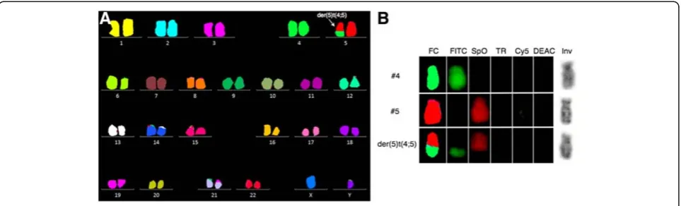
To examine the cytogenetic basis for these findings, and to disclose other possible structural balanced abnor-malities, we then performed 24-color karyotyping using 24XCyte human multicolor FISH (mFISH) probe kit according to manufacturer’s instructions (MetaSystems, Altlussheim, Germany) consisting of 24 different chromosome painting probes, each labeled with one of five fluorochromosomes or a unique combination thereof (combinatorial labeling). Image capture was done with an automated Zeiss Axio Imager.Z2 equipped with a CCD-camera (CoolCube1, MetaSystems) and appropriate filters using Isis software. Karyotyping was done using the 24-color mFISH upgrade package. Of 12 metaphases analyzed, 7 were abnormal all harboring the unbalanced translocation der(5)t(4;5) without additional structural chromosomal abnormalities (Figure 1). Re-analysis of the G-banding could not unequivocally identify the der(5)t (4;5), which also remained cryptic upon analysis of inverted gray scale image of the DAPI counterstain image channel of positively identified translocation posi-tive chromosomes (Figure 1B).
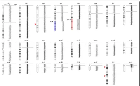
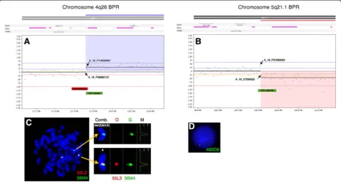
scanned at 2,5μm with GenePix 4400A microarray scan-ner. Initial analysis and normalization was done with BlueFuseMulti v2.6. For analysis and visualization nor-malized log2 probe signal values were imported into Nexus Copy Number software v. 6.1 (BioDiscovery, Cali-fornia, USA) and segmented using FASST2 segmentation algorithm with a minimum of 3 probes/segment. Regions of gain or loss contained within copy number variable regions (CNVs) were discarded. Reference genome was NCBI build 36.1 (hg18). As expected, we found the two large chromosomal imbalances: a partial gain of chromo-some 4 from q26 to q35.2 (pos. 117,710,502-190,613,014) and a partial loss of chromosome 5 q21.1 (pos. 100,418,842-180,857,866) together with three minor copy number losses (Figure 2). The analysis indicated that the breakpoint at 4q26 was between the oligo-ID’s A_16_P36882121 (pos. 117,710,502) and A_18_P14859960 (pos. 117,727,528) and that the breakpoint at 5q21.1 was between the oligo-ID’s A_16_P37295003 (pos.100,418,842) and A_16_P37295025 (pos. 100,432,041) (Figure 3A and B). To verify the break-points, FISH analysis using BAC-based BlueFISH probes (BlueGnome,Cambridge, UK) specific for the BPR were per-formed. A dual-color FISH assay on metaphases with the probes RP11-55 L3 (red) and RP11-36 M4 (green) specific for 4q26 (Figure 3A) showed one fused signal at each of the two normal chromosomes 4 and one green signal at the der (5)t(4;5) (Figure 3C, left panel). Analyzing the relative red and green fluorochrome intensities along the chromosome axis on one of the normal chromosomes 4 and the der(5)t (4;5), using the single color gallery tool in Isis (MetaSystems; Altlussheim, Germany), revealed a small portion of the red-labeled probe RP11-55 L3 present at der(5)t(4;5) (Figure 3C,
right panel). This result strongly indicates that the break-point is close to the one end of this probe and confirms the breakpoint as uncovered by the oligo-based aCGH analysis. The green-labeled FISH probe RP11-460O8 specific for 5q21.1 (Figure 3B) revealed that approximately 80 % of the nuclei had one strong green signal and one minor green sig-nal (1G1g pattern) (Figure 3D) while a minority of cells had two equally strong signals (2G pattern). This result shows that RP11-460O8 includes the breakpoint at 5q21.1 and confirms the breakpoint identified by the oligo-based aCGH analysis. Lack of material precluded further FISH analysis.
The breakpoints for the unbalanced translocation at 4q26 and at 5q21.1 is outside any known genes whereas a couple of genes are located within 1,5 Mb of the break points (Figure 3A and B, top). The MIR1973 gene is located approximately 0,3 Mb centromeric for the break-point at 4q26 in the copy number neutral region and the TRAM1L1 gene is located approximately 1,4 Mb telo-meric from the breakpoint within the gained region. At 5q21.1 the genes FAM174A, ST8SIA4 and MIR548p is located up to 0,4 Mb centromeric from the breakpoint and no known genes up to app. 1 Mb telomeric from the breakpoint. From these data we concluded that there is no apparent gene disruption and therefore it is unlikely that the unbalanced translocation has resulted in a fusion protein.
Discussion
We described a patient with T-cell ALL with a novel unbalanced karyotype 46,XY,der(5)t(4;5)(q26;q21.1)[13]/ 46,XY[12]. To our knowledge the der(5)t(4;5) has not pre-viously been described in the literature [14]. However, two cases of peripheral T-cell lymphoma with reciprocal trans-locations involving 4q26, t(4;16)(q26;p13) have been reported. In one of these cases extended analyses showed that the translocation resulted in rearrangement of inter-leukin 2 gene but this gene is located at pos. 123,59 Mb at 4q27 [15,16]. There are no reported cases with transloca-tions involving 5q21 [14]. Isolated trisomy 4 was reported in 107 cases (2 ALL, 10 bi-lineage acute leukemia and the rest mostly myeloid leukemias) with no reports on partial trisomy 4q. Whole or partial monosomy 5 has been exten-sively reported in myeloid disorders and only in two cases of chronic lymphatic leukemia.
Unlike balanced reciprocal translocations, in which the genes that become deregulated and the functional conse-quences of the rearrangements can be readily identified through analysis of the breakpoint regions, most chromo-somal imbalances have functional consequences that are unknown. This is mostly because the imbalances affect
large genomic regions containing multiple genes and the fact that tumors often have numerous unbalanced chromosomal abnormalities. This degree of genetic com-plexity has hampered delineation of the roles of individual chromosomal gains and losses. Unbalanced translocations between non-homologous chromosome arms are most often part of complex karyotypes but rarely seen as sole chromosomal aberrations making such cases important for further studies. Although we did not clone the BPR or investigate the gene expression the molecular cytogenetic results provide important clues for further clinical and diagnostic investigations. Gain of 4q or loss of 5q could re-sult in a gene dosage effect.
Alternatively, sequences close to the BPR could inter-fere with critical genes on either side of the break point as in the case for der(19)t(1;19) in B-ALL [11,12]. In our T-ALL patient we identified the BPR at 4q26 (pos. 117,710,502) with the genes MIR1973 and TRAM1L1 located on each side of the breakpoint, and the BPR at 5q21.1 (pos. 100,418,842) with the genes FAM174A, ST8SIA4 and MIR548p located centromeric to the break point. The function of these genes is mainly Figure 2High-resolution array CGH reveals the unbalanced translocation der(5)t(4;5)(q26;q21.1) and three small copy number
alterations.To the right of the individual ideograms microarray profiles of copy number gains and losses are depicted. Gain is indicated by blue color and loss is indicated by red color as an overlay on the ideogram. The log2ratios for each chromosome are−2.5, 0, and +2.5 as illustrated for
unknown, except for the gene ST8SIA4 that is one of the key enzymes of biosynthesis of polysialic acid (PSA). Interestingly, it was recently shown that polysialysa-tion of surface proteins on dendritic cells influence their T-lymphocyte interactions and that removal of PSA from their surface promoted activation and prolif-eration of T-lymphocytes [17].
The unbalanced der(5)t(4;5) as described in our case may have arisen through three different mechanisms: 1) from a balanced t(4;5) with initial loss of der(4) and subsequent doubling of the remaining normal chromosome 4, 2) from an initial trisomy 4 followed by translocation and loss of der (4) or 3) from a translocation in G2 phase of the cell cycle, with the der(4) and der(5) ending up in different daughter cells followed by selective growth advantage. All three alter-natives result in partial trisomy of 4q involving loci telo-meric to MIR1973 and partial monosomy of 5q involving loci telomeric to ST8SIA4.
Irrespective of the mechanism and what genes that are affected, the unbalanced der(5)t(4;5) may confer prolif-erative and growth advantages that contribute to neo-plastic progression.
Conclusion
We have described a novel unbalanced translocation der(5)t(4;5)(q26;q21.1) as the sole cytogenetic abnor-mality in a patient withde novoT-ALL. It is thought to play a crucial role in the pathogenesis of acute T-cell lymphoblastic leukemia either because of the gain of 4q, the loss of 5q or deregulation of genes in proximity to the BPR, as no apparent gene disruption was uncovered. One explanation that this unbalanced translocation has not been described previously could be that it is very rare or more likely that it is underreported due to the translocation’s cryptic nature. Further investigations are necessary for clarification.
Consent
Written informed consent was obtained from the patient before publication of this case report and any accom-panying images. A copy of the written consent is avail-able for review by the Editor-in-Chief of this journal.
Abbreviations
aCGH: array comparative genomic hybridization; BAC: Bacterial artificial chromosomes; BM: Bone marrow; BPR: Break point region; B-ALL: Precursor Figure 3High-resolution array CGH reveals the breakpoints of the unbalanced translocation.Vertical blue lines indicate log2ratios +0.24,
+0.60 and 2.50 and red lines indicate log2ratios−0.24, -1.00 and−2.50. At the top are indicated location of genes, CNV’s and miRNA. The X-axes at the
Author details
1Cancer Cytogenetics Laboratory, Department of Hematology, Aarhus
University Hospital, Tage-Hansensgade 2, DK-8000 Aarhus C, Denmark.
2Laboratory of Immunohematology, Department of Hematology, Aarhus
University Hospital, Tage-Hansensgade 2, DK-8000 Aarhus C, Denmark.
Received: 6 December 2011 Accepted: 13 April 2012 Published: 1 May 2012
References
1. Pui C-H, Relling MV, Downing JR:Acute lymphoblastic leukemia.
In N Engl J Med2004,350:1535–1548. 1535–1548.
2. H. Swerdlow S, Agency for Research on Cancer I, Health Organization W:
WHO classification of tumours of haematopoietic and lymphoid tissues.
pp. 439; 2008:439.
3. Vitale A, Guarini A, Ariola C, Mancini M, Mecucci C, Cuneo A, Pane F, Saglio G, Cimino G, Tafuri A,et al:Adult T-cell acute lymphoblastic leukemia: biologic profile at presentation and correlation with response to induction treatment in patients enrolled in the GIMEMA LAL 0496 protocol.In Blood2006,107:473–479. 473–479.
4. Lones MA, Heerema NA, Le Beau MM, Sposto R, Perkins SL, Kadin ME, Kjeldsberg CR, Meadows A, Siegel S, Buckley J,et al:Chromosome abnormalities in advanced stage lymphoblastic lymphoma of children and adolescents: a report from CCG-E08.In Cancer Genet Cytogenet2007,
172:1–11. 1–11.
5. Harrison CJ, Foroni L:Cytogenetics and molecular genetics of acute lymphoblastic leukemia.InRev Clin Exp Hematol, vol. 6. pp. 91–113; discussion 200–112; 2002:91–113; discussion 200–112.
6. Schneider NR, Carroll AJ, Shuster JJ, Pullen DJ, Link MP, Borowitz MJ, Camitta BM, Katz JA, Amylon MD:New recurring cytogenetic abnormalities and association of blast cell karyotypes with prognosis in childhood T-cell acute lymphoblastic leukemia: a pediatric oncology group report of 343 cases.In Blood2000,96:2543–2549. 2543–2549.
7. Adeyinka A, Smoley S, Fink S, Sanchez J, Van Dyke DL, Dewald G:
Isochromosome (X)(p10) in hematologic disorders: FISH study of 14 new cases show three types of centromere signal patterns.In Cancer Genet Cytogenet2007,179:25–30. 25–30.
8. Sanada M, Uike N, Ohyashiki K, Ozawa K, Lili W, Hangaishi A, Kanda Y, Chiba S, Kurokawa M, Omine M,et al:Unbalanced translocation der(1;7)(q10; p10) defines a unique clinicopathological subgroup of myeloid neoplasms.In Leukemia2007,21:992–997. 992–997.
9. Bacher U, Haferlach T, Schoch C:Gain of 9p due to an unbalanced rearrangement der(9;18): a recurrent clonal abnormality in chronic myeloproliferative disorders.In Cancer Genet Cytogenet2005,160:179–183. 179–183.
10. Yamamoto K, Okamura A, Wakahashi K, Katayama Y, Shimoyama M, Matsui T:A novel unbalanced whole-arm translocation der(3;10)(q10;q10) in acute monocytic leukemia.Cancer Genet Cytogenet2010,199:134–138. 134–138.
11. Paulsson K, Horvat A, Fioretos T, Mitelman F, Johansson B:Formation of der (19)t(1;19)(q23;p13) in acute lymphoblastic leukemia.In Genes
Chromosomes Cancer2005,42:144–148. 144–148.
2009,33:188–191. 188–191.
17. Curreli S, Arany Z, Gerardy-Schahn R, Mann D, Stamatos NM:Polysialylated neuropilin-2 is expressed on the surface of human dendritic cells and modulates dendritic cell-T lymphocyte interactions.In J Biol Chem2007,
282:30346–30356. 30346–30356. doi:10.1186/1755-8166-5-21
Cite this article as:Kjeldsen and Roug:A novel unbalancedde novo
translocation der(5)t(4;5)(q26;q21.1) in adult T-cell
precursor lymphoblastic leukemia.Molecular Cytogenetics20125:21.
Submit your next manuscript to BioMed Central and take full advantage of:
• Convenient online submission • Thorough peer review
• No space constraints or color figure charges • Immediate publication on acceptance
• Inclusion in PubMed, CAS, Scopus and Google Scholar
• Research which is freely available for redistribution