R E S E A R C H
Open Access
Deregulation of apoptosis-related genes is
associated with
PRV1
overexpression and JAK2
V617F allele burden in Essential
Thrombocythemia and Myelofibrosis
Raquel Tognon
1, Elainy PL Gasparotto
1, Renata P Neves
1, Natália S Nunes
1, Aline F Ferreira
1, Patrícia VB Palma
2,
Simone Kashima
2, Dimas T Covas
2,5, Mary Santana
3, Elizabeth X Souto
3, Maria Aparecida Zanichelli
4,
Belinda P Simões
5, Ana Maria de Souza
1and Fabíola A Castro
1,6*Abstract
Background:Essential Thrombocythemia (ET) and Primary Myelofibrosis (PMF) are Chronic Myeloproliferative Neoplasms (MPN) characterized by clonal myeloproliferation/myeloaccumulation without cell maturation
impairment. The JAK2 V617F mutation andPRV1gene overexpression may contribute to MPN physiopathology. We hypothesized that deregulation of the apoptotic machinery may also play a role in the pathogenesis of ET and PMF. In this study we evaluated the apoptosis-related gene and protein expression of BCL2 family members in bone marrow CD34+hematopoietic stem cells (HSC) and peripheral blood leukocytes from ET and PMF patients. We also tested whether the gene expression results were correlated with JAK2 V617F allele burden percentage, PRV1overexpression, and clinical and laboratory parameters.
Results:By real time PCR assay, we observed that A1, MCL1, BIK and BID, as well asA1, BCLWand BAKgene expression were increased in ET and PMF CD34+cells respectively, while pro-apoptotic BAXand anti-apoptotic BCL2mRNA levels were found to be lower in ET and PMF CD34+cells respectively, in relation to controls. In patients’leukocytes, we detected an upregulation of anti-apoptotic genes A1, BCL2, BCL-XLandBCLW. In
contrast, pro-apoptoticBIDand BIMELexpression were downregulated in ET leukocytes. Increased BCL-XLprotein expression in PMF leukocytes and decreased BID protein expression in ET leukocytes were observed by Western Blot. In ET leukocytes, we found a correlation between JAK2 V617F allele burden andBAX, BIK and BADgene expression and betweenA1, BAX andBIK andPRV1gene expression. A negative correlation between PRV1gene expression and platelet count was observed, as well as a positive correlation betweenPRV1 gene expression and splenomegaly.
Conclusions:Our results suggest the participation of intrinsic apoptosis pathway in the MPN physiopathology. In addition,PRV1and JAK2 V617F allele burden were linked to deregulation of the apoptotic machinery.
Keywords:Chronic Myeloproliferative Neoplasms, Apoptosis, JAK2 V617F allele burden,PRV1, BCL2 family members
* Correspondence: castrofa@fcfrp.usp.br
1Department of Clinical, Toxicological and Bromatological Analysis, University
of São Paulo, Ribeirão Preto School of Pharmaceutical Sciences, Ribeirão Preto, Brazil
Full list of author information is available at the end of the article
Background
Essential Thrombocythemia (ET) and Primary Myelofi-brosis (PMF) are disorders classified as Philadelphia chromosome-negative Myeloproliferative Neoplasms (MPN) [1]. ET is a clonal disease characterized by an increase in the platelet count associated with bone mar-row megakaryocyte hyperplasia. Thrombosis and hemor-rhagic events are the main co-morbities in ET patients. PMF is characterized by bone marrow fibrosis, as well as peripheral blood findings such as anemia, leukoery-throblastosis and the presence of dacryocytes in periph-eral blood [2].
The JAK2 V617F mutation, which leads to constitutive JAK2 activation, was shown to play an important role in MPN pathogenesis, and is found in 95% of Polycythemia Vera (PV) patients and in at least 50% of ET and PMF patients [3]. Constitutive JAK2 activation triggers several signaling pathways linked to cell survival and prolifera-tion promoting myeloproliferaprolifera-tion and resistance to cell death [4-8]. Other mutations have recently been described in ET and PMF patients, such as mutations in
JAK2 exon 12 and in the TET2, CBL, MPL and AXSL
genes [9-13]. Several studies suggest an association between MPN clinical features and the JAK2 V617F allele burden [14]. Although this knowledge and the identification of these additional mutations greatly enhanced our understanding of MPN physiopathology, a complete understanding of the cellular and molecular mechanisms involved is still lacking.
Another relevant molecular alteration described in MPN patients is the overexpression of PRV1, a surface receptor from hematopoietic cells associated with cell proliferation [15] related to JAK/STAT and SRC kinase pathways [16]. PRV1 overexpression was initially described in PV patients and in some cases of ET, but was not found elevated in other malignant hematologi-cal diseases such as Chronic Myeloid Leukemia (CML) [15,17,18]. Considering thatPRV1 gene expression is deregulated in MPN, it has been suggested that it may be used as a molecular marker in the diagnosis of these diseases [19]. Literature also supports the hypothesis thatPRV1 overexpression contributes to MPN physio-pathology, considering that there are studies showing a correlation betweenPRV1expression and patients’ clini-cal and laboratory features [15].
The apoptosis process may be triggered by two major pathways: the extrinsic or death-receptors pathway, and the intrinsic or mitochondrial pathway. The intrinsic apoptosis pathway or mitochondrial pathway is triggered by several stimuli such as ultra violet radiation, growth factor/cytokine deprivation, chemotherapeutic agents, viral infection and other stress factors related to physical and chemical injuries [20]. This pathway is mainly
regulated by BCL2 family members, classified as either anti-apoptotic (BCL2, BCL-XL, BCLW, MCL1 and A1) or pro-apoptotic (BAX, BAK, BAD, BID, BIM, Bok, BIK, BMF, BOO, BCL-XS, PUMA and NOXA) proteins [21]. The apoptotic process was found to be deregulated in several hematopoietic neoplasms, leading to resistance to therapy and progression of disease. Alterations in apoptosis have been described in CML, Myelodysplastic Syndrome (MDS), Acute Myeloid Leukemia (AML), PV, ET and PMF [20,22-24].
In this study we investigated the potential association between apoptosis deregulation, JAK2 mutation and
PRV1 overexpression in ET and PMF patients. We focused on evaluating the expression of apoptosis-related genes of the BCL2 family, JAK2 V617F allele burden and PRV1 expression in ET and PMF in bone marrow CD34+hematopoietic stem cells (HSC) and per-ipheral blood leukocytes from ET and PMF patients. The correlation between gene expression, JAK2 allele burden, clinical and laboratory parameters and PRV1
expression were also assessed.
We observed a deregulation in the expression of most of the studied genes in bone marrow CD34+ cells and peripheral leukocytes from ET and PMF patients in comparison to controls. Furthermore, a correlation between BAD, BAX and BIK expression and JAK2 V617F allele burden as well as between A1, BAX and
BIK andPRV1 expression was detected. A negative cor-relation between PRV1 expression and platelet count was also observed, as well as a positive correlation betweenPRV1expression and splenomegaly.
Subjects and methods Patients and Controls
bone marrow samples to be obtained from bone marrow donors during the bone marrow transplantation cell col-lection procedure.
JAK2 V617F allele burden detection, platelet count and spleen size determination
Detection of the JAK2 V617F mutation and the allele burden were determined as described in Tognon et al., 2011 [24], by real time allelic discrimination PCR assay. Platelets count from ET and PMF peripheral blood was determined by the Abbott Cell Dyn 3500SL Hematology Analyzer. Spleen size was determined by ultrasonogra-phy. In order to correlate gene expression and spleno-megaly, we used the increase in centimeters (cm) along the longer dimension of the patients’spleen, considering as reference, a value of 12 cm.
Cell Isolation, RNA extraction and cDNA synthesis
Cell isolation, RNA extraction and cDNA synthesis were performed as described in Tognon et al., 2011 [24]. Briefly, bone marrow CD34+ HSC were separated using the Ficoll-Hypaque protocol followed by Miltenyi CD34 isolation kit MidiMacs CD34+ Isolation Kit (MACS; Miltenyi Biotec, Bergisch Gladbach, Germany) and peripheral leukocytes were obtained by the Haes-Steril method. Total RNA from CD34+ HSC and leu-kocytes was extracted according to the Trizol™ (Invi-trogen Life Technologies, CarlsBAD, California, USA) method. One microgram of RNA was used to synthe-size cDNA using the High Capacity™ Kit from Applied Biosystems Life Technologies (Foster City, California, USA).
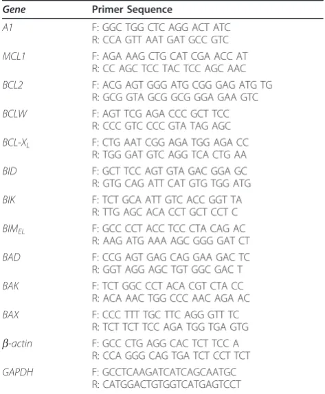
Quantification of apoptosis-related gene and PRV1 expression
The expression of anti-apoptotic genes A1, MCL1,
BCL2, BCL-XL and BCLW and pro-apoptotic genes
BAD, BAX, BAK, BID, BIKandBIMELwas evaluated by real time PCR in duplicate. For gene expression quanti-fication we used the SYBR Green PCR Master Mix Kit (Applied Biosystems) and specific oligonucleotides (Invi-trogen Life Technologies) (Table 1) on the 7500 Real Time PCR System (Applied Biosystems Life Technol-ogy). The results were normalized by the geometric mean of thebeta-actinand the GAPDHhousekeeping genes expression and represented by 2-ΔΔCtas described by Tognon et al., 2011 [24].
The PRV1gene was quantified by the TaqMan PCR Master Mix kit using TaqMan®Gene Expression Assay (PRV1 - Hs00360669_m1; GAPDH-Hs99999905_m1) on Mastercycler®ep Realplex (Eppendorf AG, Hamburg, Germany). For this gene,GAPDH was used as house-keeping gene.
Western Blot
One million leukocytes were lysated in 100 uL of sample buffer (5% mercaptoethanol, 4% sodium dodecyl sulfate - SDS, 20% glycerol and 100 mM Tris-HCl-pH 6.8), boiled at 100°C for 5 minutes and kept at -20°C until the Western-Blot analysis was performed. Twenty-five ml of patient and controls’lysate were loaded in polya-crilamide gel, the proteins were separated on 15% SDS-PAGE and transferred onto polyvinylidene difluoride (PVDF) membrane (Amersham, GE Healthcare Life Science). The membranes were incubated with the pri-mary antibody anti-BCL-XL (1:200 dilution, H-62, SC7195, Santa Cruz Biotechnology®) or anti-BID (1:500 dilution, # 611528, BD Pharmingen) diluted in TBS-Tween (20 mM Tris, 137 mM NaCl, 0.01% TBS-Tween-20) with 5% non-fat milk for 16 hours. As a control for sample loading, the blot was probed with anti-g-tubulin (1:2000 dilution, T3320, Sigma-Aldrich). Horseradish peroxidase (HRP) conjugated secondary antibodies and ECL Plus® (Amersham, GE Healthcare Life Science) were used for protein expression detection. Protein expression was measured by densitometry analysis per-formed by ImageQuant TL (Image Analysis Software, 2005, Amersham). To express the proteins densitometry results, firstly we calculated the ratio between protein test IDV (Integrated density value) and tubulin IDV, and
Table 1 Real time PCR primer sequences
Gene Primer Sequence
A1 F: GGC TGG CTC AGG ACT ATC
R: CCA GTT AAT GAT GCC GTC
MCL1 F: AGA AAG CTG CAT CGA ACC AT
R: CC AGC TCC TAC TCC AGC AAC
BCL2 F: ACG AGT GGG ATG CGG GAG ATG TG
R: GCG GTA GCG GCG GGA GAA GTC
BCLW F: AGT TCG AGA CCC GCT TCC
R: CCC GTC CCC GTA TAG AGC
BCL-XL F: CTG AAT CGG AGA TGG AGA CC
R: TGG GAT GTC AGG TCA CTG AA
BID F: GCT TCC AGT GTA GAC GGA GC
R: GTG CAG ATT CAT GTG TGG ATG
BIK F: TCT GCA ATT GTC ACC GGT TA
R: TTG AGC ACA CCT GCT CCT C
BIMEL F: GCC CCT ACC TCC CTA CAG AC
R: AAG ATG AAA AGC GGG GAT CT
BAD F: CCG AGT GAG CAG GAA GAC TC
R: GGT AGG AGC TGT GGC GAC T
BAK F: TCT GGC CCT ACA CGT CTA CC
R: ACA AAC TGG CCC AAC AGA AC
BAX F: CCC TTT TGC TTC AGG GTT TC
R: TCT TCT TCC AGA TGG TGA GTG
b-actin F: GCC CTG AGG CAC TCT TCC A
R: CCA GGG CAG TGA TCT CCT TCT
GAPDH F: GCCTCAAGATCATCAGCAATGC
R: CATGGACTGTGGTCATGAGTCCT
then we calculated the protein expression ratio between patient and controls (IDV of patients protein expression divided by controls protein expression).
Statistical analysis
The Mann-Whitney test (t-test, one-tailed) was used to compare the values of gene expression among the studied groups. The correlations between apoptosis-related gene expression, JAK2 allele burden andPRV1gene expres-sion were carried out by Spearman tests using Prisma 5.0 Software. Apvalue < 0.05 was taken as significant.
Results
Gene expression of apoptosis-related BCL2 family members is deregulated in ET and PMF patients
Gene expression analysis in CD34+HSC from ET patients showed an increased expression of anti-apoptotic genes
A1andMCL1and of pro-apoptotic genesBIDandBIK
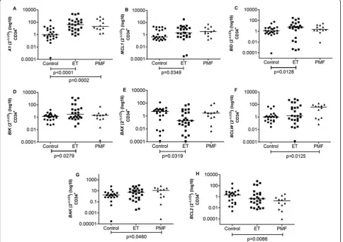
(median = 39.25, 2.07, 5.69 and 2.99, respectively) in com-parison to controls (0.986, 0.431, 1.15 and 1.36, respec-tively) (p < 0.0001, p = 0.0349, p = 0.0128 and p = 0.0279, respectively) (Figure 1A-D). The mRNA level of the pro-apoptotic gene BAX was found to be lower (0.19) in patients than controls (5.08) (p = 0.0319) (Figure 1E).
In PMF CD34+ HSC the anti-apoptotic genes A1, BCLW and pro-apoptotic BAKexpression were signifi-cantly increased (21.11, 1.55 and 11.67, respectively) compared to controls (0.99, 1.01 and 1.67, respectively) (p = 0.0002, p = 0.0125 and p = 0.0460, respectively) (Figure 1A, F and 1G).BCL2 mRNA levels were down-regulated in these cells (0.18) in comparison to controls (1.99) (p = 0.0086) (Figure 1H).
In ET patients’ leukocytes we found an overexpres-sion of the anti-apoptotic genesA1, BCL2, BCL-XLand
BCLW (16.10, 2.21, 2.71 and 2.21, respectively) when compared with controls (0.49, 0.88, 0.80 and 1.06,
respectively) (p < 0.0001, p = 0.0218, p = 0.0043 and p = 0.0342, respectively) (Figure 2A-D). The expression of pro-apoptotic genes BID and BIMELwas lower in ET leukocytes (0.62, 0.53) than in controls (1.43, 0.86) (p = 0.0004, p = 0.0116) (Figure 2E, F).
In PMF leukocytes,BCL2, BCL-XLand BCLWexpression were elevated (9.17, 16.10, 27.86, respectively) compared to controls (0.88, 0.80 and 1.07, respectively) (p = 0.0291, p < 0.0001 and p = 0.0007, respectively) (Figure 2B, C and 2D).
Pro-apoptoticBADexpression was found to be increased in ET and PMF leukocytes (3.92, 5.18) compared to con-trols (0.63) (p = 0.0130, p = 0.0276) (data not shown).
There were no differences when we comparedBCL-XL , BADandBIMELexpression between CD34+ cells from ET and PMF patients and controls, and this was also the case for MCL1, BID, BIK andBAX expression in leukocytes (p > 0.05). Furthermore, between ET and PMF leukocytes we only found a significant difference
in BCLW and BCL-XL expression (p = 0.0145, p =
0.0033) (Figure 2C and 2D).
BCL-XLandBIDprotein levels were different between controls and PMF and ET patients
We detected an elevated level of anti-apoptotic BCL-XL in PMF leukocytes and a decreased expression of
pro-apoptotic BID protein in ET leukocytes in comparison to control subjects (Figure 3A and 3B). Densitometry quantification by Integrated Density Value (IDV) showed that BCL-XLprotein level is 2.4 times higher in PMF leukocytes than in controls, and BID protein in ET Figure 2Gene expression in Control, ET and PMF leukocytes. (A) anti-apoptotic geneA1was increased in ET compared to control; (B) anti-apoptotic geneBCL2was increased in ET and PMF compared to control; (C) anti-apoptotic geneBCL-XLwas increased in ET and PMF compared
to control and also significantly different between ET and PMF; (D) anti-apoptotic geneBCLWwas increased in ET and PMF compared to control and also significantly different between ET and PMF; (E) pro-apoptotic geneBIDwas decreased in ET compared to control; (F) pro-apoptotic geneBIMELwas decreased in ET compared to control. The significant“p values”are shown in the figure. Otherwise, p > 0.05. The horizontal bars
show the median 2-ΔΔCtfor each group.
Figure 3Protein expression in Control, ET and PMF leukocytes. (A) anti-apoptotic BCL-XLshowed higher expression in PMF leukocyte
leukocytes is 0.64 fold decreased in relation to its expression in controls.
JAK2 V617F allele burden is correlated with BAD, BAX and BIK expression in ET patients’leukocytes
In JAK2 V617F-positive ET patients we detected lower levels of pro-apoptotic BAX, BIKand BADexpression (median: 0.26, 0.45 and 1.86, respectively) in leukocytes compared to JAK2 V617F-negative ET patients (1.59, 2.96 and 11.10, respectively) (p = 0.0189, p = 0.0309 and p = 0.0055, respectively) (Figure 4A-C). In addition,
BAX, BIK and BAD expression were negatively corre-lated with JAK2 V617F allele burden (BAX: r = -0.4522; p = 0.0102; BIK: r = -0.4067, p = 0.0196; BAD: r = -0.5966, p = 0.0006) in ET (Figure 4D-F).
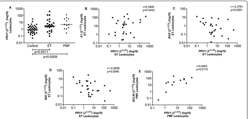
PRV1 overexpression is correlated with A1, BIK and BAX gene expression, JAK2 V617F mutation, platelet count and splenomegaly
We observed thatPRV1was overexpressed in ET (3.04) and PMF (5.12) leukocytes in comparison to controls (0.93) (p = 0.0011 and p = 0.0009) (Figure 5A). In ET leu-kocytes we also found thatPRV1expression was positively correlated withA1 expression (r = 0.3409, p = 0.0442), and negatively correlated with expression ofBAX (r=-0.3791, p = 0.0281) and BIK (r=-0.5009, p = 0.0046) (Figure 5B-D). Moreover,PRV1expression was positively correlated withBCLW(r = 0.6503, p = 0.0110) expression in PMF patients’leukocytes (Figure 5E).
We detected a positive correlation between PRV1
expression and JAK2 V617F mutation allele burden in ET and PMF patients and this result corroborates the litera-ture concerning the association betweenPRV1and JAK2 V617F described in murine myeloid cells [25] and PV patients [26]. Leukocytes from ET patients harboring the JAK2 V617F mutation showed higher expression ofPRV1
(median = 4.88) in comparison to those negative for the JAK2 V617F mutation (1.91) (p = 0.0074) (Figure 6A) and, consequently, a positive correlation betweenPRV1 expres-sion and JAK2 V617F allele burden was observed in ET patient leukocytes (r = 0.4785; p = 0.0067) (Figure 6B).
Furthermore, PRV1expression was negatively corre-lated with platelet count in ET (r=-0.3799, p = 0.0278) (Figure 7A) and PMF patients (r=-0.6713, p = 0.0084) (Figure 7B), and PRV1 expression in PMF leukocytes showed a positive correlation with the increase of the spleen size in centimeters (splenomegaly) (r = 0.6150, p = 0.0220) (Figure 7C).
Discussion
Our results indicate a deregulated expression of genes related to the intrinsic apoptosis pathway in CD34+ HSC and peripheral leukocytes from ET and PMF patients.
Our hypothesis was that higher expression levels of anti-apoptotic genes may contribute to the myeloaccu-mulation in ET and PMF. In support of this notion, in this study we found increased expression of A1, MCL1,
BCLW andBCL-XLgenes in ET and PMF patients com-pared to controls. Importantly, as previously described by our group, the cells from MPN patients are resistant to apoptosis induced by different drugs (actinomycin D, teniposide, etoposide, cytarabin, and staurosporin) [24]. These observations, and our findings in the present investigation, support our hypothesis that deregulated expression of apoptosis-related genes is linked to mye-loaccumulation and pathogenesis in ET and PMF.
The BCL2 family proteins have a central role in the process of apoptosis control. The A1, BCL2, BCLW, BCL-XLand MCL1 BCL2-family members encode anti-apoptotic molecules, while BAD, BAX, BAK, BID, BIK,
BOK, BOO, PUMA, NOXA and BIMEL encode pro-apoptotic molecules, all of them involved in the mito-chondrial apoptosis pathway [21].
A1(bfl1) expression is detected in several tissues such as hematopoietic and endothelial cells [27]. A1 gene transcription is dependent on the NF-kB pathway and its overexpression has been reported in Chronic Lym-phocytic Leukemia (CLL), particularly in CLL patients who do not respond to therapy [27].
BCL2 is an oncogene, which was first identified in Non-Hodgkin lymphoma B-cells and this gene is a pivo-tal molecule in the mitochondrial apoptosis pathway. Castro et al. (2005) described in the American Society Figure 5PRV1gene expression in Control, ET and PMF leukocytes andPRV1expression versus gene expression. (A)PRV1expression was significantly elevated in ET and PMF leukocytes; (B) anti-apoptotic geneA1showed positive correlation withPRV1expression in ET leukocytes; (C and D) pro-apoptotic genesBAXandBIKshowed negative correlation withPRV1expression in ET leukocytes; (E) anti-apoptotic geneBCLWshowed positive correlation withPRV1expression in PMF leukocytes. The significant“p values”are shown in the figure. Otherwise, p > 0.05. The horizontal bars show the median 2-ΔΔCtfor each group.
Hematology Congress [28] a decrease in BCL2 expres-sion and an increase inBCL-XL, BCLW, A1, MCL1and
cflip expression in CML and they also demonstrated that this profile of expression was correlated with CML progression. Furthermore, many publications have shown that in neoplasms such as breast or stomach can-cer, high levels of BCL2 proteins were associated with a worse prognosis [29].
Deregulation of MCL1expression was described in hepatocellular carcinoma [30] and in multiple myeloma [31]. Furthermore, the expression of this gene was also found to be correlated with prognosis in multiple mye-loma [31]. Such deregulated expression was also verified in bone marrow blasts from patients with MDS [22]. Del Poeta et al. reported increased levels of BCL2, BCL-XLand MCL1expression in AML [23] and Aichberger et al. showed that MCL1 is a BCR-ABL target gene in CML [32].
Among the pro-apoptotic proteins of the BCL2 family,
BAXhas a crucial role in apoptosis and the lack ofBAX
leads to apoptosis impairment and facilitates the develop-ment of B-cell lymphoma by c-Myc stimulation [33].
Regarding MPN pathophysiology, Zhang et al. [34] demonstrated that BCL-XLis down-regulated early dur-ingin vitrodifferentiation of megakaryocytes from ET patients and this might reflect an early entry of megakar-yocytes into a degenerating mature stage [34]. There is little data in the literature regarding apoptosis deregula-tion in PMF. On one hand, Mesa et al. (2006) showed that the levels of the anti-apoptotic and pro-apoptotic
BAX, BAK, BIMandBmfwere not different between PMF and controls [35]. On the other hand, it was demonstrated that JAK2 inhibition in a cellular model of MPN (JAK2 V617F positive cell lineage) triggers BIM activation and leads to enhanced sequestration of MCL1, furthermore, BCL-XLand MCL1 depletion by RNAi was sufficient to compromise JAK2 V617F mutant cell viabi-lity and sensitized the cells to JAK2 inhibition, indicating an association between these apoptosis-related molecules and the aberrant JAK2 signaling in these cells [36]. Figure 7Correlation ofPRV1gene expression with platelet count (PLT) and splenomegaly.PRV1expression in ET (A) and PMF (B) leukocytes showed negative correlation with platelets (PLT); (C)PRV1expression in PMF leukocytes showed positive correlation with the increase in centimeters of the spleen length (splenomegaly) determined by ultrasonography (reference value: 12 cm). The significant“p values”and the
It has been described that BCL2 family proteins interact with each other to control the intrinsic apop-tosis pathway [37]. The balance among the activities of these proteins is very important to tightly control the apoptosis process [38]. The MPN patients enrolled in this study showed overexpression of several anti-apoptotic genes such asA1andMCL1but also overex-pression of some pro-apoptotic genes such asBIK, BID
and BAKin CD34+cells. Furthermore, in CD34+ cells we observed a downregulation of BAX andBID, and
BIMEL levels were found reduced in ET leukocytes. Therefore, we postulate that the balance among these BCL2 family members is disrupted, and may contribute to the myeloaccumulation in these patients due to the increase in cell survival.
In addition, we found that mRNA levels of
pro-apopto-tic BAX, BAD and BIK mRNAs were lower in JAK2
V617F-positive patients than in those negative, and also presented a negative correlation with the JAK2 V617F allele burden. Some reviews published in the literature have already pointed out and discussed the relationship between constitutively activated JAK2/STAT signaling and deregulation of apoptosis-related genes in CML and human tumor cell lines [39,40].
Moreover, we analyzedPRV1 mRNA expression and the correlation with JAK2 V617F mutation, and with clinical laboratorial data in leukocytes from control, ET and PMF patients.PRV1is a hematopoietic cell surface receptor that has been shown to transduce intracellular signals leading to proliferation, involving JAK2 and Src kinases [16].PRV1is overexpressed in MPN patients [15,19,41,42] and seems to be associated with PV disease phenotype characterized by high erythrocyte and low pla-telet counts [15]. These studies also described a correla-tion betweenPRV1expression and the JAK2 V617F allele burden, as well as betweenPRV1overexpression and ele-vated JAK2 tyrosine kinase activity [15,19,25,41,42].
In our results we detected a correlation betweenPRV1
overexpression and the anti-apoptotic genes A1 and
BCLW, and the pro-apoptotic genesBAXandBIK expres-sion. We also found a differential expression ofPRV1
according to JAK2 V617F status and a correlation between
PRV1 expression and platelet count in ET and PMF patients, as well as splenomegaly. Thus, our results suggest a link betweenPRV1and intrinsic apoptosis pathway regu-lation and are in good agreement with previous reports about the association of the JAK2 V617F mutation with deregulation of apoptosis and disease phenotype [25].
An illustrative overview of the gene expression results in CD34+HSC and leukocytes from ET and PMF is shown in Figure 8. This figure highlights the complexity of the apop-tosis network in MPN patients. The analysis of the interac-tion of genes involved in the apoptotic machinery described here implies that apoptosis is deregulated and impaired in MPN patients since the majority of anti-apoptotic genes assessed are overexpressed, while concomitantly, some pro-apoptotic genes appear to be downregulated.
These observations are in line with previous results described by our group in Tognon et al. (2011) [24], which demonstrated that mononuclear cells from MPN patients are resistant to apoptosis, considering that patients’ cells stimulated by numerous apoptotic indu-cers, such as actinomycin and cytarabin, showed less Annexin-V FITC staining compared to controls [24]. Therefore, deregulation of the intrinsic apoptosis path-way might contribute to ET and PMF physiopathology and myeloaccumulation.
MPN do not yet have a curative therapy so it is parti-cularly relevant to consider the possibility of designing new drugs targeting apoptosis pathway. In this context, Zivny et al. (2010) reviewed this subject emphasizing the importance of developing new and more effective target cancer therapies with the potential of inhibiting the anti-apoptotic BCL2 family members or enhancing
pro-Figure 8Overview of the BCL2 family gene expression results: (A) in CD34+HSC and (B) leukocytes from patients with ET (continuous
apoptotic proteins expression [20]. These approaches might impair evasion of tumor cell to apoptosis pro-cesses or might sensitize cells to apoptosis. Taken together our results suggest that ET and PMF treatment could not be restrict to JAK2 inhibitors, considering that there are other molecular mechanisms involved in MPN pathogenesis, in addition to JAK2 mutation. Maybe in the future, JAK2 inhibitor treatment must be associated with new target therapies, such as anti-apop-totic BCL2 family members’inhibitors or pro-apoptotic enhancers, for better patients‘response to therapy.
Conclusion
CD34+ HSC and leukocytes from ET and PMF patients displayed a deregulation in expression levels of BCL2 family members, which are correlated with JAK2 V617F mutation andPRV1 mRNA levels. Our findings suggest that these alterations may contribute to increased resis-tance to apoptosis and to myeloaccumulation in ET and PMF patients.
Acknowledgements
We are really grateful to Dr. Thomas Radimerski from Novartis Institute for Biomedical Research (Basel, Switzerland) for reviewing and discussing the manuscript results. We are also grateful to Luciana Ambrosio for technical assistance. AFF, NSN and RT are recipients of a fellowship from the
Fundação de Amparo à Pesquisa do Estado de São Paulo(FAPESP) (Process
Numbers: 2008/52049-5, 2010/01756-3 and 2008/54387-5, respectively). EPLG, GLVO and RPN were recipients of fellowships from theConselho Nacional de Desenvolvimento Científico e Tecnológico(CNPq).
This work was supported by FAPESP grants Numbers 2006/50094-8 and 2008/54387-5.
Author details
1
Department of Clinical, Toxicological and Bromatological Analysis, University of São Paulo, Ribeirão Preto School of Pharmaceutical Sciences, Ribeirão Preto, Brazil.2Regional Blood Center of Ribeirão Preto, Clinical Hospital,
University of São Paulo, Ribeirão Preto School of Medicine, Ribeirão Preto, Brazil.3Brigadeiro Hospital of São Paulo, São Paulo, Brazil.4Institute for
Cancer Treatment in Children-ITACI, São Paulo, Brazil.5Department of Clinical Medicine, University of São Paulo, Ribeirão Preto School of Medicine, Ribeirão Preto, Brazil.6INCT-IF-CNPq.
Authors’contributions
RT designed and performed experiments, analyzed data and wrote the paper. EPLG, RPN, NSN, AFF and PVBP performed some of the cell isolation, RNA extraction and gene expression assays. MAZ, EXS, BPS and MS selected the patients included in this study and collected the bone marrow samples for CD34+cell isolation. NSN, MAZ, DTC, AMS and SK performed real-time
experiments, discussed the results and revised the paper. FAC conceived the project, created the study design, sought funding and wrote the paper. All authors critically reviewed the manuscript.
Competing interests
The authors declare that they have no competing interests.
Received: 28 November 2011 Accepted: 2 February 2012 Published: 2 February 2012
References
1. Tefferi A, Vardiman JW:Classification and diagnosis of myeloproliferative neoplasms: the 2008 World Health Organization criteria and point-of-care diagnostic algorithms.Leukemia2008,22:14-22.
2. Anastasi J:The myeloproliferative neoplasms: insights into molecular pathogenesis and changes in WHO classification and criteria for diagnosis.Hematol Oncol Clin North Am2009,23:693-708.
3. Vainchenker W, Delhommeau F, Constantinescu SN, Bernard OA:New mutations and pathogenesis of myeloproliferative neoplasms.Blood
2011.
4. James C, Ugo V, Le Couédic JP, Staerk J, Delhommeau F, Lacout C, Garçon L, Raslova H, Berger R, Bennaceur-Griscelli A, Villeval JL, Constantinescu SN, Casadevall N, Vainchenker W:A unique clonal JAK2 mutation leading to constitutive signalling causes polycythaemia vera. Nature2005,434:1144-1148.
5. B Baxter EJ, Scott LM, Campbell PJ, East C, Fourouclas N, Swanton S, Vassiliou GS, Bench AJ, Boyd EM, Curtin N, Scott MA, Erber WN, Green AR:
Acquired mutation of the tyrosine kinase JAK2 in human myeloproliferative disorders.Lancet2005,365:1054-1061.
6. Levine RL, Wadleigh M, Cools J, Ebert BL, Wernig G, Huntly BJ, Boggon TJ, Wlodarska I, Clark JJ, Moore S, Adelsperger J, Koo S, Lee JC, Gabriel S, Mercher T, D’Andrea A, Fröhling S, Döhner K, Marynen P, Vandenberghe P, Mesa RA, Tefferi A, Griffin JD, Eck MJ, Sellers WR, Meyerson M, Golub TR, Lee SJ, Gilliland DG:Activating mutation in the tyrosine kinase JAK2 in polycythemia vera, essential thrombocythemia, and myeloid metaplasia with myelofibrosis.Cancer Cell2005,7:387-397.
7. Kralovics R, Passamonti F, Buser AS, Teo SS, Tiedt R, Passweg JR, Tichelli A, Cazzola M, Skoda RC:A gain-of-function mutation of JAK2 in
myeloproliferative disorders.N Engl J Med2005,352:1779-1790. 8. Zhao R, Xing S, Li Z, Fu X, Li Q, Krantz SB, Zhao ZJ:Identification of an
acquired JAK2 mutation in polycythemia vera.J Biol Chem2005,
280:22788-22792.
9. Scott LM, Tong W, Levine RL, Scott MA, Beer PA, Stratton MR, Futreal PA, Erber WN, McMullin MF, Harrison CN, Warren AJ, Gilland DG, Lodish HF, Green AR:JAK2 exon 12 mutations in polycythemia vera and idiopathic erythrocytosis.N Engl J Med2007,356:459-468.
10. Delhommeau F, Dupont S, Della Valle V, James C, Trannoy S, Massé A, Kosmider O, Le Couedic JP, Robert F, Alberdi A, Lécluse Y, Plo I, Dreyfus FJ, Marzac C, Casadevall N, Lacombe C, Romana SP, Dessen P, Soulier J, Viguié F, Fontenay M, Vainchenker W, Bernard AO:Mutation in TET2 in myeloid cancers.N Engl J Med2009,360:2289-2301.
11. Grand FH, Hidalgo-Curtis CE, Ernst T, Zoi K, Zoi C, McGuire C, Kreil S, Jones A, Score J, Metzgeroth G, Oscier D, Hall A, Brandts C, Serve H, Reiter A, Chase AJ, Cross NC:Frequent CBL mutations associated with 11q acquired uniparental disomy in myeloproliferative neoplasms.Blood
2009,113:6182-6192.
12. Pikman Y, Lee BH, Mercher T, McDowell E, Ebert BL, Gozo M, Cuker A, Wernig G, Moore S, Galinsky I, DeAngelo DJ, Clark JJ, Lee SJ, Golub TR, Wadleigh M, Gilliland DG, Levine RL:MPLW515L is a novel somatic activating mutation in myelofibrosis with myeloid metaplasia.PLoS Med
2006,3:e270.
13. Carbuccia N, Murati A, Trouplin V, Brecqueville M, Adélaïde J, Rey J, Vainchenker W, Bernard OA, Chaffanet M, Vey N, Birnbaum D, Mozziconacci MJ:Mutations of ASXL1 gene in myeloproliferative neoplasms.Leukemia2009,23:2183-2186.
14. Larsen TS, Pallisgaard N, Møller MB, Hasselbalch HC:The JAK2 V617F allele burden in essential thrombocythemia, polycythemia vera and primary myelofibrosis–impact on disease phenotype.Eur J Haematol2007,
79:508-515.
15. Griesshammer M, Klippel S, Strunck E, Temerinac S, Mohr U, Heimpel H, Pahl HL:PRV-1 mRNA expression discriminates two types of essential thrombocythemia.Ann Hematol2004,83:364-370.
16. Dillon M, Minear J, Johnson J, Lannutti BJ:Expression of the GPI-anchored receptor Prv-1 enhances thrombopoietin and IL-3-induced proliferation in hematopoietic cell lines.Leuk Res2008,32:811-819.
17. Temerinac S, Klippel S, Strunck E, Röder S, Lübbert M, Lange W, Azemar M, Meinhardt G, Schaefer HE, Pahl HL:Cloning of PRV-1, a novel member of the uPAR receptor superfamily, which is overexpressed in polycythemia rubra vera.Blood2000,95:2569-2576.
18. Klippel S, Strunck E, Temerinac S, Bench AJ, Meinhardt G, Mohr U, Leichtle R, Green AR, Griesshammer M, Heimpel H, Pahl HL:Quantification of PRV-1 mRNA distinguishes polycythemia vera from secondary erythrocytosis.Blood2003,102:3569-3574.
polycythaemia vera and essential thrombocythaemia.Acta Clin Belg2009,
64:429-433.
20. Zivny J, Klener P, Pytlik R, Andera L:The role of apoptosis in cancer development and treatment: focusing on the development and treatment of hematologic malignancies.Curr Pharm Des2010,
16:11-33.
21. Thomadaki H, Scorilas A:BCL2 family of apoptosis-related genes: functions and clinical implications in cancer.Crit Rev Clin Lab Sci2006,
43:1-67.
22. Economopoulou C, Pappa V, Kontsioti F, Papageorgiou S, Kapsimali V, Papasteriadi C, Economopoulou P, Papageorgiou E, Dervenoulas J, Economopoulos T:Analysis of apoptosis regulatory genes expression in the bone marrow (BM) of adult de novo myelodysplastic syndromes (MDS).Leuk Res2008,32:61-69.
23. Del Poeta G, Bruno A, Del Principe MI, Venditti A, Maurillo L, Buccisano F, Stasi R, Neri B, Luciano F, Siniscalchi A, de Fabritiis P, Amadori S:
Deregulation of the mitochondrial apoptotic machinery and development of molecular targeted drugs in acute myeloid leukemia. Curr Cancer Drug Targets2008,8:207-222.
24. Tognon R, Gasparotto EP, Leroy JM, Oliveira GL, Neves RP, Carrara ReC, Kashima S, Covas DT, Santana M, Souto EX, Zanichelli MA, Velano CE, Simões BP, Alberto FL, Miyashiro K, de Souza AM, Amarante-Mendes GP, de Castro FA:Differential expression of apoptosis-related genes from death receptor pathway in chronic myeloproliferative diseases.J Clin Pathol
2011,64:75-82.
25. Mnjoyan Z, Yoon D, Li J, Delhommeau F, Afshar-Kharghan V:The effect of the JAK2 V617F mutation on PRV-1 expression.Haematologica2006,
91:411-412.
26. Kralovics R, Teo SS, Buser AS, Brutsche M, Tiedt R, Tichelli A, Passamonti F, Pietra D, Cazzola M, Skoda RC:Altered gene expression in
myeloproliferative disorders correlates with activation of signaling by the V617F mutation of Jak2.Blood2005,106:3374-3376.
27. Olsson A, Norberg M, Okvist A, Derkow K, Choudhury A, Tobin G, Celsing F, Osterborg A, Rosenquist R, Jondal M, Osorio LM:Upregulation of bfl-1 is a potential mechanism of chemoresistance in B-cell chronic lymphocytic leukaemia.Br J Cancer2007,97:769-777.
28. Castro FA, Jacysyn JF, Ulbrich AG, Tobo PR, Lopes LR, Colassantti MD, Camanho D, Zanichelli MA, Hamerschlak N, Cavalheiro RC, Oliveira JSR, Amarante-Mendes GP:Overexpression of the Anti-Apoptotic Genes mcl-1, bcl-w, bcl-xL and a1 Is Correlated with Chronic Myelogenous Leukemia Progression and Resistance to Gleevec [abstract].ASH Annual Meeting Abstracts2005,106:2880.
29. Frenzel A, Grespi F, Chmelewskij W, Villunger A:Bcl2 family proteins in carcinogenesis and the treatment of cancer.Apoptosis2009,14:584-596. 30. Fabregat I:Dysregulation of apoptosis in hepatocellular carcinoma cells.
World J Gastroenterol2009,15:513-520.
31. Wuillème-Toumi S, Robillard N, Gomez P, Moreau P, Le Gouill S, Avet-Loiseau H, Harousseau JL, Amiot M, Bataille R:Mcl-1 is overexpressed in multiple myeloma and associated with relapse and shorter survival. Leukemia2005,19:1248-1252.
32. Aichberger KJ, Mayerhofer M, Krauth MT, Skvara H, Florian S, Sonneck K, Akgul C, Derdak S, Pickl WF, Wacheck V, Selzer E, Monia BP, Moriggl R, Valent P, Sillaber C:Identification of mcl-1 as a BCR/ABL-dependent target in chronic myeloid leukemia (CML): evidence for cooperative antileukemic effects of imatinib and mcl-1 antisense oligonucleotides. Blood2005,105:3303-3311.
33. Eischen CM, Roussel MF, Korsmeyer SJ, Cleveland JL:Bax loss impairs Myc-induced apoptosis and circumvents the selection of p53 mutations during Myc-mediated lymphomagenesis.Mol Cell Biol2001,21:7653-7662. 34. Zhang L, Zhao H, Sun A, Lu S, Liu B, Tang F, Feng Y, Zhao L, Yang R,
Han ZC:Early down-regulation of Bcl-xL expression during
megakaryocytic differentiation of thrombopoietin-induced CD34+ bone marrow cells in essential thrombocythemia.Haematologica2004,
89:1199-1206.
35. Mesa RA, Tefferi A, Lasho TS, Loegering D, McClure RF, Powell HL, Dai NT, Steensma DP, Kaufmann SH:Janus kinase 2 (V617F) mutation status, signal transducer and activator of transcription-3 phosphorylation and impaired neutrophil apoptosis in myelofibrosis with myeloid metaplasia. Leukemia2006,20:1800-1808.
36. Rubert J, Qian Z, Andraos R, Guthy DA, Radimerski T:Bim and Mcl-1 exert key roles in regulating JAK2V617F cell survival.BMC Cancer2011,11:24.
37. Chipuk JE, Moldoveanu T, Llambi F, Parsons MJ, Green DR:The BCL-2 family reunion.Mol Cell2010,37:299-310.
38. Chipuk JE, Green DR:How do BCL-2 proteins induce mitochondrial outer membrane permeabilization?Trends Cell Biol2008,18:157-164. 39. Battle TE, Frank DA:The role of STATs in apoptosis.Curr Mol Med2002,
2:381-392.
40. Turkson J:STAT proteins as novel targets for cancer drug discovery. Expert Opin Ther Targets2004,8:409-422.
41. Puigdecanet E, Espinet B, Villa O, Florensa L, Besses C, Serrano S, Solé F:
Detection of abnormalities of PRV-1, TPO, and c-MPL genes detected by fluorescence in situ hybridization in essential thrombocythemia.Cancer Genet Cytogenet2006,167:39-42.
42. Johansson P, Andréasson B, Safai-Kutti S, Wennström L, Palmqvist L, Ricksten A, Lindstedt G, Kutti J:The presence of a significant association between elevated PRV-1 mRNA expression and low plasma
erythropoietin concentration in essential thrombocythaemia.Eur J Haematol2003,70:358-362.
doi:10.1186/1756-8722-5-2
Cite this article as:Tognonet al.:Deregulation of apoptosis-related genes is associated withPRV1overexpression and JAK2 V617F allele burden in Essential Thrombocythemia and Myelofibrosis.Journal of Hematology & Oncology20125:2.
Submit your next manuscript to BioMed Central and take full advantage of:
• Convenient online submission
• Thorough peer review
• No space constraints or color figure charges
• Immediate publication on acceptance
• Inclusion in PubMed, CAS, Scopus and Google Scholar
• Research which is freely available for redistribution