R E V I E W
Open Access
Targeting CLL-1 for acute myeloid leukemia
therapy
Hongbing Ma
1, Iyer Swaminathan Padmanabhan
2, Simrit Parmar
2*and Yuping Gong
1*Abstract
Despite major scientific discoveries and novel therapies over the past four decades, the treatment outcomes of acute myeloid leukemia (AML), especially in the adult patient population remain dismal. In the past few years, an increasing number of targets such as CD33, CD123, CLL-1, CD47, CD70, and TIM3, have been developed for immunotherapy of AML. Among them, CLL-1 has attracted the researchers’attention due to its high expression in AML while being absent in normal hematopoietic stem cell. Accumulating evidence have demonstrated CLL-1 is an ideal target for AML. In this paper, we will review the expression of CLL-1 on normal cells and AML, the value of CLL-1 in diagnosis and follow-up, and targeting CLL-1 therapy-based antibody and chimeric antigen receptor T cell therapy as well as providing an overview of CLL-1 as a target for AML.
Keywords:Acute myeloid leukemia, CLL-1, CLEC12A, DCAL-2, hMICL, CD371
Introduction
Acute myeloid leukemia (AML) is the most common and fatal hematological malignancy in adult patients where the majority have a poor prognosis. Despite major strides in the field of anti-cancer treatments and break-throughs in immunotherapies, over the past four de-cades, no significant change in the conventional chemotherapy for AML including induction and consoli-dation treatment underscores the urgency to develop new methods to improve the prognosis in this deadly
disease [1, 2]. Over the past few years, immunotherapy
has been recognized as a game changer in the field of
hematologic malignancies and solid tumors [3]; however,
due to the lack of high specificity of target antigens and heterogeneity of AML, application of a similar strategy to combat AML has been slow overall. For example, tar-geting a well-recognized surface antigen on AML cells, CD33 can also result in severe pancytopenia due to its high cross-expression on hematopoietic stem cell (HSC). In 2004, Bakker et al. used phage display technology to first identify C-type lectin-like molecule-1 (CLL-1) which is expressed on 92% AML and absent on
granulocyte-macrophage progenitors (GMPs) [4]. More
importantly, CLL-1 is also expressed on leukemic stem cell (LSC), which possesses the ability to indefinitely self-renew and produce plenty of daughter blast cells with a specific phenotype of CLL-1, CD123, CD44, CD96, CD90, CD32, CD25, and TIM-3, acting as one of
most important reasons of leukemia relapse [5–7].
Ac-cordingly, its differential characterizations allow for CLL-1 to be considered as an ideal druggable target for treatment of AML. CLL-1 is also named as c-type lectin domain family 12, member A (CLEC12A), myeloid in-hibitory c-type lectin-like receptor (MICL), dendritic cell (DC)-associated C-type lectin 2 (DCAL-2), or CD371
[8–10]. Here, we will review the advance of CLL-1 as a
therapeutic strategy for AML.
The structure and function of CLL-1
C-type lectin-like receptors play a pivotal role in the fight against infection and maintain homeostasis and self-tolerance by recognizing damage-associated—and
pathogen-associated—molecular patterns leading to
regulation of innate and adaptive immunity [11, 12]. In
contrast to classic C-type lectin receptors which are calcium-dependent, C-type lectin-like receptors are calcium-independent due to the absence of residues for calcium binding [13]. Based on the structure, C-type lec-tin and C-type-like leclec-tin receptors are categorized into type I and type II receptor, where the difference lies on
© The Author(s). 2019Open AccessThis article is distributed under the terms of the Creative Commons Attribution 4.0 International License (http://creativecommons.org/licenses/by/4.0/), which permits unrestricted use, distribution, and reproduction in any medium, provided you give appropriate credit to the original author(s) and the source, provide a link to the Creative Commons license, and indicate if changes were made. The Creative Commons Public Domain Dedication waiver (http://creativecommons.org/publicdomain/zero/1.0/) applies to the data made available in this article, unless otherwise stated.
* Correspondence:sparmar@mdanderson.org;gongyuping2010@aliyun.com
2Department of Lymphoma and Myeloma, MD Anderson Cancer Center,
Texas University, Houston, USA
1Hematology Department, West China Hospital, Sichuan University, Chengdu,
multiple and only one carbohydrate recognition domains in type I and type II, respectively [10]. The cell response depends on the balance between immune receptor tyrosine-based activation motif (ITAM) and immune re-ceptor tyrosine-based inhibitory motif (ITIM) in the intracellular NH2 terminus [14].
CLL-1 belongs to group V of C-type lectin-like receptor family. The human gene encoding CLL-1 maps to 12p13 and is within NK gene complex in which NKG2R,
low-density lipoprotein receptor-1 (LOX-1), andβglucan
receptor (BGR) are included, CLL-1 is highly homologous to LOX-1 and BGR (Fig.1a) [4,13]. The predicted size of CLL-1 gene is about 31 kDa (AY547296), encoding a
poly-peptide with 275 amino acid [10, 13]. The extracellular
feature indicates CLL-1 is a type II transmembrane glyco-protein, consisting of a single extracellular carbohydrate recognition domains with 6 N-glycosylation sites, a trans-membrane region and an intracellular NH2 terminus with
a sequence of I/VXYXXL and YXXM (Fig. 1b). The I/
VXYXXL functions as ITIM, exerting a negative role against cell activation by recruiting inhibitory Src hom-ology region 2 domain-containing phosphatase (SHP)-1 and SHP-2 [4,13,15,16]. The function of YXXM motif in CLL-1 is not clear until now. As YXXM motif carries a binding site for the p85 subunit of phosphatidylinositol 3 kinase (PI3K) which can activate down-stream signals, Darwish et al. speculated the function of CLL-1 may de-pend on the activation of ITIM or YXXM motif, which
possibly decided by the level of phosphorylation of both motifs and the efficiency of recruitment of SHP-1/2 and p85 [15,17,18]. Additionally, the YXXM and ITIM motifs have been assumed to be involved in internalization of the CLL-1 receptor upon antibody-mediated cross-linking
[19]. The exact role of YXXM in CLL-1 needs to be
ad-dressed in the future study.
Marshall et al. reported human MICL (hMICL)/CLL-1 was associated with the control of myeloid cell activation during inflammation, it was a negative regulator of
gran-ulocyte and monocyte function [9], details in this
process depended on the identification of the ligand. Al-though CLL-1 has a high sequence identity to LOX-1
and BGR which bind apoptotic cells, oxidized
low-density lipoprotein and β-glucans, respectively, the
exact ligand of CLL-1 is not completely understood [13].
In 2014, Neumann et al. revealed uric acid crystal from dead cell and unknown ligand on dead cells are ligands of CLEC12A/CLL-1, where CLEC12A/CLL-1 plays an essential role in attenuating sterile inflammation which is induced by uric acid crystal in a Syk-dependent
path-way. In Clec12a−/− mice, no difference in myeloid cells
and lymphoid cells was found from wild-type mice; how-ever, when compared with wild-type mice, monosodium urate (MSU)-induced reactive oxygen species (ROS) spe-cifically and highly increased on both bone marrow cells
and purified neutrophils of Clec12a−/− mice.
Further-more, significantly increased neutrophil infiltration
occurred in Clec12a−/− mice rather than wild-type mice when MSU crystal or dead cells were injected into peri-toneum or total body irradiation of X-ray with a dose to kill double positive thymocytes was given, accompanying
with elevated level of CXCL1,CXCL10, and TNF-α(Fig.2)
[12]. Gagne et al. showed MSU could also downregulate
expression of MICL in neutrophils, facilitating neutrophil activation and inflammatory infiltration, pretreatment with colchicine could eradicate the downregulating effect
[20]. The double-edged sword role of MSU indicates
CLL-1 plays a critical role in maintaining homeostasis and that the dysfunction of CLL-1 may elicit autoimmune dis-ease. Some studies have demonstrated CLL-1 may play a
role in the development of autoimmune diseases such as rheumatic arthritis and multiple sclerosis [21, 22]. In a
collagen antibody-induced arthritis (CAIA) model,
Clec12a−/− mice experienced more severe inflammation
during CAIA due to the over-activation of myeloid cells [23]; while in multiple sclerosis model, Sagar et al. re-vealed that CLEC12A/CLL-1 participated in the trafficking of DCs across the blood-brain barrier. The anti-CLEC12/ CLL-1 antibody can decrease the DC infiltration within the central nervous system (CNS) while restoring DC function in the peripheral blood which can result in de-layed onset of experimental autoimmune encephalomyeli-tis and alleviation of clinical symptoms as also confirmed
in CLEC12A knockout model [21]. Additionally, Begun et al. reported CLEC12A/CLL-1 to be very important in anti-bacterial defense, where Clec12a−/−mice had a higher risk to be infected due to the impaired antibacterial autophagy [24]. The inconsistent results in these studies may derive from the different target cells, variable disease model and unknown ligands, since the different C-type lectin receptor-ligand partner resulted in variant signals and
out-comes [25]. Furthermore, it was also reported that CLL-1
can mediate cell activation by an unclear mechanism [26]. More research on the ligand and pathophysiological mechanism are warranted.
Expression in normal cells and AML
In mouse model, Pyz et al. revealed the ligand of mouse MICL (mMICL) was widely expressed on bone marrow, thymus, heart, spleen, and kidney, indicating a role in
im-mune hemostasis [27]. However, although there is a
simi-larity in structure and function between mMICL and hMICL, non-hematological tissues in human seldom
ex-press CLL-1/hMICL [4, 13]. In the hematopoietic tree,
CLL1 is mainly expressed on almost all the granulocytes and monocytes, approx. 61.8% of granulocyte and mono-cyte precursors; 41.6% of progenitors and only on 2.5% of
HSC defined as CD34+CD38−, but not on T, B, NK cells
and erythrocytes and their precursors [28]. CLL1 is also
expressed on basophilic, eosinophilic granulocyte,
macro-phage, and myeloid DC [9, 10, 13, 29]. Among myeloid
progenitors defined as CD34+CD38+CD123+CD45RA+,
Marie Bill et al. reported 39.1% common myeloid progeni-tors (CMPs), 81% GMPs, and 11.9% megakaryocyte-eryth-roid progenitors (MEPs) express CLL1 whereas the
normal stem cell with lin−CD34+CD38− does not express
CLL1. In fact, the earliest CLL1+ cell in normal
hematopoiesis is CMPs and CLL1+ subsets facilitate
col-ony growth of myelomonocytic lineage [30]. In contrast,
CLL1 is present on most primary AML varying from 77.5
to 92% [28,31]. More importantly, CLL-1 can serve as a
marker of the LSC which is regarded as the source of
relapse in leukemia. Van et al. reported CLL-1+CD34
+
CD38− cells isolated from AML patients can produce
leukemia in non-obese diabetic/severe combined immuno-deficiency (NOD/SCID) mice, but CLL-1 is negative on the
CD34+CD38−cells derived from normal bone marrow,
re-generating bone marrow after chemotherapy from other diseases and mobilized peripheral blood from non-AML
patients [32]. CD123 has not only been regarded as a
marker of LSC in AML, but is also highly expressed on
non-AML regenerating bone marrow CD34+CD38− cells.
Therefore, compared with CD123, CLL-1 is a better marker for LSC. With regard to cell line, HL-60, THP-1, and U937 have a high expression of CLL-1 which facilitate to be selected as a target cell for research [4].
Diagnostic value and follow-up for minimal residual disease
In an analysis of 55 retrospective and 36 prospective samples, Larsen et al. revealed that the expression of hMICL/CLL-1 was restricted to myeloid cells as com-pared to other stem cell antigens, which indicated
CLL-1 can be used as a marker of AML diagnosis [33].
Additionally, CLL-1 expression is stable during the course of the disease and that there is no difference in expression between diagnostic and relapsed samples in
the same patients [32, 33]. Eissa et al. compared the
phenotype of bone marrow in newly diagnosed AML, in CR, and relapsed patients with ALL and healthy donors, monitoring the change after CR and relapse, they found hMICL/CLL-1 was specifically expressed in AML and showed a stable status during the course of disease [34]. Therefore, CLL-1 can be also used to monitor the min-imal residual disease (MRD) of AML in stem cell level during follow-up. Based on the analysis of 397 AML, Coustan-Smith et al. showed that combining CD371/ CLL-1 with other 21 markers facilitated distinguishing leukemic cells from normal cells, their stable expression
during the disease course made MRD more reliable [35].
Moreover, accumulating evidence demonstrate its predict-ive value in prognosis. Van et al. reported CLL-1 can be de-tected and quantified on LSC in patients at diagnosis and in CR after chemotherapy, which makes it ideal to serve as a marker of minimal residual stem cell disease [36]. High LSC frequency at diagnosis is correlated with high MRD frequency after chemotherapy and poor survival [37]. Cur-rently, CLL-1 had been integrated with abnormal makers or other lineage markers to display LSC, showing a negative
correlation with survival [36, 38]. As hMICL and CD123
were all markers of LSC, highly and stably expressed in most of AML, Rough et al. reported that combining hMICL/CLL-1 and CD123 with CD45/CD34/CD117 can sensitively detect MRD which is comparable with a real-time quantitative polymerase chain reaction,
irrespect-ive of CD34+ status [39]. High level of hMICL/CD123
MRD indicated high risk of relapse [33,39].
Of note, one needs to exclude basophils and part of DCs when CLL-1 is used as a marker to evaluate the MRD, because these cells test positive for CLL-1 and
have low CD45+ expression and low SSC on flow which
is similar to the “blast gate” [30]. Harrington et al. re-ported basophils consist of averaging 35% of bone mar-row cells of myeloproliferative disease when blast gate
was used [40]. Therefore, it needs to combine other
phenotype markers to differentiate.
Antibody-based therapy
Preclinical studies
for antibody-based therapy [4]. However, anti-CLL-1
antibody cannot inhibit the proliferation of CLL-1+
HL60 cell line, which may indicate such an antibody does not have anti-leukemic effect, the possible reason may be the absence of induction of antibody-dependent cell cytotoxicity (ADCC) and complement-dependent cytotoxicity (CDC). Van et al. proposed to combine a toxic moiety to anti-CLL-1 antibody to induce killing
ef-fect [32], where two kinds of anti-CLL-1 antibody-drug
conjugate with pyrrolobenzodiazepine (DCLL9718A) and isoquinolidinobenzodiazepine (CLT030), respect-ively, have shown powerful response to AML in animal models with no or little target off tumor toxicities [41– 43]. In contrast, Zhao et al. screened an CLL-1 anti-body from a series of candidates which showed ADCC and CDC cytotoxicity against AML cell lines and
de-layed the progress of HL-60 cells in vivo [19]. The
contradictory outcomes may derive from the difference between anti-CLL-1 antibodies. Additionally, based on the fact that tumor necrosis factor-related apoptosis-in-ducing ligand (TRAIL) can induce or increase the anti-tumor activity of neutrophil and T cell, Wiersma et al. designed a fusion protein scFvCLL1:TRAIL which can equip neutrophils with high density of TRAIL, as a result, the efficacy against AML cell line and other tumor was enhanced and more importantly, the ADCC activity of neutrophils were significantly increased when
anti-tumor monoclonal antibody was combined [44].
This provides a novel way to augment the effect of antibody-based therapy. Furthermore, bispecific antibody (T cell-dependent bispecific antibody, TDB) is another strategy. Blinatumomab, a bispecific T cell engager (BiTE) against CD3/CD19, has been approved to treat relapsed and refractory acute lymphoblastic leukemia (ALL), it can redirect and recruit unstimulated primary T cell in pa-tients against CD19-positive malignancy after binding [45]. Utilizing the same strategy, Leong et al. developed an anti-CD3/antiCLL1 T cell-dependent bispecific (TDB) antibody to treat AML and CLL-1 TDB antibody showed potent anti-leukemia activity to AML cell lines. Although high-affinity anti-CD3 TDB antibody demonstrated highly stronger effect than low-affinity anti-CD3 TDB antibody in vitro, they had almost the same effect in mice model.
Simultaneously, due to the less cytokine release,
low-affinity anti-CD3 TDB antibody was better tolerated than high-affinity anti-CD3 TDB antibody in monkey model, indicating higher safety. Therefore, low-affinity CD3 TDB antibody may be a preferable option for clinical application in the future [1]. Lu et al. also synthesized a bispecific antibody, anti-CLL1-CD3, which showed a su-perior anti-leukemia activity against AML cell lines and primary AML cells in vitro and in vivo as compared to
anti-CD33-CD3 [46]. MCLA-117, a human bispecific IgG
antibody which targets CLL-1 and CD3, was generated by
Merus B.V. and demonstrated potent cytotoxicity against primary AML cells at a low effector to target ratios in vitro [47]. Related data are summarized in Table1.
Compared with other c-type lectin receptors, DACL-2/ CLL-1 is mainly expressed on myeloid DC, it can be used as Ag capture receptor due to its internalization after lig-and binding lig-and it can also interact with TLR or CD40 to regulate the immune response. Therefore, a strategy of targeting DACL-2/CLL-1 on DCs is also a feasible way for
antibody-mediated delivery [10]. Hutten et al. showed
CLEC12A/CLL-1 on DCs was an efficient and promising
vehicle to present antigen to augment specific CD4+ and
CD8+ T cell immune response against cancer,
simultan-eously and that the antibody binding did not influence
phenotype and function of DCs [8]. However, in contrast
to in vitro results, Macri et al. reported in vivo antibody-mediated targeting CLEC12A/CLL-1 on DCs that showed an inferior response to c-type lectin domain family 9 either in cellular immunity or in humoral
im-munity [48]. Lahound et al. found that DC activation
agent could significantly enhance humoral response; moreover, OVA-conjugated with anti-CLEC12A elicited OVA-specific T cell response [49]. The reasons for the dif-ference may derive from different epitope recognition and binding efficiency of antibody or model system; further re-search is needed to elucidate [8].
Clinical trial
Up to now, there is only one clinical trial with MCLA-117 which has recruited relapsed, refractory, and newly
diag-nosed AML in old patients (≥65 years) with high-risk
cytogenetics or intolerance of induction therapy since 2016. It is a phase 1, multinational and first in a human study with a planned completion time of December 2018, where 50 patients are scheduled to be recruited with the primary goal to determine the maximum tolerated dose and then assesses the safety and efficacy based on recom-mended dose. The patients receive treatment weekly for 1 cycle, 28 days is 1 cycle, no dose, and any results are re-leased until now (NCT03038230).
Chimeric antigen receptor T cell therapy
Preclinical studies
Besides the selective expression on AML blasts and LSC, CLL-1 is also rarely expressed on non-hematological
tis-sues [4,13], making CLL-1 an ideal target for
immuno-therapy in AML. Tashiro et al., Eduardo Laborda et al., and Wang et al. developed and optimized CLL-1 CAR-T for AML; they all showed efficient and specific anti-leukemia activity to AML cell lines and primary blasts from AML patients, as well as in mouse model
[28,31, 50]. Concerning the structure of CLL-1 CAR-T,
and maintain persistent cytotoxicity after comparing one
or two combinations of CD28, 4-1BB, and OX40 [31]. It
has proved that the length of the space domain also plays a crucial role for anti-leukemia activity. Laborda et al. revealed that the shorter form is better than the
lon-ger hinge from human IgG4 in yielding cytokines [50].
In order to avoid continuous activity in vivo, inducible caspase9 suicide gene is designed in the CLL-1 CAR-T cells and can be activated by exogenous drug; a positive effect and efficiency are verified in a mouse model [31]. Kenderian et al. demonstrated that CLEC12A/CLL-1
was overexpressed on AML LSC and that the
CLEC12A+/CLL-1+AML blasts have a higher risk to be
resistant to chemotherapy than their negative counter-part. They generate second CLEC12A CAR-T with 41BB to evaluate the anti-leukemia activity, where the CAR-T cells were highly and specifically effective to CLEC12A cell lines. Although monotherapy with CLEC12A elicited modest anti-leukemia activity, a significant prolonged survival was achieved when it was sequenced after chemotherapy, indicating a preferable option for
consoli-dation to eliminate MRD and LSC [51]. Similar results
were also reported in ASH meeting 2018 [52]. Related
data are summarized in Table2.
Clinical trials
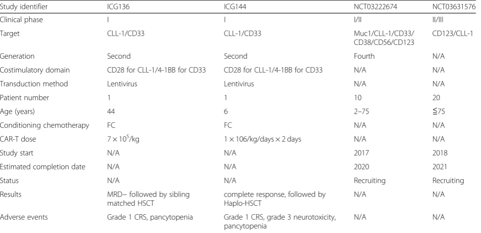
Bakker et al. reported 67% CD33-AML express CLL1,
making CLL-1 a compliment as therapeutic target [4].
On EHA meeting 2018, a team from China reported first-in-human results of a dual target combining CLL1 and CD33, where either antigen of CD33 and CLL-1 can elicit anti-leukemia activity of the compound CART (cCART). As a result, LSC and AML blasts can be eradi-cated at largest extent by the cCART and in vitro, the cCART showed specific and potent anti-leukemia effi-cacy against CLL-1 or CD33-positive on both AML cell lines and primary AML cells. In vivo experiments dem-onstrated the cCART significantly prolonged the survival of the AML mice with U937 or other cell lines. Further-more, alemtuzumab, acting as a switch, could eliminate the CAR T cells in vivo. Based on the above-mentioned results, the team designed three doses of 1 × 10⁶/kg, 3 × 10⁶/kg, and 9 × 10⁶/kg for escalation in phase I trial. In-spiringly, a 44-year-old male patient with refractory AML (AML-M4, normal karyotype, TP53 mutation)
converted to MRD− disease when a dose of 7 × 10⁵/kg
CLL-1-CD33 CAR-T cells was firstly used after T cell-depleting conditioning therapy with fludarabine 30
mg/m2 and cyclophosphamide 500 mg/m2 for 3
con-secutive days. Before receiving the CAR-T cell therapy, the patient had refractory disease to 4 cycles of chemo-therapy including DA, FLAG, and 2 cycles of priming therapy plus decitabine. The patient tolerated the ther-apy well and experienced pancytopenia, and only grade
1 cytokine release syndrome (CRS). A matched sibling allogeneic stem cell transplantation was successfully followed, and the patient is alive and disease-free at the
time of last follow-up [53]. Recently, on ASH meeting
2018, the same group reported another refractory AML with complex karyotype and FLT3-ITD mutation in a 6-year-old female patient, which was transformed from Fanconi anemia. Followed by the same conditioning
therapy, 1 × 10⁶/kg CAR-T cells were used on day 1 and
day 2, respectively and a dramatic elimination of AML cells in bone marrow within 1 week, as evidenced by 98% on day 12 and MRD on day 19, accompanying 36% and 60% CAR-T cells in PBMC and bone marrow, re-spectively, was demonstrated. The patient also experi-enced pancytopenia and grade 1 CRS, as well as grade 3 neurotoxicity. The patient went on to receive a non-myeloablative hematopoietic cell transplantation where successful hematopoiesis recovery could be seen 2 weeks after HSCT. Unfortunately, the patients
suc-cumbed to severe infection [54]. The team is enrolling
more patients to accumulate more data, the potent anti-leukemia ability implies this compound CAR-T therapy is more reasonable to act as a bridge to trans-plantation. Additionally, one phase I/II multi-CAR-T cell
therapy targeting Muc1/CLL1/CD33/CD38/CD56/
CD123 from China is enrolling patients with refractory or relapsed AML; it plans to enroll 10 patients between 2 and 75 years old and aims to evaluate the feasibility, safety, and efficacy of the fourth generation CAR-T cells, estimated completion date is December 31, 2020. Infusion dose and trial results are not yet available (NCT03222674). Another phase II/III CD123/CLL-1 CAR-T trial from China began to recruit refractory and relapsed AML pa-tients on August 15, 2018; 20 papa-tients younger than 75 years old is scheduled to assess the safety and efficacy, the primary outcome measure is leukemia-free survival of 1 year. Infusion dose is not available and estimated study completion date is August 10, 2021 (NCT03631576). Re-lated data are summarized in Table3. All the trials enroll relapsed or refractory AML in China.
Conclusion
The unique expression pattern makes CLL-1 a preferred target for AML. Although most studies regarding CLL-1 are mainly on anti-CLL-1 antibodies, the first successful utilization in human has inspired more interests in tar-geting CLL-1 for AML, it can be expected an increasing number of researches on CLL-1 CAR-T will emerge, es-pecially the combination with other markers such as CD123 or CD33 or with novel agents such as FLT3 or
BCL-2 inhibitor [2]. Considering the high cost and long
system which demonstrates several superiorities to the
previous generations [55]. The efficacy of SUPRA
CAR-T CLL-1 is merited to be investigated although there are still many challenges to be addressed in CART therapy [3, 56]. Additionally, due to high expression of CLL-1 on DCs and characteristics of internalization, DCs-based immunotherapy can potentially be exploited as another strategy. At present, more researches are needed to elucidate the exact physiological function of CLL-1; simultaneously, the outcomes from clinical inves-tigations will provide more valuable information.
Abbreviations
ADCC:Antibody-dependent cell cytotoxicity; ALL: Acute lymphoblastic leukemia; AML: Acute myeloid leukemia; BiTE: Bispecific T cell engager; CAR-T: Chimeric antigen receptor T; cCARCAR-T: Compound CAR-T;
CDC: Complement-dependent cytotoxicity; CLEC12A: c-type lectin domain family 12, member A; CLL-1: C-type lectin-like molecule-1; CMPs: Common myeloid progenitors; CNS: Central nervous system; DC: Dendritic cell; DCAL-2: Dendritic cell-associated C-type lectin 2; GMPs: Granulocyte-macrophage progenitors; hMICL: Human myeloid inhibitory C-type lectin-like receptor; HSC: Hematopoietic stem cell; LSC: Leukemia stem cell;
MEPs: Megakaryocyte-erythroid progenitors; mMICL: Mouse inhibitory C-type lectin-like receptor; MRD: Minimal residual disease; NOD/SCID: Non-obese diabetic/severe combined immunodeficiency; PI3K: Phosphatidylinositol 3 kinase; TDB: T cell-dependent bispecific; TRAIL: Tumor necrosis factor-related apoptosis-inducing ligand
Acknowledgements
The first author was supported by the China Scholarship Council for 1 year’s study at MD Anderson Cancer center.
Funding Not applicable.
Availability of data and materials Not applicable.
Authors’contributions
HBM wrote the initial drafts. YPG, SP, and SI revised the review and finalized the last version of the article. All authors checked and approved the final version.
Ethics approval and consent to participate Not applicable.
Consent for publication Not applicable.
Competing interests
The authors declare that they have no competing interests.
Publisher’s Note
Springer Nature remains neutral with regard to jurisdictional claims in published maps and institutional affiliations.
Received: 4 March 2019 Accepted: 27 March 2019
References
1. Leong SR, Sukumaran S, Hristopoulos M, Totpal K, Stainton S, Lu E, et al. An anti-CD3/anti-CLL-1 bispecific antibody for the treatment of acute myeloid leukemia. Blood. 2017;129:609–18.
2. Saygin C, Carraway HE. Emerging therapies for acute myeloid leukemia. J Hematol Oncol. 2017;10:93.
3. Im A, Pavletic SZ. Immunotherapy in hematologic malignancies: past, present, and future. J Hematol Oncol. 2017;10:94.
4. Bakker AB, van den Oudenrijn S, Bakker AQ, Feller N, van Meijer M, Bia JA, et al. C-type lectin-like molecule-1: a novel myeloid cell surface marker associated with acute myeloid leukemia. Cancer Res. 2004;64:8443–50. 5. Siveen KS, Uddin S, Mohammad RM. Targeting acute myeloid leukemia
stem cell signaling by natural products. Mol Cancer. 2017;16:13. 6. Yoshida GJ, Saya H. Therapeutic strategies targeting cancer stem cells.
Cancer Sci. 2016;107:5–11.
7. Zhou J, Chng WJ. Identification and targeting leukemia stem cells: the path to the cure for acute myeloid leukemia. World J Stem Cells. 2014;6:473–84. 8. Hutten TJ, Thordardottir S, Fredrix H, Janssen L, Woestenenk R, Tel J, et al.
CLEC12A-mediated antigen uptake and cross-presentation by human
Table 3Clinical data of CAR-T cell therapy
Study identifier ICG136 ICG144 NCT03222674 NCT03631576
Clinical phase I I I/II II/III
Target CLL-1/CD33 CLL-1/CD33 Muc1/CLL-1/CD33/
CD38/CD56/CD123
CD123/CLL-1
Generation Second Second Fourth N/A
Costimulatory domain CD28 for CLL-1/4-1BB for CD33 CD28 for CLL-1/4-1BB for CD33 N/A N/A
Transduction method Lentivirus Lentivirus N/A N/A
Patient number 1 1 10 20
Age (years) 44 6 2–75 ≦75
Conditioning chemotherapy FC FC N/A N/A
CAR-T dose 7 × 10⁵/kg 1 × 10⁶/kg/days × 2 days N/A N/A
Study start N/A N/A 2017 2018
Estimated completion date N/A N/A 2020 2021
Status N/A N/A Recruiting Recruiting
Results MRD−followed by sibling
matched HSCT
complete response, followed by Haplo-HSCT
N/A N/A
Adverse events Grade 1 CRS, pancytopenia Grade 1 CRS, grade 3 neurotoxicity, pancytopenia
N/A N/A
dendritic cell subsets efficiently boost tumor-reactive T cell responses. J Immunol. 2016;197:2715–25.
9. Marshall AS, Willment JA, Pyz E, Dennehy KM, Reid DM, Dri P, et al. Human MICL (CLEC12A) is differentially glycosylated and is down-regulated following cellular activation. Eur J Immunol. 2006;36:2159–69.
10. Chen CH, Floyd H, Olson NE, Magaletti D, Li C, Draves K, et al. Dendritic-cell-associated C-type lectin 2 (DCAL-2) alters dendritic-cell maturation and cytokine production. Blood. 2006;107:1459–67.
11. Chiffoleau E. C-type lectin-like receptors as emerging orchestrators of sterile inflammation represent potential therapeutic targets. Front Immunol. 2018;9:227. 12. Neumann K, Castineiras-Vilarino M, Hockendorf U, Hannesschlager N,
Lemeer S, Kupka D, et al. Clec12a is an inhibitory receptor for uric acid crystals that regulates inflammation in response to cell death. Immunity. 2014;40:389–99.
13. Marshall AS, Willment JA, Lin HH, Williams DL, Gordon S, Brown GD. Identification and characterization of a novel human myeloid inhibitory C-type lectin-like receptor (MICL) that is predominantly expressed on granulocytes and monocytes. J Biol Chem. 2004;279:14792–802. 14. Billadeau DD, Leibson PJ. ITAMs versus ITIMs: striking a balance during cell
regulation. J Clin Invest. 2002;109:161–8.
15. Darwish NH, Sudha T, Godugu K, Elbaz O, Abdelghaffar HA, Hassan EE, et al. Acute myeloid leukemia stem cell markers in prognosis and targeted therapy: potential impact of BMI-1, TIM-3 and CLL-1. Oncotarget. 2016;7:57811–20. 16. Robinson MJ, Sancho D, Slack EC, LeibundGut-Landmann S, Reis e Sousa C.
Myeloid C-type lectins in innate immunity. Nat Immunol. 2006;7:1258–65. 17. Wu H, Windmiller DA, Wang L, Backer JM. YXXM motifs in the PDGF-beta receptor serve dual roles as phosphoinositide 3-kinase binding motifs and tyrosine-based endocytic sorting signals. J Biol Chem. 2003;278:40425–8. 18. Songyang Z, Shoelson SE, Chaudhuri M, Gish G, Pawson T, Haser WG, et al. SH2
domains recognize specific phosphopeptide sequences. Cell. 1993;72:767–78. 19. Zhao X, Singh S, Pardoux C, Zhao J, Hsi ED, Abo A, et al. Targeting C-type
lectin-like molecule-1 for antibody-mediated immunotherapy in acute myeloid leukemia. Haematologica. 2010;95:71–8.
20. Gagne V, Marois L, Levesque JM, Galarneau H, Lahoud MH, Caminschi I, et al. Modulation of monosodium urate crystal-induced responses in neutrophils by the myeloid inhibitory C-type lectin-like receptor: potential therapeutic implications. Arthritis Res Ther. 2013;15:R73.
21. Sagar D, Singh NP, Ginwala R, Huang X, Philip R, Nagarkatti M, et al. Antibody blockade of CLEC12A delays EAE onset and attenuates disease severity by impairing myeloid cell CNS infiltration and restoring positive immunity. Sci Rep. 2017;7:2707.
22. Michou L, Cornelis F, Levesque JM, Bombardieri S, Balsa A, Westhovens R, et al. A genetic association study of the CLEC12A gene in rheumatoid arthritis. Joint Bone Spine. 2012;79:451–6.
23. Redelinghuys P, Whitehead L, Augello A, Drummond RA, Levesque JM, Vautier S, et al. MICL controls inflammation in rheumatoid arthritis. Ann Rheum Dis. 2016;75:1386–91.
24. Begun J, Lassen KG, Jijon HB, Baxt LA, Goel G, Heath RJ, et al. Integrated genomics of Crohn's disease risk variant identifies a role for CLEC12A in antibacterial autophagy. Cell Rep. 2015;11:1905–18.
25. Del Fresno C, Iborra S, Saz-Leal P, Martinez-Lopez M, Sancho D. Flexible signaling of myeloid C-type lectin receptors in immunity and inflammation. Front Immunol. 2018;9:804.
26. Hoving JC, Wilson GJ, Brown GD. Signalling C-type lectin receptors, microbial recognition and immunity. Cell Microbiol. 2014;16:185–94. 27. Pyz E, Huysamen C, Marshall AS, Gordon S, Taylor PR, Brown GD.
Characterisation of murine MICL (CLEC12A) and evidence for an endogenous ligand. Eur J Immunol. 2008;38:1157–63.
28. Wang J, Chen S, Xiao W, Li W, Wang L, Yang S, et al. CAR-T cells targeting CLL-1 as an approach to treat acute myeloid leukemia. J Hematol Oncol. 20CLL-18;CLL-1CLL-1:7. 29. Toft-Petersen M, Stidsholt Roug A, Plesner T, Ebbesen L, Brown GD, Nederby
L. The CLEC12A receptor marks human basophils: potential implications for minimal residual disease detection in acute myeloid leukemia. Cytometry B Clin Cytom. 2018;94:520–6.
30. Bill M, BvKN P, P SW, Laine Herborg L, Stidsholt Roug A, Hokland P, et al. Mapping the CLEC12A expression on myeloid progenitors in normal bone marrow; implications for understanding CLEC12A-related cancer stem cell biology. J Cell Mol Med. 2018;22(4):2311-8.
31. Tashiro H, Sauer T, Shum T, Parikh K, Mamonkin M, Omer B, et al. Treatment of acute myeloid leukemia with T cells expressing chimeric antigen receptors directed to C-type lectin-like molecule 1. Mol Ther. 2017;25:2202–13.
32. van Rhenen A, van Dongen GA, Kelder A, Rombouts EJ, Feller N, Moshaver B, et al. The novel AML stem cell associated antigen CLL-1 aids in discrimination between normal and leukemic stem cells. Blood. 2007;110: 2659–66.
33. Larsen HO, Roug AS, Just T, Brown GD, Hokland P. Expression of the hMICL in acute myeloid leukemia-a highly reliable disease marker at diagnosis and during follow-up. Cytometry B Clin Cytom. 2012;82:3–8.
34. Eissa DS, Kandeel EZ, Ghareeb M. Human myeloid inhibitory C-lectin: a highly specific and stable acute myeloid leukemia marker. Hematol Oncol. 2017;35:814–20.
35. Coustan-Smith E, Song G, Shurtleff S, Yeoh AE, Chng WJ, Chen SP, et al. Universal monitoring of minimal residual disease in acute myeloid leukemia. JCI Insight. 2018;3(9): e98561.
36. van Rhenen A, Moshaver B, Kelder A, Feller N, Nieuwint AW, Zweegman S, et al. Aberrant marker expression patterns on the CD34+CD38- stem cell compartment in acute myeloid leukemia allows to distinguish the malignant from the normal stem cell compartment both at diagnosis and in remission. Leukemia. 2007;21:1700–7.
37. van Rhenen A, Feller N, Kelder A, Westra AH, Rombouts E, Zweegman S, et al. High stem cell frequency in acute myeloid leukemia at diagnosis predicts high minimal residual disease and poor survival. Clin Cancer Res. 2005;11: 6520–7.
38. Terwijn M, Zeijlemaker W, Kelder A, Rutten AP, Snel AN, Scholten WJ, et al. Leukemic stem cell frequency: a strong biomarker for clinical outcome in acute myeloid leukemia. PLoS One. 2014;9:e107587.
39. Roug AS, Larsen HO, Nederby L, Just T, Brown G, Nyvold CG, et al. hMICL and CD123 in combination with a CD45/CD34/CD117 backbone - a universal marker combination for the detection of minimal residual disease in acute myeloid leukaemia. Br J Haematol. 2014;164:212–22.
40. Harrington AM, Olteanu H, Kroft SH. A dissection of the CD45/side scatter “blast gate”. Am J Clin Pathol. 2012;137:800–4.
41. Zheng B, Yu SF, Del Rosario G, Leong SR, Lee GY, Vij R, et al. An anti-CLL-1 antibody-drug conjugate for the treatment of acute myeloid leukemia. Clin Cancer Res. 2019;25:1358–68.
42. Leipold DD, Figueroa I, Masih S, Latifi B, Yip V, Shen BQ, et al. Preclinical pharmacokinetics and pharmacodynamics of DCLL9718A: an antibody-drug conjugate for the treatment of acute myeloid leukemia. MAbs. 2018;10(8): 1312-21.
43. Jiang YP, Liu BY, Zheng Q, Panuganti S, Chen R, Zhu J, et al. CLT030, a leukemic stem cell-targeting CLL1 antibody-drug conjugate for treatment of acute myeloid leukemia. Blood Adv. 2018;2:1738–49.
44. Wiersma VR, de Bruyn M, Shi C, Gooden MJ, Wouters MC. Samplonius DF, et al. . C-type lectin-like molecule-1 (CLL1)-targeted TRAIL augments the tumoricidal activity of granulocytes and potentiates therapeutic antibody-dependent cell-mediated cytotoxicity. MAbs. 2015;7:321–30.
45. Wu J, Fu J, Zhang M, Liu D. Blinatumomab: a bispecific T cell engager (BiTE) antibody against CD19/CD3 for refractory acute lymphoid leukemia. J Hematol Oncol. 2015;8:104.
46. Lu H, Zhou Q, Deshmukh V, Phull H, Ma J, Tardif V, et al. Targeting human C-type lectin-like molecule-1 (CLL1) with a bispecific antibody for immunotherapy of acute myeloid leukemia. Angew Chem Int Ed Engl. 2014;53:9841–5. 47. Loo PFVDR, Dolstra H, Shamsili S, Bakker L. Preclinical evaluation of
MCLA117, a CLEC12AxCD3 bispecific antibody efficiently targeting a novel leukemic stem cell associated antigen in AML. blood. 2015;126:325. 48. Macri C, Dumont C, Panozza S, Lahoud MH, Caminschi I, Villadangos JA, et
al. Antibody-mediated targeting of antigen to C-type lectin-like receptors Clec9A and Clec12A elicits different vaccination outcomes. Mol Immunol. 2017;81:143–50.
49. Lahoud MH, Proietto AI, Ahmet F, Kitsoulis S, Eidsmo L, Wu L, et al. The C-type lectin Clec12A present on mouse and human dendritic cells can serve as a target for antigen delivery and enhancement of antibody responses. J Immunol. 2009;182:7587–94.
50. Laborda E, Mazagova M, Shao S, Wang X, Quirino H, Woods AK, et al. Development of a chimeric antigen receptor targeting C-type lectin-like Molecule-1 for human acute myeloid leukemia. Int J Mol Sci. 2017;18(11): 2259.
52. Togni EDKM, Cooper ML, Ritchey J, O'Neal J, Niswonger J, DiPersio JF. Chimeric antigen receptor T cells specific for CLL-1 for treatment of acute myeloid leukemia. Blood. 2018;132(Suppl 1):2205.
53. Liu F, Pinz K, Ma Y, Wada M, Chen K, Ma G, et al. First-in-human CLL1-CD33 compound CAR T cells as a two-pronged approach for the treatment of refractory acute myeloid leukemia. HemaSphere. 2018;2(S1):29.
54. Liu F, Cao Y, Pinz K, Ma Y, Wada M, Chen K, et al. First-in-human CLL1-CD33 compound CAR-T cell therapy induces complete remission in patients with refractory acute myeloid leukemia: update on phase 1 clinical trial. Blood. 2018;132(Suppl 1):901.
55. Zhao J, Lin Q, Song Y, Liu D. Universal CARs, universal T cells, and universal CAR T cells. J Hematol Oncol. 2018;11:132.