R E S E A R C H
Open Access
Improving nelarabine efficacy in T cell
acute lymphoblastic leukemia by targeting
aberrant PI3K/AKT/mTOR signaling pathway
Annalisa Lonetti
1†, Alessandra Cappellini
2†, Alice Bertaina
3, Franco Locatelli
3, Andrea Pession
4,
Francesca Buontempo
1, Camilla Evangelisti
5, Cecilia Evangelisti
1, Ester Orsini
1, Laura Zambonin
6, Luca Maria Neri
7,
Alberto Maria Martelli
1*and Francesca Chiarini
5*Abstract
Background:Although in recent years, the introduction of novel chemotherapy protocols has improved the
outcome of T cell acute lymphoblastic leukemia (T-ALL) patients, refractory and/or relapsing disease remains a foremost concern. In this context, a major contribution was provided by the introduction of the nucleoside analog nelarabine, approved for salvage treatment of T-ALL patients with refractory/relapsed disease. However, nelarabine could induce a life-threatening, dose-dependent neurotoxicity. To improve nelarabine efficacy, we have analyzed its molecular targets, testing selective inhibitors of such targets in combination with nelarabine.
Methods:The effectiveness of nelarabine as single agent or in combination with PI3K, Bcl2, and MEK inhibitors was evaluated on human T-ALL cell lines and primary T-ALL refractory/relapsed lymphoblasts. The efficacy of signal modulators in terms of cytotoxicity, induction of apoptosis, and changes in gene and protein expression was assessed by flow cytometry, western blotting, and quantitative real-time PCR in T-ALL settings.
Results:Treatment with nelarabine as a single agent identified two groups of T-ALL cell lines, one sensitive and one resistant to the drug. Whereas sensitive T-ALL cells showed a significant increase of apoptosis and a strong down-modulation of PI3K signaling, resistant T-ALL cells showed a hyperactivation of AKT and MEK/ERK1/2 signaling pathways, not caused by differences in the expression of nelarabine transporters or metabolic activators. We then studied the combination of nelarabine with the PI3K inhibitors (both pan and dualγ/δinhibitors), with the Bcl2 specific inhibitor ABT199, and with the MEK inhibitor trametinib on both T-ALL cell lines and patient samples at relapse, which displayed constitutive activation of PI3K signaling and resistance to nelarabine alone. The combination with the pan PI3K inhibitor ZSTK-474 was the most effective in inhibiting the growth of T-ALL cells and was synergistic in decreasing cell survival and inducing apoptosis in nelarabine-resistant T-ALL cells. The drug combination caused AKT dephosphorylation and a downregulation of Bcl2, while nelarabine alone induced an increase in p-AKT and Bcl2 signaling in the resistant T-ALL cells and relapsed patient samples.
Conclusions:These findings indicate that nelarabine in combination with PI3K inhibitors may be a promising therapeutic strategy for the treatment of T-ALL relapsed patients.
Keywords:PI3K signaling, Apoptosis, Drug resistance, Combination therapy
* Correspondence:alberto.martelli@unibo.it;francesca.chiarini@cnr.it †Equal contributors
1
Department of Biomedical and Neuromotor Sciences, University of Bologna, Bologna, Italy
5Institute of Molecular Genetics, Rizzoli Orthopedic Institute, National
Research Council, Bologna, Italy
Full list of author information is available at the end of the article
Background
T cell acute lymphoblastic leukemia (T-ALL) is a hematologic malignancy resulting from the transform-ation of T cell progenitors that accounts for 15 % of pediatric and 25 % of adult ALL cases. Despite improve-ments in cure rates, the outcome of T-ALL patients with chemoresistant or relapsed leukemia is still poor [1]. T-ALL requires aggressive chemotherapy. To minimize and overcome the detrimental effects of therapeutic regi-mens, it is essential to identify novel molecular targets in T-ALL and test selective inhibitors of such targets [2]. Thus, major efforts are being made to develop targeted molecules against deregulated signaling pathways sus-taining T-ALL cell growth/survival/drug resistance. Indeed, selective inhibitors of deregulated pathways could be used together with chemotherapy, allowing for a lower dosage of chemotherapeutic drugs which should minimize toxic side effects. The nucleoside analog nelar-abine is a prodrug of the deoxyguanosine analog 9-β-D -arabinofuranosylguanine (Ara-G) [3, 4]. Nelarabine is rapidly metabolized in the plasma by an adenosine deaminase into the active metabolite Ara-G, the latter having a much longer plasma half-life and reaching higher plasma concentrations [5]. Ara-G is taken up by leukemia cells via the nitrobenzylthioinosine-sensitive nucleoside membrane transporter equilibrative nucleo-side transporter 1 (ENT1) [6]. Ara-G is then phosphory-lated to Ara-G monophosphate by the deoxycytidine and deoxyguanosine kinase (dCK and dGK, respectively). It is then phosphorylated to its triphosphate form Ara-GTP, which in turn competes with dGTP for DNA poly-merase and is subsequently incorporated into DNA, resulting in termination of DNA synthesis [7]. Since 2005, nelarabine has been approved for the treatment of both pediatric and adult T-ALL patients who have refractory or progressive disease after previous chemo-therapy regimens [5, 8]. Nelarabine is preferentially cytotoxic to T lymphoblasts through the accumulation of Ara-GTP, which occurs to a greater extent in T cells than in B cells, resulting in inhibition of ribonucleotide reductase and subsequent DNA synthesis [9, 10].
Nelara-bine as monotherapy was effective in inducing
complete/partial responses in both children and adults with refractory or relapsed T-ALL [3, 8, 11]. Given the impressive single-agent activity seen in refractory or relapsed T-ALL, substantial interest developed in evalu-ating nelarabine in the up-front treatment of T-ALL. Clinical trials have shown that nelarabine could be safely combined with intensive chemotherapy (BFM-86) in the frontline therapy of pediatric T-ALL, and in combination with hyper-CVAD in the frontline therapy for adults with T-ALL [12–14]. Of 40 patients with T-ALL or T lymphoblastic lymphoma (T-LL), the CR rate was 89 % in T-ALL and 94 % in T-LL. Overall survival at 3 years
was 63 % [14]. However, in recent years, it is emerging that nelarabine could induce a significant neurotoxicity, particularly when given in conjunction with multi-agent chemotherapy regimens [15, 16]. Patients who suffered from nelarabine neurotoxicity displayed myelopathy and severe necrotic changes in the nervous system [17], as well as irreversible paresthesia or paraplegia [17, 18]. Im-portantly, nelarabine-induced neurotoxicity depends on the dosage [19]. Therefore, a combined therapy with signal transduction modulators could allow for the use of a lower dosage of nelarabine, resulting in a lower inci-dence and/or seriousness of neurotoxicity and resistance. However, there is no information regarding how aber-rantly activated signaling pathways could influence T-ALL cell sensitivity to nelarabine. In contrast, in recent years, valuable information has been collected regarding aberrantly activated signaling pathways influ-encing leukemia cell sensitivity to fludarabine [20], a nucleoside analog which is employed in the treatment of B cell chronic lymphocytic leukemia (CLL), or to clofar-abine, a nucleoside analog used for treating acute myeloid leukemia (AML) patients [21, 22]. The effects of phosphoinositide 3-kinase (PI3K) inhibition on fludara-bine sensitivity could be related to changes in the expression of Bcl2 family proteins (Mcl-1 and Bim) [23, 24] which are also involved in clofarabine sensitivity in AML cells [22]. Importantly, PI3K/AKT/mammalian
target of rapamycin (mTOR) signaling inhibition
enhanced fludarabine-induced cell death in a T-ALL cell line (CEM-S) [25]. Moreover, a combination of clofara-bine and temsirolimus, an mTOR inhibitor, displayed synergistic cytotoxic effects in AML cell lines and primary samples, and showed an encouraging clinical activity [21, 26]. A recent study underscored a potential role for Bcl2 family proteins in determining nelarabine resistance, in an Ara-G-resistant T-ALL cell line variant, established by serial incubation with Ara-G (the active metabolite of nelarabine) [27]. In this study, anti-apoptotic Bcl-xL was augmented and pro-anti-apoptotic Bax and Bad were reduced in CEM/Ara-G cells, suggesting refractoriness to Ara-G-induced apoptosis [27].
Here, we demonstrate that in nelarabine-resistant T-ALL cell lines, there is a hyperactivation of PI3K/AKT/ mTOR, ERK1/2 and Bcl2 signaling in response to nelar-abine. Treatment with ZSTK-474 (a pan PI3K p110 inhibitor [28]), IPI-145 (Duvelisib@
a dephosphorylation of AKT and ERK1/2 and induced an increase in the expression of Bax and Bak pro-apoptotic members of Bcl2 family in T-ALL cells resist-ant to nelarabine. The combination of nelarabine with ZSTK-474 was able to induce a marked cell death in MOLT-4 cells co-cultured with HS-5 human HS-5 stromal cells, which mimic bone marrow (BM) micro-environment. These observations suggest the possibility to combine nelarabine together with selective inhibitors of the PI3K signaling pathway, to improve the efficacy of T-ALL treatment of relapsed/refractory patients.
Results
Treatment with nelarabine identifies two groups of T-ALL cell lines: one sensitive and one resistant
The effects of nelarabine on a panel of T-ALL cell lines were tested by incubating the cells for 48 h with increas-ing concentrations of the drug and then analyzincreas-ing the rate of cell viability, using MTT assays. Two significantly different groups of cell lines were outlined: a sensitive and a resistant group of cell lines. Cell viability decreased in a concentration-dependent fashion, and the IC50 values ranged between 2 and 5.5 μM for sensitive cell lines (Fig. 1a, b). However, it was observed that some cell lines were markedly resistant to nelarabine treatment. In particular, LOUCY, ALL-SIL, MOLT-16, and PEER cells did not reach an IC50 at the concentra-tions used, after 48 h of treatment (Fig. 1a, b). The most
resistant cell line was LOUCY, with an IC50 of 300 μM, calculated with the CalcuSyn software. This cell line is representative of ETP-ALL, a T-ALL subtype with a very poor prognosis [29]. In vitro studies, using T-ALL cells, demonstrated that reduction of cell viability of induced by either nelarabine or Ara-G was comparable (Fig. 1c). Importantly, nelarabine induced a significant reduction in viable cell number, in sensitive versus resistant cell lines, as documented by absolute count (Fig. 1d).
Nelarabine treatment promotes apoptosis in sensitive T-ALL cell lines
To evaluate whether the effects of nelarabine on cell viability could be related to apoptosis, flow cytometric analysis was performed on the sensitive cell lines. In response to treatment with 5 μM nelarabine (2μM for MOLT-4 cells), we detected a marked increase in the percentage of early apoptotic (positive for Annexin V) and/or late apoptotic (positive for both Annexin V and PI) cells after 48 h of treatment of T-ALL cell lines MOLT-4, JURKAT, P12-ICHIKAWA, and DND41 (Fig. 2a). Apoptosis was further investigated by western blotting, which documented a time-dependent cleavage of caspase 8, caspase 9, caspase 3, and poly(ADP-ri-bose) polymerase (PARP) in response to nelarabine treatment (Fig. 2b). All cell lines analyzed display phosphorylated AKT, which is indicative of constitutive
activation of PI3K signaling pathway [32]. We
examined the effects of nelarabine on the most import-ant signaling downstream targets of PI3K/AKT/mTOR and MEK pathways. In all T-ALL sensitive cell lines, nelarabine treatment induced a marked decrease of phosphorylated AKT at Ser473, S6 ribosomal protein (S6RP) at Ser235/236, and GSK3βat Ser9 after 48 h of treatment, suggesting that PI3K/AKT/mTOR pathway is down-modulated by nelarabine (Fig. 2c). Moreover, in all sensitive cell lines analyzed, ERK phosphorylation was impaired by the treatment, indicating also a down-regulation of the MEK pathway. In all cell lines evaluated, total protein levels were unaffected. It was demonstrated that DNA fragmentation is associated
with nucleoside analog-induced apoptosis [27, 33]. To
better understand the mechanism of
nelarabine-induced apoptosis, we have analyzed the phosphoryl-ation of H2AX. Indeed, the rapid phosphorylphosphoryl-ation of H2AX on Ser139 (γH2AX) is an early cellular response to double-strand breaks (DSBs). This phosphorylation event is one of the most well-established chromatin modifications linked to DNA damage. Moreover, the production of reactive oxygen species (ROS) coincides with the appearance of DNA damage and partially contributes to the DNA damage accumulated, followed by apoptosis at later time points [34]. MOLT-4 and Jurkat cell lines were treated with nelarabine for 7 and
24 h, and γH2AX was evaluated by flow cytometry in both control and treated cells. We observed a time-dependent increase ofγH2AX in response to nelarabine in both cell lines compared to controls (untreated cells). In addition, we measured the intracellular ROS levels in response to nelarabine treatment, and we observed an increase of ROS production following 1 and 24 h of nelarabine treatment. The induction of DNA damage by nelarabine is consistent with a reduction in cell viability and an increase in caspases/ PARP activity. These findings suggest that nelarabine-induced apoptosis could depend on these mechanisms (Additional file 1: Figure S1).
Resistance to nelarabine is not dependent on differential expression of ENT1/2 transporters and is partly due to upregulation of PI3K/AKT/mTOR, MEK, and Bcl2 signaling
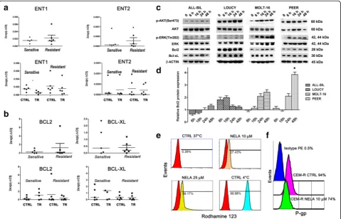
To find potential explanations for differences in nelara-bine sensitivity displayed by T-ALL cell lines, we determined mRNA expression levels of ENT1 and ENT2 nelarabine transporters, which could have a role in nelarabine cellular uptake [35]. Both ENT1 and ENT2 were expressed in all T-ALL cell lines, but there were no differences between the sensitive versus resistant group in the levels of expression of these transporters (Fig. 3a). Moreover, nelarabine treatment did not affect ENT1/2 mRNA levels in T-ALL sensitive or resistant groups
(Fig. 3a). By western blotting, we have also evaluated the expression of the two enzymes, dCK and dGK, involved in the purine metabolism. However, no significant differ-ences in the expression of these enzymes in sensitive versus resistant group were detected (Additional file 2: Figure S2).
A recent study underscored a potential role for Bcl2 family members in determining nelarabine resistance, as
a nelarabine-resistant T-ALL cell line displayed
increased anti-apoptotic Bcl-xL levels, whereas pro-apoptotic Bax and Bad levels were decreased [27]. To better understand the role of Bcl2 family, it was first investigated the gene expression of Bcl2 and Bcl-xL genes in sensitive and resistant to nelarabine cell lines. Analyses of gene expression demonstrated no significant differences in the levels of mRNA, between sensitive and resistant to nelarabine T-ALL cell lines under basal conditions (Fig. 3b, upper panel). Moreover, treatment with nelarabine did not induce significant differences in the gene expression of resistant or sensitive cell lines (Fig. 3b, lower panel). By western blotting, we analyzed the effects of nelarabine on the expression of anti-apoptotic Bcl2 and Bcl-xL proteins in resistant cell lines. Among T-ALL cell lines, LOUCY cell line showed the highest level of expression of Bcl2 under basal condi-tions (Additional file 3: Figure S3). Nelarabine treatment affected the expression of Bcl2 in all resistant cell lines analyzed. However, a significant increase of Bcl2 protein expression was observed only in PEER cells, after 48-h treatment (Fig. 3c, d).
Aberrant activation of several signal transduction pathways enhances survival, proliferation, and drug resistance of leukemic cells. PI3K/AKT/mTOR pathway is frequently overactive in T-ALL cells, and is very important for drug resistance. Therefore, it was investi-gated the status of this pathway in nelarabine resistance. T-ALL cell lines showing high IC50 to nelarabine treat-ment were treated for 6, 16, 24, 48 h with the drug (10 μM) and then analyzed by western blotting for the activation of PI3K/AKT/mTOR or MEK/ERK pathways. In ALL-SIL, LOUCY, MOLT-16, and PEER cell lines, treatment with nelarabine induced a time-dependent increase in p-AKT (Ser473), indicating an overactivation of PI3K/AKT signaling (Fig. 3c). It was also investigated the status of the MEK/ERK1/2 pathway in these resistant cell lines. This signaling cascade is also involved in leukemic cell survival and drug resistance [36]. Western blotting analysis documented that nelarabine dramatic-ally increased p-ERK (Thr202) levels in all resistant cell lines analyzed, especially in MOLT-16 and PEER cells (Fig. 3b). The results presented above indicated these
signaling cascades as attractive targets for the
development of new therapeutic strategies for T-ALL cells resistant to nelarabine treatment.
Effects of nelarabine on 170-kDa P-glycoprotein
No information is at present available regarding the effects of 170-kDa P-glycoprotein (P-gp), one of the major determinants of multidrug resistance in acute leukemias [37], on nelarabine sensitivity.
In contrast, it is known that pharmacological inhib-ition of P-gp activity increased fludarabine sensitivity in CLL cells [38]. Furthermore, P-gp expression is known to be under the control of PI3K/AKT/mTOR signaling in acute leukemia cells [39, 40]. We noticed that CEM-R cells, which overexpress P-gp [41], were less sensitive to nelarabine than parental CEM-S cells. Therefore, we set out to investigate the relevance of P-gp in nelarabine sensitivity. A functional assay for P-gp activity, based on Rhodamine 123 extrusion, demonstrated that nelarabine treatment (48 h) was able to decrease P-gp activity in CEM-R cells (Fig. 3d). Moreover, flow cytometric analysis documented that nelarabine negatively affected P-gp surface expression in these cells (Fig. 3e). There-fore, our findings indicated that nelarabine actually decreased P-gp activity and expression in CEM-R cells. Thus, it is unlikely that the different sensitivity to nelarabine displayed by CEM-S and CEM-R cells could be dependent on P-gp expression.
Nelarabine combined with ZSTK-474 affects PI3K/AKT sig-naling and modulates p-ERK and Bcl2 family members in T-ALL cells resistant to nelarabine
To better understand the role of PI3K, MEK/ERK1/2, and Bcl2 signaling, it was investigated the effect of the combination of nelarabine with the pan PI3K inhibitor ZSTK-474, the γ/δ PI3K p110 inhibitor IPI-145, the MEK inhibitor trametinib, or the Bcl2 inhibitor ABT199. The effects of the different drug combinations on nelarabine-resistant T-ALL cell lines were analyzed by treating the cells with increasing concentrations of the drugs and then measuring the rates of survival by MTT assays at 48 h. Cell lines (LOUCY, MOLT-16, PEER, and ALL-SIL) were cultured in the presence of nelarabine or
the abovementioned drugs, either alone or in
combination at a fixed ratio (Fig. 4a). The combined treatment with ZSTK-474 was highly effective in inducing cytotoxicity in all the resistant cell lines analyzed, indicating that PI3K is at least in part involved in nelarabine resistance. ZSTK-474 was synergistic with nelarabine also in P-gp overexpressing CEM-R cells (Additional file 4: Figure S4).
calculated with CalcuSyn software for dose-effect analysis, indicated the existence of a strong synergism for both drugs (CI <0.5) (Fig. 4a).
Treatment with the MEK inhibitor trametinib was also synergistic, but less potent in inhibiting cell viability in all cell lines studied. Interestingly, MOLT-16 and PEER cells, which displayed the most marked activation of MEK/ERK1/2 signaling after nelarabine treatment, were the most sensitive to trametinib. The Bcl2 inhibitor, ABT199, was very efficacious in inducing cell growth impairment, especially in ALL-SIL and LOUCY cell lines (Fig. 4a). LOUCY cells are representative of ETP-ALL and displayed the highest Bcl2 expression and sensitive-ness to Bcl2 inhibition by ABT199 [43].
To better assess the role of PI3K signaling, western blot analyses with an antibody to p-AKT (Ser473) were carried out, demonstrating a marked decrease in response to the combination of nelarabine with ZSTK-474 treatment after 48 h, in all cell lines analyzed (Fig. 4b). Also, one of the downstream substrates of mTORC1 (S6RP) was efficiently dephosphorylated by the drug combination in all cell lines. Furthermore, the combination of nelarabine with ZSTK-474 was able to induce a decrease in the Bcl2 and Bcl-xL protein expression in all cell lines resistant to nelarabine alone, except for LOUCY cells, which were not affected by the combined treatment (Fig. 4c). The combination was also capable of inducing an increase in the expression of pro-apoptotic Bax and Bak proteins after 48 h of treatment (Fig. 4c).
To assess whether nelarabine directly affected PI3K/ AKT and MEK/ERK1/2 signaling pathways, we have treated T-ALL resistant to nelarabine cells with selective inhibitors of these two axes. In particular, we employed LY294002 (PI3K inhibitor), CCI-779 (mTOR allosteric inhibitor), and trametinib (MEK1/2 inhibitor). As shown in Additional file 5: Figure S5, we confirmed that treat-ment with nelarabine as a single agent upregulated p-AKT (Ser473) and p-ERK (Thr202), whereas LY294002, CCI-779 and trametinib as single agents did not. LY294002 was not able to dephosphorylate AKT and to synergize with nelarabine, in contrast to the PI3K inhibi-tor ZSTK-474. This result could be dependent on the
LY294002 concentration we used, 10 μM to avoid
aspecific effects on signaling pathways, whereas pub-lished data employed LY294002 at higher concentrations [41]. Combining nelarabine with CCI-779 inhibitor re-sulted in upregulated PI3K and MEK signaling as dem-onstrated by an increase in AKT (Ser473) and p-ERK (Thr202), compared to untreated or CCI-779 treated cells, and indicating a major role of nelarabine in hyperactivating these axes. To ascertain CCI-779 effects, we evaluated one of its downstream key targets S6RP, which was strongly dephosphorylated by the treatment. Trametinib was able to completely dephosphorylate ERK both alone and in combination with nelarabine, but it did not perturbate significantly AKT phosphorylation. However, also in this case, the combination of trametinib with nelarabine increased p-AKT (Ser473) compared to T-ALL cells untreated or treated with nelarabine/trame-tinib as single agents. These results suggest that the up-regulation of PI3K/AKT and MEK/ERK1/2 signaling pathways are directly affected by nelarabine treatment. Moreover, ZSTK-474 strongly counteracted nelarabine effects on both PI3K and MEK/ERK1/2 signaling
path-ways compared to the other specific inhibitors
employed.
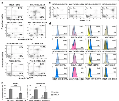
The combination of nelarabine with ZSTK-474 restored sensitiveness in MOLT-4 and P12-ICHIKAWA cells co-cultured with of HS-5 stromal cells and down-modulated PI3K and Bcl2 signaling in a system which mimics BM microenvironment
We employed the human stromal cell line HS-5, known to provide long-term support for hematopoietic progeni-tors [44]. It was investigated whether cell death induced by nelarabine treatment in sensitive cell lines could be im-paired by co-culturing T-ALL cells with HS-5, which mimic the BM microenvironment. MOLT-4 and P12-ICHIKAWA cells displayed a significant protection from apoptosis, in-duced by the co-culture with HS-5 stromal cells at 48 h of treatment, as demonstrated by staining with Annexin V/PI and flow cytometric analysis of apoptosis (Fig. 5a, b). The combination of nelarabine and ZSTK-474 was efficient in decreasing resistance to nelarabine, induced in these two cell lines by the co-culture with HS-5 stromal cells. More-over, to better assess the effectiveness of a combined treat-ment, we examined MOLT-4 cells for the levels of p-AKT (See figure on previous page.)
(Ser473) and Bcl2, with or without the support of HS-5 stromal cells, using flow cytometry. Flow cytometric ana-lysis documented a decrease in the levels of p-AKT (Ser473) in nelarabine-treated samples without the support of stromal cells (Fig. 5c). Interestingly, nelarabine induced a hyperphosphorylation of AKT in MOLT-4 cells supported by the co-culture with HS-5 cells, while the treatment with ZSTK-474 was able to in-duce a marked reduction of p-AKT and Bcl2 expres-sion. Overall, these findings demonstrated that the
combination of nelarabine with ZSTK-474 has a po-tent cytotoxic activity also in co-culture conditions which mimic the BM microenvironment.
HS-5 stromal cells protect MOLT-4 cells from nelarabine cytotoxicity at least in part through CXCL12/CXCR4 interactions
It is well established that interactions between the chemokine CXCL12, secreted by BM stromal cells, and its receptor CXCR4, expressed on the surface of
leukemic cells, are very important for protecting leukemia cells from chemotherapeutic drugs [37, 45]. HS-5 cells secrete CXCL12 [46], while MOLT-4 cells express high levels of CXCR4 [47]. Therefore, we wanted to establish whether CXCL12/CXCR interactions were involved in the protective effects exerted by HS-5 cells on nelarabine-treated MOLT-4 cells. As shown in Additional file 6: Figure S6A, AMD3100 (plerixafor), which selectively blocks the interactions between CXCL12 and CXCR4 [47], significantly increased the cytotoxicity of nelarabine in MOLT-4 cells co-cultured with HS-5 cells in a Transwell@system.
To document that the CXCR4/CXCL12 axis was indeed functional in our system, we performed western blot analysis. The presence of HS-5 cells increased p-ERK and p-CXCR4 levels in MOLT-4 cells, as expected [48]. It is worth noting that Ser339 is an amino acidic residue previously identified as a site for ligand-induced phosphorylation of CXCR4 [49] and is involved in ERK1/2 activation [50, 51]. The presence of nelara-bine did not further increase the phosphorylation or either ERK or CXCR4. However, AMD3100 dampened the phosphorylation levels of both ERK and CXCR4 (Additional file 6: Figure S6B).
T-ALL blasts are sensitive to the combination of nelarabine with ZSTK-474
To better assess the effectiveness of the combination of nelarabine with ZSTK-474 as a potential therapeutic strategy in T-ALL, we examined relapsed pediatric T-ALL patient samples isolated from the bone marrow or peripheral blood, for the sensitiveness to nelarabine alone, in vitro. These patients were previously analyzed for the activation of PI3K signaling, showing high levels of p-AKT (Ser 473). The effects of nelarabine on patient lymphoblasts were tested by incubating the cells for 72 h with increasing concentrations of the drug and then analyzing the rate of cell viability using MTT assays. Some patient samples showed high IC50(>10μM), while others were more sensitive to nelarabine alone (IC50 <5 μM) (Fig. 6a). The resistant samples were treated with nelarabine in combination with ZSTK-474, and ab-solute cell count/PI staining was assessed by flow cytom-etry for 24, 48, and 72 h for Pt-3, showing the significant effects of the combination (Fig. 6b). The combination of nelarabine with ZSTK-474 was highly effective in inducing apoptosis in the resistant patient samples analyzed, showing a marked increase in Annexin V/PI stained (Fig. 6c, Pt-3). Pt-5 and Pt-6 were also evaluated by MTT assays with nelarabine in combination with ZSTK-474 at fixed ratios (nelarabine/ZSTK-474, 4:1). The synergistic effect was assessed and the combination indexes (CIs) were calculated with the CalcuSyn software (Fig. 6d).
Finally, we evaluated the effects of single treatment with nelarabine or combination with ZSTK-474 on T-ALL patient samples on the expression levels of p-AKT (Ser473) and Bcl2. Flow cytometric analysis documented an increase in the levels of p-AKT in nelarabine-treated samples, while the treatment with the combination of nelarabine and ZSTK-474 was able to induce a marked reduction of p-AKT (Ser473) and Bcl2 expression (Fig. 6e, f ). Overall, these findings demon-strated that the combination of nelarabine with ZSTK-474 has a potent cytotoxic activity also in primary cells from relapsed T-ALL patients with upregulated PI3K/ AKT signaling and resistant to nelarabine in vitro.
Discussion
nucleoside transporters were related to in vitro nelara-bine sensitivity of T-ALL cell lines and primary samples [35]. These transporters have been shown to be import-ant for the Ara-C import, and elevated ENT1 levels have
been reported to explain the high Ara-C sensitivity of infant ALL [57]. However, a previous work on the T-ALL cell line CEM-S demonstrated that, while the cellular transport of forodesine was dependent on
Fig. 6Nelarabine combined with ZSTK-474 is able to induce a marked cytotoxic effect in relapsed T-ALL patients with upregulated PI3K signaling.
aMTT assays of lymphoblasts from relapsed T-ALL patients (Pts) with activated PI3K signaling treated with nelarabine for 72 h. Data shown are the mean of at least two different experiments ± SD.bFlow cytometric cell count with eBeads123/PI staining of T-ALL blasts treated with nelarabine alone or combined with ZSTK-474 for 24, 48,72 h.cFlow cytometric analysis of Annexin V-FITC/PI of T-ALL blasts treated with nelarabine alone or combined with ZSTK-474. The percentages of early apoptotic cells (Annexin-V FITC+/PI−;bottom right quadrant) and late apoptotic/necrotic cells (Annexin-V FITC+/PI+;top right quadrant) are plotted. The histograms are representative of two separate experiments.dCell viability assays of lymphoblasts from relapsed T-ALL patients treated for 72 h with increasing concentrations of nelarabine alone or combined with the pan PI3K inhibitor ZSTK-474 at fixed ratios. Results are the mean of two different experiments ± SD.e,fFlow cytometric analysis documenting a decrease of
ENT1/2, forodesine toxicity was not [58]. Recent studies also showed that ENT1/2 expression levels were not re-lated to forodesine toxicity levels in T-ALL cells [35]. We have analyzed the expression of ENT1/2 trans-porters in the sensitive and resistant to nelarabine groups. In all the T-ALL cells analyzed, we did not find significant differences between resistant and sensitive cells as far as the expression of ENT1/2 transporters was concerned. Furthermore, the levels of expression of ENT1/2 genes were not modulated by the treatment with nelarabine in a significant manner. In addition, Ara-G must be phosphorylated by dCK and dGK to become an intracellular metabolite and to be incorpo-rated into DNA [7]. The protein levels of dCK and dGK were not significantly different in resistant or sensitive groups, indicating that the resistance to nelarabine in T-ALL cells could be due to other mechanisms. Accumulating evidence indicates that the PI3K signaling is linked to resistance to therapy in several disorders [59, 60]. In hematological malignancies, it has been shown to support tumor proliferation, viability, and drug resist-ance. In a recent paper by Silveira et al. [61], it was shown that PI3K pathway activity was higher in T-ALL patients who underwent relapse [61]. Owing to the fundamental role of PI3K pathway in tumors and in particular in T-ALL, we investigated the phosphorylation status of AKT, a direct downstream target of PI3K, in T-ALL cells. We have shown that in resistant T-ALL cell lines and primary relapsed T-ALL blasts, nelarabine alone caused an increase of AKT phosphorylation at Ser473. Accordingly, PI3K inhibition with ZSTK-474 deeply sensitized T-ALL cells to nelarabine, while the combination of nelarabine with either MEK or Bcl2 inhibitors was less synergistic in inducing cell death in T-ALL cell lines. Because at present, it is impossible to
predict if patient could benefit from nelarabine
treatment, this study for the first time uncovered mecha-nisms responsible for resistance to nelarabine in T-ALL cells, paving the way to novel combination strategies, able to overcome resistance and facilitate the bridge to hematopoietic cell transplantation. Indeed, these findings strongly suggest that combining nelarabine with inhibi-tors of PI3K/AKT/mTOR signaling may represent a possible strategy for the treatment of relapsed or refrac-tory patients with T-ALL. In this sense, it should be emphasized that a recent phase I/II clinical trial has demonstrated that the mTOR inhibitor everolimus was moderately effective in combination with hyper-CVAD chemotherapy especially in relapsed/refractory T-ALL patients [45].
Conclusions
Therefore, data obtained from this work could improve the therapy of refractory/relapsed T-ALL through the
design of new regimens that could ameliorate clinical care of patients and their outcome, providing more effective interventions for patients that are resistant to the current therapies.
Methods
Materials
Nelarabine, ZSTK-474, IPI-145, trametinib, CCI-779, and ABT199 were kindly provided by Selleckchem (Houston, TX, USA). Ara-G, insulin, transferrin, sodium selenite (ITS), propidium iodide (PI), AMD3100, and LY294002 were purchased from Sigma-Aldrich (St. Louis, MO, USA). For western blotting analysis, all the primary antibodies were purchased from Cell Signaling Technology (Danvers, MA, USA) except for the anti-bodies to CXCR4 and p-CXCR4 (Ser339) which were from Abcam (Cambridge, UK) and antibodies to dGK and dCK which were from Thermo Scientific (Thermo Scientific, Waltham, MA, USA). AlexaFluor@-conjugated antibodies were from Cell Signaling Technology or BD Biosciences (Franklin Lakes, NJ, USA).
Cell cultures and patient samples
A panel of human T-ALL cell lines (HPB-ALL, DND41, RPMI-8402, JURKAT, MOLT-4, MOLT-16, CEM-S, CEM-R drug resistant, ALL-SIL, LOUCY, HSB-2, PEER, KOPTK1, PF382, P12-ICHIKAWA) was employed. HS-5 human stromal cells were also employed to mimic bone marrow microenvironment. All cell lines, except HS-5, were from Deutsche Sammlung von Mikroorganismen
und Zellkulturen GmbH (DSMZ, Braunschweig,
Germany). HS-5 cells were from ATCC (Manassas, VA, USA). Primary blast cells from relapsed T-ALL were ob-tained, upon written informed consent in accordance with the Declaration of Helsinki and the study has been approved by the ethics committee (Independent Ethics Committee of the University Hospital of Bologna “S. Orsola-Malpighi”). Blasts from the bone marrow and peripheral blood samples were obtained by density gradient centrifugation over Lymphoprep (Nycomed UK, Birmingham, UK). Cell cultures and primary T-ALL refractory/relapsed lymphoblasts were grown in RPMI 1640 + ITS, supplemented with 10 or 20 % fetal bovine serum (FBS), L-glutamine, and penicillin–streptomycin. HS-5 human stromal cells were grown in MEM Alpha Modification (Sigma-Aldrich) medium supplemented with 10 % FBS,L-glutamine, and penicillin–streptomycin.
Cell viability analysis
Annexin V-fluorescein isothiocyanate (FITC)/PI staining
To assess the extent of apoptosis induction, a flow cyto-metric analysis of Annexin V-FITC/PI-stained samples was performed. Analyses were performed on a FC500 flow cytometer (Beckman, Miami, FL, USA), with the appropriate software (CXP, Beckman). At least 15,000 events per sample were acquired.
Quantification of p-H2AX (Ser139) (γH2AX) by flow cytometry
Cells were treated with nelarabine (2μM MOLT-4; 5μM Jurkat) and fixed in 70 % ethanol over night at −20 °C. Afterwards, cells were rehydrated for 10 min at 4 °C in PBS containing 4 % FBS and 0.1 % Triton X-100. Perme-abilized cells were incubated with an Alexa Fluor 647 Mouse anti p-H2AX (Ser139) (BD Bioscience) at room
temperature. Fluorescence was measured by flow
cytometer on a FC500 flow cytometer (Beckman, Miami, FL, USA), with the appropriate software (CXP, Beckman). At least 10,000 events per sample were acquired.
Measurement of intracellular reactive oxygen species (ROS) levels
ROS intracellular level was evaluated by using the fluor-escent probe 2′,7′-dichlorodihydrofluorescein diacetate (H2DCFDA). Cells (0.5 × 10⁶/mL) were incubated with nelarabine for 1 and 24 h; then, cells were washed twice in HBSS and incubated with 5μM H2DCFD for 20 min at 37 °C. H2DCFDA is a small polar, non-fluorescent molecule that diffuses into the cells, where it is enzymatically deacetylated by intracellular esterases to a polar non-fluorescent compound, which is oxidized to the highly green fluorescent 2′,7′-dichlorofluorescein (DCF). The fluorescence of oxidized probe was mea-sured using a multiwell plate reader (Wallac Victor2, PerkinElmer). Excitation wavelength was 485 nm, and emission wavelength was 535 nm. Fluorescence values were reported as the percentage of intracellular ROS in respect to controls.
Western blot analysis
This was performed by standard methods, as previously reported [62].
Bcl2, Bcl-xL, and ENT1/2 expression analyses by qRT-PCR
Total RNA was extracted using the RNeasy Mini Kit (Qiagen, Venlo, The Netherlands) according to the man-ufacturer’s instructions, and 1 μg of total RNA was reverse transcribed using High-Capacity cDNA Reverse Transcription Kit (Thermo Scientific). Gene expression was assessed using The TaqMan® Gene Expression Mas-ter Mix and the assays Hs_01085704g1, Hs00155426m1, Hs00608023_m1, and Hs00236329_m1, using the 7300 real-time PCR system (Applied Biosystems, Foster City,
CA, USA). Results were normalized to the level of the ubi-quitously expressed RNA 18S ribosomal 1 gene (RN18S, Hs03928990_g1), and the Universal Human Reference RNA (Agilent) was used as control. RNA 18S ribosomal 1 gene was used as a internal control gene (RN18S, Hs03928990_g1). Results were expressed as 2−ΔCt (ΔCt = [(CT gene of interest−CT internal control) to compare the relative gene expression among samples, and as 2−ΔΔCt (ΔΔCt = [(CT gene of interest − CT internal control) sample − (CT gene of interest − CT internal control) universal]) to compare gene expression of the treated cell lines with that of untreated control [63].
Flow cytometric analysis of P-gp expression and P-gp dir-ect dye efflux assay
These were performed essentially as described [64].
Co-culture with human HS-5 stromal cells
HS-5 stromal cells were grown in the lower chamber of Transwell® six-well plates (Corning, New York, NY, USA) containing a 0.4 μm of polyester membrane, then T-ALL cells (375,000) were added to the upper chamber and treated with nelarabine (2–5 μM). After 48 h, the viability of treated cell lines grown either alone or co-cultured was evaluated. Cells were collected and analyzed for further experiments.
Combined drug effect analysis
The combination effect and a potential synergy were evaluated from quantitative analysis of dose-effect relationships as described previously [62]. For each combination experiment, a combination index (CI) number was calculated using the Biosoft CalcuSyn software (Cambridge, UK).
Flow cytometric analysis of p-AKT (Ser473), p-ERK (Thr202), and Bcl2 levels in T-ALL patient samples
Blasts from pediatric patients with T-ALL were fixed/ permeabilized in methanol 90 % for at least 30 min, then washed in PBS with 4 % FBS. Cells were incubated with primary antibodies conjugated to AlexaFluor® 488 or 647, from Cell Signaling. Isotype controls were employed as well. Cells were analyzed on a FC500 flow cytometer (Beckman Coulter) with the appropriate software (CXP, Beckman Coulter). At least 15,000 events per sample were acquired. Absolute cell count by flow cytometry was performed using eBeads123 (Affimetrix eBios-ciences, Santa Clara, CA, USA).
Statistical analysis
test was used to statistically analyze the differences in the two subgroups of sensitive/resistant to nelarabine T-ALL cells.
Additional files
Additional file 1: Figure S1.DNA damage and ROS production. A. Flow cytometric analysis ofγH2AX in MOLT-4 and Jurkat sensitive to nelarabine cells (upper panel). Percentages ofγH2AX are shown in the table (lower panel). B. Reactive oxygen species (ROS) production was assessed in MOLT-4 and Jurkat sensitive T-ALL cell lines after 1 and 24 h treatment with nelarabine. Fluorescence values (DCF) were reported as the percentage of intracellular ROS in respect to controls. ROS increased significantly in both 1- and 24-h treatment compared to controls in MOLT-4 and Jurkat cells. Statistical analyses were performed with the Dunnett’s multiple comparison test.
Additional file 2: Figure S2.Expression of dCK and dGK in T-ALL cell lines. Western blotting analyses for the expression of dCK and dGK proteins in T-ALL cell lines. Fifty micrograms of protein was blotted to each lane. Antibody toβ-actin served as a loading control. Molecular weights are indicated on the right. One representative of two different experiments is shown.
Additional file 3: Figure S3.Bcl2 expression in T-ALL cell lines. Western blotting analyses for the basal expression of Bcl2 in untreated T-ALL cell lines. Twenty micrograms of protein was blotted to each lane. Antibody toβ-actin served as a loading control. Molecular weights are indicated on the right. Densitometric analysis was performed using a Chemidoc 810 Imager with the appropriate software (UVP, Upland, CA, USA), and for each cell line, Bcl2 protein expression is indicated relatively toβ-actin expression.
Additional file 4: Figure S4.The combination of nelarabine and ZSTK-474 is synergistic in CEM-R cells, which overexpress P-gp. Cell viability assay of CEM-R cell line treated for 48 h with increasing concentrations of nelarabine alone or combined with the pan PI3K p110 inhibitor ZSTK-474. One representative of two different experiments is shown.
Additional file 5: Figure S5.Specific effects of nelarabine on PI3K/AKT and MEK/ERK1/2 pathways. Western blotting analyses for the expression of p-AKT and p-ERK in resistant T-ALL cell lines treated with the specific inhibitors LY294002 (PI3K inhibitor), CCI-779 (mTOR allosteric inhibitor) or trametinib (MEK1/2 inhibitor) alone or in combination with nelarabine. Thirty micrograms of protein was blotted to each lane. Antibody toβ-actin served as a loading control. Molecular weights are indicated on the right. CTRL: untreated cells; Nela and N: nelarabine at 10μM; LY: LY294002 at 10μM; CCI: CCI-779 at 100 nM; Tram: trametinib at 1μM. Cells were treated for 48 h.
Additional file 6: Figure S6.HS-5 stromal cells protect MOLT-4 cells from nelarabine cytotoxicity at least in part through CXCL12/CXCR4 interactions. A. MTT assays of MOLT-4 cells growing alone or in co-culture system with HS-5 cells and treated with nelarabine (2μM for 48 h) in a Transwell@system. Three hundred seventy-five thousand MOLT-4 cells were seeded in the upper chamber of the Transwell@system. Results are the mean of three different experiments ± SD. B. Western blot analysis for p-ERK, p-CXCR4, and their total forms in MOLT-4 cells grown either in the absence or the presence of HS-5 stromal cells in a Transwell@system. Nelarabine treatment (2μM) was for 24 h. Fifty micrograms of protein was blotted to each lane. Antibody toβ-actin served as a loading control. Molecular weights are indicated on the right. One representative of two different experiments is shown.
Abbreviations
AML:Acute myeloid leukemia; Bak: Bcl-2 homologous antagonist/killer; Bax: BCL2 associated X protein; Bcl2: B cell lymphoma 2; B-CLL: B cell chronic lymphocytic leukemia; Bcl-xL: B cell lymphoma-extra large; BM: Bone marrow; CEM-R: CEM overexpressing P-gp; CI: Combination index; CR: Complete remission; CVAD: Cyclophosphamide, vincristine, doxorubicin, dexamethasone; CXCL12: Chemokine C-X-C motif ligand 12;
CXCR4: Chemokine C-X-C motif receptor 4; dCK: Deoxycytidine kinase; dGK: Deoxyguanosine kinase; ENT1/2: Equilibrative nucleoside transporters1/ 2; ETP-ALL: Early T precursor acute lymphoblastic leukemia; HS-5: Human stromal cells 5; Mcl1: Myeloid cell leukemia 1; MEK: Mitogen-activated protein kinase kinase; mTOR: Mammalian target of rapamycin; PARP: Poly(ADP-ribose) polymerase; PCR: Polymerase chain reaction; P-gp: P-glycoprotein;
PI: Propidium iodide; PI3K: Phosphoinositide 3-kinase; S6RP: S6 ribosomal protein; T-ALL: T cell acute lymphoblastic leukemia; T-LL: T cell lymphocytic lymphoma
Acknowledgements
Not applicable.
Funding
This study is supported by Fondazione Del Monte di Bologna e Ravenna to AMM.
Availability of data and material
All data generated or analyzed during this study are included in this published article and its supplementary information files.
Authors’contributions
FC and AMM were the principal investigators of the study and gave final approval. FC, AL, and AC coordinated the research. FC was a major contributor in writing the manuscript. AC, LZ, LMN, FB, CE, CE, and EO performed the laboratory work for this study. AB, FL, and AP contributed to sample collection and analyses. AL, FC, and AMM contributed to data interpretation. All authors read and approved the final manuscript.
Competing interests
The authors declare that they have no competing interests.
Consent for publication
Not applicable.
Ethics approval and consent to participate
Primary blast cells from relapsed T-ALL were obtained, upon written informed consent in accordance with the Declaration of Helsinki and the study has been approved by the Independent Ethics Committee of the University Hospital of Bologna“S. Orsola-Malpighi”(Prot. N. 95.2015/U/Tess).
Author details
1
Department of Biomedical and Neuromotor Sciences, University of Bologna, Bologna, Italy.2Department of Human Social and Health Sciences, University
of Cassino, Cassino, Italy.3Department of Pediatric Hematology-Oncology, IRCCS, Bambino Gesù Children’s Hospital, Rome, Italy.4Department of
Pediatrics,“Lalla Seràgnoli”Hematology-Oncology Unit, University of Bologna, Bologna, Italy.5Institute of Molecular Genetics, Rizzoli Orthopedic
Institute, National Research Council, Bologna, Italy.6Department of Pharmacy and Biotechnology, University of Bologna, Bologna, Italy.7Department of
Morphology, Surgery and Experimental Medicine, University of Ferrara, Ferrara, Italy.
Received: 12 July 2016 Accepted: 14 October 2016
References
1. Van Vlierberghe P, Ferrando A. The molecular basis of T cell acute lymphoblastic leukemia. J Clin Invest. 2012;122(10):3398–406.
2. Litzow MR, Ferrando AA. How I treat T-cell acute lymphoblastic leukemia in adults. Blood. 2015;126(7):833–41.
3. DeAngelo DJ, Yu D, Johnson JL, Coutre SE, Stone RM, Stopeck AT, Gockerman JP, Mitchell BS, Appelbaum FR, Larson RA. Nelarabine induces complete remissions in adults with relapsed or refractory T-lineage acute lymphoblastic leukemia or lymphoblastic lymphoma: Cancer and leukemia group B study 19801. Blood. 2007;109(12):5136–42.
4. Kurtzberg J. The long and winding road of the clinical development of nelarabine. Leuk Lymphoma. 2007;48(1):1–2.
6. Prus KL, Averett DR, Zimmerman TP. Transport and metabolism of 9-beta-D-arabinofuranosylguanine in a human T-lymphoblastoid cell line:
nitrobenzylthioinosine-sensitive and -insensitive influx. Cancer Res. 1990;50(6):1817–21.
7. Rodriguez Jr CO, Mitchell BS, Ayres M, Eriksson S, Gandhi V.
Arabinosylguanine is phosphorylated by both cytoplasmic deoxycytidine kinase and mitochondrial deoxyguanosine kinase. Cancer Res. 2002;62(11): 3100–5.
8. Berg SL, Blaney SM, Devidas M, Lampkin TA, Murgo A, Bernstein M, Billett A, Kurtzberg J, Reaman G, Gaynon P, et al. Phase II study of nelarabine (compound 506U78) in children and young adults with refractory T-cell malignancies: a report from the Children’s Oncology Group. J Clin Oncol. 2005;23(15):3376–82.
9. Rodriguez Jr CO, Stellrecht CM, Gandhi V. Mechanisms for T-cell selective cytotoxicity of arabinosylguanine. Blood. 2003;102(5):1842–8.
10. Larson RA. Three new drugs for acute lymphoblastic leukemia: nelarabine, clofarabine, and forodesine. Semin Oncol. 2007;34(6 Suppl 5):S13–20. 11. Gokbuget N, Basara N, Baurmann H, Beck J, Bruggemann M, Diedrich H,
Guldenzoph B, Hartung G, Horst HA, Huttmann A, et al. High single-drug activity of nelarabine in relapsed T-lymphoblastic leukemia/lymphoma offers curative option with subsequent stem cell transplantation. Blood. 2011;118(13):3504–11.
12. Dunsmore KP, Devidas M, Linda SB, Borowitz MJ, Winick N, Hunger SP, Carroll WL, Camitta BM. Pilot study of nelarabine in combination with intensive chemotherapy in high-risk T-cell acute lymphoblastic leukemia: a report from the Children’s Oncology Group. J Clin Oncol. 2012;30(22):2753–9.
13. Winter SS, Dunsmore KP, Devidas M, Eisenberg N, Asselin BL, Wood BL, Leonard Rn MS, Murphy J, Gastier-Foster JM, Carroll AJ, et al. Safe integration of nelarabine into intensive chemotherapy in newly diagnosed T-cell acute lymphoblastic leukemia: Children’s Oncology Group Study AALL0434. Pediatr Blood Cancer. 2015;62(7):1176–83.
14. Jain P, Kantarjian H, Ravandi F, Thomas D, O’Brien S, Kadia T, Burger J, Borthakur G, Daver N, Jabbour E, et al. The combination of hyper-CVAD plus nelarabine as frontline therapy in adult T-cell acute lymphoblastic leukemia and T-lymphoblastic lymphoma: MD Anderson Cancer Center experience. Leukemia. 2014;28(4):973–5.
15. Luskin MR, Ganetsky A, Landsburg DJ, Loren AW, Porter DL, Nasta SD, Svoboda J, Luger SM, Frey NV: Nelarabine, cyclosphosphamide and etoposide for adults with relapsed T-cell acute lymphoblastic leukaemia and lymphoma. Br J Haematol 2015
16. Gollard RP, Selco S. Irreversible myelopathy associated with nelaribine in T-cell acute lymphoblastic leukemia. J Clin Oncol. 2013;31(19):e327–31. 17. Hartz B, Lobel U, Hagel C, Escherich G. Fatal neurological side-effects with
necrosis of spinal cord following nelarabine treatment in a child with relapsed T-cell acute lymphoblastic leukemia. Am J Hematol. 2013;88(12):1096–7.
18. Papayannidis C, Iacobucci I, Abbenante MC, Curti A, Paolini S, Parisi S, Baccarani M, Martinelli G. Complete paraplegia after nelarabine treatment in a T-cell acute lymphoblastic leukemia adult patient. Am J Hematol. 2010;85(8):608.
19. Cohen MH, Johnson JR, Justice R, Pazdur R. FDA drug approval summary: nelarabine (Arranon) for the treatment of T-cell lymphoblastic leukemia/ lymphoma. Oncologist. 2008;13(6):709–14.
20. Hallek M. Signaling the end of chronic lymphocytic leukemia: new frontline treatment strategies. Blood. 2013;122(23):3723–34.
21. Chiarini F, Lonetti A, Teti G, Orsini E, Bressanin D, Cappellini A, Ricci F, Tazzari PL, Ognibene A, Falconi M, et al. A combination of temsirolimus, an allosteric mTOR inhibitor, with clofarabine as a new therapeutic option for patients with acute myeloid leukemia. Oncotarget. 2012;3(12):1615–28. 22. Shigemi H, Yamauchi T, Tanaka Y, Ueda T. Novel leukemic cell lines resistant
to clofarabine by mechanisms of decreased active metabolite and increased antiapoptosis. Cancer Sci. 2013;104(6):732–9.
23. Rosich L, Saborit-Villarroya I, Lopez-Guerra M, Xargay-Torrent S, Montraveta A, Aymerich M, Villamor N, Campo E, Perez-Galan P, Roue G, et al. The phosphatidylinositol-3-kinase inhibitor NVP-BKM120 overcomes resistance signals derived from microenvironment by regulating the Akt/FoxO3a/Bim axis in chronic lymphocytic leukemia cells. Haematologica.
2013;98(11):1739–47.
24. Johnston JB, Paul JT, Neufeld NJ, Haney N, Kropp DM, Hu X, Cheang M, Gibson SB. Role of myeloid cell factor-1 (Mcl-1) in chronic lymphocytic leukemia. Leuk Lymphoma. 2004;45(10):2017–27.
25. Yu C, Mao X, Li WX. Inhibition of the PI3K pathway sensitizes fludarabine-induced apoptosis in human leukemic cells through an inactivation of MAPK-dependent pathway. Biochem Biophys Res Commun. 2005;331(2):391–7.
26. Amadori S, Stasi R, Martelli AM, Venditti A, Meloni G, Pane F, Martinelli G, Lunghi M, Pagano L, Cilloni D, et al. Temsirolimus, an mTOR inhibitor, in combination with lower-dose clofarabine as salvage therapy for older patients with acute myeloid leukaemia: results of a phase II GIMEMA study (AML-1107). Br J Haematol. 2012;156(2):205–12.
27. Yamauchi T, Uzui K, Nishi R, Shigemi H, Ueda T. Reduced drug incorporation into DNA and antiapoptosis as the crucial mechanisms of resistance in a novel nelarabine-resistant cell line. BMC Cancer. 2014;14:547.
28. Lonetti A, Cappellini A, Sparta AM, Chiarini F, Buontempo F, Evangelisti C, Orsini E, McCubrey JA, Martelli AM. PI3K pan-inhibition impairs more efficiently proliferation and survival of T-cell acute lymphoblastic leukemia cell lines when compared to isoform-selective PI3K inhibitors. Oncotarget. 2015;6(12):10399–414.
29. Pillinger G, Loughran NV, Piddock RE, Shafat MS, Zaitseva L, Abdul-Aziz A, Lawes MJ, Bowles KM, Rushworth SA. Targeting PI3Kdelta and PI3Kgamma signalling disrupts human AML survival and bone marrow stromal cell mediated protection. Oncotarget. 2016.
30. Tran KA, Cheng MY, Mitra A, Ogawa H, Shi VY, Olney LP, Kloxin AM, Maverakis E. MEK inhibitors and their potential in the treatment of advanced melanoma: the advantages of combination therapy. Drug Des Devel Ther. 2016;10:43–52.
31. Choudhary GS, Al-Harbi S, Mazumder S, Hill BT, Smith MR, Bodo J, Hsi ED, Almasan A. MCL-1 and BCL-xL-dependent resistance to the BCL-2 inhibitor ABT-199 can be overcome by preventing PI3K/AKT/mTOR activation in lymphoid malignancies. Cell Death Dis. 2015;6, e1593.
32. Lonetti A, Antunes IL, Chiarini F, Orsini E, Buontempo F, Ricci F, Tazzari PL, Pagliaro P, Melchionda F, Pession A, et al. Activity of the pan-class I phosphoinositide 3-kinase inhibitor NVP-BKM120 in T-cell acute lymphoblastic leukemia. Leukemia. 2014;28(6):1196–206.
33. Huang P, Robertson LE, Wright S, Plunkett W. High molecular weight DNA fragmentation: a critical event in nucleoside analogue-induced apoptosis in leukemia cells. Clin Cancer Res. 1995;1(9):1005–13.
34. Mah LJ, El-Osta A, Karagiannis TC. gammaH2AX: a sensitive molecular marker of DNA damage and repair. Leukemia. 2010;24(4):679–86. 35. Homminga I, Zwaan CM, Manz CY, Parker C, Bantia S, Smits WK,
Higginbotham F, Pieters R, Meijerink JP. In vitro efficacy of forodesine and nelarabine (ara-G) in pediatric leukemia. Blood. 2011;118(8):2184–90. 36. Martelli AM, Tabellini G, Ricci F, Evangelisti C, Chiarini F, Bortul R, McCubrey
JA, Manzoli FA. PI3K/AKT/mTORC1 and MEK/ERK signaling in T-cell acute lymphoblastic leukemia: new options for targeted therapy. Adv Biol Regul. 2012;52(1):214–27.
37. Zeng Z, Samudio IJ, Munsell M, An J, Huang Z, Estey E, Andreeff M, Konopleva M. Inhibition of CXCR4 with the novel RCP168 peptide overcomes stroma-mediated chemoresistance in chronic and acute leukemias. Mol Cancer Ther. 2006;5(12):3113–21.
38. Svirnovski AI, Serhiyenka TF, Kustanovich AM, Khlebko PV, Fedosenko VV, Taras IB, Bakun AV. DNA-PK, ATM and MDR proteins inhibitors in overcoming fludarabine resistance in CLL cells. Exp Oncol. 2010;32(4):258–62. 39. Cheng Z, Yang N, Liang W, Yan X, Li L, Pan L. Effect of phosphatase and
tensin homology deleted on chromosome 10 (PTEN) gene transfection on reversal of multidrug resistance in K562/ADM cells. Leuk Lymphoma. 2012;53(7):1383–9.
40. Wang H, Jia XH, Chen JR, Wang JY, Li YJ. Osthole shows the potential to overcome P-glycoproteinmediated multidrug resistance in human myelogenous leukemia K562/ADM cells by inhibiting the PI3K/Akt signaling pathway. Oncol Rep. 2016;35(6):3659–68.
41. Mantovani I, Cappellini A, Tazzari PL, Papa V, Cocco L, Martelli AM. Caspase-dependent cleavage of 170-kDa P-glycoprotein during apoptosis of human T-lymphoblastoid CEM cells. J Cell Physiol. 2006;207(3):836–44.
42. Subramaniam PS, Whye DW, Efimenko E, Chen J, Tosello V, De Keersmaecker K, Kashishian A, Thompson MA, Castillo M, Cordon-Cardo C, et al. Targeting nonclassical oncogenes for therapy in T-ALL. Cancer Cell. 2012;21(4):459–72.
44. Garrido SM, Appelbaum FR, Willman CL, Banker DE. Acute myeloid leukemia cells are protected from spontaneous and drug-induced apoptosis by direct contact with a human bone marrow stromal cell line (HS-5). Exp Hematol. 2001;29(4):448–57.
45. Chiarini F, Lonetti A, Evangelisti C, Buontempo F, Orsini E, Cappellini A, Neri LM, McCubrey JA, Martelli AM. Advances in understanding the acute lymphoblastic leukemia bone marrow microenvironment: from biology to therapeutic targeting. Biochim Biophys Acta. 2016;1863(3):449–63. 46. Braun M, Qorraj M, Buttner M, Klein FA, Saul D, Aigner M, Huber W,
Mackensen A, Jitschin R, Mougiakakos D. CXCL12 promotes glycolytic reprogramming in acute myeloid leukemia cells via the CXCR4/mTOR axis. Leukemia. 2016.
47. Hatse S, Princen K, Bridger G, De Clercq E, Schols D. Chemokine receptor inhibition by AMD3100 is strictly confined to CXCR4. FEBS Lett. 2002;527(1-3):255–62.
48. Zeng Z, Shi YX, Samudio IJ, Wang RY, Ling X, Frolova O, Levis M, Rubin JB, Negrin RR, Estey EH, et al. Targeting the leukemia microenvironment by CXCR4 inhibition overcomes resistance to kinase inhibitors and chemotherapy in AML. Blood. 2009;113(24):6215–24.
49. Woerner BM, Warrington NM, Kung AL, Perry A, Rubin JB. Widespread CXCR4 activation in astrocytomas revealed by phospho-CXCR4-specific antibodies. Cancer Res. 2005;65(24):11392–9.
50. Busillo JM, Armando S, Sengupta R, Meucci O, Bouvier M, Benovic JL. Site-specific phosphorylation of CXCR4 is dynamically regulated by multiple kinases and results in differential modulation of CXCR4 signaling. J Biol Chem. 2010;285(10):7805–17.
51. Zuo K, Kuang D, Wang Y, Xia Y, Tong W, Wang X, Chen Y, Duan Y, Wang G. SCF/c-kit transactivates CXCR4-serine 339 phosphorylation through G protein-coupled receptor kinase 6 and regulates cardiac stem cell migration. Sci Rep. 2016;6:26812.
52. Frey NV, Luger SM. How I treat adults with relapsed or refractory Philadelphia chromosome-negative acute lymphoblastic leukemia. Blood. 2015;126(5):589–96.
53. Roecker AM, Stockert A, Kisor DF. Nelarabine in the treatment of refractory T-cell malignancies. Clin Med Insights Oncol. 2010;4:133–41.
54. Foss FM, Zinzani PL, Vose JM, Gascoyne RD, Rosen ST, Tobinai K. Peripheral T-cell lymphoma. Blood. 2011;117(25):6756–67.
55. Beesley AH, Palmer ML, Ford J, Weller RE, Cummings AJ, Freitas JR, Firth MJ, Perera KU, de Klerk NH, Kees UR. In vitro cytotoxicity of nelarabine, clofarabine and flavopiridol in paediatric acute lymphoblastic leukaemia. Br J Haematol. 2007;137(2):109–16.
56. Cohen MH, Johnson JR, Massie T, Sridhara R, McGuinn Jr WD, Abraham S, Booth BP, Goheer MA, Morse D, Chen XH, et al. Approval summary: nelarabine for the treatment of T-cell lymphoblastic leukemia/lymphoma. Clin Cancer Res. 2006;12(18):5329–35.
57. White JC, Rathmell JP, Capizzi RL. Membrane transport influences the rate of accumulation of cytosine arabinoside in human leukemia cells. J Clin Invest. 1987;79(2):380–7.
58. Huang M, Wang Y, Gu J, Yang J, Noel K, Mitchell BS, Schramm VL, Graves LM. Determinants of sensitivity of human T-cell leukemia CCRF-CEM cells to immucillin-H. Leuk Res. 2008;32(8):1268–78.
59. Vivanco I, Sawyers CL. The phosphatidylinositol 3-Kinase AKT pathway in human cancer. Nat Rev Cancer. 2002;2(7):489–501.
60. Huang WC, Hung MC. Induction of Akt activity by chemotherapy confers acquired resistance. J Formos Med Assoc. 2009;108(3):180–94. 61. Silveira AB, Laranjeira AB, Rodrigues GO, Leal PC, Cardoso BA, Barata JT,
Yunes RA, Zanchin NI, Brandalise SR, Yunes JA. PI3K inhibition synergizes with glucocorticoids but antagonizes with methotrexate in T-cell acute lymphoblastic leukemia. Oncotarget. 2015;6(15):13105–18.
62. Chiarini F, Grimaldi C, Ricci F, Tazzari PL, Evangelisti C, Ognibene A, Battistelli M, Falcieri E, Melchionda F, Pession A, et al. Activity of the novel dual phosphatidylinositol 3-kinase/mammalian target of rapamycin inhibitor NVP-BEZ235 against T-cell acute lymphoblastic leukemia. Cancer Res. 2010;70(20):8097–107.
63. Schmittgen TD, Livak KJ. Analyzing real-time PCR data by the comparative C(T) method. Nat Protoc. 2008;3(6):1101–8.
64. Chiarini F, Del Sole M, Mongiorgi S, Gaboardi GC, Cappellini A, Mantovani I, Follo MY, McCubrey JA, Martelli AM. The novel Akt inhibitor, perifosine, induces caspase-dependent apoptosis and downregulates P-glycoprotein expression in multidrug-resistant human T-acute leukemia cells by a JNK-dependent mechanism. Leukemia. 2008;22(6):1106–16.
• We accept pre-submission inquiries
• Our selector tool helps you to find the most relevant journal
• We provide round the clock customer support
• Convenient online submission
• Thorough peer review
• Inclusion in PubMed and all major indexing services • Maximum visibility for your research
Submit your manuscript at www.biomedcentral.com/submit