INHIBITION OF ABL FAMILY KINASES PRODUCES A PROFOUND
CHANGE IN CELL SHAPE AND MIGRATION
Zaozao Chen
A dissertation submitted to the faculty of the University of North Carolina at Chapel Hill in partial fulfillment of the requirements for the degree of Doctor of Philosophy in the
Department of Cell Biology and Physiology.
Chapel Hill 2012 Approved by: Ken Jacobson, Ph.D. Keith Burridge, Ph.D. James E. Bear, Ph.D. Edward D. Salmon, Ph.D. Maryna Kapustina, Ph.D.
ii © 2012 Zaozao Chen ALL RIGHTS RESERVED
iii
ABSTRACT
ZAOZAO CHEN: Inhibition of Abl family kinases produces a profound change in cell shape and migration.
(Under the direction of Ken Jacobson)
Cell migration is fundamental to establishing and maintaining the proper organization
of multi-cellular organisms. In this dissertation we reviewed and discussed the biological process of single cell migration in two-dimensional and three-dimensional environments. We reported that Gleevec (Imatinib), an Abl family kinase inhibitor, produces a profound change in the shape and migration of rat bladder tumor cells (NBT-II) plated on collagen-coated substrates. Cells treated with Gleevec adopt a highly spread D-shape (similar to fish keratocytes) and migrate more rapidly with greater persistence. Our finding indicate integrin mediated adhesion changes happened with the inhibition of Abl-family kinases, while RhoA activity increased in this cases, which via myosin activation, led to an increase in the magnitude of total traction force applied to the substrate. We also discovered a special band of small punctate, rapidly turning over adhesions near the leading margin of spread D-shape NBT-II cells. Our results taken as a whole indicate Abl family kinases play an important role in the regulation of cell adhesion and migration in that their inhibition produces a profound change in cell adhesions, morphology and migration. In this dissertation, we also elucidated
iv
the mechanism of protein inactivation mediated by fluorescent protein chromophore-assisted light inactivation (FP-CALI). Our finding indicates the involvement of a reactive oxygen species (ROS) in the CALI effect. The progress towards a Bio-Field Effect Transistor (FET)-based detector of local cell adhesion in single cell was also included. Lastly, how the NBT-II cell may become a model for rapidly migrating cells and how the tools we developed may advance our current understanding of cell adhesion and migration is discussed.
v
DEDICATION
To my parents and grandma, who supported and encouraged me all the time.
And to my wife, Lily, who gives endless love and support to me.
vi
ACKNOWLEDGMENTS
The work presented in this thesis would not have been possible without the supports from following people. I’d like to express my deepest gratitude and sincere appreciation to all of them.
First of all, I would like to thank my advisor Dr. Ken Jacobson for his patient guidance during my study and research in UNC. My previous background was Biomedical Engineering. Under his supervision, I made a successful transition from an engineering student to a real Cell Biologist. Dr. Ken Jacobson has been a role model for me, and has undeniably been the driving force to positively shape me into the scientist I am today. I am particularly grateful for his unwavering supports for the project and my career, and for making possible all of the incredible collaborations and opportunities I have had throughout my graduate career. It has been an honor and a privilege studying under him.
My warmest thanks also go to all my committee members: Dr. Keith Burridge, Dr. James Bear, Dr. Ted Salmon, and Dr. Maryna Kapustina. Thank you for your enthusiasm, guidance, and support. I feel so fortunate to have a committee composed of scientists that I have great admiration for.
vii
I am deeply grateful for the help from many scientific collaborators. Much of the work contained in this thesis would have been impossible without the support from them. Dr. Veena Misra, who collaborated with us, has taught me much more knowledge about electrical biosensor than I have learned before. I would like to thank Bongmook Lee and Smita Sarkar particularly for their invaluable contributions. I appreciate all the graciously help from the members of the Misra lab during my fabrication of Bio-Field Effect Transistor devices at North Carolina State University.
I am also grateful for the help from Dr. Gorge Truskey in the department of Biomedical Engineering at Duke University, whose support has been particularly meaningful to my research. His continued advice and perceptive insight made the in-depth understanding of Abl kinase family related special cell-substrate adhesions happened.
I would like to thank all the former and current members of the Jacobson Lab. They have helped me with their support, friendship, and valuable suggestions. Particularly, I would like to thank: Bing Yang for teaching me many experiments in cell biology; Zenon Rajfur for advanced microscopy imaging skills; Michelle Itano, Ping Liu and Li Li for being great friends and colleagues; and Cai Huang for all the important groundwork for the studies on cell migration, for teaching me everything I know about the cell adhesions, and making great suggestions throughout my graduate research.
viii
I would like to show my gratitude to all of my other colleagues and collaborators, too numerous to name. Your support has been particularly meaningful to my graduate career. Thanks to all faculty and staff of the Department of Cell and Developmental Biology.
And finally, I want to express my gratitude for the daily and unconditional support I receive from my wife Lili Xu. We have been together as lovers and partners for almost 10 years. In the past, long distance, illness, and other difficulties never departed us but made us even stronger. We will be happily together forever.
ix
TABLE OF CONTENTS
List of Figures ... xiv
List of Abbreviations and Symbols ... xvi
CHAPTER 1: Introduction ... 1
CHAPTER 2: The Review of Single Cell Migration ... 4
2.1 Summary ... 4
2.2 Introduction and Context ... 5
2.3 Types of Single Cell Migration and Related Phenomena ... 5
... 2.3.1 Fibroblasts 5 ... 2.3.2 Keratocytes 7 2.3.3 Leukocytes ... 11
2.3.4 Single Cell Migration in Three Dimensions ... 12
2.4 Adhesions in Migrating Cells ... 14
2.4.1 Focal Adhesions: Composition and Structure ... 14
... 2.4.2 Focal Adhesion Dynamics 18 2.4.3 Podosomes ... 21
2.4.4 Focal Complexes ... 22
x
2.4.6 Outlook ... 25
2.5 Measurements of Tractions in Single Migrating Cells ... 25
2.5.1 Elastic Substrate Traction Measurements ... 25
... 2.5.2 Force, Cell Adhesions, and Cell Migration 27 2.6 Credits ... 29
2.7 References ... 30
CHAPTER 3: A Profound Change in Cell Shape and Migration Induced by Abl Family Kinases Inhibitor ... 41
3.1 Summary ... 41
3.2 Introduction ... 43
3.3 Materials and Methods ... 44
... 3.3.1 Antibodies and Immunofluorescence 44 ... 3.3.2 Cell Culture and Transfection 45 ... 3.3.3 Cell Migration, Surface Coating and Drug Treatment 45 ... 3.3.4 Assay for Active RhoA GTPases 46 ... 3.3.5 Cell imaging 47 ... 3.3.6 Measurement of Cell Adhesion Strength 47 ... 3.3.7 Traction Force Microscopy 48 ... 3.3.8 Data Quantification and Calculation 49 3.4 Results ... 52
3.4.1 Treatment with Gleevec Induces a D-shaped Morphology in NBT-II Cells ... 52 3.4.2 Both Gleevec Concentration and Substrate Adhesiveness
xi
Affect NBT-II Cell Miagraion ... 55
3.4.3 Gleevec treated NBT-II cells are More Adherent to Their Substrate than Control Cells ... 57
3.4.4 Punctuate Adhesions are Present at the Leading Edge of Gleevec treated D-shape NBT-II Cells ... 60
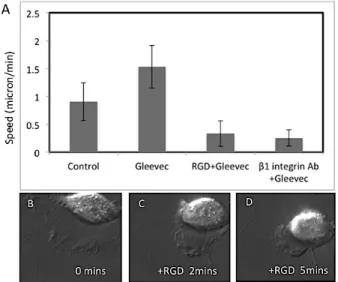
3.4.5 β1 integrin-containing cell adhesions are important for maintaining D-shape morphology and migration status ... 63
3.4.6 Gleevec induces changes in actin cytoskeleton, p-MLC localization and traction ... 64
3.4.7 Effects of RhoA family GTPases on D-shaped NBT-II cell migration ... 66
3.5 Discussion... 70
3.5.1 Mechanism of the Gleevec-induced change in morphology and migration ... 70
3.5.2 Changes in the adhesive behavior of the Gleevec treated NBT-II cells ... 71
3.6 Supplemental Materials ... 73
3.7 References ... 78
CHAPTER 4: Mechanism of Chromophore Assisted Laser Inactivation Employing Fluorescent Proteins ... 83
4.1 Summary ... 83
4.2 Introduction ... 85
4.3 Materials & Methods ... 86
xii
... 4.3.2 Protein Expression and Activity Measurement 87
... 4.3.3 Chromophore Assisted Laser Inactivation Setup 89
4.4 Results & Discussion ... 91
...4.4.1 Spatial Selectivity of FP CALI 91 ... 4.4.2 CALI Dose-Response Characteristics for XFP-GST 93
... 4.4.3Mechanism of EGFP-CALI 96
4.5 Discussion... 100
4.6 Supplemental Figures ... 104
4.7 References ... 110
CHAPTER 5: Use of microfabrication to measure local cell-substrate adhesion: An FET (Field Effect Transistor) cell adhesion sensor ... 112
5.1 Summary ... 112
5.2 Introduction ... 113
5.3 Materials and Methods ... 114
5.3.1 Cells, Staining and Microscopy ... 114
... 5.3.2 Mask design 115
... 5.3.3 FET fabrication: Process Flow 116
... 5.3.4 Electrical analyses 118 5.4 Results ... 119 ... 5.4.1 Fabric ... 5.4.2 Perfor ... 5.4.3 Charg ... 5.4.4 3T3 c 5.6 References ... 127
xiii
Chapter 6: Conclusions and Outlook... 129 6.1 Surprising Similarities Between Two Fast Migrating Cells ... 129 6.2 Future Directions ... 132
xiv
LIST OF FIGURES
Figure 2.1 Different types of cell migration ... 7
Figure 2.2 Adhesion structure and function in cells ... 17
Figure 2.3 Different type of cell adhesions ... 24
Figure 2.4 Different type of cell adhesions ... 26
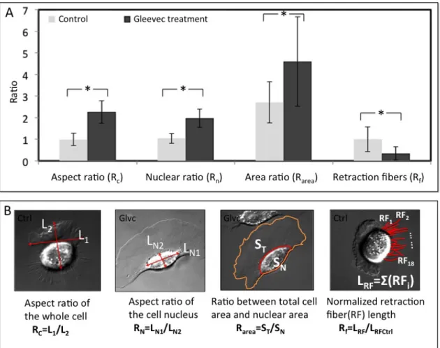
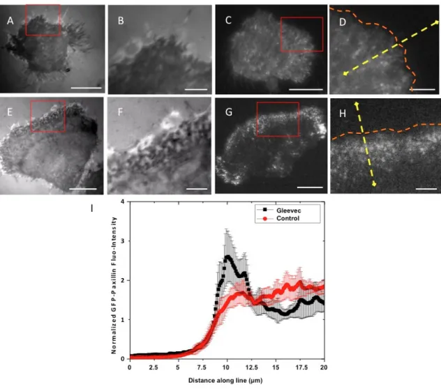
Figure 3.1 Transformation of NBT-II cells morphology and migratory phenotype after Gleevec treatment ... 53
Figure 3.2 Detailed analysis of cell morphology changes after Gleevec treatment ... 55
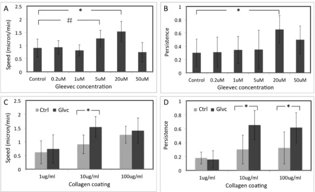
Figure 3.3 NBTII cell migration behavior depends on substrate adhesiveness and Gleevec concentration ... 57
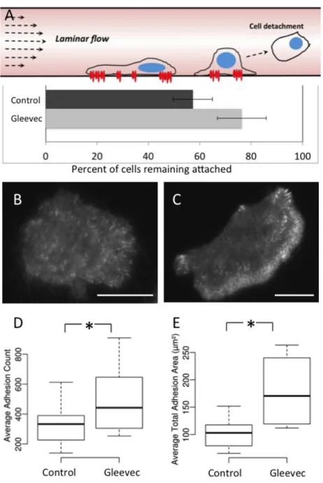
Figure 3.4 Gleevec treated cells are more adhesive than control cells ... 59
Figure 3.5 Punctuate adhesions are present at the leading edge of Gleevec treated NBT-II cells ... 62
Figure 3.6 Blocking of integrin related adhesion dramatically inhibits the migration speed of Gleevec-treated NBT-II cells. ... 63
Figure 3.7 Changes in distribution of active myosin and traction forces after Gleevec treatment ... 65
Figure 3.8 Abl-family kinase inhibition increases RhoA activity ... 67
Figure 3.9 RhoA/ROCK activity is important for the Gleevec phenotype ... 69
Figure 4.1 The CALI effect and fluorophore and chromophore photobleaching depend on the illumination dose ... 92
Figure 4.2 CALI effect of FP-GST depends on concentration of illuminated sample ... 96
Figure 4.3 Effect of inhibitors on the CALI effect and chromophore bleaching ... 98
Figure 4.4 Factors influencing the effectiveness of XFP-CALI ... 101
xv
Figure 5.2 Fabrication and structure of our BioFETs ... 119
Figure 5.3 Comparison between solid state FET and our BioFET ... 121
Figure 5.4 3T3 cell adhesion can be detected by BioFET ... 123
Figure 5.5 Multigate BioFET... 125
Figure 6.1 Comparison between fan-shape NBT-II cells and fish keratocytes ... 131
Figure 6.2 Actin and microtubule cytoskeleton of gleevec treated fan-shape NBT-II cells and fish keratocytes ... 134
xvi
LIST OF ABBREVIATIONS AND SYMBOLS
% – percent ° – degree
BSA – bovine serum albumin C – Celsius
Ca2+ – calcium cation
CALI – chromophore-assisted light inactivation cm – centimeter
C-terminus, C-term – carboxy terminus DIC– differential interference contrast DMEM– Dulbecco’s modified eagle medium DNA – deoxyribonucleic acid
DPBS – Dulbecco’s phosphate-buffered saline EGFP – enhanced green fluorescent protein EM – electron-multiplying
EMCCD – electron-multiplying charge coupled device Fab – fragment, antigen-binding
FBS – fetal bovine serum FET – Field Effect Transistor
FRET – Förster resonance energy transfer g – gram
xvii g – gravitational acceleration GST – Glutathione-S-transferase h – hour Hz – hertz kHz – kilohertz L – liter LC – light chain M – molar mg – milligram µ – micro µg – microgram µL – microliter µm – micron µM – micromole µW – microwatt min – minutes mL – milliliters mm – millimeter mM – millimole
mFP– monomeric fluorescent protein ms – millisecond
mW – milliWatt n – number of trials
xviii NBT-II – Nara Bladder Tumor No. 2
ng – nanogram
NIH – National Institutes of Health nL – nanoliter
nlines – number of lines
nm – nanometer nM – nanomole npx – number of pixels OD – optical density PBS – phosphate-buffered saline PFA – para-formaldehyde
pH – negative log (base 10) of the molar concentration of hydronium ions PSF – point spread function
RPMI – Roswell Park Memorial Institute s – seconds
SEM – standard errors of the means SNR – signal-to-noise ratio
3D – three dimensional
3T3 – mouse embryonic fibroblast TIR – total internal reflection
TIRF – total internal reflection fluorescence v – volume
CHAPTER 1 Introduction
This first chapter contains an overview of the doctoral work described in the six chapters of this dissertation. The work presented in this dissertation focuses on cell adhesion and cell migration research. The dissertation starts with a review of single cell migration, and then presents a detailed study of the migration of a particular rat bladder carcinoma cell that is regulated by Abl family kinases. Two techniques for the study of cell adhesion and cell migration have been developed by our group and my contribution to these efforts are described. Finally, a conclusion is presented.
The biological process of single cell migration in two-dimensional and three-dimensional environments is discussed in Chapter 2. Several representative examples of migrating cells are discussed: the migration of fibroblasts, the unusual movement of fish or amphibian keratocytes, and the amoeboid locomotion of leukocytes. Adhesions types, function, and their regulation are also reviewed. In addition, measurements of tractions in single migrating cells are reviewed. The work in Chapter 2 is reproduced/adapted with permission from an article in publication that was a part of the Comprehensive Physiology (Xavier Trepat, Zaozao Chen, and Ken Jacobson. Cell Migration.
2
In Chapter 3, we report that Gleevec (Imatinib), an Abl family kinase inhibitor, produces a profound change in the shape and migration of rat bladder tumor cells (NBT-II) plated on collagen-coated substrates. Cells treated with Gleevec adopt a highly spread D-shape and migrate more rapidly with greater persistence. We found this more spread state is integrin mediated and is coupled with increases in the size and number of discrete adhesions. To be noted is a band of small punctate, rapidly turning over adhesions near the leading margin of the cell. Overall, inhibition of Abl-family kinases led to an increase in global cell-substrate adhesion. Gleevec-treated cells have greater RhoA activity which, via myosin activation, led to an increase in the magnitude of total traction force applied to the substrate. Our results taken as a whole indicate Abl family kinases play an important role in the regulation of cell adhesion and migration in that their inhibition produces a profound change in cell adhesions, morphology and migration. The work in Chapter 3 is under revision by PLoS ONE (Zaozao Chen et al.
PLoS ONE, in revision. 2012)
The work presented in Chapter 4 elucidates the mechanism of protein inactivation mediated by fluorescent protein CALI (FP-CALI). Our finding indicates the involvement of a reactive oxygen species (ROS) in the CALI effect. The GST enzyme activity of purified Glutathione-S-transferase-FP (GST-EXFP) fusions was measured vitro before and after laser irradiation. We found different FP mutants fused to GST vary in their CALI efficiency in the order EGFP>EYFP>ECFP, while a GST construct that binds FlAsH results in significantly higher CALI efficiency than any of the XFPs tested. The work in this chapter is reproduced/adapted with permission from a paper published in the
3
Analytical Chemistry (Mark A. McLean, Zenon Rajfur, Zaozao Chen, David Humphrey,
Bing Yang, Stephen G. Sligar, and Ken Jacobson. 2009. Mechanism of chromophore
assisted laser inactivation employing fluorescent proteins. Anal Chem. 2009 Mar
1;81(5):1755-61. PMID: 19199572).
Progress towards a Field Effect Transistor (FET)-based detector of local cell adhesion in signal cells is presented in Chapter 5. This work was conducted in collaboration with Dr. Veena Misra’s laboratory in the Department of Electrical & Computer Engineering at North Carolina State University. The devices were fabricated in the clean room of Nano-fabrication Center of NCSU. Cell experiments and device measurements were done either at UNC or NCSU. The final FET devices had a minimum dimension 2 microns. With these devices, we demonstrated the potential feasibility of this approach to resolve the adhesions of single cells to substrates but extensive further development work is required. Several abstracts were generated describing this work.
The last Chapter presents the conclusions from my work, providing an outlook of how the NBT-II cell may become a model for rapidly migrating cells and how the tools we developed may advance our current understanding of cell adhesion and migration.
CHAPTER 2
The review of single cell migration1
2.1 SUMMARY
Cell migration is fundamental to establishing and maintaining the proper
organization of multi-cellular organisms. Morphogenesis can be viewed as a consequence, in part, of cell locomotion, from large-scale migrations of epithelial sheets during gastrulation, to the movement of individual cells during development of the nervous system. In an adult organism, cell migration is essential for proper immune response, wound repair, and tissue homeostasis, while aberrant cell migration is found in various pathologies. Indeed, as our knowledge of migration increases, we can look forward to, for example, abating the spread of highly malignant cancer cells, retarding the invasion of white cells in the inflammatory process, or enhancing the healing of wounds. This chapter is devoted to the single cell migrating in isolation such as occurs when leukocytes migrate during the immune response or when fibroblasts squeeze through connective tissue. Our research on NBT-II cell migration (in Chapter 3) is closely related to the topics reviewed here.
1 Reproduced/apdapted with permission from:
Xavier Trepat, Zaozao Chen, and Ken Jacobson. Cell Migration. Comprehensive
Physiology. 2012, in press.
Zaozao Chen primarily contributed to writing the “Single Cell Migration” portion of this manuscript and also contributed to the overall organization, writing, and editing of this manuscript, including figure preparation.
5
2.2 INTRODUCTIONS AND CONTEXT
In this section, some representative migrating cells will be introduced citing
appropriate reviews, as there is by now a vast literature on cell migration. As examples, we will focus on fibroblast migration, the unusual movement of fish or amphibian keratocytes, and amoeboid locomotion as exemplified by leukocytes. Generally, in cell migration, cells must first adhere at some point. In this review, we will focus on various types of cell adhesions, highlighting some of the structural and signaling proteins involved. It is through adhesions that the tractions required for movement are applied to the substrate and we will outline the measurement of tractions in single, migrating cells. While, we focus on migration principles for cells moving on two-dimensional substrates, there is now a great deal of interest in single cell movement in three dimensional tissue environments (1, 2). However, detailed mechanisms are more difficult to dissect in these environments as the imaging tools available provide lower resolution at this juncture.
2.3 TYPES OF SINGLE CELL MIGRATION AND RELATED PHENOMENA 2.3.1. FIBROBLASTS
In vivo, fibroblasts are typically found in connective tissue where they synthesize
collagens, glycosaminoglycans, and other important glycoproteins of the extracellular matrix (ECM) including fibronectin, for example. In vitro, these cells have been objects of extensive study because of the ease of culturing them (Figure 2.1A). Fibroblasts cultured on glass have a spread or spindle-shaped morphology, often characterized by
6
several extending processes (3, 4). In cell culture, fibroblasts move slowly with an average speed less than 1µm/min and often change direction. It is from fibroblast cell migration that the textbook paradigm for the classic steps of locomotion is derived. The locomotory cycle (e.g. Alberts et al, pp 965-1051. Molecular Biology of the Cell 5th Edition) consists of cells protruding and subsequently adhering at the leading margin, developing contractile forces between the front and trailing margins, and finally releasing trailing adhesions due to the applied tension and/or enzymatic action. Retraction generates excess dorsal surface to sustain the protrusion in a process termed retraction induced spreading (5, 6). Over the past several decades considerable work has been devoted to understanding the mechanistic steps of cell migration as exemplified by fibroblasts (7, 8).
Fibroblasts play a critical role in wound healing. In vivo (9), and in vitro (10), fibroblasts migrate into wounds, in the process cell acquiring cues that enable them to secrete ECM proteins and proliferate. However, they migrate in vitro with different speeds and morphology when compared to single fibroblasts in cell culture. Fibroblasts migrating into a wound tend to have a large lamellipod extending into the wound with few stress fibers in the cell; by contrast, stationary fibroblasts have smaller lamellipodia, and are characterized by multiple stress fibers. A typical wound healing assay is shown in figure 2.1B. It is known that many of the growth factors presented at a wound site act either as mitogens or as chemotactic factors for fibroblasts (11); these include, for example, epidermal growth factor (EGF) (12) and platelet derived growth factor (PDGF)
7
(13). Stimulation by growth factors can increase single fibroblast migration speed up to 3-fold, at the same time increasing changes in cell migration direction (12).
Figure 2.1. Different types of cell migration. (A) A stationary, spread C3H10T1/2 fibroblast triple stained with DAPI (Blue) for DNA, MitoTracker (Red) for mitochondria, and Alexa Fluor phalloidin (Green) for F-actin. (B) Fibroblasts migrating into wound. Top: initially, a wound was made in a confluent monolayer of MDA-MB-231cells by scratching using a pipette tip. Bottom: after 15 hours, migrating cells began to fill in the wound (14). (C) Migrating zebrafish keratocytes with large fan-like lamellipodia. (D) An HL-60 cell (Human promyelocytic leukemia cell) migrating on a glass substrate after differentiation with DMSO to exhibit leukocyte-like behavior on glass substrate. (Image in 1A and 1D are courtesy of Bing Yang and Zenon Rajfur, respectively.) Scale bars in A,C,D are 10µm, in B is 100um.
2.3.2 KERATOCYTES
At the other end of the spectrum of cell locomotion, fish or amphibian keratocytes
migrate in rapid, highly persistent mode in which protrusion, contraction and retraction
D)
8
are smoothly coordinated so that the cell maintains a nearly constant shape. Keratocytes are terminally differentiated epithelial cells in fish and amphibians that make good models for several aspects of migrating cells. In primary cultures of scales, keratocytes from goldfish (15, 16) were found to move away from the scale with high velocities (typically 10-15 µm/min but occasionally up to 60 µm/min). The highly directional movement of isolated keratocytes may originate from their ability to move as sheets to close wounds at the surface of the scale. Indeed they are robust migration machines, migrating for days under proper culture conditions. Even keratocytes lacking the nucleus and microtubules can migrate following a stimulus (17).
2.3.2.1 Lamellipodium structure:
Keratocytes have a large fan-like lamellipodium (figure 2.1C). The cell body at the base of lamellipodium is pulled (laterally) into to an elongated shape by actin bundles; in keratocytes microtubules and intermediate filaments do not penetrate the thin, actin-rich lamellipodium but are confined to the perinuclear region. Light, fluorescence and electron microscope images of f-actin in the lamellipodium can be reconciled and all show an oriented f-actin network (18); this presumably reflects the underlying branched actin network as described by the Dendritic Nucleation Model (7). The issue of the predominant f-actin structure is not completely settled, however, and an alternate view is offered by Urban et al (19).
9
2.3.2.2 Cytoskeletal dynamics and migration:
Considerable work has been devoted to the cytoskeletal mechanisms involved in keratocyte migration (20-22). Actin polymerization, treadmilling, retrograde actin network flow and myosin II-based contractility all play major roles in migrating keratocytes (21, 22). Indeed, the force required to stall a protruding keratocyte is consistent with an actin polymerization ratchet model (23); however, the shape of the force-velocity curve is not--indicating additional factors come into play when the elastic ratchet model (24, 25) is placed in a cellular context. Careful examination of actin flows using fluorescence speckle microscopy (FSM) reveals retrograde actin flow, smaller at the leading edge and larger at the wings (sides) of the keratocyte (26). The difference between protrusion and retrograde actin flow rates represents the net actin polymerization rate which is highest at center of the leading margin and falls off towards the wings. These flows are related to tractions exerted on the substratum (see below).
Interestingly, the myosin II network moves relative to the actin network (27). Since the myosin II inhibitor, blebistatin, reduces keratocyte locomotion, cell body translocation involves both actomyosin contraction as well as actin assembly. In fact, Theriot and coworkers (28) demonstrated a novel role for myosin II in addition to its well-known role powering contraction: by accelerating network disassembly, myosin II activity leads to network shrinkage via tension induced actin filament breakage. This action will not only directly lead to retraction but it also recycles monomeric actin for new polymerization at the front.
10
One effect of myosin II based contraction is to drive a forward flow of cytoplasm in migrating keratocytes (29). By measuring the front to rear gradient (higher in the front) in the concentration of quantum dots that had been introduced into the cytoplasm and fitting this data to a simple model for flow driven accumulation at the front, anterograde flow velocities in the cell frame of reference that were about 1/3 that of the keratocyte velocity (~0.1 µm/s vs ~0.3 µm/s) were obtained. Such flows could augment migration by feeding more actin monomer to the growing network at the leading edge and perhaps even providing pressure on the cell surface at the leading margin making network growth via actin polymerization more facile.
2.3.2.2 Shape and migration:
Recently, the shape and movement of keratocytes has been described in detail following an initial description by Lee et al (30) termed the Graded Radial Extension Model. Based on a shape and speed analysis of hundreds of cells, Theriot and coworkers proposed a model for observed keratocyte morphology and crawling behavior (29). Their model is based on the notion that actin polymerization and treadmilling drives migration but is it is resisted by the constant tension of an inextensible membrane surrounding cells of constant area. Spatial differences in the density of growing actin filament network, namely that the density of filaments is graded with highest values at the center of the leading edge, give rise to characteristic shape of the dominant modes of keratocyte locomotion. Thus, cells with higher actin density at the center than at the sides will have a larger aspect ratio defined as the ratio of the long axis (width) to short axis (length) of the keratocyte. In this model, global integration of spatially varying actin polymerization
11
powered protrusion is provided by membrane tension to specify cell shape. In addition, the model predicts that cell speed will be positively related to the aspect ratio of the cells; thus, canoe-shaped keratocytes, with a larger aspect ratio, move faster than D-shaped cells, with a smaller aspect ratio.
Mogilner and colleagues (31) have constructed in silico models of keratocyte locomotion in which several qualitative notions are incorporated mathematically. At the front of the cell, the dendritic nucleation model (7) is responsible for protrusion while at the rear, the dynamic network contraction model (21) is responsible for retraction. Recently, a model of a visco-elastic lamellipod was generated using a realistic geometry that correctly predicts measured centripetal flow of the actin network and the positive gradient of myosin II going from front to rear (32).
2.3.3 LEUKOCYTES
Leukocytes, or white blood cells (WBCs), are cells of the immune system
defending the body against infecting organisms and foreign materials. They are highly motile cells found throughout the body, including tissues, blood and the lymphatic system. The recruitment of leukocytes to the site of bacterial and viral infection involves initial attachment to vascular endothelium, rolling, weak and firm adhesion, transendothelial migration and chemotaxis (33). Leukocyte chemotaxis in vivo and vitro occurs at speeds around 4 µm/min (34). Leukocytes migrate on different substrates through adhesions that involve the integrins β2, and α4β1 (35). However, recently it has
12
(34), indicating that leukocytes employ additional mechanisms for adhesion and migration. A view of differentiated migrating HL-60 leukemia-like cell is shown in figure 2.1D.
2.3.4. SINGLE CELL MIGRATION IN THREE DIMENSIONS
Although cell migration has been studied extensively in essentially
two-dimensional (2D) cell culture conditions where cells grow on a substrate, increasing attention has been paid to the movement of cells in 3D environments. The 3D matrix acts as a scaffold that produces physical support for cells which can affect cell morphology and induce cell growth or migration (36, 37). In addition, the matrix can induce variation in signaling cascades in cells via adhesions and tensile forces (see for example, (38)).
2.3.4.1. Cell morphology and migration in 3D environments:
Most migration modes previously observed in 2D environments also occur in 3D tissue environments. However, because the distribution of ligands in 2D is generally much more uniform than in 3D matrix models where, for example, clustered ligands may exist on fibrils, cell morphology is quite different in the two environments (36). In 2D cell culture, fibroblasts have large lamellipodia and filopodia. By contrast, fibroblasts in 3D collagen gels exhibit both smaller and fewer lamellipodia and filopodia (39). Due to extensive adhesion to a flat substratum, cells in 2D show very broad, flat and thin lamellipodia whereas cells in 3D show a less exaggerated appearance. Three motile morphologies can be delineated in a 3D matrix (37): amoeboid blebby (macrophages,
13
some stem cells on soft/loose connective tissue); amoeboid pseudopodal (leukocytes, dictyostelium on loose connective tissue); and, mesenchymal (fibroblasts, and some cancer cells on loose or dense connective tissue).
2.3.4.2. Regulation of cell migration in 3D matrices:
Three important factors regulate 3D cell migration: cell-matrix adhesions, the Rho family of small GTPases, and proteases. In 2D culture, integrins are primarily responsible for cell adhesions to ECM in the form of focal adhesions, focal contacts, podosomes, etc. However, in 3D cell culture, a reduction in the number of focal adhesions and their component integrins occurs. Thus, for example, αVβ3 integrin, which is highly expressed in 2D cell culture, was not detected in the 3D-matrix adhesions of fibroblasts, and the level of FAK phosphorylation was reduced (40). Changes in the nature and strength of adhesions in 3D and 2D environments will result in differences in cell tension, morphology, and migration type (41).
The Rho family of small GTPases play a prominent role in regulating cell migration in 3D. Leukocytes employ amoeboid migration that is based on the Rho/ROCK pathway maintaining contractility at the posterior end and Rac1 mediating protrusion at the leading margin (42). However, other reports indicate that Rac1 activity is suppressed in fibroblasts and neurons in 3D culture, thus decreasing leading edge ruffling and axonal branching, respectively (43, 44).
The role of proteolysis in 3D migration in tissue has been actively investigated. Multiple proteases have collagenolytic activity but the emphasis has been on
matrix-14
metallo proteases (MMP) and these have been reported to affect both normal and cancer cell migration in vitro (see also collective cell migration below). However, clinical trials of MMP inhibitors did not impair metastasis suggesting that metastatic cells may switch from mesenchymal to ameboid locomotion (45-47).
2.4 ADHESIONS IN MIGRATING CELLS
Cells adhere to ECM or other cells by both non-specific electrostatic interactions
and specific binding of cell adhesion molecules such as selectins, integrins, and cadherins to extracellular matrix ligands and to cadherins on other cells. We will focus on cell-ECM adhesions, and divide such adhesions into focal adhesions, podosomes, focal complexes, and close contacts.
2.4.1. Focal adhesions: composition and structure
Focal adhesions were first identified in chicken heart fibroblasts by electron microscopy, as dense plaques between the cell’s ventral surface and the substrate (4). Focal adhesions are usually found at the ends of stress fibers; they have a dimension on the order of a micron, and a lifetime ranging between minutes and hours. They have been visualized by epifluorescence microscopy (Figure 2.2A), by total internal fluorescence microscopy (TIRFM), or by interference reflection contrast microscopy (IRM) (Figure 2.2B). In the past, terms such as adhesion plaques (4), or focal contacts (48) were employed, but now the field appears to have settled on the term focal adhesion (FA) (49). Focal adhesion components can be divided into four general categories: (1) ECM components, of which fibronectin, laminin, vitronectin, and the collagens are important
15
examples; (2) transmembrane proteins, of which integrins are the most prominent class; (3) structural proteins that both stabilize the FA and provide scaffolding functions; and (4) signaling proteins (50, 51). The number of proteins found in focal adhesions is now exceeds160, and the possible interactions between these components is described in what is colloquially called the “Geiger diagram” (49) which evolves as new components are identified (51, 52).
Integrins are the transmembrane proteins that recognize ECM proteins containing short amino acid sequences, such as the Arginine-Glycine-Aspartic acid (RGD), Asp-Gly-Glu-Ala (DGEA) and Glycine-Phenylalanine-Hydroxyproline-Glycine-Glutamate-Arginine (GFOGER) motifs (53, 54). Functional integrins are heterodimers containing two distinct (α and β) subunits. Currently, there are more than 24 types of α and β integrin subunits characterized in mammals (55, 56). Each type of integrin heterodimer binds distinct ligands, e.g. α5β1 integrin binds fibronectin, and α3β1 bind to laminin (57). Focal adhesions in different fibroblasts and epithelial cells that are adherent to distinct ECM materials contain integrins with various combinations of α and β subunits (58). One function of cytoskeletal proteins, including talin (59), α-actinin (60), filamin (61) and tensin (62), is to link integrins to the actin cytoskeleton. Other adaptor proteins directly or indirectly interact with integrin cytoplasmic tails and form protein complexes; examples include FAK (63, 64)], vinculin (65), paxillin (66, 67), dynamin (68), and Ena/VASP (69). As an example, an epi-fluorescence image of antibody labeled paxillin is given in Figure 2.2A and shows the extensive array of FAs in murine fibroblasts adherent to a serum coated glass substrate.
16
Signaling proteins are recruited to FA and regulate their assembly and disassembly; examples include the Src family of non-receptor tyrosine kinases (NRPTK) (70), the Abl family NRPTK (71) and the Rho family of small GTPases (72), and p21-activated kinase (73) (74). In addition, phosphorylation of paxillin by c-Jun amino-terminal kinase (JNK) or cdk 5 has been found essential for maintaining the labile adhesions required for rapid migration in both fibroblasts and neurons (14, 75). Some proteins and signaling pathways involved in FA structure and regulation and their relationship to cell adhesion and migration are diagrammed schematically in Figure 2.2C. FA appear to be an amorphous collection of interacting proteins making 3D structure determinations difficult be either light or electron microscopy. However, recently progress has been made employing photoactivation localization microscopy (PALM) in 2D (76, 77) and by iPALM, in 3D (78). Such studies are revealing the 3D organization of individual FA proteins (79).
17
Figure 2.2. Adhesion structure and function in cells
(A) An immunofluorescence image of focal adhesions in an NIH 3T3 cell stained with anti-paxillin; (B) an interference reflection microscopy (IRM) image of focal focal adhesions in a similar NIH 3T3 fibroblast on a FN coated substrate; the very dark regions (arrows) are focal adhesions; (C) Schematic figure for the relationship between cell adhesion, cell migration, and some of the corresponding adaptor and signal proteins. Cell matrix adhesion complexes are depicted a key component in single cell adhesion and migration. After activation, integrins bind ECM and provide a link to the actin cytoskeleton. Cytoplasmic adaptor proteins bind integrin cytoplasmic domains, stabilize FA, and provide scaffolding functions. Integrin activation also initiates downstream signaling. Such signaling may regulate cell adhesion turnover, internal force development, and cytoskeletal rearrangements including formation of stress fibers, lamellipodia, filopodia and podosomes. Cell migration also involves both ECM degradation and proteolysis and adhesion complex internalization (see section on focal adhesion dynamics). Scale bars in A & B are 10µm.
18
2.4.2. Focal adhesion dynamics
FAs are dynamic structures that undergo cycles of assembly and disassembly; indeed, regulated FA turnover is integral to cell migration. Thus, here we will review some the key aspects of FA dynamics.
Focal adhesion assembly: The role of integrin activation in FA assembly and in
initiating downstream signaling has been extensively investigated. With stimulation, for example, by growth factors, integrin β subunit cytoplasmic domains bind the talin phosphotyrosine-binding (PTB) domain causing integrin activation (80, 81). Activated integrins then bind ECM components and the cytoplasmic domain recruits signaling proteins; this process initiates downstream signaling, including FAK phosphorylation, MAP kinase activation, paxillin binding, and the formation of a complex containing vinculin, FAK, α-actinin, WASP, tensin, Src and zyxin (51, 82, 83). Knockouts of key recruited signaling components have demonstrable effects on cell adhesion and migration. Thus, for example, FAK null fibroblasts exhibit increased numbers of adhesions and consequent reduced cell motility (84). In addition, kinase dead Src mutants promoted both the number and size of cell adhesions, reducing the speed of cell migration. Webb et al found that Src, paxillin and FAK formed complexes in vitro and
vivo; in this study, FAK and Src were speculated to regulate cell adhesion disassembly
via paxillin and the downstream ERK and MLCK pathways (85). Abl knockdown cells also exhibited an increase in cell adhesion size and stability, and rescue of Abl kinase activity restored the cell adhesion disassembly rate (86). Rho family GTPases have also
19
been reported as key regulators of focal adhesion dynamics, e.g. active RhoA changed small peripheral adhesions (focal complexes) into elongated focal adhesions (87). External stretch induced nascent adhesions to mature into focal adhesions via a RhoA-ROCK pathway (87).
Focal adhesion disassembly: Compared with extensive studies on FA formation,
the disassembly process is not as clear. Several related pathways may contribute to focal adhesion disassembly: i) adhesion release produced by ECM degradation; ii) adhesion turnover mediated by the cytoskeleton and internalization; and iii) disassembly mediated by kinases and proteases (88, 89). It has been reported that ECM degradation is, in part, responsible for cell adhesion disassembly, cell migration, and invasion (90); thus, for example, ECM degradation by matrix metalloproteinases (MMPs) could induce the release of cell adhesions resulting in an increase cell motility and invasion (91, 92).
Cytoskeletal components are an important regulatory factor in adhesion disassembly. Microtubules (MTs) have been observed to target focal adhesions promoting their disassembly (93, 94). Moreover, MTs have been speculated to induce cell adhesion disassembly via dynamin and clathrin dependent integrin endocytosis (95, 96). Caveolin-1 was also reported to regulate FA turnover and cell migration directionality possibly via internalization (97, 98). In addition, cellular contractile machinery may also induce focal adhesion disassembly; for example, RhoA, and myosin II were found to positively regulate adhesion disassembly and cause cell rear detachment (99, 100).
20
Proteases and kinases have also been reported to regulate cell adhesion. Calpain, a calcium-dependent protease cleaves talin, FAK, and paxillin in FA(91). Cleavage of these proteins leads to disassembly of the FA and the detachment of the tail of the cell (101, 102). Moreover, recent studies have demonstrated that, Smurf1, an E3 ubiquitin ligase, degrades the talin head and controls cell adhesion stability (103). Other ubiquitin ligases, including Cbl, Smurf2, HDM2, BCA2, also play an important role in regulating cell adhesion and migration through ubiquitination of their specific substrates (89).
Methods have been developed to study the dynamics of focal adhesions. Studies using FRAP and GFP-fusion proteins or labeled microinjected proteins have shown that protein components of focal adhesions including α-actinin, vinculin and FAK slowly exchange between the cytosol and the adhesion with half-times for recovery on the order of minutes. More recently, Horwitz and co-workers measured adhesion disassembly rates of fluorescent protein (FP) conjugated -paxillin, -FAK and -zyxin; these studies indicated that the FAK-Src complex could interrupt focal adhesion maturation by promoting disassembly through the downstream ERK and MLCK pathways (85). Using the techniques of image correlation microscopy, Gratton, Wiseman, Horwitz and their co-workers measured FAK, Vinculin (Vn) and Paxillin (Pax) diffusion and binding to adhesions in mouse embryonic fibroblasts. No FAK, Vn and Pax complexes were preassembled in cytoplasm, but when the adhesions disassembled, these proteins disassociated in complexes (104, 105). Waterman and colleagues studied FA dynamics using speckle microscopy and advanced image analysis; they found that the retrograde F-actin network velocity is a fundamental regulator of traction force at FAs via the Rho and
21
myosin II pathways (106). These investigators also demonstrated that the interplay between actomyosin and FA dynamics results in a balance between adhesion and contraction in order to induce maximal migration velocity. Such studies indicated a relationship between force and FA assembly and disassembly and predicted how under certain circumstances the FA slide (83, 107).
2.4.3. Podosomes
Podosomes are specialized integrin-mediated adhesions often found in highly migratory monocytic cells that mediate the inflammatory response (92, 108). They also have the capacity for matrix degradation. Linking the ECM to the actin cytoskeleton, podosomes have a fairly uniform dimension of around 0.5um, a half-life of 2 to 20 min and are abundant (20-100 per cell) (92, 109). An image of podosomes is shown in figure 2.3B.
Podosomes have a dense actin core surrounded by a rosette-like structure containing integrins, such as αvβ3, FA proteins including talin and vinculin that play a
major structural role, other associated proteins (gelsolin, alpha-actinin and actin-related protein 2/3 (Arp2/3)), tyrosine kinases (Src, Pyk2) and phosphoinositide-3 kinase (PI3K)) and also the Rho-family GTPases (108). The podosome core also contains proteins involved in regulating actin polymerization including WASP (Wiskott-Aldrich Syndrome Protein) (92, 108). A larger, more stable but related structure, the invadopodia, plays an important role in invasive cancer cells and has been thoroughly reviewed (110-112).
22
2.4.4. Focal complexes
The term, “focal complex”, describes small adhesions that form at the leading margin of migrating cells, typically fibroblasts. Focal complexes are significantly smaller in area (<0.25µm²), and are shorter lived (often <5min but some have even shorter lifetimes) than focal adhesions (113). Focal complexes contain integrins, talin and paxillin, but fewer actin filaments are associated with them (49, 101). Migrating cells often have a large number of focal complexes at the protruding edge. Most of these focal complexes never mature, and are likely disassembled when the lamellipodium retracts. Some investigators have suggested that focal complexes might be precursors of FA because applied contractile forces can convert focal complexes into larger oval shape adhesions (114-117).
2.4.5. Close contacts in migrating cells
Close contacts appear as broad grey areas in interference reflection microscopy (IRM) (Figure 2.3A). The original definition of close contacts was based on IRM images and indicated that the separation between the ventral surface of the cell and the substratum was about 20 to 50 nm (48). By contrast, the ventral surface and substratum is separated by 10–15 nm or less in FA. Compared to FA, little is known about these adhesions. They predominate in fast moving cells such as keratocytes (118, 119) although regions of close contact also exist in fibroblasts and epithelial cells in culture. (120).
The composition of close contacts was investigated by immunofluorescence staining of fish keratocytes using antibodies against known FA components. The close
23
contact areas at the rim of leading edge were found enriched in β1-integrin and talin, with little paxillin and FAK (118). In general, close contacts appear to be mediated by integrins. Forward movement of the Xenopus keratocyte lamella could be halted by adding RGD peptide or an anti-integrin mAb while the rear of cell continued to retract (121).
Anderson and Cross (119) performed a detailed study of more mature vinculin-containing adhesions using microinjected fluorescent vinculin and combined confocal and IRM imaging. They found that these contacts formed behind the leading edge and matured beneath the lamellipodium and remained stationary while the cell passed over them. By contrast, vinculin-containing contacts in the wings of the cell grew larger before sliding inward. These large contacts are presumably transmitting the large lateral traction in keratocytes that are used for retraction of the wings. The actual mechanism for disassembly of released contacts remains an open question.
There are really no structural models for close contacts. A possible model would consist of finger-like projections of a small diameter that contact the surface using the usual repertoire of focal adhesions molecules (Figure 2.3C). In this respect, these projections would be a cross between podosomes and filopodia. The net result would be to draw the surface closer to the substratum such that the region appears grey in IRM yet the adhesion itself could be readily remodeled to accommodate rapid cell migration.
24
Figure 2.3. Different type of cell adhesions
(A) IRM image of close adhesion in migrating fish keratocytes, the adhesion pattern consists of an outer rim (r) of very close contact skirting a crescent-shaped band of alternating very close (v) and distant contacts (d). B) Epi-fluorescent image of podosomes in a human dendritic cell with F-actin labeling. C) A hypothetical view of close contacts in which small diameter projections attach to the substrate and serve to draw the ventral surface closer to the substrate such that it appears grey in IRM. Integrin, talin, F-actin have been reported to be in close adhesions (in this schematic, the actin network is depicted like that in a microvillus with parallel actin bundles but it could also be in the form of a dendritic actin network (not shown)); however, paxillin and FAK are not found in initial close contacts. Scale bars are 10µm. Image in panel A is from Lee and Jacobson 1997; image in panel B is courtesy of Aaron Neumann.
25
2.4.6. Outlook
In addition to the extensive cataloging of adhesion components, there are recent developments in super-resolution microscopy (79) and several live cell fluorescence microscopy methods that promise to enhance our understanding of structure-function relationships in the adhesive structures that enable the cell to exert traction on its environment (106, 122, 123). Also, recent developments in Rho family biosensors and detailed analysis of such data, promise to provide detailed mapping of the localization and activation pattern of these GTPases in relation to the regulation of dynamic adhesive behavior, tractions and cell migration (124-126). Overall, it appears that the next decade will produce important advances in our understanding of cell-substratum adhesions.
2.5 MEASUREMENTS OF TRACTIONS IN SINGLE MIGRATING CELLS
2.5.1. Elastic substrate traction measurements
The effects of tractions exerted by migrating chick heart fibroblasts plated on a deformable silicone substrate (a thin film of silicone cross-linked by means of glow-discharge) were visualized as visible wrinkles in the film under the cell body and perpendicular to the direction of cell movement (73). Such compression wrinkles qualitatively reflect the strong contractile forces exerted by fibroblasts on their environment but do not give the actual distribution of traction stresses under the cell.
Spatially resolved information on the distribution of tractions has been obtained in the past 15 years by following the displacements of fiduciary markers embedded in
26
deformable substrata (Figure 2.4) or the response of individual force sensing elements. This approach was first applied to fish scale keratocytes migrating on silicone rubber substrata in which small polystyrene latex beads had been embedded (120). When the tractions were calculated from the bead displacements (127), it was found, surprisingly, that the major propulsive tractions were applied in the wings of the keratocyte (128).
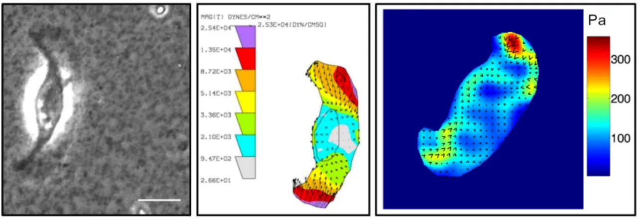
Figure 2.4. Use of elastic substrates to map tractions in migrating cells
A. Phase image showing a fish keratocytes crawling on an elastic polyacrylamide substrate. B. Tractions mapped on the same cell shown in A. The Dembo Boundary element method algorithm (127) was used to calculation of cell traction force from beads displacement; the units in the map are in Dynes/ cm2 (1 dyne=10-5N). 4C. The Fourier-transform traction cytometry (FTTC)
algorithm (129) was used to calculate tractions for another keratocyte; the right scale of color bar represents stress in units of Pa (1Pa=1N/m2). Scale bar is 10µm. Images are courtesy of Zenon Rajfur.
However, with silicone rubber films, matching the compliance to the tractions exerted by the cells and providing a defined surface coating on the film for optimal adhesion is not always easy. These difficulties were circumvented by developing polyacrylamide gel substrates with variable degrees of cross-linking onto which extracellular matrix proteins could be conjugated (130-135). An example of the use of
27
polyacrylamide substrates for examining the tractions exerted by locomoting keratocytes in seen in Figure 2.4. Moreover, these films are optically tractable so that when fluorescent beads are used as the fiduciary markers in the gel, dual channel fluorescence microscopy permits the correlation of tractions in relation to the spatial localization of fluorescently-labeled focal adhesion proteins (115).
Another approach employs special microfabricated substrates that contain an array of force sensing elements. These are flexible cantilevers of known bending stiffness so that the forces exerted by moving cells on these pads can be computed directly from the deflection of the cantilever beams (136-138). An alternate approach employs an elastomeric substrate (silicone film) that is micropatterned to give rise to a regular array of either surface indentations or projections of sub-micron dimensions (117, 139). An algorithm allows the surface distortion of the micropattern caused by cells to be directly translated to the cellular forces. Thus, a number of methods now exist that are similar in overall concept and permit calculation of traction stresses and the correlation of those stresses with the molecular constituents of the force-transmitting adhesive structures.
2.5.2. Force, cell adhesions, and cell migration
There is a clear interplay between contractile force generated by the cell, adhesion to the substrate and the traction applied to the substrate that is beginning to be investigated in detail. As stated above, force can induce focal complexes to mature into large focal adhesions near the leading edge of migrating cells; at the trailing edge, contractile forces regulate adhesion disassembly and cell detachment. Also, MT induced
28
adhesion disassembly has been observed as mentioned previously and it was speculated that the growth of stiff microtubule growth into adhesions can release the force originally exerted by the actomyosin cytoskeleton, thus promoting adhesion disassembly (87, 94).
The relationship between adhesion, traction applied to the substrate, and cell migration is under active investigation. At the outset, it is important to note that the net traction to move the cell through a low viscosity buffer is effectively zero. This leads to the conclusion that the typical tractions measured, which are much larger than what are required to move the cell, must be used to break adhesions in spatiotemporal patterns that dictate both the speed and direction of the cell.
Using keratocytes as a model, Lee and her colleagues reported that slowly migrating keratocytes are more fibroblast-like in their migration and characterized by slipping of adhesions that are coupled with retrograde actin flow; in fast moving keratocytes, adhesions have more gripping character to sustain the rapid protrusion powered by the fast-paced polymerizing actin network; these cells exhibit a much smaller rearward actin flow (140). Recently, maps of actin–substrate coupling were used to quantify differences in force transmission efficiency between different cell regions (20). Thus, a more detailed scenario about the substrate adhesion-traction-migration relationship could be proposed: At the leading edge, traction was transmitted in a manner partially independent of actin velocity (gripping) but at the cell flanks, the force transmission was mediated by the high friction between the actin network and the substrate; at the cell body, little traction was transmitted, because of low friction.
29
Undoubtedly, this relationship will be further investigated both experimentally and theoretically (32) as it is key to achieving a global understanding of how cells move.
2.6 CREDITS
This work was supported by National Institutes of Health Grant GM 41402 and the Cell Migration Consortium Grant GM 064346.
30
2.7 REFERENCES
1. Gligorijevic B and Condeelis J. (2009). Stretching the timescale of intravital imaging in tumors. Cell Adh Migr 3(4): p. 313-5
2. Friedl P and Wolf K. (2010). Plasticity of cell migration: a multiscale tuning model.
J Cell Biol 188(1): p. 11-9
3. Puck TT, Cieciura SJ, and Fisher HW. (1957). Clonal growth in vitro of human cells with fibroblastic morphology; comparison of growth and genetic characteristics of single epithelioid and fibroblast-like cells from a variety of human organs. J Exp Med 106(1): p. 145-58
4. Abercrombie M, Heaysman JE, and Pegrum SM. (1970). The locomotion of fibroblasts in culture. 3. Movements of particles on the dorsal surface of the leading lamella. Exp Cell Res 62(2): p. 389-98
5. Chen W-T. (1979). Induction of spreading during fibroblast movement. J. Cell
Biol. 81: p. 684-691
6. Dunn GA. (1980). Mechanisms of fibroblast locomotion. Cell Adhes. Mot.: p. 409-424
7. Pollard TD and Borisy GG. (2003). Cellular motility driven by assembly and disassembly of actin filaments. Cell 112(4): p. 453-65
8. Ridley AJ, Schwartz MA, Burridge K, Firtel RA, Ginsberg MH, Borisy G, Parsons JT, and Horwitz AR. (2003). Cell migration: integrating signals from front to back.
Science 302(5651): p. 1704-1709
9. Martin P. (1997). Wound healing--aiming for perfect skin regeneration. Science 276(5309): p. 75-81
10. Schreier T, Degen E, and Baschong W. (1993). Fibroblast migration and proliferation during in vitro wound healing. A quantitative comparison between various growth factors and a low molecular weight blood dialysate used in the clinic to normalize impaired wound healing. Res Exp Med (Berl) 193(4): p. 195-205
11. Werner S and Grose R. (2003). Regulation of wound healing by growth factors and cytokines. Physiol Rev 83(3): p. 835-70
12. Ware MF, Wells A, and Lauffenburger DA. (1998). Epidermal growth factor alters fibroblast migration speed and directional persistence reciprocally and in a matrix-dependent manner. J Cell Sci 111 ( Pt 16): p. 2423-32
13. Suetsugu S, Yamazaki D, Kurisu S, and Takenawa T. (2003). Differential roles of WAVE1 and WAVE2 in dorsal and peripheral ruffle formation for fibroblast cell migration. Dev Cell 5(4): p. 595-609
31
14. Huang C, Rajfur Z, Borchers C, Schaller MD, and Jacobson K. (2003). JNK phosphorylates paxillin and regulates cell migration. Nature 424(6945): p. 219-23
15. Goodrich HB. (1924). Cell behaviour in tissue cultures. . Biol. Bull (Woods Hole). (46): p. 252-262.
16. Radice GP. (1980). Locomotion and cell-substratum contacts of Xenopus epidermal cells in vitro and in situ. J Cell Sci 44: p. 201-23
17. Verkhovsky AB, Svitkina TM, and Borisy GG. (1999). Self-polarization and directional motility of cytoplasm. Curr Biol 9(1): p. 11-20
18. Verkhovsky AB, Chaga OY, Schaub S, Svitkina TM, Meister JJ, and Borisy GG. (2003). Orientational order of the lamellipodial actin network as demonstrated in living motile cells. Mol Biol Cell 14(11): p. 4667-75
19. Urban E, Jacob S, Nemethova M, Resch GP, and Small JV. (2010). Electron tomography reveals unbranched networks of actin filaments in lamellipodia. Nat
Cell Biol 12(5): p. 429-35
20. Fournier MF, Sauser R, Ambrosi D, Meister JJ, and Verkhovsky AB. (2010). Force transmission in migrating cells. J Cell Biol 188(2): p. 287-97
21. Svitkina TM, Verkhovsky AB, McQuade KM, and Borisy GG. (1997). Analysis of the actin-myosin II system in fish epidermal keratocytes: mechanism of cell body translocation. J Cell Biol 139(2): p. 397-415.
22. Small JV, Herzog M, and Anderson K. (1995). Actin filament organization in the fish keratocyte lamellipodium. J Cell Biol 129(5): p. 1275-86
23. Prass M, Jacobson K, Mogilner A, and Radmacher M. (2006). Direct measurement of the lamellipodial protrusive force in a migrating cell. J Cell Biol 174(6): p. 767-72
24. Mogilner A and Oster G. (1996). Cell motility driven by actin polymerization.
Biophys J 71(6): p. 3030-45
25. Mogilner A and Oster G. (2003). Force generation by actin polymerization II: the elastic ratchet and tethered filaments. Biophys J 84(3): p. 1591-605
26. Vallotton P, Danuser G, Bohnet S, Meister JJ, and Verkhovsky AB. (2005). Tracking retrograde flow in keratocytes: news from the front. Mol Biol Cell 16(3): p. 1223-31
27. Schaub S, Bohnet S, Laurent VM, Meister JJ, and Verkhovsky AB. (2007). Comparative maps of motion and assembly of filamentous actin and myosin II in migrating cells. Mol Biol Cell 18(10): p. 3723-32
28. Wilson CA, Tsuchida MA, Allen GM, Barnhart EL, Applegate KT, Yam PT, Ji L, Keren K, Danuser G, and Theriot JA. (2010). Myosin II contributes to cell-scale
32
actin network treadmilling through network disassembly. Nature 465(7296): p. 373-7
29. Keren K, Yam PT, Kinkhabwala A, Mogilner A, and Theriot JA. (2009). Intracellular fluid flow in rapidly moving cells. Nat Cell Biol 11(10): p. 1219-24 30. Lee J, Ishihara A, Theriot JA, and Jacobson K. (1993). Principles of locomotion
for simple-shaped cells. Nature 362(6416): p. 167-71
31. Rubinstein B, Jacobson K, and Mogilner A. (2005). Multiscale Two-Dimensional Modeling of a Motile Simple-Shaped Cell. Multiscale Model Simul 3(2): p. 413-439
32. Rubinstein B, Fournier MF, Jacobson K, Verkhovsky AB, and Mogilner A. (2009). Actin-myosin viscoelastic flow in the keratocyte lamellipod. Biophys J 97(7): p. 1853-63
33. Hynes RO and Lander AD. (1992). Contact and adhesive specificities in the associations, migrations, and targeting of cells and axons. Cell 68(2): p. 303-22 34. Lammermann T, Bader BL, Monkley SJ, Worbs T, Wedlich-Soldner R, Hirsch K,
Keller M, Forster R, Critchley DR, Fassler R, and Sixt M. (2008). Rapid leukocyte migration by integrin-independent flowing and squeezing. Nature 453(7191): p. 51-5
35. Laudanna C, Campbell JJ, and Butcher EC. (1996). Role of Rho in chemoattractant-activated leukocyte adhesion through integrins. Science 271(5251): p. 981-3
36. Even-Ram S and Yamada KM. (2005). Cell migration in 3D matrix. Curr Opin
Cell Biol 17(5): p. 524-32
37. Friedl P and Gilmour D. (2009). Collective cell migration in morphogenesis, regeneration and cancer. Nat Rev Mol Cell Biol 10(7): p. 445-57
38. Amatangelo MD, Bassi DE, Klein-Szanto AJ, and Cukierman E. (2005). Stroma-derived three-dimensional matrices are necessary and sufficient to promote desmoplastic differentiation of normal fibroblasts. Am J Pathol 167(2): p. 475-88 39. Heath JP and Peachey LD. (1989). Morphology of fibroblasts in collagen gels: a
study using 400 keV electron microscopy and computer graphics. Cell Motil
Cytoskeleton 14(3): p. 382-92
40. Cukierman E, Pankov R, Stevens DR, and Yamada KM. (2001). Taking cell-matrix adhesions to the third dimension. Science 294(5547): p. 1708-12
41. Kole TP, Tseng Y, Jiang I, Katz JL, and Wirtz D. (2005). Intracellular mechanics of migrating fibroblasts. Mol Biol Cell 16(1): p. 328-38
42. Sanz-Moreno V and Marshall CJ. (2009). Rho-GTPase signaling drives melanoma cell plasticity. Cell Cycle 8(10): p. 1484-7
33
43. Hu H, Marton TF, and Goodman CS. (2001). Plexin B mediates axon guidance in Drosophila by simultaneously inhibiting active Rac and enhancing RhoA signaling. Neuron 32(1): p. 39-51
44. Pankov R, Endo Y, Even-Ram S, Araki M, Clark K, Cukierman E, Matsumoto K, and Yamada KM. (2005). A Rac switch regulates random versus directionally persistent cell migration. J Cell Biol 170(5): p. 793-802
45. Wolf K, Mazo I, Leung H, Engelke K, von Andrian UH, Deryugina EI, Strongin AY, Brocker EB, and Friedl P. (2003). Compensation mechanism in tumor cell migration: mesenchymal-amoeboid transition after blocking of pericellular proteolysis. J Cell Biol 160(2): p. 267-77
46. Wolf K, Wu YI, Liu Y, Geiger J, Tam E, Overall C, Stack MS, and Friedl P. (2007). Multi-step pericellular proteolysis controls the transition from individual to collective cancer cell invasion. Nat Cell Biol 9(8): p. 893-904
47. Niggemann B, Drell TLt, Joseph J, Weidt C, Lang K, Zaenker KS, and Entschladen F. (2004). Tumor cell locomotion: differential dynamics of spontaneous and induced migration in a 3D collagen matrix. Exp Cell Res 298(1): p. 178-87
48. Izzard CS and Lochner LR. (1976). Cell-to-substrate contacts in living fibroblasts: an interference reflexion study with an evaluation of the technique. J Cell Sci 21(1): p. 129-59
49. Zamir E and Geiger B. (2001). Molecular complexity and dynamics of cell-matrix adhesions. J Cell Sci 114(Pt 20): p. 3583-90
50. Lo SH and Chen LB. (1994). Focal adhesion as a signal transduction organelle.
Cancer Metastasis Rev 13(1): p. 9-24
51. Zaidel-Bar R, Cohen M, Addadi L, and Geiger B. (2004). Hierarchical assembly of cell-matrix adhesion complexes. Biochem Soc Trans 32(Pt3): p. 416-20
52. Geiger B, Spatz JP, and Bershadsky AD. (2009). Environmental sensing through focal adhesions. Nat Rev Mol Cell Biol 10(1): p. 21-33
53. Reyes CD and Garcia AJ. (2003). Engineering integrin-specific surfaces with a triple-helical collagen-mimetic peptide. J Biomed Mater Res A 65(4): p. 511-23 54. Emsley J, Knight CG, Farndale RW, Barnes MJ, and Liddington RC. (2000).
Structural basis of collagen recognition by integrin alpha2beta1. Cell 101(1): p. 47-56
55. Humphries MJ. (2000). Integrin structure. Biochem Soc Trans 28(4): p. 311-39 56. Barczyk M, Carracedo S, and Gullberg D. (2010). Integrins. Cell Tissue Res