Address for correspondence
Shiji Dinakaran
E-mail: [email protected]
Funding sources
None declared
Conflict of interest
None declared
Received on May 1, 2017 Reviewed on June 23, 2017 Accepted on July 12, 2017
Abstract
Root caries is an important problem of concern for the elderly population in many countries. Due to its rapidly progressing nature and occurrence in areas of limited visibility, these lesions often go unnoticed unless they become symptomatic. Multiple systemic diseases, poor oral hygiene coupled with reduced manual dexterity of this age group add to the severity of the challenge. Limitations of available biomaterials along with reduced accessibility and isolation make the restoration of such dental lesions a clinical challenge also for dentists. Routine dental checkups and regular monitoring of risk factors can significantly reduce the occurrence of such caries to a major extent.
This article reviews the contemporary literature concerning the clinical features, etiology, risk factors and prevention of root caries. The main topic of the work is the electronic search of literature related to root caries; it was carried out in MEDLINE PubMed database using MeSH terms “root caries”, “etiology”, “prevention”. Key words: risk factors, prevention, restoration, root caries
Słowa kluczowe: czynniki ryzyka, zapobieganie, odbudowa, próchnica korzeni
DOI
10.17219/dmp/75872
Copyright
© 2017 by Wroclaw Medical University and Polish Dental Society
This is an article distributed under the terms of the Creative Commons Attribution Non-Commercial License (http://creativecommons.org/licenses/by-nc-nd/4.0/)
Root caries: A geriatric challenge
Próchnica korzenia – problem gerostomatologiczny
Shiji Dinakaran
A–F, Anupama S. Gopinathan
B,D,EDepartment of Conservative Dentistry and Endodontics, Government Dental College, Thiruvananthapuram, Kerala, India A – research concept and design; B – collection and/or assembly of data; C – data analysis and interpretation; D – writing the article; E – critical revision of the article; F – final approval of the article
Root caries is a multifactorial dynamic disease process that involves the interaction of the host, bacteria and nu-trients, along with modifying or protective factors, result-ing in the demineralization of a tooth. It was virtually the only form of caries seen in the aboriginal population in Papua-New Guinea.1 An examination of ancient skulls
has revealed that root caries was more common than enamel caries in ancient man (Egyptian, North American Indian, Early Anglo Saxon). Today, in primitive societies such as certain natives of New Guinea highlands and ab-origines of India, has been observed as the major cause of tooth loss.2
These soft, rapidly progressing destructive lesions are either confined to the root surface or involve undermin-ing the enamel at the cementoenamel junction. Often, it is seen close to or within 2 mm of the cementoenamel junction.3 Root caries lesions that occur closely adjacent
to or within 2 mm of the crest of gingiva are considered to be active, whereas lesions that occur farther from the gingival crest are more likely to be arrested.4 These
le-sions usually have less well-defined margins, are u-shaped in the cross section, often asymptomatic and have rapid progression. They can occur in areas of abrasion, erosion, abfraction, or as primary root caries or recurrent caries.5
Root caries progresses at about twice the rate of coronal caries because the critical pH of dentin is 6.7. Cementum covering the root surface is extremely thin near the ce-mentoenamel junction, so it provides less resistance to acid attack. Apatite crystals of dentin and cementum are smaller in size than the enamel, and so the reactive sur-face is bigger.6 The loss of cementum increases the rate
of penetrating the root by the lesion and also increases calcium and phosphate loss from the tooth surface.7 Early
root caries tends to be diffuse and track along the cemen-toenamel junction on the root surface. More advanced root lesions enlarge toward the pulp.8
Evidence suggests that either facial or interproximal surfaces are more affected by root caries.9 The proximal
root surface near the cementoenamel junction is often unattended by routine oral hygiene procedures because of the concave contours and occasional roughness at the termination of the enamel. These factors along with gingi-val recession lead to proximal root surface caries. Among the mandibular teeth, molars are more often affected, whereas in the case of maxillary teeth, the anteriors are more affected.10
In 1980, Root Caries Index (RCI) was introduced to overcome the limitations of DFS (Decayed, Filled Surface) index, since DFS index does not consider the number of tooth surfaces at risk of caries.11 It is calculated using the
formula below and is expressed as a percentage:
Root Caries Index =
number of root surfaces with root caries
× 100 . number of root surfaces
with gingival recession
The prevalence of root caries in the age group of 50–64 was found to be 3 times higher compared to 20–34 age group.12 Initially, Actinomyces sp. was believed to be the
pathogen responsible for initiating root caries.2 Later,
some studies suggested that Lactobacillus casei/paraca-sei/rhamnosus and Psuedoramibacter alactolyticus are as-sociated with root caries.13 With advancing age, as a result
of reduced immunity and systemic problems, oral micro-flora changes. Proteolytic bacteria represent 40% of mi-crobes in root caries lesions.14 A recent study has found Propionibacterium acidifaciens, Streptococcus mutans,
Olsenella profusa, Prevotella multisaccharivorax and
Lactobacillus crispatus to be more associated with root caries.15
Classifi cation of root caries
International Caries Detection and Assessment system (ICDAS) classifies and detects root caries based on the following criteria:
– color (light/dark brown/black); – texture (smooth/rough);
– appearance (shiny/glossy, matte/non-glossy); – perception on gentle probing (soft/leathery/hard); – cavitation (loss of anatomic contour).16
In order to detect and classify root caries using ICDAS criteria a community periodontal index (CPI) probe should be used by the examiner. One score will be as-signed per root surface. The facial/mesial/distal/lingual root surfaces of each tooth should be classified as follows: Code O Root surface does not exhibit any unusual discoloration or surface defect, has normal surface con-tour.
Code E Root surface cannot be visualized directly or by gentle air-drying.
Code 1 Clearly demarcated areas on root surface or at the cementoenamel junction that is discolored but no cavitation present (loss of anatomic contour <0.5 mm).
Code 2 Clearly demarcated area on root surface or at cementoenamel junction that is discolored and has cavi-tation (≥0.5 mm).
Code 3 Cavitated (>0.5 mm depth) carious root sur-face which is soft/leathery.
Code 4 Cavitated (>0.5 mm depth) carious root surface which is hard/glossy indicating arrested/inactive caries.
Code 6 Extensive caries involving at least half of the tooth surface and reaching the pulp.
Code 7 Filled root with no caries.
Risk factors for root caries
Since root caries is a multifactorial disease, several in-traoral, behavioral and population related factors may increase an individual’s risk of developing root caries.
Some of these factors are modifiable, whereas others are uncontrollable.17 Most studies have shown that the
ma-jority of root caries are primary lesions. This indicates that controlling/modifying risk factors can prevent car-ies.18 Risk factors can be intraoral or extraoral. The most
common intraoral risk factor for root caries in adults is increased gingival recession leading to exposed roots.17
From clinical findings it can be assumed that an indivi-dual with a high risk of coronal caries has a higher chance of developing root caries, indicating that risk factors for both type of caries are more or less the same.
Xerostomia, resulting from chronic medication and ra-diation therapy, increases the risk of root caries. Drugs such as antipsychotics, sedatives, barbiturates, antihis-tamines reduce salivary flow and increase caries risk.19
Some studies indicate that low buffering capacity, low sali vary immunoglobulin A, low salivary calcium and phosphate may also be associated with increased risk of root caries. As we age, our immune system weakens and less antimicrobial immunoglobulin is produced and found in the saliva. The level of serum immunoglobulin IgG and IgM is significantly reduced in older individuals. This may contribute to the increased susceptibility of el-derly individuals to oral diseases.20
A diet high in refined carbohydrates, poor general and dental health are also risk factors contributing to the de-velopment of root caries. Both the quantity and frequency of sugar consumption are crucial factors in the develop-ment of root caries, similarly as in the case of coronal caries. Other intraoral risk factors include periodontal disease, periodontal surgery, unrestored coronal caries, partial dentures associated with plaque accumulation and gingival recession on abutment teeth, overdenture abut-ments, removable partial dentures, malocclusion, cervi-cal non-carious lesions, tipped teeth, which make certain areas of teeth inaccessible for cleaning.21
Cardiovascular diseases have also been associated with higher caries experience, particularly in individuals of 80 years or older. Older people with hypoalbuminemia are at considerable risk of root caries.22 Diseases like Sjögren’s
syndrome, diabetes mellitus also increase caries risk sta-tus.19. Drugs used for managing these systemic conditions,
unattended oral hygiene and altered oral flora may be the contributing factors for elevated root caries level in such individuals. Low indices of socioeconomic status have been associated with an increase in caries due to reduced access to care. Another behavioral risk factor is tobacco
use. Smoking is associated with periodontal diseases and gingival recession, and chewing tobacco contains fermentable carbohydrates.23 Other behavioral risk
fac-tors include consumption of alcohol/narcotics. Males are more often affected by root caries than females. Patients with physical disabilities or with limited manual dexterity for cleaning their teeth are also at a high risk of root car-ies. Traumatic tooth brushing habits render the retained dentition susceptible to root surface caries.24
Prevention of root caries
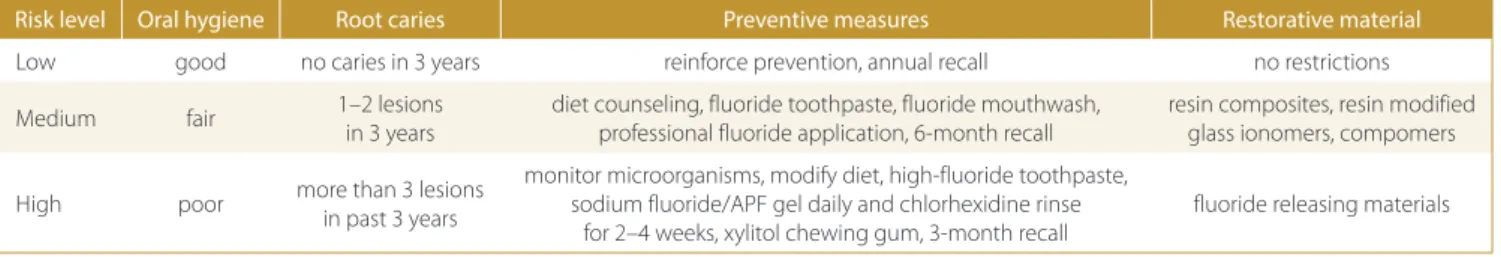
Measures to prevent root caries can be categorized into 3 levels – primary, secondary and tertiary prevention. Primary preventive measures help prevent the occur-rence of the disease. This can be done at the population level or individual level. Measures at the population level include water fluoridation, whereas individual measures include reducing or removing risk factors and enhanc-ing protective factors. Secondary preventive measures help slow down or arrest an ongoing disease process. In the case of root caries these measures include reminer-alization/minimal restoration of carious lesions. They help remineralize incipient lesions and prevent the de-velopment of new lesions. Tertiary preventive measures include placing extensive restorations so as to prevent further disease-related deterioration of the tooth and to return it to normal function. Preventive and restorative options based on the risk status of the individual are pre-sented in Table 1.21,25
In clinical practice, primary preventive measures to prevent root caries are significant, since restorative treat-ment of such lesions is extremely challenging and may lead to tooth loss in most cases due to recurrent caries or the sensitivity of the restored tooth. Therefore, such measures should be taken immediately after the exposure of root surfaces, as root caries progresses faster due to low mineral content of the covering cementum.26 Fluoride is
a widely accepted preventive agent, since it can interfere with the physiochemical and microbiological proceses of root caries. High fluoride concentration may be necessary to prevent and control root caries compared to coronal caries.27 Patient-level preventive measures include
tak-ing fluoride supplements regularly, fluoridated tooth-paste, xylitol gums/mouthwash, chlorhexidine rinses, di-amine silver chloride lozenges, altering dietary habits etc. Table 1. Experimental groups
Risk level Oral hygiene Root caries Preventive measures Restorative material
Low good no caries in 3 years reinforce prevention, annual recall no restrictions Medium fair 1–2 lesions
in 3 years
diet counseling, fluoride toothpaste, fluoride mouthwash, professional fluoride application, 6-month recall
resin composites, resin modified glass ionomers, compomers High poor more than 3 lesions
in past 3 years
monitor microorganisms, modify diet, high-fluoride toothpaste, sodium fluoride/APF gel daily and chlorhexidine rinse
for 2–4 weeks, xylitol chewing gum, 3-month recall
Professional-level preventive measures include the ap-plication of topical fluoride gels/varnishes. The list of medications taken by the patient can be checked and less xerogenic drugs can be prescribed or their dosage can be reduced. Salivary stimulants such as pilocarpine can be prescribed.28 For the primary prevention of root caries
the recommended best choice is 38% silver diamine fluo-ride solution professionally applied manually; it is recom-mended as the most cost saving treatment for root caries prevention in high risk individuals.29 If no professional
application is possible, the best alternative is the use of self-applied amorphous calcium phosphate (ACP) and fluoride toothpaste daily.
Subjects with risk of root caries should be recom-mended to use fluoride toothpaste with a concentration between 1000 and 1500 ppm of fluoride as sodium fluo-ride, monofluoro phosphate or amino fluoride. The daily use of 1100 ppm dentifrice combined with APF (acidu-lated phosphate fluoride) gel application provides added protection.30 Studies have shown that the use of fluoride
toothpaste containing 1.5% arginine and insoluble calci-um resulted in the formation of ammonia and increased pH level of the oral cavity, and thus prevented the devel-opment of root caries. Also, arginine helps maintain the normal oral flora and thereby derive the benefits of nor-mal oral flora.31 Mechanical plaque control methods may
require good manual dexterity, which is compromised at old age. Moreover, hypersensitivity is a common problem for elderly people. Therefore, antimicrobial agents may complement mechanical plaque removal for preventing root caries. The use of automated toothbrushes and the daily use of water irrigation devices can also be suggested for root caries-prone patients. In high risk patients who wear dental prostheses avoid placing restoration margins apical to the surrounding tissue to prevent plaque accu-mulation.28
Cavitated root caries that is not easily accessible for cleaning may be restored by minimally invasive tech-niques.32 Fast setting self-cure glass ionomers could be
used for such minimal restorations. Regular use of den-tifrices containing 5000 ppm fluoride and quarterly pro-fessional application of chlorhexidine or silver diamine fluoride varnishes have been found to be effective in de-creasing the progression and initiation of root caries.33
Silver nitrate and silver diamine fluoride are more effec-tive in arresting aceffec-tive caries lesions and preventing new caries than fluoride varnish. It has antimicrobial activity against Streptococcus mutans and Actinomyces naeslundii found on dentin surfaces and slows down dentin demin-eralization.34 Studies have shown that the use of 10%
sodi-um hypochlorite removes organic materials and increases the permeability of fluoride ions, and thereby increases the potential of remineralization.35 In a 15-month period
study the daily intake of milk supplemented with fluoride and, or probiotic bacteria was found to reverse soft and leathery primary root caries lesions.36
Dentin biomodifiers are agents which interact with vari-ous extracellular matrix components of dentin and increase their mechanical properties, decrease the biodegradation rate and possibly promote mineral nucleation.37 Dentin
modifiers, such as 5% glutaraldehyde, 0.5% proanthocy-anidin and 0.625% genipin, can modify dentin collagen and increase its resistance to enzymatic digestion by cariogenic organisms.38 Grape seed extract contains
proanthocyani-din which can be used instead of fluoride during minimally invasive the treatment of root caries. Proanthocyanidin-treated collagen matrices resisted enzyme digestion in vivo and in vitro. Also it increased collagen synthesis and accel-erated the conversion of soluble collagen into insoluble col-lagen during development. Grape seed extract also induced cross-links in dentin collagen and increased the ultimate tensile strength of demineralized dentin.39 The combined
use of proanthocyanidin and casein phosphopeptide-amorphous calcium fluoride phosphate (CPP-ACFP) has synergist ic effect on root caries remineralization and in-creases the hardness of root caries.40
Coating the root surface with resin-based materials that can release and recharge fluoride may be a simple and effective measure to protect it from physical, chemi-cal and biologic insults. Surface prereacted glass ionomer (S-PRG) fillers incorporated into resinous material, which have the ability to release and recharge fluoride and other ions such as aluminum, boron and strontium, can be used to enhance remineralization and reduce acid attack by oral cariogenic bacteria. RG filler containing resins were found to inhibit bacteria induced pH drop and formed a hermetic seal on the exposed dentin surface.41
Progres-sion of root caries could be inhibited by the application of antibacterial adhesive system containing 12-methacryloy-loxydodecylpyridinium bromide (MDPB). Unpolymer-ized MDPB in the primer solution penetrates deeply into the lesion to kill or inactivate the infecting bacteria. Also, it seals the lesion and prevents caries.42
A combination of casein phosphopeptide and amor-phous calcium phosphate (CCP-ACP) and photo acti-vated disinfection (PAD) proved to be very effective and holds great potential as a recommended treatment for stabilizing root surface caries in the clinical practice.43
Healozone has been proposed as a method of reversing, arresting or slowing down the progression of dental car-ies. Ozone gas is applied to the carious lesion followed by the use of a remineralizing solution, immediately after the application of ozone.44
Carious lesions should be restored taking into consider-ation the patient’s age, health status and symptoms asso-ciated with root caries. Before the teeth are restored, root caries must be controlled using preventive measures. The type of restorative material depends on the location, acces-sibility of margins of the lesion and oral hygiene. Restorative management is challenging due to difficulties in visibility, moisture control, access to carious lesions, proximity to pulp/gingival margin and high organic content of dentine.
Microleakage and poor marginal adaptation of the restora-tion or defective margins are the most common causes of failure. Restorative material with high potential for fluoride release as well as uptake should be considered.45
The root surface is cleaned with pumice, the excavation of carious tissue is done, and the restoration walls are ppared. Margins and restoration design depend on the re-storative material used. Modified composite or resin modi-fied glass ionomer are ideal for restorations in medium risk individuals. Root caries often may extend below the gingi-val margin, making it necessary to retract the gingiva with a clamp/pack to expose the cervical margin of the lesion or to utilize laser or electrosurgery to recontour the gingiva and obtain access to the lesion. A combined minor peri-odontal and restorative procedure is relatively simple and when done properly can provide excellent and affordable dentistry in the problematic areas.46 The decision to use
a particular method of retraction depends upon whether the apical extension of the lesion is located 2 mm above the height of the alveolar crest. Access can be gained by the use of a retraction cord and cervical clamp like 212, which displaces the soft tissue and exposes the lesion, or by cre-ating a mini flap.47 Coverage of a previously carious root
with subepithelial connective tissue graft is a very predict-able procedure, with results similar to those found in intact roots. This procedure may provide a definitive biological alternative for conventional treatment modalities for root caries. Moreover, the results are more esthetic, biologically acceptable and maintainable.48 In search of other effective
long-term preventive measures against caries, research is being conducted on developing caries vaccine. The main research target is the mechanism of adherence of Strepto-coccus, which can be affected by active/passive immuniza-tion and the use of DNA vaccines.49
Conclusions
With the development of newer technologies and oral hygiene measures, the incidence of dental caries has been reduced significantly. However, root caries continues to be a major challenge for both the patient and the dentist. It has been found to be a major factor which negatively affects the oral-health-related quality of life in the elderly population. In most cases, when these lesions are diagnosed, they are in the advanced stages, which necessitates the removal of the tooth. Regular dental visits and oral hygiene monitoring is essential in old age to meet this challenge.
References
1. Schamschula RG, Barmes DE, Keyes PH, Gulbinat W. Prevalence and interrelationships of root surface caries on Lufa, Papua New Guin-ea. Community Dent Oral Epidemiol. 1974;2:295–304.
2. Sumney DL, Jordan HV. Characterization of bacteria isolated from human root surface carious lesions. J Dent Res. 1974;53:343–351. 3. Aherne CA, O’Mullane D, Barrett BE. Indices of root surface caries.
J Dent Res. 1990;69:1222–1226.
4. Beighton D, Lynch E, Heath MR. A microbiological study of prima-ry root-caries lesions with different treatment needs. J Dent Res. 1993;72:623–629.
5. Jones JA. Root caries: Prevention and chemotherapy. Am J Dent. 1995;8:352–357.
6. Surmont PA, Martens LC. Root surface caries: An update. Clin Prev Dent. 1989;11:14–20.
7. McIntyre JM, Featherstone JD, Fu J. Studies of dental root surface caries. Aust Dent J. 2000;45,24–30.
8. The role of cementum in root surface caries. Aust Dent J. 2000; 45:97–102.
9. Ellen RP, Banting DW, Fillery ED. Streptococcus mutans and Lactoba-cillus detection in the assessment of dental root surface caries risk.
J Dent Res. 1985;64:1245–1249.
10. Banting DW. Epidemiology of root caries. Gerodontol. 1986;5:5–11. 11. Nisha G, Amit G. Textbook of Operative Dentistry. 2nd ed. New Delhi,
India: Jaypee Brothers Medical Publishers; 2013.
12. Katz RV. The RCI revisited after 15 years: Used, reinvented, modified, debated, and natural logged. J Public Health Dent. 1996;56:28–34. 13. Dye BA, Tan S, Smith V, et al. Trends in oral health status: United
States, 1988–1994 and 1999–2004. Vital Health Stat. 2007;11:1–92. 14. Preza D, Olsen I, Willumsen T, et al. Microarray analysis of the microflora
of root caries in elderly. Eur J Clin Microbiol Infect Dis. 2009;28:509–517. 15. Hashimoto K, Sato T, Shimauchi H, Takahashi N. Profiling of dental
plaque microflora on root caries lesions and the protein-denatur-ing activity of these bacteria. Am J Dent. 2011;24:295–299. 16. Chen L, Qin B, Du M, et al. Extensive description and comparison of
human supra-gingival microbiome in root caries and health. PLoS One. 2015;10. doi:10.1371/e 0117064.
17. Shivakumar K, Prasad S, Chandu G. International caries detection and assessment system: A new paradigm in detection of dental caries. J Conserv Dent. 2009;12:10–16.
18. Chi DL, Shyue C. Managing caries risk in adults. Dimen Dent Hyg.
2014;12:36–40.
19. Joshi A, Douglass CW, Jette A, Feldman H. The distribution of root caries in community-dwelling elders in New England. J Public Health Dent. 1994;54:15–23.
20. Lenander-Lumikari M, Loimaranta V. Saliva and dental caries. Adv Dent Res. 2000;14:40–47.
21. Fischer D, Ship JA. Effect of age on variability of parotid salivary gland flow rates over time. Age Ageing. 1999;28:557–561.
22. Shay K. Root caries in the older patient: Significance, prevention, and treatment. Dent Clin North Am. 1997;41:763–793.
23. Yoshihara A, Takano N, Hirotomi T, Ogawa H, Hanada N, Miyazaki H. Longitudinal relationship between root caries and serum albumin.
J Dent Res. 2007;86:1115–1119.
24. Ringelberg ML, Gilbert GH, Antonson DE, et al. Root caries and root defects in urban and rural adults: The Florida dental care study.
J Am Dent Assoc. 1996;127:885–891.
25. Imazato S, Ikebe K, Nokubi T, Ebisu S, Walls AW. Prevalence of root caries in a selected population of older adults in Japan. J Oral Reha-bil. 2006;33:137–143.
26. Shaker Randa E. Diagnosis, prevention and treatment of root car-ies. Saudi Dent J. 2004;16:84–86.
27. Keltjens H, Schaeken T, van der Hoeven H. Preventive aspects of root caries. Int Dent J. 1993;43:143–148.
28. Rodrigues JA, Lussi A, Seemann R, Neuhaus KW. Prevention of crown and root caries in adults. Periodontol 2000. 2011;55:231–249. 29. Gluzman R, Katz RV, Frey BJ, McGowan R. Prevention of root caries:
A literature review of primary and secondary preventive agents.
Spec Care Dent. 2013;33:133–140.
30. Schwendicke F, Göstemeyer G. Cost-effectiveness of root caries preventive treatments. J Dent. 2017;56:58–64.
31. Vale GC, Tabchoury CP, Del Bel Cury AA, Tenuta LM, ten Cate JM, Cury JA. APF and dentifrice effect on root dentin demineralization and biofilm. J Dent Res. 2011;90:77–81.
32. ten Cate JM, Cummins D. Fluoride toothpaste containing 1.5% argi-nine and insoluble calcium as a new standard of care in caries pre-vention. J Clin Dent. 2013;24:79–87.
33. Heasman PA, Ritchie M, Asuni A, Gavillet E, Simonsen JL, Nyvad B. Gingival recession and root caries in the ageing population: a critical evaluation of treatments. J Clin Periodontol. 2017;44(Suppl. 18):S178–S193.
34. Wierichs RJ, Meyer-Lueckel H. Systematic review on noninvasive treatment of root caries lesions. J Dent Res. 2015;94:261–271. 35. Mei ML, Chu CH, Lo EC, Samaranayake LP. Preventing root caries
development under oral biofilm challenge in an artificial mouth.
Med Oral Patol Oral CirBucal. 2013;18:e557–563.
36. Inaba D, Duschner H, Jongebloed W, Odelius H, Takagi O, Arends J. The effects of a sodium hypochlorite treatment on demineralized root dentin. Eur J Oral Sci. 1995;103:368–374.
37. Petersson LG, Magnusson K, Hakestam U, Baigi A, Twetman S. Reversal of primary root caries lesions after daily intake of milk sup-plemented with fluoride and probiotic lactobacilli in older adults.
Acta Odontol Scand. 2011;69:321–327.
38. Bedran-Russo AK, Pereira PN, Duarte WR, Drummond JL, Yam-auchi M. Application of crosslinkers to dentin collagen enhances the ultimate tensile strength. J Biomed Mater Res B Appl Biomater. 2007;80:268–272.
39. Jayakrishnan A, Jameela SR. Glutaraldehyde as a fixative in biopros-theses and drug delivery matrices. Biomaterials. 1996;17:471–484. 40. Han B, Jaurequi J, Tang BW, Nimni ME. Proanthocyanidin:
A natu-ral crosslinking reagent for stabilizing collagen matrices. J Biomed Mater Res A. 2003;65:118–124.
41. Epasinghe DJ, Yiu C, Burrow MF. Synergistic effect of proanthocy-anidin and CPP-ACFP on remineralization of artificial root caries. Aust Dent J. 2015;60:463–470.
42. Ma S, Imazato S, Chen JH, et al. Effects of a coating resin contain-ing S-PRG filler to prevent demineralization of root surfaces. Dent Mater J. 2012;31:909–915.
43. Rolland SL, McCabe JF, Imazato S, Walls AW. A randomised trial comparing the antibacterial effects of dentine primers against bacteria in natural root caries. Caries Res. 2011;45:574–580. 44. Vlacic J, Meyers IA, Walsh LJ. Combined CPP-ACP and
photoacti-vated disinfection (PAD) therapy in arresting root surface caries: A case report. Br Dent J. 2007;203:457–459.
45. Brazzelli M, McKenzie L, Fielding S, et al. Systematic review of the effectiveness and cost-effectiveness of HealOzone for the treat-ment of occlusal pit/fissure caries and root caries. Health Technol Assess. 2006;10:75–80.
46. Anusavice KJ. Dental caries: Risk assessment and treatment solu-tions for an elderly population. Compend Contin Educ Dent. 2002; 23(Suppl. 10):12–20.
47. Burgess JO, Gallo JR. Treating root-surface caries. Dent Clin North Am. 2002;46:385–404.
48. Turner EW, Shook LW, Lackey M. Accessing and restoring root car-ies: A case report. J Tenn Dent Assoc. 2007;87:20–24.
49. Goldstein M, Nasatzky E, Goultschin J, Boyan BD, Schwartz Z. Cov-erage of previously carious roots is as predictable a procedure as coverage of intact roots. J Periodontol. 2002;73:1419–1426.