ORIGINAL PAPERS
Agnieszka Przystańska
1, 2, A–D, F, Dorota Lorkiewicz-Muszyńska
1, A–C, E, F,
Michał Rychlik
3, B, C, E, F, Mariusz Glapiński
4, A–C, F, Marzena Łabęcka
1, B, E, F,
Paweł Świderski
1, C, E, F, Czesław Żaba
1, A, E, FThe Effectiveness of 2D and 3D Methods
in the Analysis of Experimental Bite Marks
Skuteczność metod 2D i 3D w analizie
eksperymentalnych śladów ugryzień
1 Department of Forensic Medicine, Poznań University of Medical Sciences, Poznań, Poland 2 Department of Anatomy, Poznań University of Medical Sciences, Poznań, Poland
3 Institute of Combustion Engines and Transport, Division of Virtual Engineering, Poznań University
of Technology, Poznań, Poland
4 Oral Rehabilitation Clinic, Poznan University of Medical Sciences, Poznań, Poland
A – research concept and design; B – collection and/or assembly of data; C – data analysis and interpretation;
D – writing the article; E – critical revision of the article; F – final approval of article
Abstract
Background. A bite mark left on different materials and surfaces such as food, chewing gum or the skin of dead or living persons and found at a crime scene can provide the crucial evidence required to secure a conviction in cases where neither fingerprints nor DNA are found. The most challenging factor in bite mark analysis is the interpreta-tion of human bite marks left on the skin. This is due to both the specific character of skin – its elasticity and ability to distort – and the different shape and curvature of body parts and regions.
Objectives. To investigate the possibility of identifying a biter using 2D and 3D analysis of experimental bite marks and to check to what extent the characteristics of the material bitten influence the identification rate.
Material and methods. The volunteers were asked to bite various materials of different structure and shape. The impressions of individuals’ dentition and wax bite registrators were taken in order to prepare dental casts. The samples of dental casts, corresponding bite registrators and bite marks from different materials were then scanned with 2D and 3D scanners, photographed, and finally examined and compared using computer methods.
Results. By comparing the characteristics of the teeth and experimental bite marks, it was possible to positively iden-tify all individuals, but with different degrees of possibility. Negative results were obtained when comparing charac-teristics of teeth from one individual with the bite marks created by another individual who took part in the study.
Conclusions. When applied to experimental bite marks, both 2D and 3D methods proved effective in the positive identification of the biter (Dent. Med. Probl. 2015, 52, 1, 86–92).
Key words: forensic odontology, bite marks, identification.
Słowa kluczowe: odontologia sądowa, ślady ugryzień, identyfikacja.
Dent. Med. Probl. 2015, 52, 1, 86–92
ISSN 1644-387X © Copyright by Wroclaw Medical University and Polish Dental Society
Teeth, apart from their primary function of biting and mastication, are used occasionally by humans as a weapon to attack another person and also as a defense against an attack. The mecha-nism of biting leaves imprints on the skin. Sur-prisingly, the number of cases with human bites is not significantly lower than the number of cases with animal bites [1]. The bite marks observed on
the skin are most often related to sexual assaults and thus they are usually localized over the breast or external genitalia. Bite marks are also observed in children as the result of domestic violence or child abuse. Occasionally, bite marks are revealed on different types of food found at the crime scene (fruit, sweets, chewing gum) and also other mate-rials, such as paper and plastic coverings on
a bot-tle tops [2–4]. Biting is a dynamic process and is influenced by several factors: the mutual relation-ship between the maxillary and mandibular teeth, the action of muscles of mastication, and more specifically, a person’s reaction to being bitten by an aggressor [2, 5, 6]. The bite mark left by one person can vary in the depth and accuracy of the impression, which is caused by the variable dy-namics of the biting process itself. Bite marks are expressed on different materials with varying de-grees of accuracy. To a large extent, the impres-sion depends on the properties of the materials on which the bite marks are present. The complicated nature of bite marks is manifested not only by the impressions left by incisors, canines and premo-lars but also by the accompanying abrasions and sugillations. The molars, whose primary function is chewing, do not leave impressions during bit-ing. “Biting into” a material causes an interruption in the surface of the material; however, it does not cause the separation of the fragments of the bitten material. The indentations represent the impres-sions of part of the arch with various individual characteristics of the teeth in contact with the ma-terial. “Biting off” causes a disruption in the con-tinuity of the material and separation of parts of the material and leaves an impression of the arch-es and the characteristics of the teeth. Sometimarch-es, impressions are created by the mechanical shift of the teeth superficially along the material. Most of-ten, these are the traces left behind by the man-dibular and maxillary teeth. It is also possible that the maxillary teeth work as an anchor and the mandibular teeth, due to the mobility of the lower jaw, they move along the surface, leaving scratch-es and abrasions [7]. It is the rscratch-esponsibility of fo-rensic experts (fofo-rensic odontologists) to identify a bite mark, take care of its proper preservation and documentation, and finally to adequately an-alyze and interpret the evidence. Identification based on bite mark analysis is based on two as-sumptions: first, that the characteristics of an in-dividual’s dentition are unique, and second, that these traits leave unique impressions in the bitten material. Bite mark analysis includes the exami-nation of physical characteristics of both the bite mark wound and the suspect’s teeth, particular-ly the distance from canine to canine, the shape of the mouth arch, the evidence of tooth rotation, tooth width, reciprocal high and thickness, spac-ing between teeth, the curves of bitspac-ing edges, and unevenness of teeth edges. Comparative analy-sis is conducted comparing tooth-tooth and arch- -arch interrelations, size of teeth and the size of the dental arch, the arrangement of teeth in the dental arch, height of the teeth and unevenness of occlu-sal line and also characteristics of teeth
contain-ing restorations, prosthetic restorations (bridges, crowns) and anomalies [8, 9]. The initial step in preserving bite marks is photographic documenta-tion: this should be performed in accordance with the principles accepted in criminology (camera lens should be positioned at an angle of 90 degrees in relation to the surface with the bite mark and a bite mark should be localized in the center of the photo frame) [3, 10 – 12]. Unfortunately, minimal distortions during photographic documentation are a universal problem in analysing bite marks, even when following standard protocols. Because the preservation of the bite mark can be crucial for further investigation, different methods, such as pouring moulds, performing scans, saving saliva swabs for DNA examination or scanning electron microscopy, have been recognized as effective [13, 14]. In order to avoid distortion of the preserved bite marks, and in accordance with the American Board of Forensic Odontology (ABFO) guidelines, the roller-scale ABFO No. 2 should be used [3, 15]. The use of 2D and 3D scanning of the bite marks seems to be a satisfactory means of preserving the shape and form of the bite marks prior to inves-tigation procedures. The aim of the study was to investigate whether the experimental bite marks preserved by 2D and 3D scanning can be properly analyzed and whether they can lead to the identi-fication of the biter. The second goal was to check to what extent the characteristics of material bit-ten influenced the identification rate.
Material and Methods
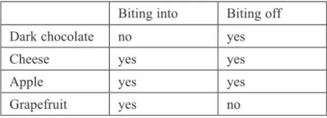
Experimental bite marks were obtained from 10 individuals (volunteers). The individuals in-volved in the study were asked to leave bite marks of two types (“bite-in” and “bite-off”) on 4 dif-ferent types of material, characterized by varying structure, coherence, hardness, elasticity and sur-face shape (Table 1).
Anatomical impressions and wax bite regis-trators were obtained from the individuals taking part in the study, and gypsum models were formed on the basis of the impressions. The models, along with the bite registrations, were used as experimen-tal elements in further steps of the examination.
Table 1. Type of bitemarks collected on various materials. “Bite-off” – a portion of testing material was separated by the teeth; “bite-into” – clear marks were left on the material
Biting into Biting off Dark chocolate no yes
Cheese yes yes
Apple yes yes
2D Analysis
The gypsum models, along with the bite reg-istrations were scanned using an Epson Perfec-tion 4990 Photo flatbed scanner with a resoluPerfec-tion of 1200. During scanning, the ABFO No. 2 scale was used. In analyzing the mandible and maxil-lary casts, the graphic program CorelDrawX3 was used to obtain incisal edge contours and outlines of the chewing surfaces of the cusps from central incisor to second premolar. A mirror image of the teeth of the gypsum models was created. It was compared with the teeth impressions in the wax bite registration, taken from the individuals tak-ing part in the study.
Photographic documentation of the bite marks was obtained with the Canon 20D digital SLR camera. During photo documentation, the ABFO No. 2 scale was used. To create a uniform surface between the bite marks and the measuring device the scale was positioned on “modeling pedestals”. The pedestals were positioned, taking into account their height, so that the scale would find itself at the height and location of the bite marks. The pho-tos were taken from a vertical projection, at a 90 degree angle and subsequently, they were saved to a computer hard drive. The previously collected characteristics of each individual tooth were com-pared with the bite marks on the different test ma-terials. A comparative analysis of the unique mor-phological features of the teeth and characteristics of the bite marks was performed with the use of CorelDraw X3. The comparison method was the superimposition of the images. Multilayered pag-es were created, starting with importing photo-graphs of the materials containing the bite marks onto the first layer. On the second layer, outlines and contours of the gypsum cast were import-ed, from the central incisor to the second premo-lar. The characteristics of the width and shape of the maxillary and mandibular arches, width and shape of the projection of the line and incisor edg-es of the individual cusps of the teeth, all the fea-tures of the teeth in the arch (position, rotations, etc.) were analysed.
3D Analysis
The gypsum casts and also the materials con-taining the experimental bite marks were scanned to obtain corresponding 3D models. The scanning was performed using an optical scanner from the company GOM Atos II Rev. 01 with a measuring volume of 120 × 96 × 80 mm. The resolution of the camera was 1.4 megapixels. The distance be-tween points during scanning was 0.18 mm. The objects and models were placed on an automated rotation table, which rotates until the whole ob-ject is scanned. The 3D model gypsum casts were
placed on the bite marks corresponding to the 3D scanned objects, lining up the borders of the inci-sors and canines and also the cusp tips of the pre-molars of the maxilla and mandible to the rein-forced bite marks to a scale of 1 : 1. Polygon mesh-es (3D models) of the models of the dentitions and of the bite marks were created, using the ATOS software. In the comparison of the dentitions with the bite marks, the ATOS modelling software was applied. The meshes of the maxilla and the mandi-ble were transformed into the corresponding po-sitions of the bite mark. This allowed for conclu-sive results regarding the correspondence between the dentition and the bite mark. In this 3D pattern comparison, the structure of several teeth, as well as the relationship to the surrounding teeth was compared. The meshes of the maxilla and mandi-ble were transformed into the corresponding po-sitions of the bite marks. This allows for conclu-sions to be made between the dentition and the bite marks. The width and shape of the maxillary and mandibular arches, width and shape of the projection of the incisive edges and cusps of the teeth, and other characteristics of the teeth (posi-tion, rotations, restorations) were analysed.
The conclusions were made based on the ABFO standards. To describe the relation of a bite mark to a biter the below terms were used:
– the biter;
– the probable biter; – not excluded as the biter; – excluded as the biter; – inconclusive [15].
Results
The registered bite marks exposed the various impressions of the teeth depending on the materi-al. The most obvious and clear bite mark impres-sions were observed on the cheese. The bite marks were less distinct on apples and grapefruits. With the latter materials, there were distortions due to the material characteristics: in certain places the disturbance of the fruit skin was uneven and slightly distorted the imprinted bite mark. The ex-periment with dark chocolate revealed that at the initial stages of biting, the teeth immersed them-selves into the material, after which the chocolate broke during further biting. Distortion of the orig-inal bite marks arose in most cases.
The result of the added outline of the border of the anterior maxillary teeth on the bite marks (“bite-on” and “bite-off”) using the graphic pro-gram CorelDraw X3 is presented in Fig. 1. Thanks to the accurate impression of the bite marks, it was possible to perform a detailed analysis in a wide
range of evaluated characteristics of the arches and individual teeth along with the characteristics of the bite marks.
The analysis using 2D and 3D methods, which compared the characteristics of the teeth and ex-perimental bite marks, allowed for identification of the biters in most of the cases (Tab. 2, 3). The best results (positive and possible identification in
all cases) were achieved when analysing the bite marks on the cheese and apple. The analysis of the bite marks left in the cheese (Fig. 2) allowed for an unambiguous identification of all biters. Nega-tive results were obtained when comparing char-acteristics of teeth from one individual with the bite marks created by another one involved in the experiment.
The comparative analysis (both methods 2D and 3D) of the bite marks left in the apple (Fig. 3) allowed for unambiguous identification of 8 in-dividuals. Two individuals were positively identi-fied, however without absolute certainty (possible biters).Negative results were obtained when com-paring characteristics of teeth from one individu-al with the bite marks created by another one in-volved in the experiment.
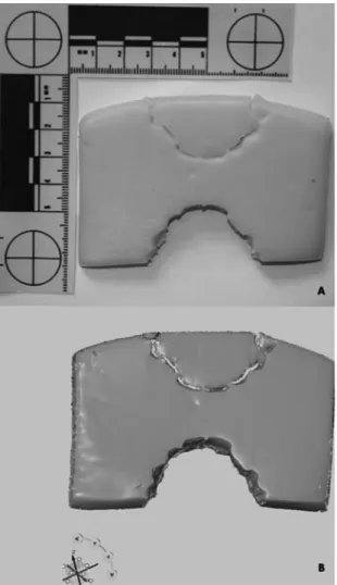
Fig. 2. The polygon mesh (3D model) of bite marks on the apple – “bite-off” (A) and “bite-into” (B)
Fig. 1. Bitemarks (“bite-into” and “bite-off”) on cheese saved using 2D method (A), and the polygon mesh (3D model) of bitemarks on the cheese (B)
Table 2. Results of biter’s identification based on experi-mental bitemarks 2D analysis
Identified
as biter Identified as possible biter
Not exclud-ed as biter Dark
choc-olate 6 2 2
Cheese 10 – –
Apple 8 2 –
Grapefruit 4 5 1
Table 3. Results of biter’s identification based on experi-mental bitemarks 3D analysis
Identified
as biter Identified as possible biter
Not exclud-ed as biter Dark
chocolate 6 2 2
Cheese 10 – –
Apple 8 2 –
Even though the results of the analysis of bite marks left in the grapefruit (both methods 2D and 3D) allowed for identification of all 10 indi-viduals, an unambiguous identification was con-cluded in 4 cases only. The 6 individuals were identified; however, 5 were identified as possible biters and 1 was not excluded as the biter. The bite marks did not present enough characteristic fea-tures to reach an unambiguous positive conclu-sion. Nonetheless, negative results were obtained when comparing characteristics of teeth from one individual with the bite marks created by another one involved in the experiment.
Also the experiment with dark chocolate al-lowed for the identification of all the individuals. Unambiguous identification was concluded in 6 individuals. The 4 individuals were identified, however with only partial certainty (2 possible biters and 2 not excluded as biters). Bite marks observed on the dark chocolate did not present enough characteristic features to reach an un-ambiguous positive conclusion, but negative re-sults were obtained when comparing character-istics of teeth from one individual with the bite marks created by another one involved in the ex-periment.
Discussion
The complex processes of biting and mastica-tion characterized by variable dynamics cause the bite marks left behind by one individual to vary in shape, size and depth of impression [5, 16].Every contact of a tooth with a surface creates a differ-ent and unique case of biting [5, 6]. The impres-sions left behind by the teeth depend on the an-gle and arrangement of the maxillary and man-dibular arches in motion relative to the surface that has been bitten and result in “bite-into”, “bite-off” or abrasions. Moreover, the character of the im-pression to a large extent depends on the prop-erties of the material (shape, structure, elasticity, etc.).There are many challenging aspects to bite mark analysis. The problem that is faced most of-ten deals with original and subsequent bite mark distortions. The original (primary) distortions are caused by both the dynamics and force exerted during biting and the characteristics of the materi-al and then there are the distortions caused by the passage of time and by the technicalities involved in recording them as evidence. It is during this passage of time that “subsequent” malformations occur [6]. The rapid changes that occur to a body
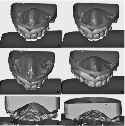
Fig. 3. A 3D compari-son of features of the maxillary teeth of an individual participating in the experiment and bitemarks (“bite- -off”) on cheese using GOM Inspect V7 SR2 software. Different sequences of com-parison in frontal view (A–D) and from a back view (E and F)
postmortem and, where food is concerned, its to-tal deterioration, can lead to significant secondary distortion to bite marks in a reasonably short time, to the extent that they significantly decrease their use in the identification process. It is an absolute condition for any effective identification of a per-petrator that bite mark evidence is effectively and efficiently recorded as quickly as possible; there-fore, the initial phase of any investigation should be the recognition, recording and protection of bite mark evidence. Photography, the standard method of securing the evidence can lead to distortions of the bite marks because of inaccurate angles and camera positioning as well as the curvature of the material surface [5, 6]. Despite following rigorous standards during photographic documentation minimal distortions are unfortunately universal problems in bite mark analysis [6]. The use of 3D methods allows for the elimination of problems related to the distortions arising from 2D photo-graphic documentation. The third dimension is very useful for bite mark investigation in different food products [16–19]. It was proved that the 3D optical surface scanning is an appropriate meth-od for the documentation of bite marks and denti-tions for the purpose of bite mark analysis [20, 21]. One of the advantages of 3D analysis is the abili-ty to manoeuvre the models in 3D space. This sig-nificantly facilitates bite mark analysis by allow-ing the assessment of bite marks and the maxillary and mandibular arches from different angles. It al-so allows the depressions of the teeth in the mate-rial to be observed. The polygon meshes (3D mod-els) of the scanned foodstuffs displayed high reso-lution and high accuracy resulting in visualization
of all details and delicate depressions on the sur-face of foodstuff models. Bite marks preserved with a 3D scan were clear and easily deciphered. This allowed the analysis to be controlled and the appropriate angulation of the maxilla and mandi-ble to the bitten surface to be obtained. Also, bite mark analysis and characteristics of the teeth with varied depths of the teeth in the material was per-formed. This considerably increased the possibili-ties achieved by the 2D method. Most important-ly, an event that occurred in 3D space was able to be replicated in an experimental 3D space. Foras-much as the 3D method confirmed the identifica-tion of the biter in most of the cases, it seems to be useful in situations where the process of biting leads to the disruption of the continuity of the ma-terial, and the marks have a character of “biting- -into” and “biting-off”. The method proved less ef-fective in experiments with materials of skin-sim-ilar characteristics, which implies that the method will not be useful in the analysis of the bite marks left on the skin with accompanying sugillations and/or abrasion and scratches.
Bite marks registered on different materials leave impressions with varying degrees of preci-sion and these differences have a relative impact on the identification of the biter. The 3D models of the scanned foodstuffs display high resolution and high accuracy of bite marks. The methods can be useful and effective in some identification cas-es based on discovering bite marks. The authors conclude that the use of 2D and 3D methods in situations concerning bite marks on the skin with accompanying sugillations and/or abrasions and scratches can be questionable.
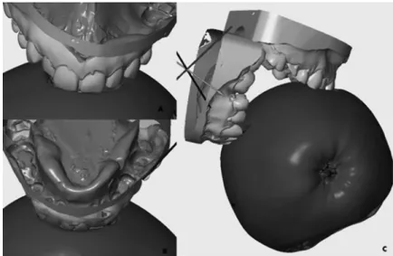
Fig. 4. A 3D comparison of features of the maxillary teeth (A), of the mandible (B) separately, and maxilla and man-dible together (C) of individual partici-pating in the experiment and bitemarks (“bite-into”) on apple using GOM Inspect V7 SR2 software
Acknowledgment. The authors wish to thank Mr. T. Danyluk and Mr. A. Stanowski from ITA company in Poznań for optical scanning. Thanks are also due to Miss Anna Kruszelnicki and Mrs. Ruth Hounam for their gratuitous lan-guage consultation.
References
[1] Łabęcka M., Lorkiewicz-Muszyńska D., Przystańska A., Kondrusiewicz K.: Injuries due to human and an-imal aggression in humans. Ann. Agric Environ. Med. 2013, 20, 91–95.
[2] Bernitz H., Van Heerden W.F.P., Solheim T., Owen J.H.: A technique to capture, analyse, and quantify ante-rior teeth rotations for application in court cases involving tooth marks. J. Forensic Sci. 2006, 51, 624–629. [3] Thali M.J., Braun M., Markwalder T.H., Brueschweiler W., Zollinger U., Malik N.J., Yen K.,
Dirn-hofer R.: Bitemark documentation and analysis: 3D/CAD supported photogrammetry approach. Forensic Sci. Int. 2003, 135, 115–121.
[4] Żaba C., Lorkiewicz-Muszyńska L., Glapiński M., Smoluch K., Świderski P.: Identification of perpetrator based on bitemarks left on the victim’s body. Arch. Med. Sąd. Kryminol. 2010, 60, 22–26 [in Polish].
[5] Martin-de-las-Heras S., Tafura D.: Comparison of simulated human dermal bitemarks possessing three-di-mensional attributes to suspected biters using a proprietary three-dithree-di-mensional comparison. Forensic Sci. Int. 2009, 190, 33–37.
[6] Sheasby D.R., MacDonald D.G.: A forensic classification of distortion in human bitemarks. Forensic Sci. Int. 2001, 122, 75–78.
[7] Bernitz H., Piper S.E., Solheim T., Van Niekerk P.J., Swart T.J.P.: Comparison of bitemarks left in foodstuffs with models of the suspects dentitions as a means of identifying a perpetrator. J. Odontostomatol. 2000, 18, 27–31. [8] Al-Talabani N., Al-Moussawy N.D., Baker F.A., Mohammed H.A.: Digital analysis of experimental human
bitemarks: application of two new methods. J. Forensic Sci. 2006, 51, 1372–1375.
[9] Martin-de las Heras S., Valenzuela A., Ogayar C., Eng M., Valverde A.J., Torres J.C.: Computer-based production of comparison overlays from 3D-scanned dental casts for bitemark analysis. J. Forensic Sci. 2005, 50, 127–133.
[10] Bush M.A., Miller R.G., Bush P.J., Dorion R.B.J.: Biomechanical factors in human dermal bitemarks in a cadav-er model. J. Forensic Sci. 2009, 54, 167–176.
[11] Keiser J.A., Bernal V., Wadell J.N., Raju S.: The uniqueness of the human anterior dentition: a geometric and morphometric analysis. J. Forensic Sci. 2007, 52, 671–677.
[12] Solheim T., Leidal T.I.: Scanning electron microscopy in the investigation of bitemarks in foodstuffs. J. Forensic Sci. 1975, 6, 205–215.
[13] Sweet D., Hildebrand D.: Saliva from cheese bite yields DNA profile of burglar: a case report. Int. J. Leg. Med. 1999, 112, 201–203.
[14] Hyzer W.G., Krauss T.C.: The bitemarks standard reference scale-ABFO No. 2. J. Forensic Sci. 1988, 33, 498-506. [15] American Board of Forensic Odontology [Internet]. ABFO Diplomates Reference Manual p. 119 [cited 2013 Apr 5]. Available from: http://www.abfo.org/wp-content/uploads/2012/08/ABFO-Reference-Manual-1-22-2013-revision.pdf [16] Pretty I.A., Sweet D.: The scientific basis for human bitemark analyses – a critical review. Sci. Justice 2001, 41, 85–92. [17] Martin-de-las Heras S., Valenzuela A., Valverde A.J., Torres J.C., Luna-del-Castillo J.D.: Effectiveness of
comparison overlays generated with DentalPrint© Software in bitemark analysis. J. Forensic Sci. 2007, 52, 151–156. [18] Bernitz H., Owen J.H., Van Heerden W.F.P., Solheim T.: An integrated technique for the analysis of skin
bite-marks. J. Forensic Sci. 2008, 53, 194–198.
[19] Blackwell S.A., Taylor R.V., Gordon I., Ogleby C.L., Tanijiri T., Yoshino M., Donald M.R., Clement J.G.: 3-D imaging and quantitive comparison of human dentitions and simulated bitemarks. Int. J. Leg. Med. 2007, 121, 9–17.
[20] Lorkiewicz-Muszyńska D., Glapiński M., Żaba C., Łabęcka M.: Comparative analysis of the bitemarks and teeth characteristics with use of 2D and 3D methods. Arch. Med. Sąd. Kryminol. 2011, 61, 107–114 [in Polish]. [21] Naether S., Buck U., Campana L., Breitbeck R., Thali M.: The examination and identification of bitemarks in
foods using 3D scanning and 3D comparison methods. Int. J. Legal. Med. 2012, 26, 89–95.
Address for correspondence
Dorota Lorkiewicz-Muszyńska Department of Forensic Medicine Poznań University of Medical Sciences Święcickiego 6
60-781 Poznań Poland
Tel.: 618 546 415
E-mail: [email protected]. Conflict of Interest: None declared Received: 23.07.2014
Revised: 15.09.2014 Accepted: 17.09.2014