R E V I E W A R T I C L E
Intraoral and extraoral autologous bone block graft
techniques: A review of the recent literature
Gianluca Idrontino1, Nicola Alberto Valente2
1School of Medicine, Catholic University of the Sacred Heart, Rome, Italy, 2Department of Periodontics, School of Dental Medicine, State University of New York, Buff alo, New York, USA
Abstract
The bone block grafting is a technique that has been used for many years for the implant treatment and rehabilitation of partially or fully edentulous patients. This type of graft can be autologous (taken from another individual of the same species), heterologous (taken from an individual of a diff erent species), or synthetic (using artifi cial materials). Considering the osteogenetic, osteoinductive, and osteoconduction properties, the best technique is still considered the autograft, which can be intraoral or extraoral. The purpose of this work is to review the diff erent bone harvesting techniques, both intraoral and extraoral, most commonly used in recent years.
Keywords: Autografts, bone regeneration, dental implants
Correspondence
Nicola Alberto Valente, Department of Periodontics, School of Dental Medicine, State University of New York, Buff alo, New York, USA. Phone: +39 3385932069. E-mail: nicolaal@buff alo.edu
Received 05 March 2016; Accepted 06 April 2016
doi: 10.15713/ins.ijcdmr.99
How to cite the article:
Gianluca Idrontino, Nicola Alberto Valente, “Intraoral and extraoral autologous bone block graft techniques: A review of the recent literature,” Int J Contemp Dent Med Rev, vol. 2016, Article ID: 030316, 2016. doi 10.15713/ins.ijcdmr.99
Introduction
The rehabilitation of partially or fully edentulous patients is of considerable importance today. In these patients, there are often situations of insuffi cient bone volume, loss of height or width of the ridge, and loss of normal interarch relations.
The use of autologous bone block grafts is a technique that has shown effi cient results in those patients with atrophic maxillary and mandibular arch in which implant placement is complicated and sometimes impossible.[1]
The reconstruction of atrophic jaws, for implant purposes, with non-vascularized bone grafts was originally described by Brånemark et al.[2] and has become a widespread and predictable procedure.
These surgical procedures are intended to obtain a volumetric augmentation of atrophic ridges on the horizontal aspect, the vertical aspect or a combination of both, by the use of autologous bone.
The surgery consists in the transposition of a bone fragment which is fi xed on the receiving site which, in turn, is prepared to accommodate the graft in a stable manner, ensuring maximum contact surface between the two parts.
The bone block can be composed by only cortical bone, cancellous bone, or corticocancellous bone. The diff erent bone types depend on the location and thickness of the donor site.
The block autograft can be harvested either from extraoral sites or intraoral sites. Using an extraoral harvesting technique, we can defi nitely obtain a greater amount of tissue. This type of graft, therefore, has a greater application for reconstructions of very extensive atrophies. The problems of this type of graft are related to the nature of the bone harvested that, in most cases, tends to be resorbed and to the fact that the patients need to be hospitalized. Furthermore, it is a procedure that requires two distinct surgeries and general anesthesia.[3]
On the other hand, the intraoral harvesting procedure may be performed on an outpatient basis and with local anesthesia or conscious sedation. This type of harvesting is also facilitated by the reduced morbidity and lack of cutaneous scars.
instead is characterized by a slow angiogenesis, increased resorption and increased risk of infection.[4]
Intraoral Grafts [Table 1]
The most commonly used intraoral sites are: · Symphysis
· Ramus of the mandible and retromolar trigone · Tuber maxillae.
The graft from the mandibular symphysis [Figures 1 and 2] guarantees a corticocancellous type of bone formed by rectangular blocks of 2-5 cc of volume.[5] This harvesting area provides a good surgical access, and a minimum resorption of the graft obtained. It is, however, characterized by a high esthetic risk (ptosis of the chin), by a high incidence of edema
and post-operative pain, and possible sensory alterations to both lower incisors and surrounding soft tissues. Furthermore, dehiscence of the fl ap may be occasionally experienced.[6] According to recent studies, it has been noticed that the most frequent complications of this type of graft are temporary and permanent sensory alterations of teeth, skin, and mucosa.[7] In addition to the sensitivity alterations, in another study, this type of graft was found to increase of lamina dura in the adjacent teeth (incisors, canines, fi rst and second premolar) and to cause possible periapical disease to the lower incisor.[8]
The harvesting from the ramus of the mandible and the retromolar trigone are characterized by the presence of a cortical graft formed from rectangular blocks often subtle with volumes from 10 to 25 cc. They are characterized by a discrete surgical access and a lower risk of esthetic complications
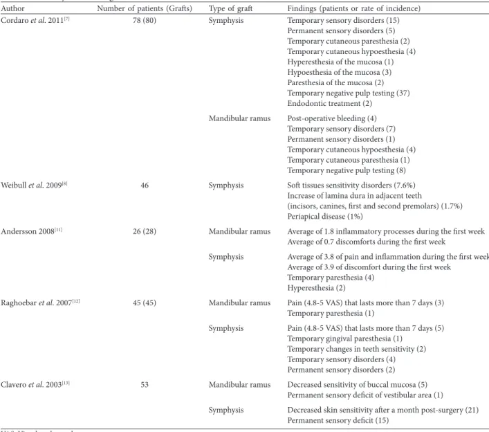
Table 1: Summary of intraoral graft s studies
Author Number of patients (Graft s) Type of graft Findings (patients or rate of incidence)
Cordaro et al. 2011[7] 78 (80) Symphysis Temporary sensory disorders (15)
Permanent sensory disorders (5) Temporary cutaneous paresthesia (2) Temporary cutaneous hypoesthesia (4) Hyperesthesia of the mucosa (1) Hypoesthesia of the mucosa (3) Paresthesia of the mucosa (2) Temporary negative pulp testing (37) Endodontic treatment (2)
Mandibular ramus Post-operative bleeding (4)
Temporary sensory disorders (7) Permanent sensory disorders (1) Temporary cutaneous hypoesthesia (4) Temporary cutaneous paresthesia (1) Temporary negative pulp testing (8)
Weibull et al. 2009[8] 46 Symphysis Soft tissues sensitivity disorders (7.6%)
Increase of lamina dura in adjacent teeth
(incisors, canines, fi rst and second premolars) (1.7%) Periapical disease (1%)
Andersson 2008[11] 26 (28) Mandibular ramus Average of 1.8 infl ammatory processes during the fi rst week
Average of 0.7 discomforts during the fi rst week
Symphysis Average of 3.8 of pain and infl ammation during the fi rst week
Average of 3.9 of discomfort during the fi rst week Temporary paresthesia (4)
Hyperesthesia (2)
Raghoebar et al. 2007[12] 45 (45) Mandibular ramus Pain (4.8-5 VAS) that lasts more than 7 days (3)
Temporary paresthesia (1)
Symphysis Pain (4.8-5 VAS) that lasts more than 7 days (5)
Temporary gingival paresthesia (1) Temporary changes in teeth sensitivity (2) Temporary sensory disorders (4) Permanent sensory disorders (2)
Clavero et al. 2003[13] 53 Mandibular ramus Decreased sensitivity of buccal mucosa (5)
Permanent sensory defi cit of vestibular area (1)
Symphysis Decreased skin sensitivity aft er a month post-surgery (21)
Permanent sensory defi cit (15)
than the harvest from mandibular symphysis. They are also characterized by a minimum resorption. Compared to the symphysis, this technique is characterized by minor edema and post-operative pain, by a lower risk of sensory alterations
of the teeth.[9] Sensory disturbances of the neighboring
tissues and dehiscence of the fl ap are, in fact, rare. Anyway, possible complications of the mandibular ramus graft are inferior alveolar nerve or buccal nerve damage, trismus and fractures of the mandible.[10] Cordaro et al.[7] in a study published, in 2011, has found for this type of graft, a minimal post-operative bleeding and less sensory alterations of teeth and skin compared to the symphysis graft. According to a diff erent study, this type of harvesting technique has a lower rate of infl ammation and discomfort compared to mandibular symphysis grafting.[11] Furthermore according to Raghoebar
et al.[12] this graft has less sensory disturbances and a lower rate of pain that lasts more than 7 days compared to grafts from the mental symphysis. Similarly, Clavero et al.[13] had noticed few alterations in the buccal mucosa in this type of harvesting procedure while more sensory abnormalities were recorded, mainly aff ecting the skin, in the case of graft from the mandibular symphysis.
The graft from the tuber maxillae, while being an easily accessible surgical area, is less used because it provides mainly cancellous bone, rich in cells, but with weak consistency.
Extraoral Grafts [Table 2]
The extraoral donor sites are: • Iliac crest
• Parietal cranial bone (calvaria) • Tibial plateau
• Ribs.
The fi rst authors to describe the use of iliac crest bone grafts for implant purpose were Kratochvil and Boyne,[14] in 1972, and subsequently Breine and Brånemark[15] in 1980.
The graft from the iliac crest has the advantage of providing large blocks (both cancellous and cortical), granting a good surgical accessibility, a discrete plasticity, and adaptability to the recipient site and a low surgical risk. Unfortunately, this type of harvesting procedure is characterized by a greater morbidity than the intraoral grafts.[3] Nkenke andNeukam,[16] in a recent study, compared the grafts from anterior iliac crest with the grafts from
Figure 1: Chin symphysis block graft in situ. Th e horizontal and vertical osteotomies have been performed on the cortical aspect of the chin of a patient before detaching the block
Figure 2: Donor site aft er harvesting. Th e bone block has been harvested from its site leaving an empty area on the chin that can be left as is or fi lled with collagen sponges
Table 2: Summary of extraoral graft s studies
Author Number of
patients
Type of graft
Findings (number of patients or rate of incidence)
Kuik et al. 2016[22]
27 Iliac crest Severe post-operative pain
Calvaria Considerable lasting scars
Obvious profi le defi cit (contours defi cits) Dura mater exposure (3)
Kang et al. 2015[20]
36 Iliac crest Immediate vertical bone
resorption compared to a slower bone resorption in intraoral graft s
Nkenke and Neukam 2014[16]
24 (examined studies)
Iliac crest Minor morbidity rate in the
graft from anterior iliac crest than the posterior iliac crest
Boven et al. 2014[21]
40 Iliac crest Seroma (1)
Hematoma (2)
Lateral cutaneous femoral nerve sensory disorders (1)
Mertens et al. 2013[23]
23 Iliac crest Complication rate: 33.3%
Severe graft resorption (24.16%)
Calvaria Complication rate: 35.7%
the posterior iliac crest experiencing a lower morbidity rate in the latter.
Another fundamental aspect is that the bone from the iliac crest tends to be resorbed by up to 50% of the initial volume.[17] It is not clear whether the marked resorption of the iliac crest grafts depends on their diff erent embryogenic origin from intraoral grafts or rather by their architecture, which is essentially cancellous.[18,19] Kang et al.[20] revealed a more immediate vertical bone loss, when the iliac crest is used, compared to the grafts harvested from intraoral sites where the vertical bone loss seems to be slower. Boven et al.[21] found that the most frequent complications in this type of graft are: Seroma, hematoma, and sensorial disturbances of the lateral femoral cutaneous nerve.
This technique also requires patient hospitalization and the use of general anesthesia to perform the surgery. The post-operative course is characterized by temporary disability and, more rarely, lameness for 6-8 months if the insertion of the inguinal ligament is traumatized (anterior superior iliac spine). From the esthetic point of view, this technique will cause the permanence of a skin scar of modest size.
The bone harvesting from calvaria (parietal bone or occipital skull) is a technically simple procedure to perform but is nevertheless burdened with higher morbidity. The patient usually has no edema, post-operative pain, and temporary disability, so he can quickly return to his daily activities. This bone is, however, very hard and therefore less manageable in complexes reconstructions. It is also very poor in cells and, as a consequence, it has less intrinsic regenerative capacities. Within the complications, although rare, there is the possibility, during the surgery, to accidentally invade the intracranial compartment perforating the dura mater. This intervention, however, cannot be performed by the dentist but only by the maxillofacial surgeon.[22,23]
Kuik et al., in a recent study, compared the bone harvesting from calvaria with that from the iliac crest noticing less post-operative pain in the fi rst, but a greater occurrence of cutaneous scars than that from the iliac crest as well as a greater number of profi le defi cit (contours defi cits). Another complication observed in this study was the dura mater exposure occurred in three patients.[22]
Mertens et al.[23] comparing again the graft from calvaria with that from iliac crest has found a higher rate of complications but less bone resorption rate in calvaria.
Grafts from tibial plateau and ribs are used infrequently for obvious disadvantages in terms of surgical accessibility, bone architecture, intraoperative risk, and post-operative complications.[4]
Conclusions
With this review, we have described the most common autologous bone harvesting techniques for the treatment of maxillary atrophies, considered by some author, the “gold standard” for regenerative oral and maxilla-facial surgery. We
also highlighted the positive aspects of the autologous intraoral grafts in terms of hospitalization, anesthetic techniques, bone resorption, and esthetic aspects. The authors, therefore, recommend the intraoral graft compared to the extraoral one, unless the clinician is dealing with very severe cases of maxillary atrophies and therefore with greater need of bone graft of bigger dimensions.
Referenc es
1. Tarquini G, Reconstruction of maxillary atrophies using autologous intraoral bone block graft s. Symp Odontoiatrico 2012;1:32-7.
2. Brånemark PI, Lindström J, Hallén O, Breine U, Jeppson PH, Ohman A. Reconstruction of the defective mandible. Scand J Plast Reconstr Surg 1975;9:116-28.
3. Misch CE, editor. Bone augmentation for implant placement: Keys to bone graft ing. In: Contemporary Implant Dentistry. St Louis, MO: Mosby; 1999. p. 457.
4. Gherlone EF. Prosthetic implant rehabilitations and minimally invasive surgery. Como: Ariesdue; 2013.
5. Neiva RF, Gapski R, Wang HL. Morphometric analysis of implant-related anatomy in Caucasian skulls. J Periodontol 2004;75:1061-7.
6. Nkenke E, Schultze-Mosgau S, Radespiel-Tröger M, Kloss F, Neukam FW. Morbidity of harvesting of chin graft s: A prospective study. Clin Oral Implants Res 2001;12:495-502. 7. Cordaro L, Torsello F, Miuccio MT, di Torresanto VM,
Eliopoulos D. Mandibular bone harvesting for alveolar reconstruction and implant placement: Subjective and objective cross-sectional evaluation of donor and recipient site up to 4 years. Clin Oral Implants Res 2011;22:1320-6.
8. Weibull L, Widmark G, Ivanoff CJ, Borg E, Rasmusson L. Morbidity aft er chin bone harvesting –A retrospective long-term follow-up study. Clin Implant Dent Relat Res 2009;11:149-57. 9. Misch CM. Comparison of intraoral donor sites for onlay
graft ing prior to implant placement. Int J Oral Maxillofac Implants 1997;12:767-76.
10. Nkenke E, Radespiel-Tröger M, Wiltfang J, Schultze-Mosgau S, Winkler G, Neukam FW. Morbidity of harvesting of retromolar bone graft s: A prospective study. Clin Oral Implants Res 2002;13:514-21.
11. Andersson L. Patient self-evaluation of intra-oral bone graft ing treatment to the maxillary frontal region. Dent Traumatol 2008;24:164-9.
12. Raghoebar GM, Meijndert L, Kalk WW, Vissink A. Morbidity of mandibular bone harvesting: A comparative study. Int J Oral Maxillofac Implants 2007;22:359-65.
13. Clavero J, Lundgren S. Ramus or chin graft s for maxillary sinus inlay and local onlay augmentation: Comparison of donor site morbidity and complications. Clin Implant Dent Relat Res 2003;5:154-60.
14. Kratochivil FJ, Boyne PJ. Th e combined use of a subperiosteal
implant and bone marrow graft s in effi cient mandibole.
J Prosthet Dent 1972;27:645-53.
implants. Scand J Plast Reconstr Surg 1980;14:23-48.
16. Nkenke E, Neukam FW. Autogenous bone harvesting and graft ing in advanced jaw resorption: Morbidity, resorption and implant survival. Eur J Oral Implantol 2014;7 Suppl 2:S203-17. 17. Zins JE, Whitaker LA. Membranous versus endochondral bone:
Implications for craniofacial reconstruction. Plast Reconstr Surg 1983;72:778-85.
18. Rabie AB, Dan Z, Samman N. Ultrastructural identifi cation of cells involved in the healing of intramembranous and endochondral bones. Int J Oral Maxillofac Surg 1996;25:383-8.
19. Kusiak JF, Zins JE, Whitaker LA. Th e early revascularization of
membranous bone. Plast Reconstr Surg 1985;76:510-6.
20. Kang YH, Kim HM, Byun JH, Kim UK, Sung IY, Cho YC, et al. Stability of simultaneously placed dental implants with
autologous bone graft s harvested from the iliac crest or intraoral jaw bone. BMC Oral Health 2015;15:172.
21. Boven GC, Meijer HJ, Vissink A, Raghoebar GM. Reconstruction of the extremely atrophied mandible with iliac crest onlay graft s followed by two endosteal implants: A retrospective study with long-term follow-up. Int J Oral Maxillofac Surg 2014;43:626-32. 22. Kuik K, Putters TF, Schortinghuis J, van Minnen B, Vissink A,
Raghoebar GM. Donor site morbidity of anterior iliac crest and calvarium bone graft s: A comparative case-control study. J Craniomaxillofac Surg 2016. pii: S1010-518200005-6.
23. Mertens C, Decker C, Seeberger R, Hoff mann J, Sander A,
Freier K. Early bone resorption aft er vertical bone