Thenucleotidesequenceofanuntranslated but conserved domain at the 3' end of the avian sarcomavirusgenome
A.P.Czernilofsky, W.DeLorbe, R.Swanstrom,H.E.Varmus,J.M.Bishop, E.Tischer* and H.M.Goodman*
DepartmentofMicrobiology and Immunology,and*Department of Biochemistry and Biophysics, Universityof California, San Francisco,CA94143, USA
Received10April 1980 ABS.T
The genarnes of mnmerous avian retroviruses contain at their 3' termini a conserved danain denoted "c". The precise boundaries and
function
of "c" have been enigmas. In an effort to resolve these is-sues, we determined thesequence
of ovrer 900 rncleotides at the 3' end of the genom of theSchmidt-Ruppin
subgroup A strain of avian sarcoa virus (ASV). We obtained the sequencefrom
a suitablefragment
of ASV IX that had been cloned into thesingle-stranded
Ephage
M13np2.
Coputer-assisted analysis of the sequence revealed the following structural features: i) the length of mc" - 473 nucleotides; ii) the 3' terminal domain of src, ending in an amber codon at the 5' boundary of"c";
iii) terminator Cdons that preclude continuous translation from "c"; iv) suitably located sequences that my serve as signals for the initiaticn of viral RIA synthesis and for the processing and/or polyadenylaticn of viral M; v) a repeated sequence that flanks src and that could facilitate deletion of this gene; vi) repeated se quences within wc"; and vii) unexplained hanologies between sequences in "c" andsequences
in several other reicacids, including the 5' terminaldmairt.W
the ASV genomae,tR
and its inversion, thecoo-plenwt
of tR4 and itsinversion,
and the 18S MA ofeukaryotic
rib--ones. We conclude that "c" probably does not encode a protein, but its sequence may nevertheless serve several essential functions in viral replication.m1e
haploidgerxne
of aviansaroma
virus (ASV) is a single-stranded RIconpoxed
of ca. 9500 nucleotides (1). This RW fills tw roles during the viral life cycle. First, it serves as niM for thesynthesis
of several viralpolypeptides
(1);
accordingly,
theRM
isfor reverse transcriptase (6); and a sequence of 16-21 nucleotides, repeated directly at the tw ends of the viral genome
(7,8,28),
medi-atesearly
events inpropagation
of the nascent C chain(4,7-9,28;
R.S.,manuscript
inpreparation).
Theintegrated
E (or"provirus")
of ASV is coextensive with viral RPM but, inaddition,
contains a330p
terminal redundancycopoed
of nucleotide sequences derived from both the 3' and 5' ends of the viral genome(10,11).
The redun-dant sequences may serve either or both of two purposes: they may mediate integration; and they may harbor thepromoter
for viral RA synthesis.mEhe genome of ASV contains four identified genes, arranged as follows on the viral MA: 5'- g-ag-env-src -3' (1). The genes
M,
Et and env are required for viral replication, whereas src is devotedexclusively
to necplastic transformation of the host cell (12). Inaddition, structural studies have identified a conserved dcmain locat-ed to the right of src,
coupoed
of ca. 500 nucleotides, and denoted "cm because of its presence in numerous strains of avian retroviruses(13,14).
Tw distinctive functions have been attributed to "c". On the one hand, recarbination analysis indicated that the sequence may be
inpor-tant for viral replication (15,16). On the other hand, some investi-gators have sought to inplicate "c" in leukemogenesis by avian leu-kosis viruses, which otherwise have no apparent genetic locus devoted to tunmrigenesis (1,17,18). Inorder to explore the structural boun-daries and possible functions of "c", we have
determined
the sequence of over 900 rucleotides at the 3' end of thegenome
of theSchmidt-RLppin
sugroup A strain of ASV (SR-A ASV).Our
experimetal
strategy exploited the fact that we had previ-ouslycloned
the entire gerxne of SR-A ASV into both phage and plasmid vectors (19). From theseclones,
we isolated a 3.2kbEBo
RI fragment of MA that contained all but 65 nucleotides of "c", the entirety ofa-dditicn, we have found suitably located nucleotide sequences that may serve as signals for the initiation of viral RNA synthesis and the po-lyadenylaticn of viral RNA. We encountered unexpected and non-random homnlogies between sequences in "c" and sequences in
tR7r
, its po-lyrucleotide conplement, and the 5' end of the ASV genome. Several of our conclusions conform to a recent report that contained thesequence
of 217 nucleotides in the "c" domain of theSchmidt-Ruppin
subgroup D strain of ASV (24).MATORIALS AND M E Materials
Most of the materials have been described previously (19-23,25). All of the restriction enzymes were from NewEngland Biolabs Inc. and were used as reccmended. The synthetic dodecanucleotide
d(VACNGCiTWrG)
was obtained from Collaborative Research. This oli-gonucleotide is conplementary to a sequence within the lac region of M13np2 and can be used as a universal primer for thesequencing
of DNA joined to the lac region at the Eco RI cloning site (20,21). The Klenow fragment of polymerase I and polynucleotide kinase were from Boehringer Mannheim, and the ei32PP and)32PATP
from Amersham(specific
activity ca. 400rCi/nml
and 3000Ci/nurl
respectively).Clcnipg_ceure:
The viral DNA of SR-A was cloned previously intothe vector
Xgt%EW4B
(19). Treatment of the cloned DM with Eoo RI generates the following fragments: 2.0kb, 3.9kb, 3.2kb, 0.33kb, and 0.26kb. We used for our cloning purposes the fragment of 3.2kb. DM for cloning was usually obtained by Boo RI treatment of the recm-binantphage
DN and subsequentseparation
of thefragments
on a 1% agarose gel. The DN was localized by staining with ethidium bromide and by reference to appropriate markers and was then recovered by electroeluticn (25). C was cloned intoMl3np2
and recovered asrecombinant phage
genome
or replicative form as described previously (20,21,25). The phage vector and its host, E. coli strain JM101,of regative polarity (ie., conpiementary to the ASV genome) and ws used for screening recuxrbinant plaques and for identification of the polarity of the single-stranded viral DNA cloned into M13np2. ii) Aprobe for nucleotide sequences represented in the 3.2kb Eboo RI frag-ment of ASV E was
prepared by transcribing
the denaturedfragment
with reverse transcriptase in the presence of random oligodecoynucleo-tide primers, as described previously (19,27,28). EIAs weretransferred from agarose gels to nitrocellulose filters and
hybridized
with radioactive cEls (29).Isolaticn of primers for sequencing reactions: The 3.2kb fragment of ASV El was isolated from the lantbda clone described above, from a sub-clone in pBR322, or from the replicative form of a sub-clone in M13np2.
mfhe
purified fragment was cleaved with suitable restriction endonucleases; the products of digestion were fractionated byelectro-phoresis
in gels of polyacrylamide or agarose, located by staining with ethidium bromide and by reference to standard markers, and recovered from slices of the gel by electroelution (25).Sequencing techniques: We generally used chain termination with
di-deoxyrucleotides
exactly as described by Sanger et al (22), except that we usedc32P-dAP as the radioactive precursor in all reactions.At the conclusion of the polymerase reactions, primers were usually renwved from the newly synthesized
DM@
by cleavage with the appropri-ate restriction endonuclease (one unit of enzyme per 20 microliters of reaction mixture, five minutes at 37 ). Three fragments were also se-quenced by the Maxam-Gilbert technique (23), which was used without any nodification. ffte DMZ fragments were labeled on the 5'-end withy32P_JP
and polynucleotide kinase under standard conditions(23), or they were labeled on their 3'-ends
by
copying
the5'
"over-hangs" of restriction sites withpolymerse
I(Klenow
fragment)
under the same conditions as we used for thesequencing
reaction described above.RESULTS
Isolation of ASV DNA for sequencing: molecular cloning in the single-stranded ENA p Ml3np2.
cleavage
is a 3.2 kb DAfragment
that extends frcan the 65th residue of "c"(nunbering
from the 3' terminus of the viral RAgenome)
to ca.l.Okb to the 5' side of src (19). In work to be reported elsewhere, we used transfection of cultured fibroblasts to demonstrate that this DE fragment is capable of directing the synthesis of the protein en-coded in src, and thus, of transforming recipient cells to a
necplas-tic phenotype (P. Luciw et al., manuscript in preparation); hence, the structure and function of the src gene (and by inference, the remainder of the DNA fragment) survived cloning and propagation in prokaryotic vectors.We isolated the 3.2kb IA
fragment
as described under Materials and Methods; analysisby
electrophoresis in an agarose gel denonstrat-ed that the isolated fragment was homogeneous and pure (Figure la, lane 1). The fragment was then cloned into the Eco RI site of ML3mp2. In order to document thecc6position
of positive clones, we isolated the replicative form DNA of recambinant phage, cleaved the DA withEco RI, and demonstrated that the products of cleavage included a 3.2kb fragment that hybridized with ASV
cDNArep
(Figure la, lane 2). We concluded that the viral DNA had survived the sub-cloning withoutsustaining an appreciable deletion.
In preliminary studies of a set of sub-clones, we found that the single-stranded genane of the chimeric phage invariably contained only the negative strand of ASV
[E;
the predaninance of one polarity in recombinant clones of M13 has been a recurrent problem in the use of this vector (unpublished observations of the authors). In an effort to obtain both polarities of ASV DA in the phage genane, we isolated the replicative form of one of the recombinant clones, cleaved theDtA
with Eco RI, religated the DA and used it to transform the bacterial host JM 101. Clones containing viral DNAwere again isolated, and some werefoundl
to contain a 3.2kb Eco RI fragment that hybridized withcDL rep (data not shown).a
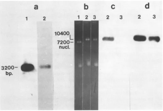
b
c
d
1
2
1
2
3
2
3
2
3
10400
7200-__
_
nucl.
3200-
dI+
bp.
Figure 1. Preparation of cloned WS for sequence analysis.
I^swere isolated, cleaved with restriction endonucleases,
frac-tionated
by
electrcphoresis
in agarose gels, transferred to nitrocel-lulose filters and analysed by molecular hybridization as outlined uder Materials and Methods.Panel a.
Sub-clning
the Eoo RI 3.2kb fragment of ASV Dt. Lane 1, the 3.2kb fragment prior to sub-cloning into Ml3np2. bhe 3.2kb frag-ment of ASV M waspurified following
cleavage
of recombinant CMcontaining the entire ASV genome with Boo RI. Aportion of the iso-lated fragnt was analysed by
electrcphoresis
in a gel of 1% agarose. The figure illustrates an autoradiogram obtained following transfer of the [W to a nitrocellulose filter and hybridization with cUt rLane 2, the 3.2kb fragment following sub-cloning into M13up2. Te
Sik
fragment analysed in lane 1 was sub-cloned into Ml3np2 at the EBo RI site. Replicative form of thesub-clone
was isolated,cleavWwith
EBo RI and analysed as in lane 1.
71e
figure illustrates anautora-no-gram
obtainedfollowing hybridization
of theinmmbilized
CM withPaneEB:
Detection of inserts in the gerou of Ml3np2. Single-stranded M was extracted fran phage, fractionated by electrophoresis in a gel of 1% agarose and detected by staining with ethidium bromide. Lane1,
thegenrxe
of M13np2. Lanes 2 and 3, the gencmes of phagecontaining
inserts of ASV E.Panel c. Detection of
renccbinant
clones of M13np2 containing posi-tive strands of ASVEH.
The DtNs of lanes 2 and 3 in panel b were transferred to nitrocellulose filters andhybridized
withcE1
; the figure illustrates autoradiogramobtainedsubsequent
to thefiEridi-zation.
strarnds of ASV Et. The filters used in
panel
c weresubsequently
hy-bridized with radioactive E representing both strands of the 3.2kb Ebo RIfragment
of ASV EA; the figure illustrates an autoradiogram obtained after the hybridization.We determined the polarity of the ASV inserts in the genanes of chimeric phage by hybridizing the fractionated DE with either
cESrep
(which reacts principally with ASV nucleic acids of positive polarity) or a radioactive arnd denatured preparation of the original 3.2kb ASV DN fragment (which will react with ASV nucleic acids of both polari-ties). Representative results are illustrated in Figure lc and d: the genome of one recombinant phage hybridized withcEDNArep
and thus con-taimed ASV DNA of positive polarity (Figure lc); another reccbrinant genome hybridized only with the cDNAmaie with the 3.2kb fragment and thus contained ASV DNA of negative polarity (Figure ld, right lane). Subsequent analyses of rucleotide sequence confirmed these assign-ments. Assured that we now possessed reccmbinant clones that could be used for sequencing both strands of the ASV DMA fragment under study, we proceeded with the sequence analysis.Nucleotide sequence of the "c" domain of the ASV genome.
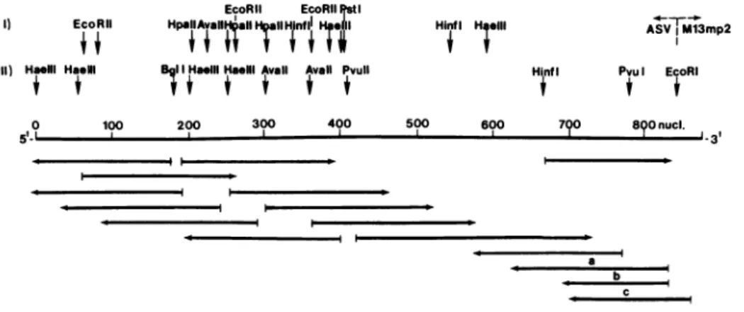
Figure 2 illustrates our experimental strategies for determining the rucleotide sequence reported below. We relied principally upon chain termination with dideoxynucleotides, as described by Sanger and colleagues (22). We
attenpted
to inprove the accuracy of our results by using at least two independent means to reproduce all of the se-quence: usually, we sequencedoverlapping
regions by using two dif-ferent primers in the terminator technique; in all regions, the se-quence was determined in both polarities; and in a few instances, we obtained supplementary data with the procedure of Maxam and Gilbert(23). The 3.2kb fragment of IYNA used in the present
analyses
does not include the last 65 nucleotides at the 3' terminus of the ASV gencme. The sequence of these residues has been determined in a separate study which will be reported elsewhere (R.S., manuscript in preparation),and similar results have been obtained previously for the Prague sub-group C strain of ASV (unpublished results of A.P.C. and J. Shine, and personal comiunication from D.Schwartz and W. Gilbert); for clarity, these data will be included in the discussion that follows.
il-EcoRli EcoRIlIstI
I) EcoRIlI HpallAvall
AalHrIIT
nflH,a il Hinf H;elll ASV %Ml3mp211) Haeill HasNI IIHa.Ili HaelllAvail Avail Pvull HinfI Pvul EcoRI
0 100 200 300 400 500 600 790 8()0 nucl.
5 ~~~~~~~~~~~~~~~~~~~~~~~~~~~~3
I I
a
e c L
Figure 2. Strategy for determining the nucleotide sequence of c". The map coordinates
begin arbitrarily
with the first nucleotide at the 5' boandary of the sequence determined in the presentstudy
and extend beyond thejunction
between the cloned ASV CM and the EBo RI site of the M13 vector EN. The map is aligned according to i po-larity of the ASV genone (plus strand).Restriction
sites are located by arroas above the map: row II gives the positions of sites used insequencing
the CM, row I gives additional sites deduced from the fi-nalsequence.
The arrows below the map define theregions
sequen with individal primers andfragments.
The direction of the arrows designates the polarity of [AIA used asteiplate
in the terminator pro-cedure: to the left,minus
strand; to the right, plus strand.Addi-tional
designations: a, a sequence determined with the Maxam-Gilbert technique; the fragment was labelled at the 3' terminus; b, asequence
determined with the Maxam-Gilbert technique; thefragment
was labelled at the 5' terminus; c, asequence
obtained by using a synthetic primer that initiates Csynthesis
20 nucleotides to theright
of the junc-ticn between ASV IS and M13 Ws.lustrates this sequence and joins it to the sequence found at the 5' end of ASV
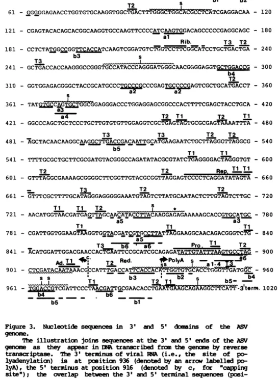
IRNA
(8,30); we chose this aligmennt for illustration in order to facilitate discussion of important structural and functional aspects of the nucleotide sequence. The gerxne of ASV has a direct terminal redundancy (7,8); this sequence appears once in Figure 3 (po-sitions 917-936), linking the 3' and 5' domains of the viral genane, in accord with the structure of ENA transcribed from viral INA (R.S., manuscript in preparation).Structural features of the sequence.
i.) The topography of the sequence was
mct
disturbed by molecular cloning.mapped
extensively by site-specific cleavage with restriction endonu-cleases (19). Our sequence contains all of the sites located previ-ously by this means. In the process of preparing suitable primers for sequencing with the terminator technique, we identified a number of additicnal restriction sites; these also appear at appropriate posi-ticns in the the final sequence. Since we prepared most of the pri-mers from the original clcne of [NA in lambda phage rather than from our sub-clones in M13, we are further assured that the sub-cloning did mot cause major deletions or rearrangements in the DE. Figure 2 sum-marizes the locations of cleavage sites for most of the available res-triction endonucleases; accrplete
catalogue of restriction sites within the sequence is available from the authors on request.ii.) Correlaticns withprevious data.
Previous reports have described oligonucleotides obtained from the "c"
region
byhydrolysis
of ASV RA with Tl RNase (13-15). The mst characteristic of these is an oligonucleotide whosecpmpoition
has proven to be remarkably constant among numerous strains of avian retroviruses (13-15,31,32). The sequence illustrated in Figure 3 on-tains this oligonucleotide at positions 904 to 915, a location that is in accord with previous, less precise efforts to map the oligonucleo-tide on the viral gename (13,14). We were able to detect other simi-larities between the regions of the sequence reported here and oli-gonucleotides described in the previous literature, but the hcmologies were frequently
inccmplete
and are not located in Figure 3. These findings conform to previous reports that changes in single nucleo-tides are frequent among the genomes of related retroviruses(13,14 ,31,32) .
iii.) The boundaries of "c".
5tterm.
T3 s1 - GGCCTATGTGGAGAGGAXAACTACGTGC AGACcTGcGGCGGCCAACATCCTGGT - 60
T2 bl b2
T2 s
61 - GGGGGAGAACCTGGTGTGCAAGGTGGCTGACTTTfG-GCTGCALCOUC2TCGAGGACAA - 120 121 - CGAGTACACAGCACGGCAAGGTGCCAAGTTCCCCATCAAGTGGACAGCCCCCGAGGCAGC - 180
al
Rib. T3 T2
181 - CCTCTATGGrCGGTTCACCATCAAGTCGGATGTCTGGTCCTTCGGCATCCTGCTGACTGA - 240
b3 s
T3 L
241 - GCTGACCACCAAGGGCCGGGTGCCATACCCAGGGATGGGCAACGGGGAGGTGCTGGACCG - 300 b4
T2
310 - GGTGGAGACGGGCTACCGCATGCCCT
CCCGAGTGCCCCGAGTCGCTGCATGACT
- 360s ~~~a2~ a2
361 - TATG T CGAGGGACCCTGGAGGAGCGGCCCACTTTTCGAGCTACCTGCA - 420
a4 T2 Ti T2 Ti
421 - GGCCCAGCTGCTCCCTGCTTGTGTGTTGGAGGTCGCTGAGTAGTGCGCGAGTAAAATTTA - 480
T3 T3 T2 T2
481 - AGCTACAACAAGGCAAGGCTTGACCGACAATTGCATGAAGAATCTGCTTAGGGTTAGGCG - 540 b5
Ti TI
541 - TTTTGCGCTGCTTCGCGATGTACGGGCCAGATATACGCGTATCT AGGGGACTAGGGTGT - 600
T2 T2
Rep.
TIT1
601 - GTTTAGGCGAAAAGCGGGGCTTCGGTTGTACGCGGTTAGGAGTCCCCTCAGGATATAGTA - 660
_ . T2 T2
661 - GTTTCGCTTTTGCATAGGGAGGGGGAAATGTAGTCTTATGCAATACTCTTGTAGTCTTGC - 720
Ti
TI T2 i *721 - AACATGGTAACGATGAGTTAGCAACATACCTTACAAGGAGAGAAAAAGCACCGTGCATGC - 780
a5 a3
Ti Ti Ti
781 - CGATTGGTGGAAGTAAGGTGGTACGATCGTGCCTTATTAGGAAGGCAACAGACGGGTCTG - 840 a5
T3 b6 ' a6 Pro. T1 T2
841 -
ACATGGATTGGACGAACCACTMATTCCGCATCGCAGAGATATTPTA
- 900AdT1
c T2 Red. PlyAo s -a -6-
U~~~~~~~~
Ia-901 -
CTCGATACAATAAACGCCATTTGACCATTCACCAATGGTTGCACCTaFGLATGGC
- 9608
s-T
b3 T T s b4----L ---- Ti T2T1 b5S
961 -
TCGATTCCCTAACGCCTGI'4XAiCAAGGCTTCACT
-3term.1020b4 b6
b5 bl
Figure 3. Nucleotide sequences in 3' and 5'
d*mains
of the ASVgen9ma.
(posi-ticns 917-936, denoted Red.) represents the terminal
redundancy
of the viralgenomes, only
one coy of which appears in the Esynthesized
by
reverse transcriptase (R.S., mauscript inpreparation).
The se-quences were derived as follows: positions 1-868, from the present study; positions 869-936, from a separate analysis of cloned ASV -t that will be described elsewhere (R.S., manuscript inpreparation);
andpositions
937-1020, fromprevious analyses
of EA transcribed from the 5' end of the genrme of the Prague-C strainof ASV (30). Addi-tional syrbols: i.) Tl, T2 and T3, termination codons in three reading frames; ii.) s, regions of dyad syrmetry; iii.) Rib., a sequence that is onnplemsntary bo the 3' end of 18S ribosoml I; iv.) Rep., a se-quence repeated to the 5' side of src; v.) Pro., a possiblecoupoent
of the promoter for viral RH synthils; vi.) underlinings marked a" denote olignxucleotides repeated within the 3' domain of the squence; ruubers identify specific oligcnucleotides; vii.) underlinings marked Obw denote oligonucleotides found in both the 3' and 5' domains of the sequence;nuibers
identify specific oligonucleotides; viii.) Ad., the sequence alleged to signal processing and/or polyadenylation of nRAs(positions
908-914); ix.) the underlining of positions 870-912 demar-cates a region that can fold into a stable hairpin structure to in-clude the allegedprooter sequence; and x.) the * at position 759 denotes an ambiguity in the sequence (G or T).codon is likely to mark the 3' terminus of src; our provisional se-quence of the entire srcgene sustains this suggestion (work in pro-gress). We conclude that the "c" region of SR-A ASV is probably 473 nucleotides in length. This conclusion is in accord with previous es-timates obtained by mapping deletion mutants with restriction endonu-cleases (27), whereas analysis of heteroduplexes by electron
micros-oopy
hadsuggested
a scmewhatlarger
value(33).
iv.) Can the "c" regicn be translated?
The distribution of termination codons within the "c" region pre-cludes continuous translation from any of the reading frames. This concl1vsion is based on the assunption that termination at the codon UGA - which is the predaninant terminator codon in the sequence - is not suppressed at an appreciable frequency; recent findings have raised the possibility of such suppression in eukaryotic cells but have not assessed its prevalence (34; and B. Cordell et al, manuscript submitted). If translation is circumscribed by UGX codons, three
re-gions within "c" could nevertheless give rise to polypeptides of ap-preciable size: (a) Positions 515-586; the polypeptide would consist of 23
amino
acids with the following sequence: met lys asn leu leu arg val arg arg phe ala leu leu arg asp val arg ala arg tyr thr arg ile .with the following sequence: met tyr gly pro asp ile arg val ser glu gly thr arg val cys leu gly glu lys arg gly phe gly cys thr arg leu gly val pro ser gly tyr ser ser phe ala phe ala. (c) Positions 777-863; the polypeptide would consist of 28 amino acids with the
follow-ing
sequence: met pro ile gly gly ser lys val val arg ser cys leu ile arg lys ala thr asp gly ser asp met asp trp thr asn his. The first of these possiblepolypeptides
would be notably hydrephobic and basic, the corrposition of the others would be less distinctive.It is of course possible that FMA splicing could rearrange the sequence of "c" so as to facilitate translation into a protein of
rea-sonable size, but we and others have failed to detect a suitable mTN in infected cells (35,36). We therefore conclude that "c" is unlikely
to encode a protein larger than 39 amimo acids unless UGA codons within the sequence are suppressed in eukaryotic cells. Previous pro-posals that translation from "c" might acoount for leuke4mogenesis are probably incorrect. It remains possible, however, that the danain gives rise to at least one of the small polypeptides described above; the factors that stinulate growth of eukaryotic cells include
proteins
of this size (37).v.) Polyadenylation of viral RN&.
The sequence AAUAAA
appears
near the 3' terminus of most, if not all, eukarytic mRns and is thought to be the signal for sane or all of the events that generate thepolyaderylated
3' terminus of maturenmik
(38,39). The sequence of "c" contains this alleged signal at po-sitions 909-914, or 23-28 residues from the 3' terminus of the viral genome as represented in Figure 3. This location is similar to thatfound
in other eukaryotic mRAs (38-42).vi.) A signal for the initiation of viral RNA synthesis?
A portion of "c" is contained within the
330bp
terminal redundan-cy that brackets the integrated provirus of ASV and allegedly harbors the promoter for viral RMA synthesis (10,11). We have isolated the terminal redundancy and analysed its nucleotide sequence; the results of these studies will be reported and discussed in full elsewhere (R.S., manuscript in preparation). Here we explore the relationship of these findings to thecorposition
of "c".R4A synthesis might begin at the 5' terminus of the genome
proper
(po-siticn 917) in response to signals possibly contained within the 3' domain of the terminal redundancy. We have therefore searched the se-quence of "c" for oligcnucleotides implicated previously in the ini-tiaticn of nmRN synthesis by eukaryotic cells (38-42). A sequence bearing features of the canonical eukaryotic "promoter site" occurs at positicrs 880 to 893 in Figure 3, 24 nucleotides from the alleged site of initiation of RNA synthesis. The candidate signal for initiation can be included in a large hair-pin structure that spans positions 881 to 912 (see Figure 3). Several elements of dyad synmmetry (denoted s in Figure 3) are located downstrean from the sequence. These findings are all in accord with previous descriptions of sites where the syn-thesis ofeukaryotic
nRN
is initiated and encourage us to examine morerigorously
thepossibility
that the promoter for ASV RNA syn-thesis resides in the terminal redundancy of viral DNA.vii.)
Secondary
structure in "c".Conputer-assisted
analysis of the sequence reported here revealed ca. 200 possibilities for the formation of hairpin duplexes containing eight or more base-paired nucleotides (not illustrated). We have nomeans at present by which to discern whether any of these structures actually exist in the native RNA, or whether any are of functional significance. We also found a number of regions that display
appreci-able dyad synmnetry; Figure 3 illustrates ten of the more extensiveex-anples.
viii.) A repeated nucleotide sequence that flanks src.
By carparison of the sequence presented here with our unpublished data, we have identified a sequence of at least twenty nucleotides (positions 643 to 662) that is repeated directly and most likely pre-cisely in an
apparently
untranslated region to the 5' side of src. Moreover, the precise repeats are each extended in both directions by adjacent, partially repeated (90%) sequences caposed of ca. 20nu-cleotides. Hence, the repeated domains include at least 60 nucleo-tides. We tentatively propose that this repeated sequence might medi-ate deletion of src by hmologous recombination within the ASV genome, or by some other mechanism. This proposal could account for the
re-markable frequency with which src is deleted during the propagation of nmst strains of ASV (12).
It is thought that
canplementary
interactions between mRA and 18S ribosomal RNA may figure in the synthesis of proteins in eukaryot-ic cells (43). We therefore searched the nucleotide sequence inFig-ure 3 for
relationships
to 18S ribosomal iA and found that these-quence at positions 215 through 228 is cosplemantary at 11 out of 14 nucleotides to the sequence at the 3' terminus of 18S ribosonal RNA. The significance of this finding is presently moot because we have no evidence that ribosane binding occurs anywhere within the damain of
c.
x.) Homologies with NA.
A nurrber of tRIAs from the host cell can base-pair with the genome of ASV (44,45). The significance and location of this base-pairing has been established in only one instance -
t.WTrp,
which is situated near the 5' terminus of the viral genane and serves as primer for reverse transcriptase (6). (Available data do not exclude the possiblility thattTrP
also binds to the genome at other sites.) We searched the sequence in Figure 3 for relationships to t1Trp and discovered non-random homologies with the sequence oftRATrp,
its inversion, the complemmt oftNTrp,
and its inversion; these homolo-gies are illustrated in Figure 4. We also searched published se-quences of the untranslatedregions
ofglobin
mRNA and SV40 DNA for similar relationships totRtTrp
and faod none. Other investigators, hwever, have reported partial homologies with t1Ms in the gene for 23S ribosomal MA of bacteria (46,47), in sequences that bracket the gene for cytochrome II oxidase in mintochondrial DNtA of mice (38), and in the genane of polycmna virus (48).xi.) Homologies with the 5' end of the ASV genome.
The genane of ASV contains a direct terminal redundancy (7,8). This fact pronpted us to search for further evidence of relatedness between the 3' and 5' domains of viral RNA. Although our search was
constrained
by the limited amount of data available for the 5' end of the viral genane, we nevertheless found six regions ofappreciable
homology
between the 5' and 3' domains (designated "b" in Figure 3). The significance of these findings is presentlynoot.
xii.) Repeated nucleotide sequences within the 3' domain.
a) 5'-
GACCTCGTGGCGCAACGGTAGCGCGTCTGACTCCAGATCAGAAGGCTGCGTGTTCGMTCACGTCGGGGTCACCA
-3'277 285 186 200
511 523
785 826
b)5'- CTGGAGCACCGCGTTGCCATCGCGCAGACTGAGGTCTAGTCTTCCGACGCACAAGCTTAGTGCAGCCCCAGTGGT -3'
315 332 782 788
188 178408 428
86 101 284 276
105 113
C) 5'- ACCACTGGGGCTGCACTAAGCTTGTGCGTCGGM.GACTAGACCTCAGTCTGCGCGATGGCAACGCGGTGCTCCAG -3'
614 623 205 221 191 200
718 - 732
19 32
821 829
d) 5'- TGGTGACCCCGACGTGATTCGAACACGCAGCCTTCTGATCTGGAGTCAGACGCGCTACCGTTGCGCCACGAGGTC -3'
29 40 201 - 212
167 176 306 - 318
337 347
Figujp4.
Houlogies
between the 3' domain of the ASV genane and tRN~AUnderlinings denote sequences that are also present in the se-quence illustrated in Figure 3; nuitr s denote the positions in Figure 3. a) The sequence of chicken tR , rendered in
deoxynucleotides;
b) thecczplement
of the sequence in a; c) inversion of the sequencein a; d)
inversion
of the sequence in b.fcund both within "c" and in the portion of the sequence that we be-lieve to be the 3' domain of src.
Concluding remarks
It appears unlikely that "c" encodes a protein, but the sequence may nevertheless serve essential functions in viral replication:
recently identified in the genane of avian
myeloblastosis
virus and one of its helper viruses (unpublished results of T. Gonda andl J.M.B.). It may prove instructive to caipare the nucleotide sequences of these three forms of "c".The avian leukosis viruses readily induce
lynphomas
in birds, yetapparently
do not have a genetic locus devoted specifically toon-cogenesis (1,12). Moreover, deletion of src from the genane of ASV deprives the virus of its ability bo induce sarcomas, but gives rise to a leukosis virus (12,50). These findings have been attributed in the past to the possibility that "c" might encode an oncogenic pro-tein; the present report calls this explanation into serious doubt. It remains possible, however, that "c" participates in leukemogenesis. We have argued elsewhere that the promoter for retrovirus RNA syn-thesis may, on occasion, induce transcription of otherwise silent cel-lular genes (in the manner of the "floating promoter" effect of some tranposable elements in bacteria (51)), and that induction of cellular genes in this manner could contribute to both leukenogenesis and
co-carcinogenesis by retroviruses that do notpossess specific oncogenes (52). If this suggestion is correct, and if the cOoposition of "c" bears on the relative efficiency of retrovirus promoters, then sane forms of "c" may be associated with
leukenuogenesis
and others not. Again, orparative studies of "c" in the genanes of leukemogenic andnon-leukemogenic
avian retroviruses may be informative.CKNOWEG
This research was supported by grants from the National Cancer Institute and the American Cancer Society. The authors thank Barbara Baker, Paul Luciw, Greg Payne and Richard Parker for gifts of cloned DEA, Hugo Martinez for assistance in the use of his
coiputer
program, R. Friedrich for assistance with statistical conputations, J. Messingfor
Ml3mp2
and E. coli JM101, and D. Schwartz for personalonuunica-tions of data
prTor-to
publication. We also thank B. Cook for helping uswith the manuscript.1.
Bishop,
J.M. (1978). Ann. Rev. Biochem. 47, 35-88.2. Furuichi, Y., Shatkin, A.J., Stavnezer, E. and
Bishop,
J.M.(1975).
Nature 257, 618-620.3. Wang, L.H. and Duesberg, P. (1974). J. Virol. 14, 1515-1529. 4. Haseltine, W.A. and Baltimore, D. (1976) in ICN-tXIA Symposia on
5. Varmus, H.E., Vogt, P.K. and Bishop, J.M. (1973). Proc. Nat. Acad. Sci. U.S.A. 70, 3067-3071.
6. Taylor, J.M. (1977). Biochim.
Biophys.
Acta 473, 57-72.7. Schwartz, D.E., Zamecnik, P.C. and Weith, H.L. (1977). Natl. Acad. Sci. U.S.A. 74, 994-998.
8. Haseltire, W.A., Maxam, A.M. and Gilbert, W. (1977). Proc. Nat. Acad. Sci. U.S.A. 74, 989-993.
9. Coffin, J.M. and Haseltire, W.A. (1977). Proc. Nat. Acad. Sci. U.S.A. 74, 1909-1912.
10. Hughes, S.H., Vogt, P.K., Shank, P.R., Spector, D., Kung, H.-J., Breitman, M.L., Bishop, J.M. and Varmus, H.E. (1978). Cell 15,
1397-1410.
11. Sabran, J.L., Hsu, T.W., Yeater, C., Kaji, A., Mason, W.S. and Taylor, J.M. (1979). J. Virol. 29, 170-178.
12. Vogt, P.K. (1977) in Conprehensive Virology, H. Fraenkel-Conrat and R.R. Wagner, Eds. Vol. 9, 341-456 (Plenum Press).
13. Wang, L.H., Duesberg, P., Beemon, K. and Vogt, P.K. (1975). J. Virol. 16, 1051-1070.
14. Coffin, J.M. and Billeter, M.A. (1976). J. Mol. Biol. 100, 293-318. 15. Tsichlis, P.N. and Coffin, J.M. (1980). J. Virol. 33, 238-249. 16. Tsichlis, P.N. and Coffin, J.M. (1979). Proc. Nat. Acad. Sci. U.S.A.
76, 3001-3005.
17. Purchio, A.F., Erikson, E. and Erikson, R.L. (1977). Proc. Nat. Acad. Sci. U.S.A. 74, 4661-4665.
18. Crittenden, L.B., Hayward, W.S., Hanafusa, H. and Fadly, A.M. (1980). J. Virol. 33, 915-918.
19. DeLorbe, W.J., Luciw, P., Varmus, H.E. and Bishop, J.M. (1980). Submitted to J. Biol. QCem.
20. Gronenborn, B. and Messing, J. (1978). Nature 272, 375-377.
21. Messing, J., Gronenborn, B., Muller-Hill, B. and Hofschneider, P.H. (1977). Proc. Nat. Acad. Sci. U.S.A. 71, 3612-3616.
22. Sanger, F., Nicklen, S. and Coulson, A.R. (1977). Proc. Nat. Acad. Sci. U.S.A. 74, 5463-5467.
23. Maxam, A.M. and Gilbert, W. (1977). Proc. Nat. Acad. Sci. U.S.A. 74, 560-564.
24. Yamanoto, T., Jay, G. and Pastan, I. (1980). Proc. Nat. Acad. Sci. U.S.A. 77, 176-180.
25. Cordell, B., Bell, G., Tischer. E., De Noto, E.M., Ullrich, A., Pictet, R., Rutter, W.J. and Goodman, H.M. (1979). Cell 18, 533-543. 26. Friedrich, R., Kung, H.-J., Baker, B., Varmus, H.E., Goodman, H.M.
and Bishop, J.M. (1977). Virology 79, 198-215.
27. Shank, P.R., Hughes, S.H., Kung, H.-J., Majors, J.E., Quintrell, N., Guntaka, R.V., Bishop, J.M. and Varmus, H.E. (1978). Cell 15, 1383-1395. 28. Taylor, J.M. (1977). Biochem. Biophys. Acta 473, 57-51.
29. Southern, E.M. (1975). J. Mol. Biol. 98, 503-517.
30. Shire, J., Czernilofsky, A.P., Friedrich, R., Bishop, J.M. and Goodman, H.M. (1977). Proc. Nat. Acad. Sci. U.S.A. 74, 1473-1477. 31. Wang, L.-H., Duesberg, P.H., Kawai, S. and Hanafusa, H. (1976).
Proc. Nat. Acad. Sci. U.S.A. 73, 447-451.
32. Wang, L.-H., Duesberg, P., Mellon, P. and Vogt, P.K. (1976). Proc. Nat. Acad. Sci. U.S.A. 73, 1073-1077.
33. Junghans, R.P., Hu, S., Knight, C.A. and Davidson, N. (1977). Proc. Nat. Acad. Sci. U.S.A. 74, 477-481.
34. Geller, A.I. and Rich, A. (1980). Nature 283, 41-46.
35. Weiss, S.R., Varnus, H.E. and BisIhp, J.M. (1977). Cell 12, 983-992. 36. Hayward, W.S. (1977). J. Virol. 241, 47-63.
38. Barrell, , B.C., Bakier, A.T. and Drouin, J. (1979). Nature 282, 189-194. 39. Hcnjo, T., Obata, M., Yamawaki-Kataoka, Y., Kataoka, T., Kwakami, T.,
Takahashi, N. and MadM, Y. (1979). Cell 18, 559-568.
40. Soeda, E., Arrand, J.R., Snmlar, N. and Griffin, B.E. (1979). Cell 17, 357-370.
41. Arrand, J.R., Soeda, E., Walsh, J.E., Smolar, N. and Griffin, B.E. (1980). J. Virol. 33, 606-618.
42. Gannon, F., O'Hare, K., Perrin, F., Le Pennec, J.P., Benoist, C., Cochet, M., Breathnach, R., Ilyal, A., Garapin, A., Cami, B. and Chanbai, P. (1979). Nature 278, 428-434.
43. Hagenbuchle, O., Santer, M., Steitz, J.A. and Mans, R.J. (1978). Cell 13, 551-563.
44. Sawyer, R.C. and Dahlberg, J.E. (1973). J. Virol. 12, 1226-1237. 45. Taylor, J.M., Cordell, B., Rohde, W., Gooclman, H.M. and Bishop, J.M.
(1975).
Virology
65, 248-259.46. Dahlberg, J.E., Kintner, C. and Lund, E. (1978). Proc. Nat. Acad. Sci. U.S.A. 75, 1071-1075.
47. Brosius, J., Dull, T.J. and Noller, H.F. (1980). Proc. Nat. Acad. Sci. U.S.A. 77, 201-204.
48. Soeda, E., Arrand, J.R., Snolar, N., Walsh, J.E. and Griffin, B.E. (1980). Nature 283, 445-453.
49. Coffin, T.M., Chanpicn, M. and Chabot, F. (1978). J. Virol. 28, 972-991. 50. Biggs, P.M., Milne, B.S., Graf, T. and Bauer, H. (1973). J. Gen. Virol.
18, 399-403.
51. Starlinger, P. and Saedler, H. (1977) in Current Topics Microbiol.