TGF-TGF-
βββββ
signaling in
signaling in
signaling in
signaling in
signaling in
cervical cancer
cervical cancer
cervical cancer
cervical cancer
cervical cancer
Pr
Pr
Pr
Pr
Proefschrift
oefschrift
oefschrift
oefschrift
oefschrift
ter verkrijging van
de graad van Doctor aan de Universiteit Leiden, op gezag van Rector Magnificus prof. mr. P.F. van der Heijden,
volgens besluit van het College voor Promoties te verdedigen op woensdag 28 januari 2009
klokke 13.45 uur
door
Judith Nathalie Kloth
Judith Nathalie Kloth
Judith Nathalie Kloth
Judith Nathalie Kloth
Judith Nathalie Kloth
Promotores Prof. Dr. G. J. Fleuren Prof. Dr. G. G. Kenter
Co-promotor Dr. A. Gorter
Referent Prof. Dr. S. Smola-Hess
Universität zu Köln, Duitsland
Overige commissieleden Dr. S. H. van der Burg
Prof. Dr. C. J. Cornelisse Prof. Dr. P. ten Dijke Dr. E. de Heer Dr. E. S. Jordanova
The research described in this thesis was performed at the Department of Pathology Leiden University Medical Center
The studies described in this thesis were supported by the Dutch Cancer Society (grant no RUL2001-2465).
Financial support of this thesis was provided by: Dutch Cancer Society
Stichting Nationaal Fonds tegen Kanker J.E. Jurriaanse Stichting
Chapter 1 Chapter 1 Chapter 1 Chapter 1
Chapter 1 General Introduction 7
Chapter 2 Chapter 2 Chapter 2 Chapter 2
Chapter 2 Elevated expression of SerpinA1 and SerpinA3 in HLA 29
positive cervical carcinoma
Journal of Pathology. 2008; 215(3): 222-230
Chapter 3 Chapter 3 Chapter 3 Chapter 3
Chapter 3 Lack of TNFαmRNA expression in cervical cancer is not 47
associated with Loss of Heterozygosity at 6p21.3, inactivating mutations or promoter methylation Molecular Immunology. 2008; 45(1):152-159
Chapter 4 Chapter 4 Chapter 4 Chapter 4
Chapter 4 Substantial changes in gene expression of Wnt, MAPK 61
and TNFαpathways induced by TGF-β1 in cervical cancer cell lines
Carcinogenesis. 2005; 26(9): 1493-1502
Chapter 5 Chapter 5 Chapter 5 Chapter 5
Chapter 5 Expression of Smad2 and Smad4 in cervical cancer: absent 79
nuclear Smad4 expression correlates with poor survival Modern Pathology. 2008; 21(7): 866-875
Chapter 6 Chapter 6 Chapter 6 Chapter 6
Chapter 6 Combined array-comparative genomic hybridization and 97
single- nucleotide polymorphism-loss of heterozygosity analysis reveals complex genetic alterations in cervical cancer
BMC Genomics. 2007; 8(53)
Chapter 7 Chapter 7 Chapter 7 Chapter 7
Chapter 7 Summary and General Discussion 117
Chapter 8 Chapter 8 Chapter 8 Chapter 8
Chapter 8 Nederlandse Samenvatting 133
Curriculum Vitae 141
List of publications 143
Dankwoord 145
1
11
11
General Introduction
General Introduction
General Introduction
General Introduction
General Introduction
1 11
11 Cervical Cancer: PathogenesisCervical Cancer: PathogenesisCervical Cancer: PathogenesisCervical Cancer: PathogenesisCervical Cancer: Pathogenesis
1.1 Incidence and Epidemiology
1.2 Classificifation
1.3 Carcinogenesis
Human Papillomavirus Genetic factors
Factors associated with the immune system
2 22
22 The immune systemThe immune systemThe immune systemThe immune systemThe immune system
2.1 Immune response
2.2 Immune response in cervical cancer
3 33
33 Immune escapeImmune escapeImmune escapeImmune escapeImmune escape
3.1 Alterations in HLA class I expression
3.2 Immunosuppressive cytokines
4 44
44 TGF-TGF-TGF-TGF-TGF-βββββ
4.1 Signal transduction
4.2 Role in cancer
TGF-β1 as a tumor suppressor TGF-β1 as a tumor promoter 4.3 TGF-βin cervical cancer
5 55
1 Cervical Cancer: pathogenesis
1 Cervical Cancer: pathogenesis
1 Cervical Cancer: pathogenesis
1 Cervical Cancer: pathogenesis
1 Cervical Cancer: pathogenesis
1.1 Incidence and Epidemiology 1.1 Incidence and Epidemiology 1.1 Incidence and Epidemiology 1.1 Incidence and Epidemiology 1.1 Incidence and Epidemiology
Cervical cancer is the second most common cancer amongst women world-wide with an incidence of around 500,000 new cases each year [1]. Developing countries account for 80% of cervical cancer cases [1,2]. The incidence rates vary among geographical areas and depend on factors like early age at sexual initiation, high number of sexual partners, high frequency of exposure to other risk factors(e.g., smoking, other sexually transmitted diseases and poor hygiene) and especially access to routine screening programs [3]. In developed countries, the incidence of cervical cancer is markedly reduced because of the introduction of large-scale cytological screening programs that allow detection and treatment of premalignant disease.
1.2 Classification 1.2 Classification 1.2 Classification 1.2 Classification 1.2 Classification
Cervical cancer generally originates in the transformation zone, the area of the cervix where the ectocervix and endocervix meet. Carcinomas of the cervix can be classified into squamous carcinomas, adenocarcinomas, adenosquamous carcinomas and neuro-endocrine tumors [4]. About 80% of epithelial cervical tumors are squamous cell carcinomas [5]. The development to cervical cancer is a multi-step process which commences with precursor lesions. The preneoplastic dysplastic changes of squamous carcinoma are referred to as cervical intraepithelial neoplasia (CIN). CIN is divided into three grades, known as CIN1, CIN2 and CIN3. These lesions are classified histologically on the basis of the proportion of epithelial cells with atypia that increases with grade. Most instances of CIN do not progress to invasive carcinoma, probably because human papillomavirus (HPV) infections and CIN are cleared by the immune system [6,7]. Only 1% of cases with CIN1, 5% of cases with CIN2 and 12% of cases with CIN3 will ultimately progress to cancer [8]. Adenocarcinomas represent 10-15% of cervical tumors. These have a glandular origin and their precursor lesion is referred to as cervical glandular intra-epithelial neoplasia (CGIN) and adenocarcinomain situ. Staging of cervical carcinomas is based on clinical evaluation using the ‘International Federation of Gynecology and Obstetrics’ (FIGO) classification system.
1.3 Carcinogenesis 1.3 Carcinogenesis 1.3 Carcinogenesis 1.3 Carcinogenesis 1.3 Carcinogenesis Human Papillomavirus
Persistent infection with high-risk HPV is the basic cause of cervical cancer [9,10]. More than 150 different HPV genotypes have been defined including 40 anogenital types, at least 13 of these are oncogenic [11,12]. HPV 16 and 18 are the most common HPV types in cervical cancer.
the basal cells of the cervical epithelium. For its life cycle, HPV depends on the differentiation of the keratinocytes. The expression of 3 viral proteins, E5, E6 and E7, occurs as the cervical epithelial cells go through differentiation. These viral early proteins stimulate cell cycle reentry, to guarantee a DNA replicative machinery, on which the virus relies for replication of its genome. E6 and E7 viral proteins interfere with the function of p53 and proteins of the retinoblastoma family. These proteins control cell cycle proliferation and represent important tumor suppressor genes [13]. By deregulating the normal cell cycle, the virus initiates premalignant lesions. The HPV DNA is usually extrachromosomal in benign cervical precursor lesions. The integration of HPV DNA into host-cell DNA of proliferating cells, leading to increased stability of E6 and E7 viral transcripts [14], is likely to be a critical event in cervical carcinogenesis [15].
Genetic factors
Only a small fraction of women with HPV infection in the transformation zone will ultimately develop cervical cancer, indicating that other factors are involved in carcinogenesis. Cervical tumors are characterized by complex genetic alterations throughout the genome. These might involve inactivation of tumor suppressor genes, resulting in loss of function, and amplification or activation of oncogenes, resulting in a gain of function. The most common chromosomal regions with amplification are 1q, 3q, 5p and 8q. Candidate oncogenes in these regions are PI-3 kinase/AKT (3q), c-Myc (8q) and TERT (5p). These genes are involved in cell growth and survival and become activated when amplified [16-18]. Loss of genetic material occurs frequently at chromosomal region 2q, 3p, 4p, 4q, 5q, 6p, 6q, 11q, 13q, and 18q [19]. Possible tumor suppressor genes in these chromosomal regions are FHIT (3p) [20], involved in cell cycle control and apoptosis, NOL7 (6p), possibly involved in inhibition of angiogenesis [21], and human leukocyte antigens (HLA) class I (6p), involved in antigen presentation [22]. Inherited susceptibility to cervical cancer may have some influence. Families in which more than one woman has cervical cancer are rare, although there is evidence that having a sister or a mother with cervical cancer increases a woman’s risk of cervical cancer two-fold [23].
Factors associated with the immune system
involved in antigen processing [28]. Also, certain alleles of the polymorphic HLA class I and II genes of the major histocompatibility complex (MHC), involved in antigen presentation, seem to display protection or susceptibility in terms of high-grade CIN development [29,30]. Carriers of commonly reported protective HLA class II alleles displayed lower viral load, short-term HPV infection and a decreased risk of cervical carcinomain situ [30].
2 The immune system
2 The immune system
2 The immune system
2 The immune system
2 The immune system
2.1 Immune response 2.1 Immune response 2.1 Immune response 2.1 Immune response 2.1 Immune response
called apoptosis [33]. Thus a cell-mediated immune response is essential for killing of virus-infected cells, intracellular bacteria, fungi and cancer cells.
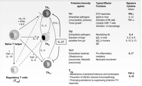
Besides recognition of antigen, a second signal is usually required for activation of B- and T lymphocytes. In case of T lymphocytes, co-stimulation occurs through engagement of CD28 with B7 molecules on APC [34]. Cytokines, secreted by activated T helper lymphocytes, are co-stimulatory signals necessary for growth and differentiation of B lymphocytes and CTL. In the absence of co-stimulation, anergy, the inability of immune cells to mount an adequate immune response against an antigen, can occur. Different T helper cell subsets, designated Th1, Th2 and Th17 can be distinguished by the cytokines they secrete (Fig. 1). Th1 cytokines include IFNγthat activates CTL, thereby stimulating cell-mediated immunity. In contrast, Th2 cytokines, such as IL-4, IL-5, and IL-10, function as a helper for B lymphocyte activation. A Th2 response is often elevated in allergic disease and parasitic infections [35]. Th17 is a recently discovered T cell subset that is involved in the control of infections against some extracellular bacteria [36,37]. The characteristic cytokine expressed by Th17 cells is IL-17. IL-17 mediates a pro-inflammatory response by inducing the expression of other cytokines (e.g. IL-6) and chemokines (e.g. IL-8) [38]. Furthermore, other subpopulations of T lymphocytes, named suppressor or “regulatory” T lymphocytes can suppress the immune system by producing inhibitory cytokines such as IL-10 and TGF-β. T lymphocytes can regulate the intensity of an immune response by switching from immune-promoting cytokines (e.g. IFNγ) to inhibiting cytokines (IL-10) in a later stage of the response.
2.2 Immune response in cervical cancer 2.2 Immune response in cervical cancer 2.2 Immune response in cervical cancer 2.2 Immune response in cervical cancer 2.2 Immune response in cervical cancer
HPVs do not provoke an adequate humoral immune response. Naturally arising antibodies against HPV do not seem to be effective in preventing subsequent infections of homologous or genetically related HPV types [39]. On the contrary, antibodies against HPV capsid proteins that arise after vaccination can be protective at preventing infections of homologous or genetically related HPV types [40,41]. Nevertheless, antibodies do not play an important role in the regression of established HPV infections and cervical lesions [42]. Cellular immune responses however are likely to be an important effector mechanism for the clearance of established infections [43]. HPV-specific T cell immunity is frequently detected in healthy individuals. Presence of T cell immunity therefore plays a role in protecting against persistent HPV infection and hence in the development of cervical malignancies [44]. In patients with cervical lesions and cervical cancer, tumor-specific T lymphocytes against the two HPV encoded oncoproteins, E6 and E7, were detected at low levels in the peripheral blood of approximately 50% of the patients [45-47]. HPV-specific systemic immunity in patients may not be beneficial since it has been associated with a non-inflammatory cytokine profile and did not correlate with prognostic factors [47,48]. In contrast, presence of high numbers of tumor-infiltrating lymphocytes has been associated with a survival advantage in patients with cervical cancer [49,50]. However, recent data suggests that the ratio between infiltrating CTL and infiltrating regulatory T lymphocytes is also an important determinant for prognosis [48],[51]. By suppressing proliferation and cytokine production of CTL, these regulatory T lymphocytes may well interfere with the tumor-specific immune response [52].
3 Immune escape
3 Immune escape
3 Immune escape
3 Immune escape
3 Immune escape
will be discussed more in detail in the next 2 paragraphs.
3.1 Alterations in HLA class I expression 3.1 Alterations in HLA class I expression 3.1 Alterations in HLA class I expression 3.1 Alterations in HLA class I expression 3.1 Alterations in HLA class I expression
An important mechanism of immune escape in cervical cancer is HLA class I loss. HLA class I comprises a highly polymorphic heavy chain, encoded by a HLA-A, HLA-B or HLA-C genes located on chromosome region 6p21.3 and a light chain, β 2-mircoglobulin. The extracellular part of the heavy chain contains a peptide-binding groove, in which peptides bind in an allele-specific manner. Since HLA molecules are responsible for the presentation of foreign antigens, these molecules play a central role in the immune recognition and subsequent clearance of virus-infected or other deviating cells [57]. Thus loss of HLA class I enables tumor cells to evade recognition and lyses by CTL. Especially in case of tumors with a viral origin such as cervical-cancer, development of a powerful immune response could be hindered since viral antigen is not or insufficiently presented. Defects in HLA class I are caused by multiple mechanisms [58]. Total HLA class I loss can be the result of mutations in the
β2-mircoglobulin-gene or defects in the molecules of the HLA class I processing and presentation pathway such as peptide transporter TAP1 or TAP2. Low expression of TAP1 and TAP2 have been observed in cervical cancer cell lines and tumors [59,60]. Partial HLA class I loss can be explained by (structural) defects in the particular HLA proteins which can be caused by locus-specific downregulation, possibly owing to shortcomings at the transcriptional level. Also loss of a single class I allele by mutations or deletions and loss of HLA class I haplotype by loss of heterozygosity (LOH) can be the cause for partial HLA class I loss. LOH can be due to mitotic recombination, deletion or chromosomal nondisjunction [58]. A detailed study of HLA class I aberrations in cervical cancer showed that approximately 90% of the cervical tumors had HLA class I defects [22]. The major mechanism for loss of expression was LOH (50%). Also HPV might interfere with HLA expression as a mechanism to escape from host immune surveillance. HPV16 E5 was shown to impede transport of HLA class I complexes to the cell surface and downregulate surface expression of HLA-A and HLA-B [61]. The importance of HLA class I expression in cervical cancer is emphasized by the findings that reduced expression of HLA class I correlates with worse prognosis and overall survival [62,63].
3.2 Immunosuppressive cytokines 3.2 Immunosuppressive cytokines 3.2 Immunosuppressive cytokines 3.2 Immunosuppressive cytokines 3.2 Immunosuppressive cytokines
inflammatory diseases are frequently associated with increased risk of cancer [64,66,67]. During a chronic inflammatory process, besides many cytokines (including growth and angiogenic factors), reactive oxygen and nitrogen species are generated that may cause DNA damage and predispose to neoplasia [67]. In the process of cancer development, cytokines produced by tumor cells, immune cells or stroma, facilitate cancer growth [68]. However in immunogenic cancer types, vaccination with tumor cells in combination with proinflammatory cytokines, including TNFα, IFNγ, IL-2 and GM-CSF, does show therapeutic benefit [69-7IL-2]. Evidence exists that this is associated with anti-tumor activity by T lymphocyte- mediated responses [72]. The contribution of proinflammatory cytokines to the initiation and development of cancer or prevention of cancer and anti-tumor activities depends on factors like the type of cancer, the tumor stage and the tumor micro-environment. In cervical cancer, several studies provided evidence that the cause of a failing anti-HPV immune response was a decrease in proinflammatory cytokines such as TNFα[47,73-75]. Also, several studies reported a predominant Th2 cytokine pattern in cervical lesions and cancer [73,76]. Polarization towards a Th2 cytokine profile signifies downregulation of a pro-inflammatory immune response and therefore a decreased susceptibility to the effect of CTL.
Expression of immunosuppressive cytokines like IL-10 and transforming growth factorβ(TGF-β)is another strategy used by tumors to escape immunosurveillance [77]. These cytokines inhibit the function of CTL and promote a shift towards Th2 cytokine expression, thereby downregulating anti-tumor immunity [76,78,79]. Tumor-derived IL-10 and TGF-βhave been shown to induce regulatory T lymphocytes and skew the immune response to Th2 polarity in cervical cancer [76]. It is conceivable that cervical cancers circumvent the development of a potent HPV-specific anti-tumor immune response by not providing the appropriate proinflammatory environment and instead stimulate the expression of immunosuppressive cytokines as TGF-βand IL-10. TGF-βwill be discussed in more detail in the next chapter, since a substantial part of this thesis concerns research into this cytokine.
4
4
4
4
TGF-4 TGF-
βββββ
4.1 Signal transduction 4.1 Signal transduction 4.1 Signal transduction 4.1 Signal transduction 4.1 Signal transduction
TGF-β1 is secreted as part of a latent complex into the extracellular matrix [81].
“TGF-βactivators” such as thrombospondin, matrix metalloproteinases, plasmin and certain integrins release TGF-β1 from its latent state [78,82]. Once activated, TGF-βfamily members initiate signaling by interacting with receptor serine/threonine kinases, type I and type II (Fig. 2). Upon ligand binding, the TGF-β receptor type II (TβRII) phosphorylates the TGF-βreceptor type I (TβRI) which leads to the activation of its kinase domain and subsequent regulation of the mothers against decapentaplegic drosophila homolog (Smad) proteins. Activated TβRI phosphorylates Smad2 and Smad3 proteins, which form hetero-oligomeric complexes with Smad4. These complexes translocate to the nucleus and, depending on the cell type and their
Figure 2. Figure 2. Figure 2. Figure 2.
interactions with certain transcription factors, activate or repress transcription via direct DNA binding [83].
Evidence exists for a strong integration of Smad signaling within a complex network with other signaling pathways that largely contribute to modifying the initial Smad signals and allows diverse responses to TGF-β1 [84,85]. Smad signaling may even be dispensable for some of the responses initiated by TGF-β1 [86,87]. TGF-β1 has been shown in various cell types to activate extracellular signal-regulated kinase (ERK), p38 and jun kinases (JNK) which are mitogen activated protein kinases (MAPK) [88,89]. ERK-mediated pathways are involved in proliferation and usually anti-apoptotic. In contrast, JNK and p38-signaling, activated by stress stimuli, often induce apoptosis. Activation of MAPK by TGF-β1 can occur directly by TGF-βactivated kinase-1 (TAK1) or indirectly, possibly resulting from Smad-dependent transcriptional responses [90]. MAPK can positively and negatively influence TGF-β1 signaling [84]. MAPK negatively influences TGF-β1 signaling by stimulating expression of Smad7, an inhibitor of Smad signaling [91]. Furthermore, phosphorylation of the linker region of R-Smads may block nuclear translocation and signaling by Smads [92]. MAPK are positively implicated in TGF-β1 signaling by interaction of AP-1 transcription factors, downstream components of the MAPK pathway, with Smad complexes to regulate transcription [93]. Also other signaling pathways, including the PP2A/p70S6, Rho GTPases, PI-3 kinase/AKT and Wnt pathways, can either be induced by TGF-β1, or can modulate the outcome of TGF-β-induced Smad signaling [94-97].
4.2 Role in cancer 4.2 Role in cancer 4.2 Role in cancer 4.2 Role in cancer 4.2 Role in cancer
TGF-β1 as a tumor suppressor
colorectal cancer [108]. Mutations in TβRI and Smad2 occur at low frequency, whereas Smad3 mutations have not been detected so far [80]. Loss of expression of these components was often found to be associated with worse prognosis [109,110]. However, some alterations of the TGF-β1 pathway do not lead to a complete loss of signaling and could result in loss of tumor suppressive effects while retaining the tumor promoting effects [80]. An alternative way of inactivating the tumor suppressor function of TGF-β1 is by overexpressing or amplification of its inhibitors. For example, Smad7 amplification in colorectal cancer and high expression of SMURF2 in esophageal squamous cell carcinoma were found to be poor prognostic markers [111,112]. The proto-oncogene product Ski was shown to promote tumor growth in pancreatic cancer by abrogation of TGF-β1 signaling [113]. Furthermore, oncoproteins c-Myc and Ras were shown to antagonize the growth-inhibitory response of TGF-β1 signaling in epithelial cells [92,114-116].
Figure 3. Figure 3. Figure 3. Figure 3.
Figure 3. TGF-β1 switches from tumor suppressor in normal and premalignant stages of tumorigenesis to proto-oncogene at later stages of disease. Progression to metastatic disease is usually accompanied by decreased or altered TGF-β1 responsiveness and increased expression/ activation of TGF-β1. Besides changes in the responsiveness to TGF-β1 in tumor cells, TGF-β1 effects tumor stroma in such a manner that tumor growth and metastasis is facilitated. From Roberts et al., 2003 [117].
TGF-β1 as a tumor promoter
an epithelial-to-mesenchymal transition (EMT) [118,119]. Cells undergoing EMT exhibit changes in morphology, loose cell-cell and cell-matrix adhesive properties accompanied by degradation of the surrounding extracellular matrix (ECM). A number of studies have shown that the Smad pathway as well as other pathways in TGF-β1 signaling such as ERK and PI-3 kinase/AKT signaling are required for EMT [120-123]. Increased production and activation of TGF-β1 by tumor cells stimulates synthesis of ECM proteins and chemokine production as well as activation of fibroblasts [124,125]. By providing growth factors, ECM proteins and matrix metalloproteinases, fibroblasts support tumor progression [126,127]. Furthermore, TGF-β1 stimulates tumor angiogenesis. Angiogenesis is crucial for tumor growth and invasion, as blood vessels deliver nutrients and oxygen to the tumor cells and allow migration of tumor cells via the blood system, resulting in metastasis. TGF-β1 induces expression of vascular endothelial growth factor (VEGF) [128], thereby stimulating proliferation and migration of endothelial cells [129] and capillary formation of endothelial cells [130]. In addition, TGF-β1 possesses chemoattractant activity for monocytes which release angiogenic cytokines. Escape from immunosurveillance is mediated by inhibition of T lymphocyte proliferation through expression of cell cycle regulators and blockade of the production of IL-2, a cytokine known to activate T lymphocytes, NK cells and other types of the immune system [131]. TGF-β1 also controls T- and NK-cell effector functions by attenuating the cytolytic activity of these cells, inhibiting expression of IFN-γ, necessary for the stimulation of a Th1 response, and inhibiting the expression of perforin [132-134]. Furthermore, antigen presentation by differentiating dendritic cells, which are powerful APC, is hampered by inhibiting the expression of HLA class II and costimulatory molecules [135].
4.3 4.3 4.3 4.3
TGF-4.3 TGF-βββββ1 in cervical cancer1 in cervical cancer1 in cervical cancer1 in cervical cancer1 in cervical cancer
During cervical carcinoma development, a progressive loss of sensitivity to TGF-β 1-mediated growth inhibition was found [136]. This seems to be accompanied by increased expression levels of TGF-β1 during the progression from benign to malignant lesions [137-139]. Whereas TGF-β1 did not hinder proliferation of cervical cancer cells [138], an inverse relationship was found between its expression and the amount of tumor infiltrate [140]. Furthermore, TGF-β1 mRNA expression correlated with the amount of intratumoral stroma [140]. Consequently, the tumorsuppressive characteristics of TGF-β1, such as growth inhibition, seem to be lost, whereas the
tumor-promoting features of TGF-β1, such as immunosuppression and ECM
suppression of c-myc transcription via pRb. This has been observed in human foreskin keratinocytes transformed with HPV16 and was associated with resistance to the growth inhibitory effects of TGF-β1 [101]. However, TGF-β1 itself has been shown to inhibit expression of HPV16 E6 and E7, accompanied with cessation of cell proliferation in immortalized genital epithelial cell lines [147]. After malignant transformation, cells became partially resistant to the inhibitory effects of TGF-β1 on cell growth and gene expression of HPV16 E6 and E7 [147]. Thus far, it is unknown whether resistance to TGF-β1-induced growth inhibition is due to mutations in the Smad signaling pathway, interference by HPV oncoproteins or changes in signal transduction due to crosstalk with other pathways. Further insights into the signaling pathways downstream of TGF-β1, and the contributions of these pathways to the specific cellular and context-dependent effects of TGF-β1, may lead to more specific targeting of this pathway in the treatment of cancer.
5 Outline of the thesis
5 Outline of the thesis
5 Outline of the thesis
5 Outline of the thesis
5 Outline of the thesis
Cervical cancer is a virus-induced tumor that has developed several escape mechanisms to avoid eradication by the immune system. In this thesis we have investigated several of the immune escape routes that cervical cancer cells have acquired. A common feature of cervical cancer cells, occurring in ± 90% of tumors, is their ability to prevent recognition by the immune system through the loss of HLA class I protein. We hypothesized that tumors without HLA class I loss have developed other ways to prevent elimination by the immune system. In these latter cases, expression of immunosuppressive cytokines, such as TGF-β1 or IL-10, could be a mechanism to escape immune surveillance. In chapter 2chapter 2chapter 2chapter 2chapter 2, tumors positive and negative for HLA class I were compared, based on gene expression profile, to investigate escape routes in tumors with HLA class I expression. Microarray was used as a platform to study genomewide gene expression changes between tumors positive and negative for HLA class I. Subsequently, real-time PCR and immunohistochemistry were used to confirm expression changes. An alternative mechanism for immune escape in cervical cancer, besides loss of HLA class I and expression of immunosuppressive cytokines, is loss of proinflammatory cytokine expression. The presence of the proinflammatory cytokine TNFα may also be detrimental for the development of an adequate immune response against HPV.
Therefore, in chapter 3chapter 3chapter 3chapter 3chapter 3, loss of TNFαgene expression, as a mechanism to escape immune clearance, was investigated. The (epi)-genetic characteristics associated with the loss of TNFα expression were studied to investigate if TNFα could be a candidate tumor suppressor gene.
TGF-β1, a cytokine with potent immuno-inhibitory qualities, is excessively produced by cervical cancer cells. As discussed earlier, TGF-β1 suppresses the immune response by several mechanisms. However TGF-β1 also suppresses cell growth, therefore cervical cancer cells need to first acquire insensitivity to TGF-β1 induced cell growth inhibition, thus providing the tumor cells a growth advantage. In chapterchapterchapterchapterchapter 4,
4, 4, 4,
different sensitivity towards TGF-β1 was investigated. To investigate if TGF-β1 insensitivity in cervical cancer can be due to alterations in the canonical Smad pathway, loss of Smad2, Smad2-P and Smad4 protein expression was investigated by immunohistochemistry in a large cohort of clinical samples as described in chapterchapterchapterchapterchapter 5
55
55. Tumors with low expression of Smad2-P and Smad4 were investigated for genetic inactivation, loss of heterozygosity and in case of Smad4 for promoter methylation to determine if these genes are candidate tumor suppressor genes in cervical carcinogenesis.
Cervical tumors are characterized by complex genetic alterations involving deletions and amplifications that can affect many chromosomal regions. Also genes involved in immune responses can be affected, as has been shown for HLA class I genes. Currently, large-scale DNA copy number, genotyping and gene expression profiling methods are available to study the genome. To obtain an overview of genome-wide molecular alterations in cervical cancer and to uncover genes involved in cervical carcinogenesis, including genes related to the immune response, we investigated DNA copy number and genotype alterations in cervical cancer cell lines in detail using array comparative genomic hybridization (CGH) and single nucleotide polymorphisms (SNP) array, as described in chapter 6chapter 6chapter 6chapter 6. Furthermore, an effect ofchapter 6 copy number alteration on gene expression was investigated. Finally, in chapter 7chapter 7chapter 7chapter 7chapter 7 the results of the above mentioned studies are discussed and summarized.
References
References
References
References
References
1. Parkin DM, Bray F, Ferlay J, Pisani P. Global cancer statistics, 2002. CA Cancer J Clin 2005;55:74-108.
2. Sankaranarayanan R, Ferlay J. Worldwide burden of gynaecological cancer: the size of the problem. Best Pract Res Clin Obstet Gynaecol 2006;20:207-225.
3. Bosch FX, de Sanjose S. The epidemiology of human papillomavirus infection and cervical cancer. Dis Markers 2007;23:213-227.
4. Tiltman AJ. The pathology of cervical tumors. Best Pract Res Clin Obstet Gynaecol 2005;19:485-500.
5. Green J, Berrington dG, Sweetland S, Beral V, Chilvers C, Crossley B, et al. Risk factors for adenocarcinoma and squamous cell carcinoma of the cervix in women aged 20-44 years: the UK National Case-Control Study of Cervical Cancer. Br J Cancer 2003;89:2078-2086. 6. Bontkes HJ, de Gruijl TD, Walboomers JM, Schiller JT, Dillner J, Helmerhorst TJ, et al. Immune responses against human papillomavirus (HPV) type 16 virus-like particles in a cohort study of women with cervical intraepithelial neoplasia. II. Systemic but not local IgA responses correlate with clearance of HPV-16. J Gen Virol 1999;80 ( Pt 2):409-417. 7. Nakagawa M, Stites DP, Farhat S, Sisler JR, Moss B, Kong F, et al. Cytotoxic T lymphocyte
responses to E6 and E7 proteins of human papillomavirus type 16: relationship to cervical intraepithelial neoplasia. J Infect Dis 1997;175:927-931.
8. Ostor AG. Natural history of cervical intraepithelial neoplasia: a critical review. Int J Gynecol Pathol 1993;12:186-192.
9. Bosch FX, de Sanjose S. Chapter 1: Human papillomavirus and cervical cancer—burden and assessment of causality. J Natl Cancer Inst Monogr 2003;3-13.
10. Walboomers JM, Jacobs MV, Manos MM, Bosch FX, Kummer JA, Shah KV, et al. Human papillomavirus is a necessary cause of invasive cervical cancer worldwide. J Pathol 1999;189:12-19.
11. Munoz N, Bosch FX, de Sanjose S, Herrero R, Castellsague X, Shah KV, et al. Epidemiologic classification of human papillomavirus types associated with cervical cancer. N Engl J Med 2003;348:518-527.
13. Chakrabarti O, Krishna S. Molecular interactions of ‘high risk’ human papillomaviruses E6 and E7 oncoproteins: implications for tumor progression. J Biosci 2003;28:337-348. 14. Jeon S, Lambert PF. Integration of human papillomavirus type 16 DNA into the human
genome leads to increased stability of E6 and E7 mRNAs: implications for cervical carcinogenesis. Proc Natl Acad Sci U S A 1995;92:1654-1658.
15. Melsheimer P, Vinokurova S, Wentzensen N, Bastert G, von Knebel DM. DNA aneuploidy and integration of human papillomavirus type 16 e6/e7 oncogenes in intraepithelial neoplasia and invasive squamous cell carcinoma of the cervix uteri. Clin Cancer Res 2004;10:3059-3063.
16. Harris CP, Lu XY, Narayan G, Singh B, Murty VV, Rao PH. Comprehensive molecular cytogenetic characterization of cervical cancer cell lines. Genes Chromosomes Cancer 2003;36:233-241.
17. Abba MC, Laguens RM, Dulout FN, Golijow CD. The c-myc activation in cervical carcinomas and HPV 16 infections. Mutat Res 2004;557:151-158.
18. Zhang A, Maner S, Betz R, Angstrom T, Stendahl U, Bergman F, et al. Genetic alterations in cervical carcinomas: frequent low-level amplifications of oncogenes are associated with human papillomavirus infection. Int J Cancer 2002;101:427-433.
19. Martin CM, Astbury K, O’Leary JJ. Molecular profiling of cervical neoplasia. Expert Rev Mol Diagn 2006;6:217-229.
20. Hidalgo A, Baudis M, Petersen I, Arreola H, Pina P, Vazquez-Ortiz G, et al. Microarray comparative genomic hybridization detection of chromosomal imbalances in uterine cervix carcinoma. BMC Cancer 2005;5:77.
21. Hasina R, Pontier AL, Fekete MJ, Martin LE, Qi XM, Brigaudeau C, et al. NOL7 is a nucleolar candidate tumor suppressor gene in cervical cancer that modulates the angiogenic phenotype. Oncogene 2006;25:588-598.
22. Koopman LA, Corver WE, van der Slik AR, Giphart MJ, Fleuren GJ. Multiple genetic alterations cause frequent and heterogeneous human histocompatibility leukocyte antigen class I loss in cervical cancer. J Exp Med 2000;191:961-976.
23. Magnusson PK, Sparen P, Gyllensten UB. Genetic link to cervical tumors. Nature 1999;400:29-30.
24. Frisch M, Biggar RJ, Goedert JJ. Human papillomavirus-associated cancers in patients with human immunodeficiency virus infection and acquired immunodeficiency syndrome. J Natl Cancer Inst 2000;92:1500-1510.
25. Birkeland SA, Storm HH, Lamm LU, Barlow L, Blohme I, Forsberg B, et al. Cancer risk after renal transplantation in the Nordic countries, 1964-1986. Int J Cancer 1995;60:183-189. 26. Sasadeusz J, Kelly H, Szer J, Schwarer AP, Mitchell H, Grigg A. Abnormal cervical cytology
in bone marrow transplant recipients. Bone Marrow Transplant 2001;28:393-397. 27. Ivansson EL, Rasmussen F, Gyllensten UB, Magnusson PK. Reduced incidence of cervical
cancer in mothers of sons with allergic rhinoconjunctivitis, asthma or eczema. Int J Cancer 2006;119:1994-1998.
28. Mehta AM, Jordanova ES, van Wezel T, Uh HW, Corver WE, Kwappenberg KM, et al. Genetic variation of antigen processing machinery components and association with cervical carcinoma. Genes Chromosomes Cancer 2007;46:577-586.
29. Carrington M, Wang S, Martin MP, Gao X, Schiffman M, Cheng J, et al. Hierarchy of resistance to cervical neoplasia mediated by combinations of killer immunoglobulin-like receptor and human leukocyte antigen loci. J Exp Med 2005;201:1069-1075.
30. Beskow AH, Gyllensten UB. Host genetic control of HPV 16 titer in carcinoma in situ of the cervix uteri. Int J Cancer 2002;101:526-531.
31. Weiner GJ. Monoclonal antibody mechanisms of action in cancer. Immunol Res 2007;39:271-278.
32. Choo SY. The HLA system: genetics, immunology, clinical testing, and clinical implications. Yonsei Med J 2007;48:11-23.
33. Young LH, Liu CC, Joag S, Rafii S, Young JD. How lymphocytes kill. Annu Rev Med 1990;41:45-54.
34. Allison JP. CD28-B7 interactions in T-cell activation. Curr Opin Immunol 1994;6:414-419. 35. Chabalgoity JA, Baz A, Rial A, Grille S. The relevance of cytokines for development of
protective immunity and rational design of vaccines. Cytokine Growth Factor Rev 2007;18:195-207.
37. Malley R, Srivastava A, Lipsitch M, Thompson CM, Watkins C, Tzianabos A, et al. Antibody-independent, interleukin-17A-mediated, cross-serotype immunity to pneumococci in mice immunized intranasally with the cell wall polysaccharide. Infect Immun 2006;74:2187-2195. 38. Shen F, Hu Z, Goswami J, Gaffen SL. Identification of common transcriptional regulatory
elements in interleukin-17 target genes. J Biol Chem 2006;281:24138-24148.
39. Viscidi RP, Schiffman M, Hildesheim A, Herrero R, Castle PE, Bratti MC, et al. Seroreactivity to human papillomavirus (HPV) types 16, 18, or 31 and risk of subsequent HPV infection: results from a population-based study in Costa Rica. Cancer Epidemiol Biomarkers Prev 2004;13:324-327.
40. Suzich JA, Ghim SJ, Palmer-Hill FJ, White WI, Tamura JK, Bell JA, et al. Systemic immunization with papillomavirus L1 protein completely prevents the development of viral mucosal papillomas. Proc Natl Acad Sci U S A 1995;92:11553-11557.
41. Harper DM, Franco EL, Wheeler C, Ferris DG, Jenkins D, Schuind A, et al. Efficacy of a bivalent L1 virus-like particle vaccine in prevention of infection with human papillomavirus types 16 and 18 in young women: a randomised controlled trial. Lancet 2004;364:1757-1765.
42. Sun Y, Eluf-Neto J, Bosch FX, Munoz N, Walboomers JM, Meijer CJ, et al. Serum antibodies to human papillomavirus 16 proteins in women from Brazil with invasive cervical carcinoma. Cancer Epidemiol Biomarkers Prev 1999;8:935-940.
43. Tsukui T, Hildesheim A, Schiffman MH, Lucci J, III, Contois D, Lawler P, et al. Interleukin 2 production in vitro by peripheral lymphocytes in response to human papillomavirus-derived peptides: correlation with cervical pathology. Cancer Res 1996;56:3967-3974.
44. Welters MJ, de Jong A, van den Eeden SJ, van der Hulst JM, Kwappenberg KM, Hassane S, et al. Frequent display of human papillomavirus type 16 E6-specific memory t-Helper cells in the healthy population as witness of previous viral encounter. Cancer Res 2003;63:636-641.
45. de Gruijl TD, Bontkes HJ, Walboomers JM, Stukart MJ, Doekhie FS, Remmink AJ, et al. Differential T helper cell responses to human papillomavirus type 16 E7 related to viral clearance or persistence in patients with cervical neoplasia: a longitudinal study. Cancer Res 1998;58:1700-1706.
46. Bontkes HJ, de Gruijl TD, van den Muysenberg AJ, Verheijen RH, Stukart MJ, Meijer CJ, et al. Human papillomavirus type 16 E6/E7-specific cytotoxic T lymphocytes in women with cervical neoplasia. Int J Cancer 2000;88:92-98.
47. de Jong A, van Poelgeest MI, van der Hulst JM, Drijfhout JW, Fleuren GJ, Melief CJ, et al. Human papillomavirus type 16-positive cervical cancer is associated with impaired CD4+ T-cell immunity against early antigens E2 and E6. Cancer Res 2004;64:5449-5455. 48. Piersma SJ, Jordanova ES, van Poelgeest MI, Kwappenberg KM, van der Hulst JM,
Drijfhout JW, et al. High number of intraepithelial CD8+ tumor-infiltrating lymphocytes is associated with the absence of lymph node metastases in patients with large early-stage cervical cancer. Cancer Res 2007;67:354-361.
49. Bethwaite PB, Holloway LJ, Thornton A, Delahunt B. Infiltration by immunocompetent cells in early stage invasive carcinoma of the uterine cervix: a prognostic study. Pathology 1996;28:321-327.
50. Chao HT, Wang PH, Tseng JY, Lai CR, Chiang SC, Yuan CC. Lymphocyte-infiltrated FIGO Stage IIB squamous cell carcinoma of the cervix is a prominent factor for disease-free survival. Eur J Gynaecol Oncol 1999;20:136-140.
51. Jordanova ES, Gorter A, Ayachi O, Prins F, Durrant LG, Kenter GG, et al. Human leukocyte antigen class I, MHC class I chain-related molecule A, and CD8+/regulatory T-cell ratio: which variable determines survival of cervical cancer patients? Clin Cancer Res 2008;14:2028-2035.
52. van der Burg SH, Piersma SJ, de Jong A, van der Hulst JM, Kwappenberg KM, van den HM, et al. Association of cervical cancer with the presence of CD4+ regulatory T cells specific for human papillomavirus antigens. Proc Natl Acad Sci U S A 2007;104:12087-12092.
53. Frazer IH, Thomas R, Zhou J, Leggatt GR, Dunn L, McMillan N, et al. Potential strategies utilised by papillomavirus to evade host immunity. Immunol Rev 1999;168:131-142. 54. Tindle RW. Immune evasion in human papillomavirus-associated cervical cancer. Nat Rev
Cancer 2002;2:59-65.
56. Kanodia S, Fahey LM, Kast WM. Mechanisms used by human papillomaviruses to escape the host immune response. Curr Cancer Drug Targets 2007;7:79-89.
57. Moss DJ, Khanna R. Major histocompatibility complex: from genes to function. Immunol Today 1999;20:165-167.
58. Aptsiauri N, Cabrera T, Mendez R, Garcia-Lora A, Ruiz-Cabello F, Garrido F. Role of altered expression of HLA class I molecules in cancer progression. Adv Exp Med Biol 2007;601:123-131.
59. Evans M, Borysiewicz LK, Evans AS, Rowe M, Jones M, Gileadi U, et al. Antigen processing defects in cervical carcinomas limit the presentation of a CTL epitope from human papillomavirus 16 E6. J Immunol 2001;167:5420-5428.
60. Vermeulen CF, Jordanova ES, Ter Haar NT, Kolkman-Uljee SM, de Miranda NF, Ferrone S, et al. Expression and genetic analysis of transporter associated with antigen processing in cervical carcinoma. Gynecol Oncol 2007;105:593-599.
61. Ashrafi GH, Haghshenas MR, Marchetti B, O’Brien PM, Campo MS. E5 protein of human papillomavirus type 16 selectively downregulates surface HLA class I. Int J Cancer 2005;113:276-283.
62. van Driel WJ, Tjiong MY, Hilders CG, Trimbos BJ, Fleuren GJ. Association of allele-specific HLA expression and histopathologic progression of cervical carcinoma. Gynecol Oncol 1996;62:33-41.
63. Mehta AM, Jordanova ES, Kenter GG, Ferrone S, Fleuren GJ. Association of antigen processing machinery and HLA class I defects with clinicopathological outcome in cervical carcinoma. Cancer Immunol Immunother 2007.
64. Philip M, Rowley DA, Schreiber H. Inflammation as a tumor promoter in cancer induction. Semin Cancer Biol 2004;14:433-439.
65. Mantovani A. Cancer: inflammation by remote control. Nature 2005;435:752-753.
66. Balkwill F, Mantovani A. Inflammation and cancer: back to Virchow? Lancet 2001;357:539-545.
67. Coussens LM, Werb Z. Inflammation and cancer. Nature 2002;420:860-867.
68. Pollard JW. Tumor-educated macrophages promote tumor progression and metastasis. Nat Rev Cancer 2004;4:71-78.
69. van Horssen R, Ten Hagen TL, Eggermont AM. TNF-alpha in cancer treatment: molecular insights, antitumor effects, and clinical utility. Oncologist 2006;11:397-408.
70. Rosenberg SA, Yang JC, Topalian SL, Schwartzentruber DJ, Weber JS, Parkinson DR, et al. Treatment of 283 consecutive patients with metastatic melanoma or renal cell cancer using high-dose bolus interleukin 2. JAMA 1994;271:907-913.
71. Kirkwood JM, Strawderman MH, Ernstoff MS, Smith TJ, Borden EC, Blum RH. Interferon alfa-2b adjuvant therapy of high-risk resected cutaneous melanoma: the Eastern Cooperative Oncology Group Trial EST 1684. J Clin Oncol 1996;14:7-17.
72. Eager R, Nemunaitis J. GM-CSF gene-transduced tumor vaccines. Mol Ther 2005;12:18-27.
73. Mota F, Rayment N, Chong S, Singer A, Chain B. The antigen-presenting environment in normal and human papillomavirus (HPV)-related premalignant cervical epithelium. Clin Exp Immunol 1999;116:33-40.
74. Giannini SL, Al Saleh W, Piron H, Jacobs N, Doyen J, Boniver J, et al. Cytokine expression in squamous intraepithelial lesions of the uterine cervix: implications for the generation of local immunosuppression. Clin Exp Immunol 1998;113:183-189.
75. El Sherif AM, Seth R, Tighe PJ, Jenkins D. Quantitative analysis of IL-10 and IFN-gamma mRNA levels in normal cervix and human papillomavirus type 16 associated cervical precancer. J Pathol 2001;195:179-185.
76. Sheu BC, Lin RH, Lien HC, Ho HN, Hsu SM, Huang SC. Predominant Th2/Tc2 polarity of tumor-infiltrating lymphocytes in human cervical cancer. J Immunol 2001;167:2972-2978. 77. Salazar-Onfray F. Interleukin-10: a cytokine used by tumors to escape immunosurveillance.
Med Oncol 1999;16:86-94.
78. de Visser KE, Kast WM. Effects of TGF-beta on the immune system: implications for cancer immunotherapy. Leukemia 1999;13:1188-1199.
79. Bellone G, Turletti A, Artusio E, Mareschi K, Carbone A, Tibaudi D, et al. Tumor-associated transforming growth factor-beta and interleukin-10 contribute to a systemic Th2 immune phenotype in pancreatic carcinoma patients. Am J Pathol 1999;155:537-547.
81. Khalil N. TGF-beta: from latent to active. Microbes Infect 1999;1:1255-1263.
82. Munger JS, Huang X, Kawakatsu H, Griffiths MJ, Dalton SL, Wu J, et al. The integrin alpha v beta 6 binds and activates latent TGF beta 1: a mechanism for regulating pulmonary inflammation and fibrosis. Cell 1999;96:319-328.
83. Massague J, Seoane J, Wotton D. Smad transcription factors. Genes Dev 2005;19:2783-2810.
84. Javelaud D, Mauviel A. Crosstalk mechanisms between the mitogen-activated protein kinase pathways and Smad signaling downstream of TGF-beta: implications for carcinogenesis. Oncogene 2005;24:5742-5750.
85. Rahimi RA, Leof EB. TGF-beta signaling: A tale of two responses. J Cell Biochem 2007;102:593-608.
86. Hu PP, Shen X, Huang D, Liu Y, Counter C, Wang XF. The MEK pathway is required for stimulation of p21(WAF1/CIP1) by transforming growth factor-beta. J Biol Chem 1999;274:35381-35387.
87. Hocevar BA, Brown TL, Howe PH. TGF-beta induces fibronectin synthesis through a c-Jun N-terminal kinase-dependent, Smad4-independent pathway. EMBO J 1999;18:1345-1356. 88. Wakefield LM, Roberts AB. TGF-beta signaling: positive and negative effects on
tumorigenesis. Curr Opin Genet Dev 2002;12:22-29.
89. Derynck R, Zhang YE. Smad-dependent and Smad-independent pathways in TGF-beta family signalling. Nature 2003;425:577-584.
90. Takekawa M, Tatebayashi K, Itoh F, Adachi M, Imai K, Saito H. Smad-dependent GADD45beta expression mediates delayed activation of p38 MAP kinase by TGF-beta. EMBO J 2002;21:6473-6482.
91. Brodin G, Ahgren A, ten Dijke P, Heldin CH, Heuchel R. Efficient TGF-beta induction of the Smad7 gene requires cooperation between AP-1, Sp1, and Smad proteins on the mouse Smad7 promoter. J Biol Chem 2000;275:29023-29030.
92. Kretzschmar M, Doody J, Timokhina I, Massague J. A mechanism of repression of TGFbeta/ Smad signaling by oncogenic Ras. Genes Dev 1999;13:804-816.
93. Zhang Y, Feng XH, Derynck R. Smad3 and Smad4 cooperate with c-Jun/c-Fos to mediate TGF-beta-induced transcription. Nature 1998;394:909-913.
94. Petritsch C, Beug H, Balmain A, Oft M. TGF-beta inhibits p70 S6 kinase via protein phosphatase 2A to induce G(1) arrest. Genes Dev 2000;14:3093-3101.
95. Kamaraju AK, Roberts AB. Role of Rho/ROCK and p38 MAP kinase pathways in transforming growth factor-beta-mediated Smad-dependent growth inhibition of human breast carcinoma cells in vivo. J Biol Chem 2005;280:1024-1036.
96. Wilkes MC, Mitchell H, Penheiter SG, Dore JJ, Suzuki K, Edens M, et al. Transforming growth factor-beta activation of phosphatidylinositol 3-kinase is independent of Smad2 and Smad3 and regulates fibroblast responses via p21-activated kinase-2. Cancer Res 2005;65:10431-10440.
97. Nishita M, Hashimoto MK, Ogata S, Laurent MN, Ueno N, Shibuya H, et al. Interaction between Wnt and TGF-beta signalling pathways during formation of Spemann’s organizer. Nature 2000;403:781-785.
98. Hannon GJ, Beach D. p15INK4B is a potential effector of TGF-beta-induced cell cycle arrest. Nature 1994;371:257-261.
99. Datto MB, Li Y, Panus JF, Howe DJ, Xiong Y, Wang XF. Transforming growth factor beta induces the cyclin-dependent kinase inhibitor p21 through a p53-independent mechanism. Proc Natl Acad Sci U S A 1995;92:5545-5549.
100. Iavarone A, Massague J. Repression of the CDK activator Cdc25A and cell-cycle arrest by cytokine TGF-beta in cells lacking the CDK inhibitor p15. Nature 1997;387:417-422. 101. Pietenpol JA, Holt JT, Stein RW, Moses HL. Transforming growth factor beta 1 suppression
of c-myc gene transcription: role in inhibition of keratinocyte proliferation. Proc Natl Acad Sci U S A 1990;87:3758-3762.
102. Kang Y, Chen CR, Massague J. A self-enabling TGFbeta response coupled to stress signaling: Smad engages stress response factor ATF3 for Id1 repression in epithelial cells. Mol Cell 2003;11:915-926.
103. Kim SG, Jong HS, Kim TY, Lee JW, Kim NK, Hong SH, et al. Transforming growth factor-beta 1 induces apoptosis through Fas ligand-independent activation of the Fas death pathway in human gastric SNU-620 carcinoma cells. Mol Biol Cell 2004;15:420-434.
105. Ramjaun AR, Tomlinson S, Eddaoudi A, Downward J. Upregulation of two BH3-only proteins, Bmf and Bim, during TGF beta-induced apoptosis. Oncogene 2007;26:970-981. 106. Hahn SA, Schutte M, Hoque AT, Moskaluk CA, da Costa LT, Rozenblum E, et al. DPC4, a
candidate tumor suppressor gene at human chromosome 18q21.1. Science 1996;271:350-353.
107. Woodford-Richens KL, Rowan AJ, Gorman P, Halford S, Bicknell DC, Wasan HS, et al. SMAD4 mutations in colorectal cancer probably occur before chromosomal instability, but after divergence of the microsatellite instability pathway. Proc Natl Acad Sci U S A 2001;98:9719-9723.
108. Parsons R, Myeroff LL, Liu B, Willson JK, Markowitz SD, Kinzler KW, et al. Microsatellite instability and mutations of the transforming growth factor beta type II receptor gene in colorectal cancer. Cancer Res 1995;55:5548-5550.
109. Fukai Y, Fukuchi M, Masuda N, Osawa H, Kato H, Nakajima T, et al. Reduced expression of transforming growth factor-beta receptors is an unfavorable prognostic factor in human esophageal squamous cell carcinoma. Int J Cancer 2003;104:161-166.
110. Xie W, Mertens JC, Reiss DJ, Rimm DL, Camp RL, Haffty BG, et al. Alterations of Smad signaling in human breast carcinoma are associated with poor outcome: a tissue microarray study. Cancer Res 2002;62:497-505.
111. Fukuchi M, Fukai Y, Masuda N, Miyazaki T, Nakajima M, Sohda M, et al. High-level expression of the Smad ubiquitin ligase Smurf2 correlates with poor prognosis in patients with esophageal squamous cell carcinoma. Cancer Res 2002;62:7162-7165.
112. Boulay JL, Mild G, Lowy A, Reuter J, Lagrange M, Terracciano L, et al. SMAD7 is a prognostic marker in patients with colorectal cancer. Int J Cancer 2003;104:446-449.
113. Heider TR, Lyman S, Schoonhoven R, Behrns KE. Ski promotes tumor growth through abrogation of transforming growth factor-beta signaling in pancreatic cancer. Ann Surg 2007;246:61-68.
114. Alexandrow MG, Kawabata M, Aakre M, Moses HL. Overexpression of the c-Myc oncoprotein blocks the growth-inhibitory response but is required for the mitogenic effects of transforming growth factor beta 1. Proc Natl Acad Sci U S A 1995;92:3239-3243.
115. Stroschein SL, Wang W, Zhou S, Zhou Q, Luo K. Negative feedback regulation of TGF-beta signaling by the SnoN oncoprotein. Science 1999;286:771-774.
116. Kleeff J, Ishiwata T, Maruyama H, Friess H, Truong P, Buchler MW, et al. The TGF-beta signaling inhibitor Smad7 enhances tumorigenicity in pancreatic cancer. Oncogene 1999;18:5363-5372.
117. Roberts AB, Wakefield LM. The two faces of transforming growth factor beta in carcinogenesis. Proc Natl Acad Sci U S A 2003;100:8621-8623.
118. Saika S, Kono-Saika S, Ohnishi Y, Sato M, Muragaki Y, Ooshima A, et al. Smad3 signaling is required for epithelial-mesenchymal transition of lens epithelium after injury. Am J Pathol 2004;164:651-663.
119. Tian F, Byfield SD, Parks WT, Stuelten CH, Nemani D, Zhang YE, et al. Smad-binding defective mutant of transforming growth factor beta type I receptor enhances tumorigenesis but suppresses metastasis of breast cancer cell lines. Cancer Res 2004;64:4523-4530. 120. Itoh S, Thorikay M, Kowanetz M, Moustakas A, Itoh F, Heldin CH, et al. Elucidation of Smad
requirement in transforming growth factor-beta type I receptor-induced responses. J Biol Chem 2003;278:3751-3761.
121. Valcourt U, Kowanetz M, Niimi H, Heldin CH, Moustakas A. TGF-beta and the Smad signaling pathway support transcriptomic reprogramming during epithelial-mesenchymal cell transition. Mol Biol Cell 2005;16:1987-2002.
122. Peinado H, Quintanilla M, Cano A. Transforming growth factor beta-1 induces snail transcription factor in epithelial cell lines: mechanisms for epithelial mesenchymal transitions. J Biol Chem 2003;278:21113-21123.
123. Bakin AV, Tomlinson AK, Bhowmick NA, Moses HL, Arteaga CL. Phosphatidylinositol 3-kinase function is required for transforming growth factor beta-mediated epithelial to mesenchymal transition and cell migration. J Biol Chem 2000;275:36803-36810.
124. Postlethwaite AE, Keski-Oja J, Moses HL, Kang AH. Stimulation of the chemotactic migration of human fibroblasts by transforming growth factor beta. J Exp Med 1987;165:251-256. 125. Taipale J, Saharinen J, Keski-Oja J. Extracellular matrix-associated transforming growth
factor-beta: role in cancer cell growth and invasion. Adv Cancer Res 1998;75:87-134. 126. Orimo A, Tomioka Y, Shimizu Y, Sato M, Oigawa S, Kamata K, et al. Cancer-associated
127. Kalluri R, Zeisberg M. Fibroblasts in cancer. Nat Rev Cancer 2006;6:392-401.
128. Nakagawa T, Li JH, Garcia G, Mu W, Piek E, Bottinger EP, et al. TGF-beta induces proangiogenic and antiangiogenic factors via parallel but distinct Smad pathways. Kidney Int 2004;66:605-613.
129. Pertovaara L, Kaipainen A, Mustonen T, Orpana A, Ferrara N, Saksela O, et al. Vascular endothelial growth factor is induced in response to transforming growth factor-beta in fibroblastic and epithelial cells. J Biol Chem 1994;269:6271-6274.
130. Choi ME, Ballermann BJ. Inhibition of capillary morphogenesis and associated apoptosis by dominant negative mutant transforming growth factor-beta receptors. J Biol Chem 1995;270:21144-21150.
131. Ma A, Koka R, Burkett P. Diverse functions of IL-2, IL-15, and IL-7 in lymphoid homeostasis. Annu Rev Immunol 2006;24:657-679.
132. Ahmadzadeh M, Rosenberg SA. TGF-beta 1 attenuates the acquisition and expression of effector function by tumor antigen-specific human memory CD8 T cells. J Immunol 2005;174:5215-5223.
133. Rook AH, Kehrl JH, Wakefield LM, Roberts AB, Sporn MB, Burlington DB, et al. Effects of transforming growth factor beta on the functions of natural killer cells: depressed cytolytic activity and blunting of interferon responsiveness. J Immunol 1986;136:3916-3920. 134. Bellone G, Aste-Amezaga M, Trinchieri G, Rodeck U. Regulation of NK cell functions by
TGF-beta 1. J Immunol 1995;155:1066-1073.
135. Geissmann F, Revy P, Regnault A, Lepelletier Y, Dy M, Brousse N, et al. TGF-beta 1 prevents the noncognate maturation of human dendritic Langerhans cells. J Immunol 1999;162:4567-4575.
136. De Geest K, Bergman CA, Turyk ME, Frank BS, Wilbanks GD. Differential response of cervical intraepithelial and cervical carcinoma cell lines to transforming growth factor-beta 1. Gynecol Oncol 1994;55:376-385.
137. El Sherif AM, Seth R, Tighe PJ, Jenkins D. Decreased synthesis and expression of TGF-beta1, beta2, and beta3 in epithelium of HPV 16-positive cervical precancer: a study by microdissection, quantitative RT-PCR, and immunocytochemistry. J Pathol 2000;192:494-501.
138. Xu Q, Wang S, Xi L, Wu S, Chen G, Zhao Y, et al. Effects of human papillomavirus type 16 E7 protein on the growth of cervical carcinoma cells and immuno-escape through the TGF-beta1 signaling pathway. Gynecol Oncol 2006;101:132-139.
139. Soufla G, Sifakis S, Baritaki S, Zafiropoulos A, Koumantakis E, Spandidos DA. VEGF, FGF2, TGFB1 and TGFBR1 mRNA expression levels correlate with the malignant transformation of the uterine cervix. Cancer Lett 2005;221:105-118.
140. Hazelbag S, Gorter A, Kenter GG, van den BL, Fleuren G. Transforming growth factor-beta1 induces tumor stroma and reduces tumor infiltrate in cervical cancer. Hum Pathol 2002;33:1193-1199.
141. Baldus SE, Schwarz E, Lohrey C, Zapatka M, Landsberg S, Hahn SA, et al. Smad4 deficiency in cervical carcinoma cells. Oncogene 2005;24:810-819.
142. Maliekal TT, Antony ML, Nair A, Paulmurugan R, Karunagaran D. Loss of expression, and mutations of Smad 2 and Smad 4 in human cervical cancer. Oncogene 2003;22:4889-4897. 143. Chu TY, Lai JS, Shen CY, Liu HS, Chao CF. Frequent aberration of the transforming growth factor-beta receptor II gene in cell lines but no apparent mutation in pre-invasive and invasive carcinomas of the uterine cervix. Int J Cancer 1999;80:506-510.
144. Lee S, Cho YS, Shim C, Kim J, Choi J, Oh S, et al. Aberrant expression of Smad4 results in resistance against the growth-inhibitory effect of transforming growth factor-beta in the SiHa human cervical carcinoma cell line. Int J Cancer 2001;94:500-507.
145. Habig M, Smola H, Dole VS, Derynck R, Pfister H, Smola-Hess S. E7 proteins from high-and low-risk human papillomaviruses bind to TGF-beta-regulated Smad proteins high-and inhibit their transcriptional activity. Arch Virol 2006;151:1961-1972.
146. Lee DK, Kim BC, Kim IY, Cho EA, Satterwhite DJ, Kim SJ. The human papilloma virus E7 oncoprotein inhibits transforming growth factor-beta signaling by blocking binding of the Smad complex to its target sequence. J Biol Chem 2002;277:38557-38564.
2
22
22
Elevated expr
Elevated expr
Elevated expr
Elevated expr
Elevated expression of SerpinA1 and SerpinA3 in
ession of SerpinA1 and SerpinA3 in
ession of SerpinA1 and SerpinA3 in
ession of SerpinA1 and SerpinA3 in
ession of SerpinA1 and SerpinA3 in
HLA positive cervical car
HLA positive cervical car
HLA positive cervical car
HLA positive cervical car
HLA positive cervical carcinoma
cinoma
cinoma
cinoma
cinoma
JN Kloth, A Gorter, GJ Fleuren, J Oosting, S Uljee, N ter Haar, E Dreef, GG Kenter, ES Jordanova
Journal of Pathology. 2008; 215(3): 222-230
Intr
Intr
Intr
Intr
Introduction
oduction
oduction
oduction
oduction
The development of cervical cancer is a step-wise process, preceded by dysplastic lesions known as cervical intraepithelial neoplasia (CIN) stage I, II and III. Infection with high-risk human papilloma virus (HPV) plays an essential role in the development of cervical carcinoma [1]. In addition to persistent HPV infection, other factors are necessary and contribute towards the progression of CIN lesions to invasive cancer. The host immune response in particular plays a critical role in determining the progression or regression of HPV-associated CIN lesions [2,3].
Upon infection with HPV, the host initiates a virus-specific cell-mediated immune response, which in most instances leads to regression of the CIN lesions [4]. Cytokines play an important role in regulating the immune response towards either anti-tumor immunity or immune suppression [5]. In addition to the involvement of certain immunostimulatory cytokines, the development of an adequate cell-mediated immune response requires the proper presentation of HPV-associated proteins by human leukocyte antigens (HLA) class I proteins on the cell surface of infected keratinocytes.
Many tumor types have developed mechanisms to escape immune surveillance that involve abnormal HLA class I antigen expression; this may range from loss of a single HLA class I allele to complete loss of HLA class I antigen expression [6]. Loss of HLA class I expression occurs in the majority of cervical cancers via several underlying mechanisms, of which loss of heterozygosity in the HLA region is the most frequent event (50%) [7].
Cervical cancer cells that do express HPV peptides in HLA class I molecules may maintain survival by creating an anergic environment in which the recruited cells from the immune system are incapable of eradicating the tumor cells. Expression of immunosuppressive cytokines, such as TGF-βand IL-10 by tumor cells or suppressor T cells, has been shown to inhibit the generation and function of cytotoxic T cells [8]. Thus far, immune escape mechanisms in cervical carcinoma have been studied primarily individually. Therefore it is unknown whether different immune escape mechanisms occur simultaneously or arise independently. We hypothesize that tumors that retain HLA class I expression employ alternative defense mechanisms to prevent eradication by the immune system.
Materials and methods
Materials and methods
Materials and methods
Materials and methods
Materials and methods
Tissue samples Tissue samples Tissue samples Tissue samples Tissue samples
assessed on HE slides and scored as mild, moderate or extensive. A separate group of 117 formalin-fixed, paraffin-embedded cervical carcinomas embedded in a tissuearray in triplicate, obtained between 1985 and 1999, was used to associate immunohistochemical data with clinicopathological parameters. There was no overlap between the tumors on the tissue array and the tumors used for gene expression profiling. HPV DNA detection was performed by the INNO-LIPA prototype research genotyping assay (Innogenetics, Gent, Belgium) as described previously [9]. Correlation of expression data with clinicopathological parameters or survival was performed withχ2and Kaplan Meier tests. All tissues were retrieved from the archives
of the Department of Pathology, Leiden University Medical Center. None of the patients had received radio- or chemotherapy prior to surgery. The local Institutional Review Board approved the use of human tissue material for research.
Immunohistochemistr Immunohistochemistr Immunohistochemistr Immunohistochemistr Immunohistochemistryyyyy
Immunohistochemistry was performed on formalin-fixed paraffin-embedded tissue sections as described [10]. For HLA class I staining, antibodies against HLA-A (HCA2); HLA-B and HLA-C heavy chains (HC10, both antibodies were a kind gift from Prof. J. Neefjes, Dutch Cancer Institute); andβ2m (A 072; DAKO, Copenhagen, Denmark) were used. Normal epithelium, stroma and infiltrating leukocytes served as an internal positive control. Polyclonal antibodies against SerpinA1 (1:12800, A0012, DakoCytomation, Glostrup, Denmark), with proteinase K as antigen retrieval, and SerpinA3 (1:20000, A0022, DakoCytomation) were used for validation of expression microarray data. Immunohistochemical staining was scored based on intensity and percentage of positive tumor cells [11]. The intensity of each marker was scored as 0, 1, 2, or 3, indicating absent, weak, clear or strong expression, respectively. The percentage of positive cells was scored on a scale of 0 (0%), 1 (1-5%), 2 (5-2(1-5%), 3 (25-50%), 4 (50-7(1-5%), and 5 (75-100%). Samples were categorized in one of three categories of expression based on the sum of the scores: strong expression (total score 6-8), partial loss (3-5) and total loss (0-2). Fisher’s exact tests were computed to determine differences between groups.
Micr Micr Micr Micr
Microsatellite analysisosatellite analysisosatellite analysisosatellite analysisosatellite analysis
Genomic DNA was isolated from flow-sorted tumor cell subpopulations derived from cervical carcinomas as previously described [12]. Eight fluorescein-labeled primer pairs for microsatellite markers, including MOGC, D6S265, C125, TY2A, TNF, D6S273, D6S294 and D6S1666 (http://www.GDB.org/) were used. PCR, electrophoresis and analysis was performed as described previously [13]. In comparing normal and tumor DNA, a reduction of more than 50% of one allele was assigned as LOH. Tumors were considered to have LOH if all or 2 of the 3 markers spanning the HLA class I region (MOGC, D6S265 and C125) harboured LOH.
RNA extraction, labelling and micr RNA extraction, labelling and micr RNA extraction, labelling and micr RNA extraction, labelling and micr
RNA extraction, labelling and microarray hybridizationoarray hybridizationoarray hybridizationoarray hybridizationoarray hybridization
Agilent 2100 Bioanalyzer conductor with the RNA 6000 Nano Assay Kit (Agilent Technologies, Palo Alto, CA). cDNA was synthesized and transcribed into cRNA using the Illumina Totalprep RNA amplification kit following the manufacturer’s instructions (Ambion, Austin, Texas). Labeled cRNA was hybridized to Illumina Sentrix-human 6 expression bead-chips. These chips have genome-wide coverage with around 47,000 50-mer probes, at approximately 30-fold.
Micr Micr Micr Micr
Microarray analysisoarray analysisoarray analysisoarray analysisoarray analysis
Gene expression levels were quantified using beadstudio gene expression module 2.1, and data were subsequently normalized using the VSN method [15]. Access to complete microarray datasets are available at GEO, accession number GSE10372. Only the well-annotated genes were selected for further analysis (n = 26091). Differential gene expression analysis was performed first of all by a nested F-analysis using limma which is comparable to ANOVA [16]. After combining group B and C (see Results) T-tests in limma were performed. An FDR threshold of 0.05 was used, according to Benjamini and Hochberg [17], to select differentially expressed genes. The Global test was used [18] to determine differences between the tumor groups in certain Gene Ontology Biological processes. The influence of a different degree of inflammatory infiltrate between tumors on gene expression was tested as confounding factor in the Global test.
Quantitative r Quantitative r Quantitative r Quantitative r
Quantitative real-time PCR (qReal-time PCR (qReal-time PCR (qReal-time PCR (qRTeal-time PCR (qRTTTT-PCR)-PCR)-PCR)-PCR)-PCR)
qRT-PCR was performed as described previously [14]. Gene expression was normalized using the Genorm program [19]. Primers for the internal control genes EEF1A1, RPL11 and RPL13 were previously reported [14]. Commercially available primers were used for SerpinA1, SerpinA3 and CCL4 amplification (Superarray, Frederick, USA). Independent T-tests were performed to determine significant differences in expression between the groups. Spearman’s nonparametric correlation coefficients were calculated for the relationship between microarray and real-time expression levels.
Immunofluor Immunofluor Immunofluor Immunofluor
Immunofluorescenceescenceescenceescenceescence
Deparaffinized tissue sections were incubated with a mix of HC10 (mouse IgG2a) and SerpinA3 (rabbit polyclonal) antibodies. Proteins were visualized using a combination of fluorescent antibody conjugates (goat-anti-rabbit-IgG-Alexa-546 and goat-anti-mouse-IgG2a-Alexa-488). The images were captured with a confocal laser scanning microscope (Zeiss LSM510; Zeiss, Jena, Germany).
Results
Results
Results
Results
Results
Patients Patients Patients Patients Patientssize≥ 40 mm, or the presence of vaso-invasion. Twenty-four patients had lymph node metastases. Twenty-one patients (31%) were HPV16-positive, 18 (27%) were HPV18 positive, 10 (15%) were positive for other HPV types and from 19 patients HPV type was unknown. Prognostic relevance of the findings after microarray analysis was studied with a different group of cervical carcinomas on the tissue array; clinical features from this group are shown in Table 1.
T TT
TTable 1able 1able 1able 1able 1. Clinicopathological features of patients and tumors and association with SerpinA1 and SerpinA3 staining.
N*, number of patients/cervical carcinomas. The total number of reported cases is affected by incidental missing cases. ‡, Association of HPV status with Serpin expression was determined for HPV16 and HPV18. **, Association with histology was performed for adenosquamous/adenocarcinomas versus squamous cell carcinomas. Concerning SerpinA1 and SerpinA3 expression, tumors with less than 2 replicates were excluded from analysis.
HLA class I pr HLA class I pr HLA class I pr HLA class I pr
T TT
TTable 2.able 2.able 2.able 2.able 2. LOH of the HLA class I genomic region at 6p21.3 in squamous cervical carcinoma (n = 59) and expression of HLA class I heavy chains (n = 68).
Microsatellite analysis and immunohistochemistry was performed as described in Material and Methods. * Tumors without expression of HLA class I are negative for HLA-A and HLA-B/C. ** Weak expression of HLA-A and/or HLA-B/C or strong expression for HLA-A but not for HLA-B/C or vice versa. *** Strong expression forβ2m, HLA-A and HLA-B/C.
expression. Eight of the 68 cases (12%) showed normal expression of HLA class I, 43 cases (63%) showed partial downregulation of HLA class I, and 17 cases (25%) displayed total loss or less than 5% of HLA class I expression (β2m, HLA-A and HLA-B/C negative; Table 2). Since LOH is a major mechanism for loss of HLA class I expression and because positive immunohistochemical staining does not reveal allelic loss, we determined LOH at 6p21.3, the locus of the HLA-A, HLA-B and HLA -C genes. Fifty-six percent (33/59) of the tumors showed loss of 6p21.3 at the HLA class I region (Supplementary Figure 1 and Table 2). Thirty-two of the 68 tumors were selected for gene expression profiling based on HLA class I expression and LOH of the respective locus.
Micr Micr Micr Micr
Microarray analysisoarray analysisoarray analysisoarray analysisoarray analysis
The 32 selected tumors were divided in three groups: group A, tumors with less than 5% or no HLA class I positive tumor cells (n = 11); group B, tumors with no or partial loss of HLA class I expression and LOH at 6p21.3 (n = 11); and group C, tumors with normal HLA class I expression and no LOH at 6p21.3 (n = 10). Group C included 5 cases with minimal loss, defined as weak expression of HLA-A or HLA-B/C. Statistical analysis revealed no differentially expressed genes between group B and C. Only 15 genes were significantly differentially expressed between group A and B, and 15 genes between group A and C (Supplementary Figure 2). Since no differences in gene expression were observed between the groups with HLA expression (B and C), we combined the samples of group B and C into one group. Overall, 150 genes were significantly differentially expressed between the HLA negative group (group A, n = 11) and the HLA positive group (group B and C, n = 21).
Dif Dif Dif Dif
Differferferferences in gene exprferences in gene exprences in gene exprences in gene exprences in gene expression and gene ontologyession and gene ontologyession and gene ontologyession and gene ontologyession and gene ontology
Figur Figur Figur Figur
Figure 1.e 1.e 1.e 1.e 1. Categorization of genes with statistically significant differences in gene expression (SSDG) into biological processes (using Gene Ontology terms). The numbers behind the bars depict the total number of annotated genes represented on the array for the biological processes.
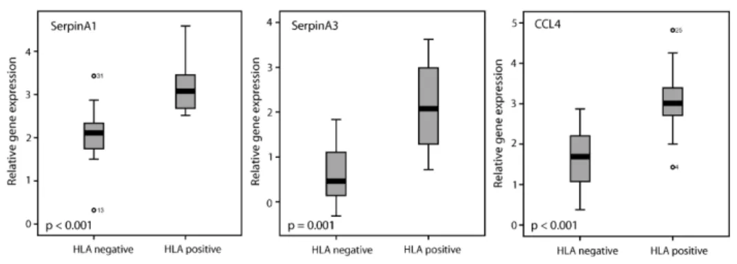
included 1032 genes, p = 0.0008) as well as the subprocess ‘immune response’ (925 genes, p = 0.0008) showed significant differences (data not shown). The most prominent subprocesses within the immune response cluster were the “inflammatory response” (256 genes, p = 0.0003, data not shown) and “acute phase response” (33 genes, p = 0.0006, Figure 2). Differences in the amount of inflammatory infiltrate did not influence the differences observed in immune (sub)processes between HLA negative and HLA positive tumors. A correction for the amount of inflammatory infiltrate in the Global test even improved the P-values (data not shown). In the inflammatory response cluster, all SSDGs (11 genes) were upregulated in HLA positive tumors (Table 3). Five of these represented chemokines, such asCCL3 and CCL4. The highest of the SSDGs was SerpinA3, which showed an 8.2 fold change in gene expression (Table 3). In the acute phase response (Figure 2),SerpinA1 and SerpinA3 were both found to be SSDGs.
qR qR qR qR
qRTTTTT-PCR verification of micr-PCR verification of micr-PCR verification of micr-PCR verification of micr-PCR verification of microarray dataoarray dataoarray dataoarray dataoarray data
Because gene expression differences between the HLA negative and HLA positive group were the most prominent in the inflammatory and acute phase responses, we chose gene products from these processes to validate the microarray results using
qRT-PCR. The expression of SerpinA1, SerpinA3 and CCL4 was significantly