R E V I E W
Open Access
STAT3, stem cells, cancer stem cells and p63
Michaela Galoczova
*, Philip Coates and Borivoj Vojtesek
* Correspondence:
Regional Centre for Applied Molecular Oncology, Masaryk Memorial Cancer Institute, Zluty kopec 7, 656 53 Brno, Czech Republic
Abstract
Signal Transducer and Activator of Transcription 3 (STAT3) is a transcription factor
with many important functions in the biology of normal and transformed cells. Its
regulation is highly complex as it is involved in signaling pathways in many different
cell types and under a wide variety of conditions. Besides other functions, STAT3 is
an important regulator of normal stem cells and cancer stem cells. p63 which is a
member of the p53 protein family is also involved in these functions and is both
physically and functionally connected with STAT3. This review summarizes STAT3
function and regulation, its role in stem cell and cancer stem cell properties and
highlights recent reports about its relationship to p63.
Keywords:
STAT3, Stem cells, Cancer stem cells, p63
This article was specially invited by the editors and represents work by leading
researchers.
Background
Our team is working on expression and functional properties of p63, which is a
mem-ber of the p53 protein family with diverse roles in carcinogenesis that include
tumor-suppressing and oncogenic effects [
1
–
4
]. Several recent studies link p63 with STAT3
that is one of the seven members of the Signal Transducer and Activator of
Transcrip-tion (STAT) family of transcripTranscrip-tion factors [
5
]. STAT3 and p63 are important
regula-tors of cell proliferation and survival, and have major roles in the maintenance of stem
cells and their differentiation, and are involved in carcinogenesis of many cell types.
STAT3 is known to act through its ability to regulate both oncogenes and tumor
sup-pressor genes, as well as influencing tumor microenvironments [
6
–
9
]. It exerts a
pleth-ora of different and sometimes contrasting functions in normal and transformed cells.
This multifaceted function can be partly explained by its involvement in signaling
pathways in many different types of cells and conditions [
10
]. p63 is most commonly
linked with epithelial malignancies, particularly squamous cancers [
9
,
11
]. Like STAT3,
p63 acts to transcriptionally regulate a wide variety of genes in cancer that are involved
in proliferation, survival and differentiation, and also has major roles in cell adhesion
and motility [
3
,
4
]. This review will provide basic information about STAT3 and its
regulation and will focus on its role(s) in stem cells and cancer stem cells. We will also
briefly discuss its relationship with p63 which is also involved in many pathways
connected with self-renewal and differentiation properties of stem cells and cancer
stem cells [
8
,
12
,
13
].
Structure of STAT3
All STATs share similar functional domains, including an N-terminal domain, a
coiled coil domain which enables protein-protein interactions, a central DNA-binding
domain, a linker domain that affects DNA-binding stability and a classic SRC homology
2 (SH2) domain. STAT3 has two important phosphorylation sites
–
a tyrosine residue
at amino acid position 705 (Tyr705) within the SH2 domain and a serine
phosphoryl-ation site at position 727 (Ser727) within the C-terminal transactivphosphoryl-ation domain, which
is absent in the alternatively spliced STAT3
β
variant [
14
].
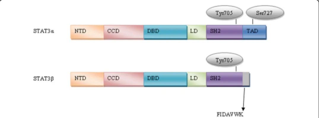
The full-length isoform (isoform 1), STAT3
α
, which is the most commonly expressed
form, encodes a protein of predicted mass 88 kDa [
15
,
16
]. The truncated STAT3
β
iso-form (isoiso-form 3) (83 kDa) is produced by alternative splicing of a 3' splice acceptor site
in exon 23 of the
STAT3
gene. STAT3
α
is 770 amino acids in length and STAT3
β
is
identical in sequence with the exception of 55 amino acids at the C-terminal tail that
are replaced with a unique seven amino acid sequence (Fig.
1
) [
15
–
17
]. STAT3
β
was
initially thought to be a negative regulator of STAT3
α
target genes because it lacks the
transactivation domain [
15
]. However, it was demonstrated
in vivo
that STAT3
β
is not
a dominant negative factor and seems to be involved in lipopolysaccharide-mediated
in-duction of the interleukin-10 promoter [
18
]. Another two isoforms have been
de-scribed, produced by limited proteolysis during granulocytic differentiation; a 72 kDa
C-terminal-truncated form known as STAT3
γ
, and a 64 kDa truncated isoform known
as STAT3
δ
[
19
–
21
]. Another isoform (isoform 2) was identified with a deleted amino
acid at position 701 (Del-Ser701) by global phosphoproteomic approaches [
22
,
23
]. The
validity and function of these latter variants remains to be determined.
Regulation of STAT3
STAT3 activity is regulated by multiple activators and negative regulators, reflecting its
varied functions in a wide range of cell types. The main mechanism of activation is
phosphorylation of Tyr705 by upstream kinases, although residue Ser727 can also be
phosphorylated. Moreover, STAT3 may be transcriptionally active in its
unphosphory-lated form and its activity is reguunphosphory-lated also by other posttranslational modifications
such as acetylation, methylation or ubiquitination. Negative regulation of STAT3 is
provided by protein phosphatases and specific protein inhibitors
–
Suppressors of
Cytokine Signaling (SOCS) and Protein Inhibitors of Activated STAT (PIAS).
Further-more, its expression is regulated by several miRNAs.
Activation of STAT3
STAT3 is mainly activated by phosphorylation of the conserved Tyr705 residue, which leads
to dimerization by reciprocal phosphotyrosine-SH2 interactions of two monomers [
24
].
Ac-tivated STAT3 dimers translocate to the nucleus through interactions with importins and
bind to the GAS (Interferon-
γ
-Activated Sequence) motif within target gene promoters to
activate transcription [
25
–
27
]. Most STATs including STAT3 bind to GAS motifs with a
consensus TTCN
2-4GAA [
28
]. The STAT3 consensus binding site is illustrated in Fig.
2
[
29
]. Besides STAT3 homodimers, STAT1/STAT3 heterodimers have been reported, with
transcriptional potential that differs from STAT1 or STAT3 homodimers [
30
].
STAT3 Tyr705 phosphorylation is primarily mediated by Janus Kinases (JAKs)
associ-ated with cytokine stimulassoci-ated receptors [
31
]. The most well-known activator is interleukin
6 (IL-6). However, other members of the IL-6 family are also able to activate STAT3,
in-cluding IL-10 [
32
], IL-11 [
33
], Ciliary Neurotrophic Factor (CNTF) [
34
], Leukemia
Inhibi-tory Factor (LIF) [
35
] and Oncostatin [
36
]. Phosphorylation of Tyr705 is also rapidly
increased by receptor tyrosine kinases including Epidermal Growth Factor Receptor
(EGFR) [
37
], Vascular Endothelial Growth Factor Receptor (VEGFR) [
38
], Platelet-derived
Growth Factor Receptor (PDGFR) [
39
] and Insulin-like Growth Factor 1 Receptor (IGFR)
[
39
,
40
] as well as by non-receptor tyrosine kinases like Src-family kinases (Src, Hck, Lyn,
Fyn, Fgr) [
41
], Bcr-Abl [
42
] and Bone Marrow X-linked non-receptor tyrosine kinase
(BMX) [
43
]. Recent studies also identified Toll-like receptors as Tyr705 activators
[
44
,
45
]. Moreover, Tyr705 can be indirectly activated by G-protein coupled receptors
such as Sphingosine-1-phosphate Receptor 1 (S1PR1) [
46
], BV8 [
47
] or angiotensin II
[
48
]. Engagement of cadherins was also shown to activate STAT3 through up-regulation
of IL-6 family cytokines [
49
].
In addition, STAT3 is phosphorylated at serine 727 (Ser727) by members of the
Mitogen-activated Protein Kinases (MAPK) like p38MAPK [
50
] or Extracellular Signal
Regulated Kinases (ERK) [
51
], by c-Jun N-terminal Kinase families (JNK) [
52
] and by
Protein Kinase C (PKC) [
53
]. The Mammalian Target of Rapamycin (mTOR) may also
phosphorylate STAT3 at Ser727 [
54
]. It was generally believed that phosphorylation of
Tyr705 is necessary for STAT3 activation, whereas Ser727 phosphorylation is required
for its maximum activity, presumably by recruiting transcriptional co-factors [
55
,
56
].
However, Ser727 phosphorylation can also reduce p-Tyr705 [
57
] and recent studies have
suggested that STAT3 can be activated through Ser727 phosphorylation in the absence of
Tyr705 phosphorylation. For example, a correlation was found between Ser727
phosphor-ylation in the absence of Tyr705 phosphorphosphor-ylation and survival of neuronal stem cells [
58
].
Moreover, constitutive activation of Ser727 is essential for the survival of primary human
in vitro
differentiated macrophages [
59
] and drives prostate carcinogenesis independently
of Tyr705 phosphorylation [
60
]. Above that, STAT3 is constitutively phosphorylated at
Ser727 while not at Tyr705 in chronic lymphocytic leukemia [
61
,
62
]. In addition, STAT3
can localize in mitochondria, where is serine phosphorylated and regulates mitochondrial
functions independently from its transcriptional activity [
63
,
64
].
Unphosphorylated STAT3 (U-STAT3) may also activate gene transcription. STAT3
nuclear import is independent of tyrosine phosphorylation and is mediated by
importin-
α
3 [
65
], Ran and importin-beta1 [
66
]. U-STAT3 dimerization is influenced by
disulfide bonds between cysteines [
67
] and dimers bind to the same GAS DNA-binding
site as phosphorylated STAT3 but also bind AT-rich DNA structures to influence
chro-matin organization [
68
]. Moreover, U-STAT3 core protein (lacking the N-terminal
do-main) binds to target
ds
DNA [
69
]. Several genes (
Cdc2
,
Cyclin B, Mras
,
E2f-1, Rantes)
do not respond directly to phosphorylated STAT3 but are activated in the late phases
of IL-6 driven responses when there is an accumulation of U-STAT3 [
70
]. Some
STAT3-responsive genes have kappa B elements, and these genes are activated by a
transcription factor complex formed when U-STAT3 binds to unphosphorylated
Nuclear Factor Kappa B (NF-
κ
B) [
71
].
STAT3 dimerization is also positively regulated by reversible acetylation of residue
Lys685 by its co-activator p300/CREB-binding protein [
72
–
74
] and by tri-methylation
of Lys180 [
75
].
Negative regulation of STAT3
Dephosphorylation of STAT3 by protein phosphatases plays a major role in regulating
STAT3. Multiple protein tyrosine phosphatases such as MEG2 [
76
], CD45 [
77
],
Src-homology Region 2 Domain-containing Phosphatase 1/2 (SHP1/2) or T-cell Protein
Tyrosine Phosphatases (TC-PTP) [
78
] have been shown to dephosphorylate Tyr705 of
STAT3. STAT3 Ser727 can be dephosphorylated by Protein Phosphatase 1 (PP1) [
79
]
or Dual Specificity Protein Phosphatase 2 (DUSP2) [
80
].
STAT3 regulation by miRNAs
Several studies have indicated that miRNAs are critical regulators of STAT3. A number
of miRNAs have been identified that affect STAT3 signaling in various types of cancers
(reviewed in [
86
]). Moreover, some miRNAs have been shown to play a role in
regulat-ing stem cells and cancer stem cell properties.
miR-124
was found to directly target
STAT3
mRNA to regulate cardiomyocyte differentiation of bone marrow-derived
mes-enchymal stem cells [
87
].
miR-1181
inhibits stem cell-like phenotypes and suppresses
STAT3 in human pancreatic cancer [
88
], whilst
miR-7
indirectly inhibits STAT3 and
thereby decreases the number of breast cancer stem cells [
89
].
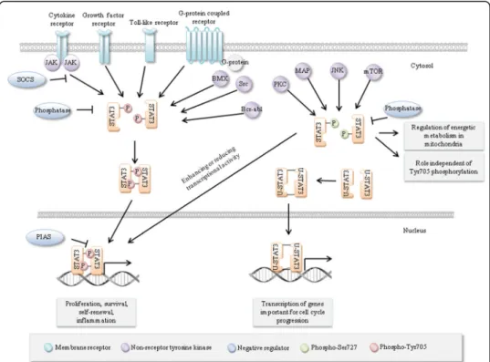
To summarize the data above, it is evident that STAT3 expression and activation are
regulated by multiple signals and they play a role in many signaling pathways. This
en-ables STAT3 to be a flexible and adaptable regulator of cell function in different types
of cells under different conditions and regulate gene expression directly or indirectly
through other transcription factors [
90
]. An overview of STAT3 regulation is shown in
Fig.
3
. This review will now focus on STAT3
’
s involvement in signaling pathways
regu-lating stem cells and cancer stem cells.
STAT3 and stem cells
Stem cells are defined by their ability to self-renew and to generate progenitor cells that
can subsequently divide and differentiate into the different types of cells of a particular
tissue [
91
]. There are two main types of naturally occurring stem cells: embryonic stem
cells, which are isolated from the inner cell mass of blastocysts, and adult stem cells,
which are found in various tissues. Embryonic stem cells are considered to be
toti-potent and can give rise to all cell types in the organism whereas adult stem cells are
pluripotent, showing lineage restriction according to the particular tissue in which they
reside. The other type of stem cells are induced pluripotent stem cells (iPSCs) that can
be generated from adult cells [
92
].
Mouse stem cells
Mouse embryonic stem cells (mESCs)
LIF and its close relatives (IL-6) family are known to maintain pluripotency of mESCs
[
93
]. The effect of LIF on JAK/STAT3 signaling is mediated through the LIF receptor
(LIFR) which consists of two subunits: gp130, which is common for all types of
cyto-kine receptors, and LIF receptor beta (LIFR
β
). LIF induces heterodimerization and
phosphorylation of these subunits [
94
] leading to rapid phosphorylation of intracellular
non-receptor JAKs that phosphorylate STAT3 on tyrosine residues [
95
]. This
phosphor-ylation is responsible for STAT3 activation necessary for self-renewal of mESCs. It
seems that activated STAT3 is sufficient to maintain the undifferentiated state of
mESCs. However, there is a threshold, and mESC lines with lower expression of
consti-tutively active STAT3 are not able to fully inhibit differentiation [
96
]. STAT3 activation
in mESCs leads to expression of genes that are known regulators of pluripotency, such
as
Myc
or
Bcl3
[
97
,
98
] and activated STAT3 is known to cooperate with Nanog,
an-other key component of pluripotency [
99
]. Thus, STAT3 plays a major role in
regulat-ing mESCs fate. The regulation is possibly enabled by existence of the two different
phosphorylation sites Tyr705 and Ser727, whose modification could switch between
self-renewal and differentiation [
100
].
Mouse adult stem cells
Gu et al. [
101
] demonstrated that suppression of STAT3 promotes neurogenesis and
in-hibits astrogliogenesis in neural stem cells. Moreover, Kamakura et al. [
102
] showed a
crosstalk between differentiation pathways where STAT3 is activated in the presence of
active Notch as well as the Notch effectors Hes1 and Hes5. However, STAT3 plays a role
not only during postnatal development of the mouse neocortex, the interaction between
the JAK/STAT3 and Notch ligand Delta-like1-Notch signaling pathways plays an essential
role in maintaining neural precursors during early neocortical development [
103
]. STAT3
also seems to be an important regulator of hematopoietic regeneration [
104
], self-renewal
of adult muscle satellite cells during injury-induced muscle regeneration [
105
],
regener-ation of airway ciliated cells from basal stem cells [
106
], adipogenesis [
107
], differentiation
of multiciliated [
106
] and hair cells [
108
] in mouse models. Furthermore, STAT3 is
re-quired to maintain the full differentiation potential of mouse mammary stem cells and the
proliferative potential of mammary luminal progenitors [
109
].
Human stem cells
Human embryonic stem cells (hESCs)
nuclear transportation through gp130/LIFR
β
, but is unable to maintain the pluripotent
state of hESCs [
110
]. However, Yang et al. [
111
] showed that increased STAT3
activa-tion is sufficient to convert epiblast-derived stem cells to naive pluripotency and Chen
et al. [
112
] observed that temporarily increasing STAT3 activity is sufficient to
repro-gram hESCs to naive-like pluripotent cells. Hence, STAT3 activation is a limiting factor
in somatic cell reprogramming.
Human adult stem cells
There are numerous studies that have investigated the role of STAT3 in human
mesenchy-mal stem cells (hMSCs). hMSCs are a heterogeneous population of non-hematopoietic
pre-cursor cells predominantly found in the bone marrow. Matsui et al. [
113
] demonstrated
that hMSCs protect against obstruction-induced renal fibrosis by decreasing STAT3
activa-tion and STAT3-dependent Matrix Metallopeptidase 9 producactiva-tion. Interesting to note is
the interaction of hMSCs with cancer cells via cytokine networks. Hsu et al. [
114
] showed
that the IL-6/JAK/STAT3 pathway could be activated by hMSCs when they are co-cultured
with lung cancer cells to enhance lung cancer initiation. Rattigan et al. [
115
] illustrated that
IL-6, which is produced and secreted at high levels by breast cancer cells in response to
hypoxia, regulates hMSC migration towards cancer cells. IL-6 binds to its receptor on
hMSCs, leading to STAT3 activation to promote hMSCs migration and survival.
STAT3, cancer and cancer stem cells
STAT3 is constitutively activated in many types of human solid tumors and hematological
malignancies [
116
]. For example, STAT3 activation occurs in more than 40% of breast
cancers, most often in the triple negative subtype that lack estrogen receptor,
progester-one receptor and Human Epidermal Growth Factor 2 (HER2) amplification [
5
,
117
].
How-ever, the
STAT3
gene is very rarely altered in human malignancies by copy number
variation, point mutation or methylation and rarely by gene expression according to the
Catalogue of Somatic Mutations in Cancer (COSMIC) database (Fig.
4
) [
118
].
In the absence of genetic alterations, constitutive activation occurs through upstream
factors such as growth factor or cytokine production acting through paracrine or
auto-crine pathways; amplification or activating mutations in related receptors; mutations in
kinase signaling cascade pathway genes; and/or the loss of negative regulators of STAT3
activity. Constitutive activation of STAT3 is predictive of poor prognosis in many types of
cancer [
119
–
121
] and although STAT3 is only rarely altered by gene expression changes,
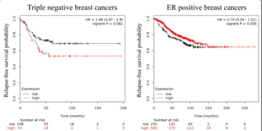
mRNA levels show a similar trend. Using publicly available array profiling data, we could
show here that in triple negative breast cancer, which is a representative cancer with
con-stitutive STAT3 activation, higher mRNA levels show a trend for worse relapse-free
sur-vival (RFS). Conversely, in ER
+breast cancer where the activation of STAT3 is low, high
STAT3
mRNA levels indicate a better RFS probability (Fig.
5
) [
122
].
Patient outcomes in relation to
STAT3
mRNA expression are described by logrank
p
value. Affymetrix ID for
STAT3
was 225289_at. Relapse-free survival (RFS) was
ana-lysed in breast cancer, progression-free survival (PFS) in ovarian cancer and
first-progression (FP) in lung and gastric cancers. JetSet best probe set and auto select best
cut-off were used for analysis from Kaplan-Meier plot [
123
]. Upward arrows indicate that
Fig. 4STAT3 mutational status. The table showing the distribution of mutations across the primary tissue types that are curated by COSMIC database was used as template. Only cancer types with all mutation types included were chosen for this picture [118]
higher
STAT3
levels correlate with improved survival; downward arrows indicate that
lower
STAT3
levels associate with improved survival.
As mentioned at the beginning, STAT3 is also important in tumor biology for its
ability to promote cancer through regulating cancer stem cell activities. It is widely
ac-cepted that tumors contain a sub-population of cells that share properties with normal
tissue stem cells, called cancer stem cells or cancer stem-like cells (CSCs) [
124
–
128
].
However, the CSC theory remains controversial because of the variety of differences
between CSCs and normal stem cells. As stem cells, CSCs have the ability to
self-renew, however while normal stem cells are able to differentiate into multiple distinct
cell types, most CSCs differentiate into only a single cell type, the cells which form the
bulk of the tumor. However, an evidence for multilineage differentiation potential of
CSCs was reported in colon carcinomas and leukemia. Another difference is that while
the phenotypes of normal stem cells seem to be fixed, the phenotypes of CSCs vary
from one tumor to another tumor of the same molecular/pathological type, most likely
because they are affected by the abnormalities resulting from the process of neoplastic
transformation [
127
]. CSC identification and understanding of their biology could
have critical clinical relevance, because CSCs are uniquely able to reform the tumor
and exhibit enhanced resistance to cancer treatments [
124
–
128
]. Notably, as
men-tioned above, STAT3 is often constitutively activated in triple negative breast cancers
and these cancers display a profile of cell surface markers that is similar to that of
breast CSCs [
117
,
126
,
129
]. Below, we summarize the evidence for the role of STAT3
in CSCs properties in the common human malignancies.
STAT3 was reported to have an essential role in maintaining the expression of genes
that are important for stem cell phenotype and are used as markers of CSCs. Many
pu-tative CSC markers have been identified, the most commonly used are expression of
transmembrane glycoproteins CD24, CD34, CD38, CD44, CD90 and CD133, together
with Aldehyde Dehydrogenase (ALDH), the ability to form spheroids in suspension
in
vitro
and the ability to exclude cell permeable dyes such as Hoechst33342 for side
population assessment [
127
]. The STAT3 pathway is preferentially active in
subpopula-tions of cells enriched for CSC markers and its inhibition decreases cell viability and
tumorsphere formation. On the other hand, several proteins that stimulate cell growth
and proliferation reduce its activity [
117
,
130
,
131
]. Importantly, STAT3 can form a
complex with internalized CD44 and acetyltransferase p300, inducing STAT3
acetyl-ation at Lys685, dimer formacetyl-ation and translocacetyl-ation to the nucleus where it binds to the
promoters of genes including cell cycle regulators
cyclin D1
[
47
] or
Myc
and
Twist1
Table 1
Patient outcomes in relation to
STAT3
mRNA expression in breast, ovarian, lung and
gastric cancers
Breast cancer All types (n=1764) Triple negative (n=161) ER positive (n=762)
RFS probability 0.0530↓ 0.0620↓ 0.0560↑
Ovarian cancer All types (n=614) Serous (n=483) Endometrioid (n=44)
PFS probability 0.0160↓ 0.0150↓ 0.0720↑
Lung cancer All types (n=596) Adenocarcinoma (n=443) Squamous cell (n=141)
FP probability 0.020↓ 0.0058↓ 0.0002↑
Gastric cancer All types (n=522)
[
132
]. Furthermore, STAT3 was found to physically interact with CD44 and NF-
κ
B and
activate the catalytic subunit of telomerase to prolong proliferative potential [
133
].
Moreover, activated STAT3 can increase CD133 expression through functional
cooper-ation with NF-
κ
B and Hypoxia Inducible Factor 1 Alpha (HIF-1
α
) [
134
].
Nowadays, epithelial-mesenchymal transition (EMT) and tumor microenvironments
are highly discussed topics in the context of CSCs. There is increasing evidence
point-ing to plasticity between CSCs and their more differentiated derivatives. It is considered
that whereas CSCs can differentiate into non-CSCs, the reverse process is also possible
via EMT, which is a transdifferentiation program required for tissue morphogenesis
during embryonic development [
135
,
136
]. Over and above that, several studies
re-ported direct links between EMT and gain of CSC properties [
137
,
138
]. EMT and CSC
formation is a dynamic process triggered by multiple shared signaling pathways, such
as Transforming Growth Factor
β
(TGF-
β
), Wnt/
β
-catenin, Hedgehog, Notch, NF-
κ
B
and others [
139
,
140
].
It was reported that hepatocellular carcinoma could arise from IL-6/STAT3 driven
transformed stem cells with inactivated TGF-
β
signaling and that human hepatocellular
cancer cells expressing STAT3 and the putative stem cell markers
Octamer
-binding
Transcription Factor 4 (OCT4) and Nanog lost pro-differentiation proteins TGF-
β
-receptor type II and Embryonic Liver Fodrin [
141
]. In addition to this role in
hepatocel-lular cancer, STAT3 activation plays a role in EMT induction in different types of
tumors. STAT3 can be activated by IL-6 dependent or independent mechanisms such
as a non-canonical Frizzled 2 pathway [
142
] or TGF-
β
/LIF [
143
]. Moreover, induction
of EMT after STAT3 activation and expansion of the CSC population were observed in
relation to resistance to cisplatin or trastuzumab [
144
,
145
]. The mechanisms of
trastu-zumab resistance, which is a HER2-targeting antibody used to treat HER2
+breast
can-cer, are well-documented. Inactivation of Phosphatase and Tensin Homolog (PTEN)
leads to increased resistance to this drug and it seems that STAT3 is a negative
regula-tor of PTEN among trastuzumab-resistant cells [
146
,
147
]. An IL-6 loop was found,
where IL-6 activates the AKT, STAT3 and NF-
κ
B pathways while suppressing PTEN
expression [
146
]. Recently, it was found that trastuzumab resistance in this type of
cancer is promoted through activation of a STAT3/HIF-1
α
/Hes1 axis via
down-regulation of PTEN [
147
].
STAT3 activation was observed also in HER2 negative breast cancer, where activation
of STAT3 also correlates with CSC properties. The STAT3 pathway is positively
regu-lated by mTOR signaling in this context, whereas PTEN serves as a negative regulator
of both STAT3 and mTOR [
148
]. Moreover, PTEN appears to function as a crucial
in-hibitor of glioblastoma stem cells through mediating cooperative perturbation of AKT
and STAT3 signals [
149
].
Furthermore, STAT3 is a critical transcription factor in angiogenesis; it participates in
expression and protein stability of HIF-1
α
and regulates or is itself regulated by VEGF.
This involvement was also shown to play a role in maintaining the self-renewal
proper-ties of CSCs [
38
]. Interestingly, VEGF-mediated angiogenesis was reported to link
EMT-induced cancer stemness to tumor initiation [
150
].
immune cells [
7
,
47
,
134
]. Among tumor-associated immune cells, tumor-associated
mac-rophages were found to promote CSC-like phenotypes through Milk Fat Globule-EGF
Factor 8 (MGF-E8)/STAT3 and Sonic Hedgehog pathways, or through EGFR/STAT3/
Sox2 [
151
,
152
]. Moreover, it was also shown that mast cells modulate proliferation,
migration and stemness through down-regulation of GSK3
β
and inhibition of STAT3
activation [
153
].
STAT3 can be activated and thereby contribute to CSCs properties by the BMX [
43
]
and Ras homolog family member C [
154
] and it can be activated also epigenetically by
the histone-lysine N-methyltransferase Enhancer of Zeste Homolog 2 (EZH2) [
75
].
Taking the above observations together, the roles STAT3 in promoting and
maintain-ing CSC properties are highly complex. STAT3 directly interacts with transmembrane
glycoproteins which are expressed by normal stem cells and are widely used as markers
to identify and isolate CSCs. STAT3 is involved in pathways that are connected with
EMT, which is one of the major proposed mechanisms of generating CSCs. Moreover,
it plays a critical role in angiogenesis and participates in regulating the tumor
micro-environment that provides signals for differentiation or proliferation especially through
its involvement in inflammatory NF-
κ
B pathway. In addition, feedback activation of
STAT3 may play a prominent role in mediating drug resistance to a broad spectrum of
targeted cancer therapies and chemotherapies [
155
]. Although it seems to be an ideal
target for anti-cancer therapy, effective approaches to inhibit STAT3 are still missing.
This lack is caused presumably because of the complexity of STAT3
’
s biology in normal
as well as cancer cells and also because it lacks enzymatic activity, making it a
challen-ging target [
86
]. Inhibitors of STAT3 which are currently tested are reviewed in [
155
],
although no inhibitor that directly targets STAT3 has yet been approved by the US
Food and Drug Administration for clinical use. However, several tyrosine kinase
inhibi-tors are in the clinic such as sorafenib and sunitinib that can inhibit STAT3 signaling
indirectly, leading to tumor cell cycle arrest and apoptosis [
156
,
157
].
As was mentioned in the beginning, there is increasing evidence that STAT3
activa-tion and p63 expression are connected; hence their relaactiva-tionship will be briefly discussed
in the following part of this review.
The connections between STAT3 and p63
p63 is a member of the p53 family of transcription factors that consists of p53, p63 and
p73 proteins. All family members have important functions in tumorigenesis and
mor-phogenesis and share the same domain organization including an N-terminal
transcrip-tion activatranscrip-tion domain (TAD), a DNA binding domain (DBD) and a C-terminal
oligomerization domain (OD). They act as tetramers and due to their partial homology
in the oligomerization domain they may form heterotetramers. They also have highly
homologous DNA binding domains, indicating that they are able to bind to the other
family members target genes. The
TP63
gene is localized on chromosome 3 and gives
rise to multiple isoforms due to differential promoter selection (full-length TA and
N-terminal truncated
Δ
Np63) and alternative splicing of the 3´end of the mRNA (
α
,
β
,
γ
,
have transactivation activity due to the presence of an alternative TAD [
158
,
159
].
Among C-terminal isoforms, p63
α
isoforms have a sterile alpha motif (SAM) that is
known to be involved in proteprotein interactions and they have a transcription
in-hibitory domain (TID), which inhibits its transcriptional activity [
160
,
161
].
p63 is well known for its role in epidermal development. The importance of this
pro-tein during this process was established using p63 null mice. Those mice showed a
complete lack of stratified squamous epithelia and their derivatives or they had
strati-fied but disrupted epidermis depending on the used model. They also had absent or
truncated limbs and craniofacial abnormalities [
162
,
163
]. The predominant isoform in
epithelial tissues is
Δ
Np63
α
which is highly expressed in the basal cells of stratified and
glandular epithelia, including epidermis, and its levels decrease with cellular
differenti-ation. Oppositely, TAp63 positive cells are located suprabasally in stratified epithelia,
indicating a switch between isoforms during differentiation [
1
,
2
,
9
].
Δ
Np63 protein
ex-pression is restricted to other basal cells including those in breast, prostate, bladder and
colorectum. Thus, it is widely used as a marker for this type of cells [
1
,
11
,
12
,
164
].
Inter-estingly, activated STAT3 plays a role in promoting the regeneration of airway ciliated
cells from basal stem cells [
106
] and is also involved in malignant transformation of
fore-gut basal progenitor cells [
165
]. Furthermore, it was shown using ChIP-Seq analysis that
p63 co-operates with STAT3 in human keratinocytes [
166
].
p63
’
s role in tumorigenesis is complex, also because it seems that TAp63 and
Δ
Np63
isoforms play opposite roles in this process. Like
STAT3
,
TP63
is rarely mutated in
hu-man cancer, but p63 activity is often increased. One mechanism for increased activity
of p63 is gene amplification, and many tumors with amplification show increased p63
expression [
9
].
Δ
Np63 is supposed to behave as oncoprotein and is up-regulated in
squamous cell carcinomas [
11
,
167
] and triple negative basal-like breast tumors [
4
]
amongst other tumor types. It also plays roles in a variety of pathways that are
im-plicated in CSC properties, reviewed in [
8
]. In addition,
Δ
Np63 increases the
ex-pression of Wnt receptor Frizzled 7 thereby enhancing Wnt signaling which leads
to promotion of normal mammary stem cell activity and tumor initiating activity
in the basal-like subtype of breast cancer [
168
]. Further, Memmi et al. [
13
] showed
a positive modulation of Hedgehog signaling pathway by
Δ
Np63 to maintain
self-renewal potential of mammary CSCs. On the other hand, TAp63 shares the
abil-ities of the
“
guardian of the genome
”
p53 to induce cell cycle arrest and apoptosis
and TAp63 may thus act as tumor suppressor. However, there are also reports that
it could behave as oncogene. For example, TAp63 is the predominant isoform
expressed in hematological malignancies, and it was shown that TAp63
over-expression leads to increased tumor progression of head and neck squamous cell
carcinoma. It is also expressed in colon carcinoma [
1
,
169
].
STAT3 is often given in connection with
Δ
Np63. Both
Δ
Np63 [
3
,
4
,
13
,
129
] and
STAT3 [
117
,
170
,
171
] were suggested as CSC markers and are associated with
triple negative breast tumors that show more CSC markers than non-triple negative.
STAT3 is also frequently constitutively activated in squamous cell carcinomas [
121
,
172
], where
Δ
Np63 is often over-expressed. Furthermore, they were both reported as
master regulators of mammary cancer stem cell maintenance [
13
,
173
]. Additionally,
the dual-regulatory effect
Δ
Np63 on its own promoter is dependent on STAT3
activa-tion and it was confirmed that STAT3 binds to the
Δ
Np63 promoter [
159
,
174
] and
regulates proliferation and differentiation of rabbit limbal epithelial cells via a
Δ
Np63
mechanisms [
175
]. There is a possible mechanism involving EGFR signaling pathway
that could regulate STAT3 and
Δ
Np63 activation and expression. It was observed that
Δ
Np63 expression is regulated by EGFR/STAT3 axis and this is crucial for proliferation
of CSCs [
176
]. Oppositely, we found that
Δ
Np63 activates EGFR signaling in triple
negative breast cancer [
4
]. Moreover, STAT3 is activated by mTOR and thereby p63
ex-pression is induced and in turn activates Notch signaling through stimulation of
Jag1
gene expression and impedes murine and human cell differentiation [
177
]. Besides the
above-mentioned evidence that STAT3 and
Δ
Np63 are closely linked,
Δ
Np63 is also
in-volved in inflammatory NF-
κ
B pathway [
178
,
179
], angiogenesis through VEGF [
180
],
and EMT. There are numerous papers about
Δ
Np63 involvement in signaling pathways
connected with EMT (reviewed in [
181
]). Some papers claim that
Δ
Np63 promotes
EMT and reduces the opposite process of mesenchymal-epithelial transition (MET),
whereas others provide evidence for an opposite role of
Δ
Np63. To help with
explaining contradictory reports could serve a paper [
182
] where they describe that
p63 can trigger the Notch signaling pathway in neighboring cells to potentially
promote EMT. Interestingly, Su et al. [
183
] recently showed that TAp63 is crucial
for the transition of mammary cancer cells to acquire characteristic of
tumor-initiating cells.
Conclusion
STAT3 signaling is a major regulatory pathway of mouse embryonic stem cell fate and
also a limiting factor in human somatic cell reprogramming and plays important roles
in the maintenance and proliferation of adult stem cells. STAT3 activation occurs
dur-ing many aspects of carcinogenesis, includdur-ing involvement in regulatdur-ing CSC
proper-ties. Together with
Δ
Np63 it was suggested as a marker of CSCs, a major regulator of
mammary CSCs maintenance and both are mostly associated with triple negative
tu-mors. They were found to directly interact and they are both involved in several
com-mon pathways regulating CSC properties, however their relationship is still not well
established and remains to be determined.
Abbreviations
ALDH:Aldehyde Dehydrogenase; BMX: Bone Marrow X-linked non-receptor tyrosine kinase; CCD: Coiled Coil Domain; CNTF: Ciliary Neurotrophic Factor; COSMIC: Catalogue of Somatic Mutations in Cancer; CSCs: Cancer Stem Cells; DBD: DNA-binding Domain; DUSP2: Dual Specificity Protein Phosphatase 2; EGFR: Epidermal Growth Factor Receptor; EMT: Epithelial-mesenchymal Transition; ER: Estrogen Receptor; ERK: Extracellular Signal Regulated Kinase;
EZH2: Enhancer of Zeste Homolog 2; FP: First Progression; GAS: Interferon-Gamma-Activated Sequence; HER2: Human Epidermal Growth Factor Receptor 2; hESCs: Human Embryonic Stem Cells; HIF-1α: Hypoxia Inducible Factor Alpha; hMSCS: Human Mesenchymal Stem Cells; IGFR: Insulin-like Growth Factor 1 Receptor; IL: Interleukin; iPSCs: Induced Pluripotent Stem Cells; JAK: Janus Kinase; JNK: c-Jun N-terminal Kinase; LD: Linker Domain; LIF: Leukemia Inhibitory Factor; LIFR: Leukemia Inhibitory Factor Receptor; LIFRβ: Leukemia Inhibitory Factor Receptor Beta; MAPK: Mitogen Activated Protein Kinase; mESCs: Mouse Embryonic Stem Cells; MET: Mesenchymal-epithelial Transition; MGF-E8: Milk Fat Globule-EGF Factor 8; mTOR: Mammalian Target of Rapamycin; NF-κB: Nuclear Factor Kappa B; NTD: NH2-terminal Domain; OCT4: Octamer-binding Transcription Factor 4; OD: Oligomerization Domain; PDGFR: Platelet-derived Growth Factor Receptor; PFS: Progression-free Survival; PIAS: Protein Inhibitors of Activated STAT; PKC: Protein Kinase C; PP1: Protein Phosphatase 1; PTEN: Phosphatase and Tensin Homolog; RFS: Relapse-free Survival; S1PR1: Sphingosine-1-phosphate Receptor 1; SAM: Sterile Alpha Motif; SH2: Src-homology 2; SHP1/2: Src-homology Region 2 Domain-containing Phosphatase 1 and Src-homology Region 2 Domain-Domain-containing Phosphatase 2; SOCS: Suppressors of Cytokine Signaling; STAT: Signal Transducer and Activator of Transcription; TAD: Transcription Activation Domain; TC-PTP: T-cell Protein Tyrosine Phosphatase; TGF-β: Transforming Growth Factor Beta; TID: Transcription Inhibitory Domain; U-STAT3: Unphosphorylated STAT3; VEGFR: Vascular Endothelial Growth Factor Receptor
Acknowledgements
Not applicable.
Funding
The work was supported by the project MEYS–NPS I–LO1413, by the Czech Science Foundation (P206/12/G151) and by MH CZ–DRO (MMCI, 00209805). MG is Brno Ph.D. Talent Scholarship holder - Funded by the Brno City Municipality.
Availability of data and materials
Not applicable.
Authors’contributions
MG reviewed the literature and wrote the manuscript. PC and BV revised the manuscript. All authors read and approved the final manuscript.
Ethics approval and consent to participate
Not applicable.
Consent for publication
Not applicable.
Competing interests
The authors declare that they have no competing interests.
Publisher
’
s Note
Springer Nature remains neutral with regard to jurisdictional claims in published maps and institutional affiliations.
Received: 15 December 2017 Accepted: 7 March 2018
References
2. Nekulova M, Holcakova J, Nenutil R, Stratmann R, Bouchalova P, Müller P, et al. Characterization of specific p63 and p63-N-terminal isoform antibodies and their application for immunohistochemistry. Virchows Arch. 2013;463:415–25.
3. Orzol P, Nekulova M, Holcakova J, Muller P, Votesek B, Coates PJ.ΔNp63 regulates cell proliferation, differentiation, adhesion, and migration in the BL2 subtype of basal-like breast cancer. Tumour Biol. 2016;37:10133–40. 4. Holcakova J, Nekulova M, Orzol P, Nenutil R, Podhorec J, Svoboda M, et al.ΔNp63 activates EGFR signaling to
induce loss of adhesion in triple-negative basal-like breast cancer cells. Breast Cancer Res Treat. 2017;163:475–84. 5. Banerjee K, Resat H. Constitutive activation of STAT3 in breast cancer cells: A review. Int J Cancer. 2016;138:2570–8. 6. Carpenter RL, Lo HW. STAT3 Target Genes Relevant to Human Cancers. Cancers (Basel). 2014;6:897–925. 7. Yu H, Pardoll D, Jove R. STATs in cancer inflammation and immunity: a leading role for STAT3. Nat Rev Cancer.
2009;9:798–809.
8. Nekulova M, Holcakova J, Coates P, Vojtesek B. The role of p63 in cancer, stem cells and cancer stem cells. Cell Mol Biol Lett. 2011;16:296–327.
9. Orzol P, Holcakova J, Nekulova M, Nenutil R, Vojtesek B, Coates PJ. The diverse oncogenic and tumour suppressor roles of p63 and p73 in cancer: a review by cancer site. Histol Histopathol. 2015;30:503–21.
10. Avalle L, Camporeale A, Camperi A, Poli V. STAT3 in cancer: A double edged sword. Cytokine. 2017;98:42–50. 11. Bishop JA, Teruya-Feldstein J, Westra WH, Pelosi G, Travis WD, Rekhtman N. p40 (ΔNp63) is superior to p63 for the
diagnosis of pulmonary squamous cell carcinoma. Mod Pathol. 2012;25:405–15.
12. Pellegrini G, Dellambra E, Golisano O, Martinelli E, Fantozzi I, Bondanza S, et al. p63 identifies keratinocyte stem cells. Proc Natl Acad Sci U S A. 2001;98:3156–61.
13. Memmi EM, Sanarico AG, Giacobbe A, Peschiaroli A, Frezza V, Cicalese A, et al. p63 Sustains self-renewal of mammary cancer stem cells through regulation of Sonic Hedgehog signaling. Proc Natl Acad Sci U S A. 2015;112:3499–504. 14. You L, Wang Z, Li H, Shou J, Jing Z, Xie J, et al. The role of STAT3 in autophagy. Autophagy. 2015;11:729–39. 15. Caldenhoven E, van Dijk TB, Solari R, Armstrong J, Raaijmakers JA, Lammers JW, et al. STAT3beta, a splice variant
of transcription factor STAT3, is a dominant negative regulator of transcription. J Biol Chem. 1996;271:13221–7. 16. The UniProt Consortium. UniProt: the universal protein knowledgebase. Nucleic Acids Res. 2017;45:D158–D69. 17. Schaefer TS, Sanders LK, Nathans D. Cooperative transcriptional activity of Jun and Stat3 beta, a short form of
Stat3. Proc Natl Acad Sci U S A. 1995;92:9097–101.
18. Maritano D, Sugrue ML, Tininini S, Dewilde S, Strobl B, Fu X, et al. The STAT3 isoforms alpha and beta have unique and specific functions. Nat Immunol. 2004;5:401–9.
19. Kato T, Sakamoto E, Kutsuna H, Kimura-Eto A, Hato F, Kitagawa S. Proteolytic conversion of STAT3alpha to STAT3gamma in human neutrophils: role of granule-derived serine proteases. J Biol Chem. 2004;279:31076–80. 20. Hevehan DL, Miller WM, Papoutsakis ET. Differential expression and phosphorylation of distinct STAT3 proteins
during granulocytic differentiation. Blood. 2002;99:1627–37.
21. Nishiki S, Hato F, Kamata N, Sakamoto E, Hasegawa T, Kimura-Eto A, et al. Selective activation of STAT3 in human monocytes stimulated by G-CSF: implication in inhibition of LPS-induced TNF-alpha production. Am J Physiol Cell Physiol. 2004;286:C1302–11.
22. Rush J, Moritz A, Lee KA, Guo A, Goss VL, Spek EJ, et al. Immunoaffinity profiling of tyrosine phosphorylation in cancer cells. Nat Biotechnol. 2005;23:94–101.
23. Bian Y, Song C, Cheng K, Dong M, Wang F, Huang J, et al. An enzyme assisted RP-RPLC approach for in-depth analysis of human liver phosphoproteome. J Proteomics. 2014;96:253–62.
24. Levy DE, Darnell JE. Stats: transcriptional control and biological impact. Nat Rev Mol Cell Biol. 2002;3:651–62. 25. Fagard R, Metelev V, Souissi I, Baran-Marszak F. STAT3 inhibitors for cancer therapy: Have all roads been explored?
JAKSTAT. 2013;2:e22882.
26. Zhong Z, Wen Z, Darnell JE. Stat3: a STAT family member activated by tyrosine phosphorylation in response to epidermal growth factor and interleukin-6. Science. 1994;264:95–8.
27. Darnell JE, Kerr IM, Stark GR. Jak-STAT pathways and transcriptional activation in response to IFNs and other extracellular signaling proteins. Science. 1994;264:1415–21.
28. Decker T, Kovarik P, Meinke A. GAS elements: a few nucleotides with a major impact on cytokine-induced gene expression. J Interferon Cytokine Res. 1997;17:121–34.
29. Khan A, Fornes O, Stigliani A, Gheorghe M, Castro-Mondragon JA, van der Lee R, et al. JASPAR 2018: update of the open-access database of transcription factor binding profiles and its web framework. Nucleic Acids Res. 2017; 30. Khatib H, Huang W, Mikheil D, Schutzkus V, Monson RL. Effects of signal transducer and activator of transcription (STAT) genes STAT1 and STAT3 genotypic combinations on fertilization and embryonic survival rates in Holstein cattle. J Dairy Sci. 2009;92:6186–91.
31. Decker T, Kovarik P. Transcription factor activity of STAT proteins: structural requirements and regulation by phosphorylation and interacting proteins. Cell Mol Life Sci. 1999;55:1535–46.
32. Gupta M, Han JJ, Stenson M, Maurer M, Wellik L, Hu G, et al. Elevated serum IL-10 levels in diffuse large B-cell lymphoma: a mechanism of aberrant JAK2 activation. Blood. 2012;119:2844–53.
33. Ernst M, Najdovska M, Grail D, Lundgren-May T, Buchert M, Tye H, et al. STAT3 and STAT1 mediate IL-11-dependent and inflammation-associated gastric tumorigenesis in gp130 receptor mutant mice. J Clin Invest. 2008;118:1727–38. 34. Peterson WM, Wang Q, Tzekova R, Wiegand SJ. Ciliary neurotrophic factor and stress stimuli activate the Jak-STAT
pathway in retinal neurons and glia. J Neurosci. 2000;20:4081–90.
35. Kunisada K, Hirota H, Fujio Y, Matsui H, Tani Y, Yamauchi-Takihara K, et al. Activation of JAK-STAT and MAP kinases by leukemia inhibitory factor through gp130 in cardiac myocytes. Circulation. 1996;94:2626–32.
36. Yanagisawa M, Nakashima K, Taga T. STAT3-mediated astrocyte differentiation from mouse fetal neuroepithelial cells by mouse oncostatin M. Neurosci Lett. 1999;269:169–72.
37. Wang Y, van Boxel-Dezaire AH, Cheon H, Yang J, Stark GR. STAT3 activation in response to IL-6 is prolonged by the binding of IL-6 receptor to EGF receptor. Proc Natl Acad Sci U S A. 2013;110:16975–80.
39. Vignais ML, Sadowski HB, Watling D, Rogers NC, Gilman M. Platelet-derived growth factor induces phosphorylation of multiple JAK family kinases and STAT proteins. Mol Cell Biol. 1996;16:1759–69.
40. Zong CS, Chan J, Levy DE, Horvath C, Sadowski HB, Wang LH. Mechanism of STAT3 activation by insulin-like growth factor I receptor. J Biol Chem. 2000;275:15099–105.
41. Schreiner SJ, Schiavone AP, Smithgall TE. Activation of STAT3 by the Src family kinase Hck requires a functional SH3 domain. J Biol Chem. 2002;277:45680–7.
42. Coppo P, Flamant S, De Mas V, Jarrier P, Guillier M, Bonnet ML, et al. BCR-ABL activates STAT3 via JAK and MEK pathways in human cells. Br J Haematol. 2006;134:171–9.
43. Guryanova OA, Wu Q, Cheng L, Lathia JD, Huang Z, Yang J, et al. Nonreceptor tyrosine kinase BMX maintains self-renewal and tumorigenic potential of glioblastoma stem cells by activating STAT3. Cancer Cell. 2011;19:498–511. 44. Kortylewski M, Kujawski M, Herrmann A, Yang C, Wang L, Liu Y, et al. Toll-like receptor 9 activation of signal
transducer and activator of transcription 3 constrains its agonist-based immunotherapy. Cancer Res. 2009;69:2497–505. 45. Eyking A, Ey B, Rünzi M, Roig AI, Reis H, Schmid KW, et al. Toll-like receptor 4 variant D299G induces features of
neoplastic progression in Caco-2 intestinal cells and is associated with advanced human colon cancer. Gastroenterology. 2011;141:2154–65.
46. Lee H, Deng J, Kujawski M, Yang C, Liu Y, Herrmann A, et al. STAT3-induced S1PR1 expression is crucial for persistent STAT3 activation in tumors. Nat Med. 2010;16:1421–8.
47. Lee JL, Wang MJ, Chen JY. Acetylation and activation of STAT3 mediated by nuclear translocation of CD44. J Cell Biol. 2009;185:949–57.
48. Zheng L, Jia X, Zhang C, Wang D, Cao Z, Wang J, et al. Angiotensin II in atrial structural remodeling: the role of Ang II/JAK/STAT3 signaling pathway. Am J Transl Res. 2015;7:1021–31.
49. Geletu M, Arulanandam R, Chevalier S, Saez B, Larue L, Feracci H, et al. Classical cadherins control survival through the gp130/Stat3 axis. Biochim Biophys Acta. 2013;1833:1947–59.
50. Turkson J, Bowman T, Adnane J, Zhang Y, Djeu JY, Sekharam M, et al. Requirement for Ras/Rac1-mediated p38 and c-Jun N-terminal kinase signaling in Stat3 transcriptional activity induced by the Src oncoprotein. Mol Cell Biol. 1999;19:7519–28.
51. Chung J, Uchida E, Grammer TC, Blenis J. STAT3 serine phosphorylation by ERK-dependent and -independent pathways negatively modulates its tyrosine phosphorylation. Mol Cell Biol. 1997;17:6508–16.
52. Lim CP, Cao X. Serine phosphorylation and negative regulation of Stat3 by JNK. J Biol Chem. 1999;274:31055–61. 53. Jain N, Zhang T, Kee WH, Li W, Cao X. Protein kinase C delta associates with and phosphorylates Stat3 in an
interleukin-6-dependent manner. J Biol Chem. 1999;274:24392–400.
54. Kim JH, Yoon MS, Chen J. Signal transducer and activator of transcription 3 (STAT3) mediates amino acid inhibition of insulin signaling through serine 727 phosphorylation. J Biol Chem. 2009;284:35425–32.
55. Wen Z, Zhong Z, Darnell JE. Maximal activation of transcription by Stat1 and Stat3 requires both tyrosine and serine phosphorylation. Cell. 1995;82:241–50.
56. Zhang X, Blenis J, Li HC, Schindler C, Chen-Kiang S. Requirement of serine phosphorylation for formation of STAT-promoter complexes. Science. 1995;267:1990–4.
57. Shi X, Zhang H, Paddon H, Lee G, Cao X, Pelech S. Phosphorylation of STAT3 serine-727 by cyclin-dependent kinase 1 is critical for nocodazole-induced mitotic arrest. Biochemistry. 2006;45:5857–67.
58. Androutsellis-Theotokis A, Leker RR, Soldner F, Hoeppner DJ, Ravin R, Poser SW, et al. Notch signalling regulates stem cell numbers in vitro and in vivo. Nature. 2006;442:823–6.
59. Liu H, Ma Y, Cole SM, Zander C, Chen KH, Karras J, et al. Serine phosphorylation of STAT3 is essential for Mcl-1 expression and macrophage survival. Blood. 2003;102:344–52.
60. Qin HR, Kim HJ, Kim JY, Hurt EM, Klarmann GJ, Kawasaki BT, et al. Activation of signal transducer and activator of transcription 3 through a phosphomimetic serine 727 promotes prostate tumorigenesis independent of tyrosine 705 phosphorylation. Cancer Res. 2008;68:7736–41.
61. Frank DA, Mahajan S, Ritz J. B lymphocytes from patients with chronic lymphocytic leukemia contain signal transducer and activator of transcription (STAT) 1 and STAT3 constitutively phosphorylated on serine residues. J Clin Invest. 1997;100:3140–8.
62. Hazan-Halevy I, Harris D, Liu Z, Liu J, Li P, Chen X, et al. STAT3 is constitutively phosphorylated on serine 727 residues, binds DNA, and activates transcription in CLL cells. Blood. 2010;115:2852–63.
63. Wegrzyn J, Potla R, Chwae YJ, Sepuri NB, Zhang Q, Koeck T, et al. Function of mitochondrial Stat3 in cellular respiration. Science. 2009;323:793–7.
64. Gough DJ, Corlett A, Schlessinger K, Wegrzyn J, Larner AC, Levy DE. Mitochondrial STAT3 supports Ras-dependent oncogenic transformation. Science. 2009;324:1713–6.
65. Liu L, McBride KM, Reich NC. STAT3 nuclear import is independent of tyrosine phosphorylation and mediated by importin-alpha3. Proc Natl Acad Sci U S A. 2005;102:8150–5.
66. Cimica V, Chen HC, Iyer JK, Reich NC. Dynamics of the STAT3 transcription factor: nuclear import dependent on Ran and importin-β1. PLoS One. 2011;6:e20188.
67. Butturini E, Gotte G, Dell'Orco D, Chiavegato G, Marino V, Canetti D, et al. Intermolecular disulfide bond influences unphosphorylated STAT3 dimerization and function. Biochem J. 2016;473(19):3205.
68. Timofeeva OA, Chasovskikh S, Lonskaya I, Tarasova NI, Khavrutskii L, Tarasov SG, et al. Mechanisms of unphosphorylated STAT3 transcription factor binding to DNA. J Biol Chem. 2012;287:14192–200.
69. Nkansah E, Shah R, Collie GW, Parkinson GN, Palmer J, Rahman KM, et al. Observation of unphosphorylated STAT3 core protein binding to target dsDNA by PEMSA and X-ray crystallography. FEBS Lett. 2013;587:833–9.
70. Yang J, Chatterjee-Kishore M, Staugaitis SM, Nguyen H, Schlessinger K, Levy DE, et al. Novel roles of unphosphorylated STAT3 in oncogenesis and transcriptional regulation. Cancer Res. 2005;65:939–47.
71. Yang J, Liao X, Agarwal MK, Barnes L, Auron PE, Stark GR. Unphosphorylated STAT3 accumulates in response to IL-6 and activates transcription by binding to NFkappaB. Genes Dev. 2007;21:1396–408.
73. Wang R, Cherukuri P, Luo J. Activation of Stat3 sequence-specific DNA binding and transcription by p300/CREB-binding protein-mediated acetylation. J Biol Chem. 2005;280:11528–34.
74. Dasgupta M, Unal H, Willard B, Yang J, Karnik SS, Stark GR. Critical role for lysine 685 in gene expression mediated by transcription factor unphosphorylated STAT3. J Biol Chem. 2014;289:30763–71.
75. Kim E, Kim M, Woo DH, Shin Y, Shin J, Chang N, et al. Phosphorylation of EZH2 activates STAT3 signaling via STAT3 methylation and promotes tumorigenicity of glioblastoma stem-like cells. Cancer Cell. 2013;23:839–52. 76. Su F, Ren F, Rong Y, Wang Y, Geng Y, Feng M, et al. Protein tyrosine phosphatase Meg2 dephosphorylates signal
transducer and activator of transcription 3 and suppresses tumor growth in breast cancer. Breast Cancer Res. 2012;14:R38. 77. Kumar V, Cheng P, Condamine T, Mony S, Languino LR, McCaffrey JC, et al. CD45 Phosphatase Inhibits STAT3 Transcription
Factor Activity in Myeloid Cells and Promotes Tumor-Associated Macrophage Differentiation. Immunity. 2016;44:303–15. 78. Kim DJ, Tremblay ML, Digiovanni J. Protein tyrosine phosphatases, TC-PTP, SHP1, and SHP2, cooperate in rapid
dephosphorylation of Stat3 in keratinocytes following UVB irradiation. PLoS One. 2010;5:e10290.
79. Zgheib C, Zouein FA, Chidiac R, Kurdi M, Booz GW. Calyculin A reveals serine/threonine phosphatase protein phosphatase 1 as a regulatory nodal point in canonical signal transducer and activator of transcription 3 signaling of human microvascular endothelial cells. J Interferon Cytokine Res. 2012;32:87–94.
80. Lu D, Liu L, Ji X, Gao Y, Chen X, Liu Y, et al. The phosphatase DUSP2 controls the activity of the transcription activator STAT3 and regulates TH17 differentiation. Nat Immunol. 2015;16:1263–73.
81. Yuan J, Zhang F, Niu R. Multiple regulation pathways and pivotal biological functions of STAT3 in cancer. Sci Rep. 2015;5:17663.
82. Nicholson SE, De Souza D, Fabri LJ, Corbin J, Willson TA, Zhang JG, et al. Suppressor of cytokine signaling-3 preferentially binds to the SHP-2-binding site on the shared cytokine receptor subunit gp130. Proc Natl Acad Sci U S A. 2000;97:6493–8. 83. Lee JH, Kim C, Sethi G, Ahn KS. Brassinin inhibits STAT3 signaling pathway through modulation of PIAS-3 and SOCS-3
expression and sensitizes human lung cancer xenograft in nude mice to paclitaxel. Oncotarget. 2015;6:6386–405. 84. Chung CD, Liao J, Liu B, Rao X, Jay P, Berta P, et al. Specific inhibition of Stat3 signal transduction by PIAS3.
Science. 1997;278:1803–5.
85. Yang J, Huang J, Dasgupta M, Sears N, Miyagi M, Wang B, et al. Reversible methylation of promoter-bound STAT3 by histone-modifying enzymes. Proc Natl Acad Sci U S A. 2010;107:21499–504.
86. Yu H, Lee H, Herrmann A, Buettner R, Jove R. Revisiting STAT3 signalling in cancer: new and unexpected biological functions. Nat Rev Cancer. 2014;14:736–46.
87. Cai B, Li J, Wang J, Luo X, Ai J, Liu Y, et al. microRNA-124 regulates cardiomyocyte differentiation of bone marrow-derived mesenchymal stem cells via targeting STAT3 signaling. Stem Cells. 2012;30:1746–55.
88. Jiang J, Li Z, Yu C, Chen M, Tian S, Sun C. MiR-1181 inhibits stem cell-like phenotypes and suppresses SOX2 and STAT3 in human pancreatic cancer. Cancer Lett. 2015;356:962–70.
89. Zhang H, Cai K, Wang J, Wang X, Cheng K, Shi F, et al. MiR-7, inhibited indirectly by lincRNA HOTAIR, directly inhibits SETDB1 and reverses the EMT of breast cancer stem cells by downregulating the STAT3 pathway. Stem Cells. 2014;32:2858–68. 90. Pawlus MR, Wang L, Hu CJ. STAT3 and HIF1αcooperatively activate HIF1 target genes in MDA-MB-231 and RCC4
cells. Oncogene. 2014;33:1670–9.
91. Reya T, Morrison SJ, Clarke MF, Weissman IL. Stem cells, cancer, and cancer stem cells. Nature. 2001;414:105–11. 92. Takahashi K, Yamanaka S. Induction of pluripotent stem cells from mouse embryonic and adult fibroblast cultures
by defined factors. Cell. 2006;126:663–76.
93. Humphrey RK, Beattie GM, Lopez AD, Bucay N, King CC, Firpo MT, et al. Maintenance of pluripotency in human embryonic stem cells is STAT3 independent. Stem Cells. 2004;22:522–30.
94. Davis S, Aldrich TH, Stahl N, Pan L, Taga T, Kishimoto T, et al. LIFR beta and gp130 as heterodimerizing signal transducers of the tripartite CNTF receptor. Science. 1993;260:1805–8.
95. Matsuda T, Nakamura T, Nakao K, Arai T, Katsuki M, Heike T, et al. STAT3 activation is sufficient to maintain an undifferentiated state of mouse embryonic stem cells. EMBO J. 1999;18:4261–9.
96. Raz R, Lee CK, Cannizzaro LA, d'Eustachio P, Levy DE. Essential role of STAT3 for embryonic stem cell pluripotency. Proc Natl Acad Sci U S A. 1999;96:2846–51.
97. Cartwright P, McLean C, Sheppard A, Rivett D, Jones K, Dalton S. LIF/STAT3 controls ES cell self-renewal and pluripotency by a Myc-dependent mechanism. Development. 2005;132:885–96.
98. Chen CY, Lee DS, Yan YT, Shen CN, Hwang SM, Lee ST, et al. Bcl3 Bridges LIF-STAT3 to Oct4 Signaling in the Maintenance of Naïve Pluripotency. Stem Cells. 2015;33:3468–80.
99. Torres J, Watt FM. Nanog maintains pluripotency of mouse embryonic stem cells by inhibiting NFkappaB and cooperating with Stat3. Nat Cell Biol. 2008;10:194–201.
100. Huang G, Yan H, Ye S, Tong C, Ying QL. STAT3 phosphorylation at tyrosine 705 and serine 727 differentially regulates mouse ESC fates. Stem Cells. 2014;32:1149–60.
101. Gu F, Hata R, Ma YJ, Tanaka J, Mitsuda N, Kumon Y, et al. Suppression of Stat3 promotes neurogenesis in cultured neural stem cells. J Neurosci Res. 2005;81:163–71.
102. Kamakura S, Oishi K, Yoshimatsu T, Nakafuku M, Masuyama N, Gotoh Y. Hes binding to STAT3 mediates crosstalk between Notch and JAK-STAT signalling. Nat Cell Biol. 2004;6:547–54.
103. Yoshimatsu T, Kawaguchi D, Oishi K, Takeda K, Akira S, Masuyama N, et al. Non-cell-autonomous action of STAT3 in maintenance of neural precursor cells in the mouse neocortex. Development. 2006;133:2553–63.
104. Chung YJ, Park BB, Kang YJ, Kim TM, Eaves CJ, Oh IH. Unique effects of Stat3 on the early phase of hematopoietic stem cell regeneration. Blood. 2006;108:1208–15.
105. Zhu H, Xiao F, Wang G, Wei X, Jiang L, Chen Y, et al. STAT3 Regulates Self-Renewal of Adult Muscle Satellite Cells during Injury-Induced Muscle Regeneration. Cell Rep. 2016;16:2102–15.
106. Tadokoro T, Wang Y, Barak LS, Bai Y, Randell SH, Hogan BL. IL-6/STAT3 promotes regeneration of airway ciliated cells from basal stem cells. Proc Natl Acad Sci U S A. 2014;111:E3641–9.
108. Chen Q, Quan Y, Wang N, Xie C, Ji Z, He H, et al. Inactivation of STAT3 Signaling Impairs Hair Cell Differentiation in the Developing Mouse Cochlea. Stem Cell Reports. 2017;9:231–46.
109. Staniszewska AD, Pensa S, Caffarel MM, Anderson LH, Poli V, Watson CJ. Stat3 is required to maintain the full differentiation potential of mammary stem cells and the proliferative potential of mammary luminal progenitors. PLoS One. 2012;7:e52608.
110. Dahéron L, Opitz SL, Zaehres H, Lensch MW, Lensch WM, Andrews PW, et al. LIF/STAT3 signaling fails to maintain self-renewal of human embryonic stem cells. Stem Cells. 2004;22:770–8.
111. Yang J, van Oosten AL, Theunissen TW, Guo G, Silva JC, Smith A. Stat3 activation is limiting for reprogramming to ground state pluripotency. Cell Stem Cell. 2010;7:319–28.
112. Chen H, Aksoy I, Gonnot F, Osteil P, Aubry M, Hamela C, et al. Reinforcement of STAT3 activity reprogrammes human embryonic stem cells to naive-like pluripotency. Nat Commun. 2015;6:7095.
113. Matsui F, Babitz SA, Rhee A, Hile KL, Zhang H, Meldrum KK. Mesenchymal stem cells protect against obstruction-induced renal fibrosis by decreasing STAT3 activation and STAT3-dependent MMP-9 production. Am J Physiol Renal Physiol. 2017;312:F25–32.
114. Hsu HS, Lin JH, Hsu TW, Su K, Wang CW, Yang KY, et al. Mesenchymal stem cells enhance lung cancer initiation through activation of IL-6/JAK2/STAT3 pathway. Lung Cancer. 2012;75:167–77.
115. Rattigan Y, Hsu JM, Mishra PJ, Glod J, Banerjee D. Interleukin 6 mediated recruitment of mesenchymal stem cells to the hypoxic tumor milieu. Exp Cell Res. 2010;316:3417–24.
116. Buettner R, Mora LB, Jove R, Activated STAT. signaling in human tumors provides novel molecular targets for therapeutic intervention. Clin Cancer Res. 2002;8:945–54.
117. Marotta LL, Almendro V, Marusyk A, Shipitsin M, Schemme J, Walker SR, et al. The JAK2/STAT3 signaling pathway is required for growth of CD44+CD24-stem cell-like breast cancer cells in human tumors. J Clin Invest. 2011;121:2723–35.
118. Forbes SA, Beare D, Boutselakis H, Bamford S, Bindal N, Tate J, et al. COSMIC: somatic cancer genetics at high-resolution. Nucleic Acids Res. 2017;45:D777–D83.
119. Xiong H, Du W, Wang JL, Wang YC, Tang JT, Hong J, et al. Constitutive activation of STAT3 is predictive of poor prognosis in human gastric cancer. J Mol Med (Berl). 2012;90:1037–46.
120. Mora LB, Buettner R, Seigne J, Diaz J, Ahmad N, Garcia R, et al. Constitutive activation of Stat3 in human prostate tumors and cell lines: direct inhibition of Stat3 signaling induces apoptosis of prostate cancer cells. Cancer Res. 2002;62:6659–66. 121. Masuda M, Suzui M, Yasumatu R, Nakashima T, Kuratomi Y, Azuma K, et al. Constitutive activation of signal
transducers and activators of transcription 3 correlates with cyclin D1 overexpression and may provide a novel prognostic marker in head and neck squamous cell carcinoma. Cancer Res. 2002;62:3351–5.
122. Györffy B, Lanczky A, Eklund AC, Denkert C, Budczies J, Li Q, et al. An online survival analysis tool to rapidly assess the effect of 22,277 genes on breast cancer prognosis using microarray data of 1,809 patients. Breast Cancer Res Treat. 2010;123:725–31.
123. Lánczky A, Nagy Á, Bottai G, Munkácsy G, Szabó A, Santarpia L, et al. miRpower: a web-tool to validate survival-associated miRNAs utilizing expression data from 2178 breast cancer patients. Breast Cancer Res Treat. 2016;160:439–46. 124. Rosen JM, Jordan CT. The increasing complexity of the cancer stem cell paradigm. Science. 2009;324:1670–3. 125. Korkaya H, Liu S, Wicha MS. Regulation of cancer stem cells by cytokine networks: attacking cancer's inflammatory
roots. Clin Cancer Res. 2011;17:6125–9.
126. Badve S, Nakshatri H. Breast-cancer stem cells-beyond semantics. Lancet Oncol. 2012;13:e43–8.
127. Pattabiraman DR, Weinberg RA. Tackling the cancer stem cells - what challenges do they pose? Nat Rev Drug Discov. 2014;13:497–512.
128. Nassar D, Blanpain C. Cancer Stem Cells: Basic Concepts and Therapeutic Implications. Annu Rev Pathol. 2016;11:47–76. 129. Foulkes WD, Smith IE, Reis-Filho JS. Triple-negative breast cancer. N Engl J Med. 2010;363:1938–48.
130. Lin L, Fuchs J, Li C, Olson V, Bekaii-Saab T, Lin J. STAT3 signaling pathway is necessary for cell survival and tumorsphere forming capacity in ALDH+/CD133+stem cell-like human colon cancer cells. Biochem Biophys Res Commun. 2011;416:246–51.
131. Schroeder A, Herrmann A, Cherryholmes G, Kowolik C, Buettner R, Pal S, et al. Loss of androgen receptor expression promotes a stem-like cell phenotype in prostate cancer through STAT3 signaling. Cancer Res. 2014;74:1227–37. 132. Su YJ, Lai HM, Chang YW, Chen GY, Lee JL. Direct reprogramming of stem cell properties in colon cancer cells by
CD44. EMBO J. 2011;30:3186–99.
133. Chung SS, Aroh C, Vadgama JV. Constitutive activation of STAT3 signaling regulates hTERT and promotes stem cell-like traits in human breast cancer cells. PLoS One. 2013;8:e83971.
134. Won C, Kim BH, Yi EH, Choi KJ, Kim EK, Jeong JM, et al. Signal transducer and activator of transcription 3-mediated CD133 up-regulation contributes to promotion of hepatocellular carcinoma. Hepatology. 2015;62:1160–73. 135. Gupta PB, Chaffer CL, Weinberg RA. Cancer stem cells: mirage or reality? Nat Med. 2009;15:1010–2.
136. Singh A, Settleman J. EMT, cancer stem cells and drug resistance: an emerging axis of evil in the war on cancer. Oncogene. 2010;29:4741–51.
137. Mani SA, Guo W, Liao MJ, Eaton EN, Ayyanan A, Zhou AY, et al. The epithelial-mesenchymal transition generates cells with properties of stem cells. Cell. 2008;133:704–15.
138. Morel AP, Lièvre M, Thomas C, Hinkal G, Ansieau S, Puisieux A. Generation of breast cancer stem cells through epithelial-mesenchymal transition. PLoS One. 2008;3:e2888.
139. Wang SS, Jiang J, Liang XH, Tang YL. Links between cancer stem cells and epithelial-mesenchymal transition. Onco Targets Ther. 2015;8:2973–80.
140. Vazquez-Santillan K, Melendez-Zajgla J, Jimenez-Hernandez L, Martínez-Ruiz G, Maldonado V. NF-κB signaling in cancer stem cells: a promising therapeutic target? Cell Oncol (Dordr). 2015;38:327–39.
141. Tang Y, Kitisin K, Jogunoori W, Li C, Deng CX, Mueller SC, et al. Progenitor/stem cells give rise to liver cancer due to aberrant TGF-beta and IL-6 signaling. Proc Natl Acad Sci U S A. 2008;105:2445–50.
142. Gujral TS, Chan M, Peshkin L, Sorger PK, Kirschner MW, MacBeath G. A noncanonical Frizzled2 pathway regulates epithelial-mesenchymal transition and metastasis. Cell. 2014;159:844–56.
![Fig. 2 STAT3 consensus binding site from JASPAR database [ 29 ]](https://thumb-us.123doks.com/thumbv2/123dok_us/7875719.2099566/3.892.175.721.842.1084/fig-stat-consensus-binding-site-from-jaspar-database.webp)