Defining the Active Fraction of Daptomycin
against Methicillin-Resistant
Staphylococcus
aureus
(MRSA) Using a Pharmacokinetic and
Pharmacodynamic Approach
Samira M. Garonzik1¤, Justin R. Lenhard1,2, Alan Forrest1,3, Patricia N. Holden1,2, Jϋrgen B. Bulitta1,4‡, Brian T. Tsuji1,2‡*
1Laboratory for Antimicrobial Pharmacodynamics, School of Pharmacy and Pharmaceutical Sciences Buffalo, Buffalo, NY, United States of America,2New York State Center of Excellence in Bioinformatics & Life Sciences, University at Buffalo, Buffalo, NY, United States of America,3Department of
Pharmacotherapy and Experimental Therapeutics, School of Pharmacy, University of North Carolina, Chapel Hill, NC, United States of America,4Center for Pharmacometrics and Systems Pharmacology, College of Pharmacy, University of Florida, Orlando, FL, United States of America
¤ Current address: Bristol Myers Squibb, Jersey City, NJ, United States of America
‡These authors are joint senior authors on this work. *[email protected]
Abstract
Our objective was to study the pharmacodynamics of daptomycin in the presence of varying concentrations of human serum (HS)in vitroto quantify the fraction of daptomycin that is
‘active’. Time kill experiments were performed with daptomycin (0 to 256 mg/L) against two MRSA strains at log-phase growth, in the presence of HS (0%, 10%, 30%, 50%, 70%) com-bined with Mueller-Hinton broth. Daptomycin2 mg/L achieved 99.9% kill within 8 h at all HS concentrations; early killing activity was slightly attenuated at higher HS concentrations. After 1 h, bacterial reduction of USA300 upon exposure to daptomycin 4 mg/L ranged from -3.1 to -0.5 log10CFU/mL in the presence of 0% to 70% HS, respectively. Bactericidal activ-ity was achieved against both strains at daptomycin4 mg/L for all fractions of HS expo-sure. A mechanism-based mathematical model (MBM) was developed to estimate the active daptomycin fraction at each %HS, comprising 3 bacterial subpopulations differing in daptomycin susceptibility. Time-kill data were fit with this MBM with excellent precision (r2>0.95). The active fraction of daptomycin was estimated to range from 34.6% to 25.2% at HS fractions of 10% to 70%, respectively. Despite the reported low unbound fraction of daptomycin, the impact of protein binding on the activity of daptomycin was modest. The active fraction approach can be utilized to designin vitroexperiments and to optimize thera-peutic regimens of daptomycin in humans.
a11111
OPEN ACCESS
Citation:Garonzik SM, Lenhard JR, Forrest A, Holden PN, Bulitta JB, Tsuji BT (2016) Defining the Active Fraction of Daptomycin against Methicillin-ResistantStaphylococcus aureus(MRSA) Using a Pharmacokinetic and Pharmacodynamic Approach. PLoS ONE 11(6): e0156131. doi:10.1371/journal. pone.0156131
Editor:Suzan HM Rooijakkers, University Medical Center Utrecht, NETHERLANDS
Received:February 21, 2016
Accepted:April 11, 2016
Published:June 10, 2016
Copyright:© 2016 Garonzik et al. This is an open access article distributed under the terms of the Creative Commons Attribution License, which permits unrestricted use, distribution, and reproduction in any medium, provided the original author and source are credited.
Data Availability Statement:All of the relevant data (colony counts used for the investigation) are included either within the paper (as time-killing figures) or the Supporting Information files (an excel sheet listing the numeric values).
Introduction
Significant controversy revolves around the impact of protein binding on antimicrobial activ-ity, particularly for drugs which display a similar affinity to their bacterial target and serum proteins [1,2]. Since the 1970’s, antibiotic activity has been generally understood to be depen-dent on free drug concentrations, or inversely related to protein binding [3]. However, there have also been reports of discrepancies between the observed increases in MIC in the presence of proteins (such as albumin), and the difference between total and unbound antibiotic concen-trations determined by ultrafiltration [2,3].
Daptomycin is a cyclic lipopeptide that binds to Gram-positive cell membranes causing rapid depolarization and loss of membrane potential, ultimately leading to rapid bacterial cell death. The novel mechanism of action utilized by daptomycin makes the agent an attractive option for the treatment of infections caused by Gram-positive species such asStaphylococcus
aureus, particularly strains with decreased susceptibility to vancomycin [4]. Due to
daptomy-cin’s high protein binding of 90–93%, the free fraction of drug found in human plasma is much lower than total plasma concentrations (www.cubicin.com). Severalin vitroandin vivostudies for daptomycin have reported disproportionately higher pharmacodynamic (PD) activity againstS.aureusthan is expected based on free-drug concentrations alone [2,5,6]. Qualitative
in vitroreports have also suggested that the presence of protein only impacts the rate of killing,
and not the overall activity expressed by daptomycin [2,6–9].
Based on the discrepancies between daptomycin’s activity and the free fraction of drug, we propose that an‘active fraction’of daptomycin may exist that differs from the‘free-drug frac-tion’calculated from reported protein binding values. The active fraction may provide a more appropriate characterization of the extent of protein binding based on bactericidal activity, and guide the translation ofin vitroactivity studies into optimal dosage regimens in humans. Therefore, our objectives were to evaluate the impact of protein binding on the bactericidal activity and time course of killing by daptomycin, and also to develop a mechanism based mathematical model capable of estimating the active fraction of daptomycin in various serum concentrations.
Materials and Methods
Ethics Statement
Bacterial isolates used for in vitro investigations were laboratory strains collected from aS.
aureusdatabase. None of theS.aureusstrains utilized in the present study were clinical isolates
obtained from patients. Tryptic soy agar with 5% sheep blood (TSA II) was commercially pur-chased from Fisher Scientific, with specifications available athttps://www.fishersci.com/shop/ products/bd-bbl-rodac-trypticasesoyagar-5-sheep-blood-tsa-ii-prepared-plated-media/ l97759#sthash.Wzi1zwJR.dpuf. Serum was commercially purchased from Sigma Chemical Co. (Lot#098K8712), with specifications available atSigma-Aldrich.com. Serum was not collected from human patients and no animals were used in the investigation.
Bacterial Strains
Two bacterial isolates were utilized including i) a vancomycin intermediateS.aureusstrain (VISA), Mu50 (NRS 4, HIP5836, daptomycin MIC = 1.0 mg/L), and ii) an MRSA strain, USA300 (NRS 384, FRP3757, daptomycin MIC = 0.5); both strains were obtained from the Network on Antimicrobial Resistance inS.aureus.
the National Institute of Allergy and Infectious Diseases of the National Institutes of Health under award number R01AI111990 (description at:http:// www.rdatlas.com/portal/portal.cfm?page= grants&applicationid=8826683). The content is solely the responsibility of the authors and does not necessarily represent the official views of the National Institutes of Health. NIH did not have any role in the study design, data collection and analysis, decision to publish, or preparation of the manuscript.
Antibiotic, susceptibility testing and medium
Daptomycin analytical grade powder was obtained from Cubist Pharmaceuticals (Lexington, MA). Stock solutions were freshly prepared immediately prior to each experiment. MIC values were determined by broth microdilution in Mueller–Hinton broth (Difco Laboratories, Detroit, Mich.) supplemented with calcium and magnesium (12.5 mg/L;“supplemented Muel-ler Hinton broth”: SMHB) according to standard methods from the Clinical Laboratory Stan-dards Institute. Human Serum (Lot #098K8712, Sigma Chemical Co., St. Louis, MO) was added to the SMHB to achieve desired concentrations of serum / SMHB. The human serum was heat inactivated at 56°C for 1 hour to inactivate compliment-mediated cell lysis. The final concentrations of human serum / SMHB (v/v) were 0%, 10%, 30%, 50% and 70%. Due to the dependence of daptomycin on calcium for its mechanism of action, the final calcium con-centration in each batch of human serum and SMHB was titrated to physiologic conditions (1.1–1.3 mmol/L). Tryptic Soy Agar plates with 5% sheep blood (TSA II) were used to quantify bacterial colony counts (Difco, Detroit, MI).
Time
–
kill experiments
Static time kill experiments were performed as previously described [5] in log phase growth against a starting inoculum of 106CFU/mL. In brief, fresh bacterial colonies were grown over-night then added to SMHB broth to provide a bacterial suspension of approximately 108CFU/ mL; the bacterial suspension was further diluted with SMHB broth to achieve a starting inocu-lum of approximately 106CFU/mL. Time kill experiments were conducted for daptomycin against both isolates at concentrations of 0, 0.125 (only for USA300), 0.25, 0.5, 1, 2, 4, 8, 16, 32, 64, and 128 mg/L, in the presence of 0%, 10%, 30%, 50%, and 70% heat inactivated human serum (v/v ratios combined with SHMB) over a period of 24 h. Samples were withdrawn at 0, 1, 2, 4, 8, and 24 hours after dosing, and viable counts were determined by plating 50μL sample aliquots diluted with saline onto TSA plates with 5% sheep’s blood using an automated spiral dispenser (WASP; Don Whitley Scientific Limited, West Yorkshire, England). Plates were incubated at 35°C for 24 h and viable colonies were quantified using a laser bacteria colony counter (ProtoCOL; Version 2.05.02, Synbiosis, Cambridge, UK). The limit of detection was 102CFU/mL (equivalent to 5 colonies for an agar plate from an undiluted sample) [10]. Bacte-ricidal activity (99.9% kill) was associated with3.0 log10CFU/mL decrease in bacterial density
compared to the initial inoculum.
Pharmacodynamic analyses
To accommodate all available data generated for each concentration tested, and to avoid conclusions based on bacterial counts at a single time point, an integrated pharmacokinetic / pharmacodynamic area measure (log ratio area) was applied to all data as previously described [5]. For each regimen tested, the area under the log10CFU/mL versus time curve from 0 to
24 h (AUCFU0–24) was calculated via the linear trapezoidal rule for both growth control
(AUCFUgrowth control) and drug containing regimens (AUCFUdrug). The AUCFU0–24was
nor-malized by the AUCFU0–24of the growth control, and the logarithm of this ratio was used to
quantify the drug effect (E) as shown inEq 1A.
E¼Log10 AUCFUdrug AUCFUgrowth control
" #
ðEquation 1aÞ
according to a Hill type model (Eq 1B).
E¼E0
Emax ½CH
½EC50Hþ ½CH ðEquation 1bÞ
The dependent variable (E) is the effect described by the log ratio area, E0is the measured
effect at zero drug concentration, Emaxis the maximal drug effect, C is the drug concentration
expressed as a multiple of the MIC, EC50is the C:MIC for which there is 50% maximal effect,
and H is the Hill or sigmoidicity constant.
Mechanism Based Mathematical Pharmacodynamic Model
Candidate models were simultaneously fit to all viable count profiles for daptomycin against USA300, which was selected for additional analyses as the most common pulsed-field gel elec-trophoresis type in the USA. Estimation was performed in NONMEM VI (level 1.2; NON-MEM Project Group, Icon Development Solutions, Ellicott City, MD) with the first order conditional estimation method. All residual variability was modeled as described previously [11] using additive and Poisson error models. Model discrimination was based on the curve fits, NONMEM's objective function, and the plausibility of parameter estimates.
Model for Effect of Human Serum Albumin
The effect of human serum albumin was estimated by calculating the‘active fraction’of dapto-mycin for each level of supplemented human serum. The active fraction is analogous to the free fraction of drug but is a better representation of daptomycin’s activity in the presence of plasma proteins. The factiveparameter multiplied by the total concentration of daptomycin
yielded the‘effective’daptomycin concentration at each concentration of human serum albu-min studied (Eq 2). The effect of human serum albumin on bacterial growth rates was also explored.
Ef f ective DaptomycinðDAPEFÞ ¼factiveð% Human SerumÞ Total DaptomycinðEquation 2Þ
Model for Bacterial Life Cycle
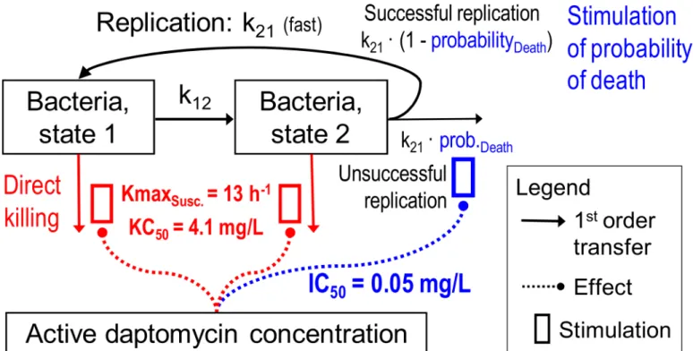
A simplified life cycle model (Fig 1) was used for bacterial replication in which the bacterial life cycle, for each subpopulation, was described by two states. State1 (S1) represents the vegetative
state and State2 (S2) the replicating state, as described previously [12]. The k12represents the
first order transition rate constant from S1to S2, while MTT12determines the mean generation
time (MTT12= 1/k12). Since the doubling was assumed to be very fast, the rate constant for the
transition from S2to S1(k21) was fixed to 50 h-1[12]. k21is the sum of two processes, namely
successful replication and bacterial death. The observed lag time in bacterial growth kinetics was described usingEq 3[13]. In this equation, klagrepresents the first order dissipation of the
lag time whileβis a sigmoidicity constant.
Lag¼1eðklagtÞb ðEquation 3Þ
Models with one, two, or three subpopulations of different daptomycin susceptibilities for each strain were considered, as previously described [11,14–17]. The following equations rep-resent a model with three bacterial subpopulations, susceptible (S1and S2), intermediate (I1
during growth is modeled usingEq 5, where CFUmrepresents the maximum achievable CFU/
ml count at which the success probability of replication is 50%.Eq 6was used to model success-ful replication.
CFUtotal¼S1þS2þI1þI2þR1þR2 ðEquation 4Þ
Plateau¼ 1 CFUtotal
CFUtotalþCFUm
ðEquation 5Þ
The resistant subpopulation(s) were allowed to have slower growth rates compared to the susceptible subpopulation. Growth rate is decreased at high bacterial burden (Eqs5and6), thus a saturable growth rate function was used to describe the saturation of bacterial growth rate at high bacterial density (Eq 7). Imaxk12is the maximal possible inhibition of growth as a
function of high bacterial burden and IC50k12is the CFU/mL associated with 50% inhibition of
MTTK12.
REP¼2Plateau ðEquation 6Þ
Growth kð 12esÞ ¼Lagk12 1
Imaxk12CFUtotal
CFUtotalþIC50k12
ðEquation 7Þ
Pharmacodynamic Modeling of Daptomycin Activity
Since models with a single killing function were unable to characterize the pharmacodynamic activity of daptomycin, bacterial killing was assumed to be due to stimulation on the
Fig 1. Structural mathematical model for bacterial growth and killing by daptomycin showing both states of the susceptible population (intermediate and‘resistant’population not shown).
probability of death (Eq 8), in addition to direct killing caused by daptomycin (Eq 9). Smaxs
represents the maximal stimulation of the probability of death for the sensitive subpopulation and SC50 represents the effective daptomycin concentration required to achieve 50% maximal stimulation on the probability of death.Eq 9determines the fractional inhibition of replication efficiency. Kmaxsand KC50sinEq 10represent the rate constant for maximal direct killing
and the effective daptomycin concentration required to achieve 50% of this effect.
STIS¼
SmaxsDAPEF
DAPEFþSC50
ðEquation 8Þ
IREPS¼1STIS ðEquation 9Þ
KillS¼
KmaxsDAPEF
DAPEFþKC50s
ðEquation 10Þ
Eqs11and12below represent the differential equations used to model the susceptible sub-population. In the absence of drug, at low bacterial burden, the replication fraction (REP) approaches 2. At high bacterial burden, REP decreases to 1 which causes a maximum popula-tion size. The apparent growth rate constant k12esdepends on the bacterial burden. Bacterial
killing by daptomycin manifested as stimulation on the probability of death as well as direct killing. The initial conditions (IC) for each equation are provided below:
dS1
dt ¼REPk21S2IREPsk12esS1KillsS1 ðIC¼CFU0I1R1Þ ðEquation 11Þ
dS1
dt ¼ k21S2þk12esS1KillsS2ðIC¼0Þ ðEquation 12Þ
Results
Time kill experiments
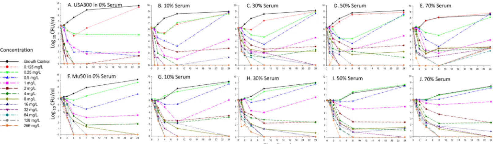
Fig 2illustrates the observed bacterial killing curves for both strains at all fractions of heat inac-tivated human serum studied, highlighting the concentration-dependent killing activity of
Fig 2. Bacterial killing activity of daptomycin against MRSA USA300 (panels A to E) and VISA Mu50 (panels F to J). Each panel represents increasing v/v ratios of human serum and MHB (0 to 70%).
daptomycin. In general, we observed that as the percent of human serum in broth increased, daptomycin activity was attenuated in a nonlinear fashion. For example, upon exposure to dap-tomycin 4mg/L, bacterial counts of USA300 after 1 h were -3.1 log10CFU/ml (Fig 2A, 0%
Human Serum), -1.8 log10CFU/ml (Fig 2B, 10% Human Serum), -0.9 log10CFU/ml (Fig 2C,
30% Human Serum), -0.8 log10CFU/ml (Fig 2D, 50% Human Serum) and -0.5 log10CFU/ml
(Fig 2E, 70% Human Serum). Similar tendencies were noted for strain Mu50 (Fig 1F–1J). Nota-bly after 24 h, in the absence of serum, bactericidal activity (99.9% kill, -3 log10CFU/mL
reduc-tion) was achieved against both strains USA300 and Mu50 upon exposure to daptomycin concentrations of>0.25 mg/L and>0.5 mg/L, respectively (Fig 2A and 2F). However, in the presence of all fractions of human serum exposure, higher daptomycin concentrations 2mg/L were required to yield bactericidal effects against both strains after 24 h (Fig 2B–2E and 2G–2J). A trend was also noted whereby the time point at which bactericidal activity was reached increased in the presence of higher human serum concentrations for both strains (Fig 2).
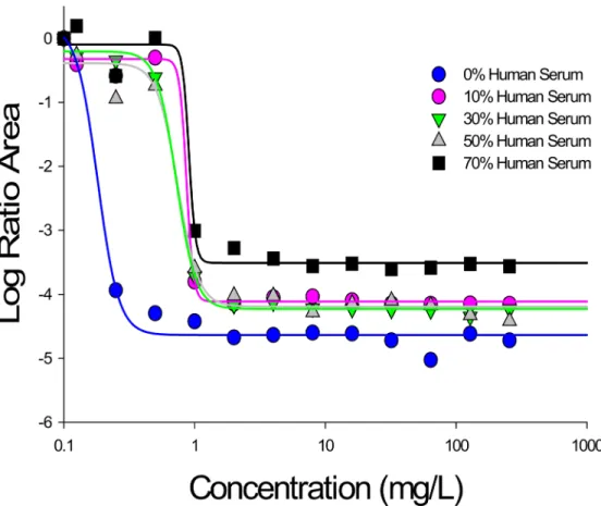
The antibacterial effect was further quantified as the overall difference in log ratio area for daptomycinversusgrowth control; using this integrated area measure, a gradual reduction in daptomycin activity was quantitatively demonstrated with increasing concentrations of human serum for both strains. For example, when daptomycin 4 mg/L was used against strain
USA300, the magnitude of the log ratio area decreased from -4.63 to -3.44 in the presence of 0% to 70% human serum, respectively. In a similar fashion, as the fraction of human serum increased from 0% to 70%, treatment with daptomycin 32 mg/L resulted in an overall area reduction ranging from -4.72 to -3.61, respectively. Identical patterns were noted for strain Mu50. These pharmacodynamic relationships of daptomycin were well fit to a Hill type model (r2>0.97) using the log ratio area approach as a measure of effect.Fig 3visually demonstrates the shift in Emaxand EC50parameters that occurs with increasing concentrations of human
serum, while PD parameters are further presented inTable 1for both strains.
Mechanism based modeling
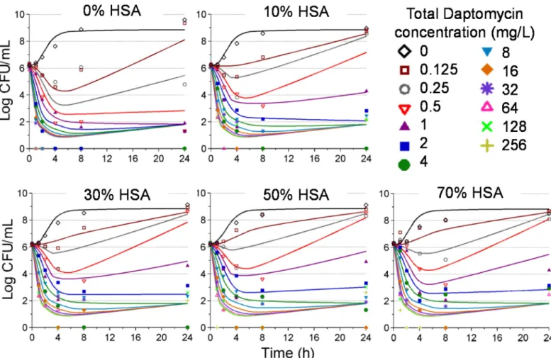
The mechanism based model characterizing the observed pharmacodynamic activity in-corporated a mixture model, comprising three bacterial subpopulations differing in daptomycin susceptibility (Fig 1). This model was able to describe all viable count profiles simultaneously with excellent precision, demonstrated by an overall r2value of 0.95 for our population fits.Fig 4represents the model fitted and observed bacterial counts (log10CFU/mL)
of USA300 following exposure to each daptomycin concentrations in the presence of increas-ing human serum fractions. All parameter estimates as well as standard errors are presented in
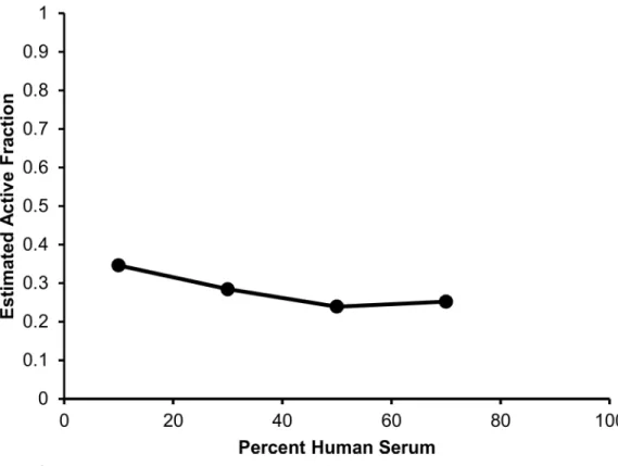
Table 2; relative standard errors were below 41% for all parameters. The mechanism based model estimated a daptomycin active fraction of 35%, 28%, 24% and 25%, in the presence of human serum fractions of 10%, 30%, 50%, and 70%, respectively (Table 1).Fig 5depicts the relationship of human serum exposure and the model estimated active fraction, demonstrating that at high concentrations of human serum, the active fraction value attains a plateau whereby the pharmacodynamics of daptomycin are not further altered by additional human serum exposure.
Estimates for SC50s(0.050 mg/L) and KC50s(4.8 mg/L) indicate that low‘effective’
concen-trations of DAP (DAPEF) reduced the probability of successful replication, while higher DAPEF
concentrations were required to cause direct killing. This observation suggests that the killing function with the greatest pharmacodyamic impact for daptomycin is the inhibition of the probability of successful replication. The key parameter in this function is the SC50s.The
Discussion
The emergence of MRSA isolates with decreased susceptibility to empiric vancomycin therapy has been increasing steadily [18,19], particularly in difficult to-treat bloodstream infections. Daptomycin has consequently been highlighted as an alternative treatment option for infec-tions caused by antibiotic resistant Gram positive strains, including MRSA [20]. However, questions are often raised regarding the pharmacodynamics of daptomycin in the presence of protein, owing to the agent’s high protein binding value (90 to 93%,www.cubicin.com). Indeed, the effect of protein binding on antibiotic pharmacodynamics has been a subject of controversy for many years [2,21,22]. Although the ultrafiltrate method is traditionally used for protein binding measurements, discrepancies exist between results derived from
Fig 3. Pharmacodynamic relationship between total daptomycin concentration and the log ratio area at each condition of human serum exposure against USA300.All r2values were>0.97. Similar results were obtained for Mu50.
doi:10.1371/journal.pone.0156131.g003
Table 1. Pharmacodynamic Hill-parameters (Emax, EC50, H) for both strains at varying human serum fractions (0% to 70%).
Human Serum USA300 Mu50
Emax EC50 H Emax EC50 H
0% 4.63 0.189 6.19 4.42 0.362 5.82
10% 4.12 0.354 7.17 4.18 0.692 6.34
30% 4.22 0.694 5.29 4.09 0.903 5.98
50% 4.20 0.68 4.07 3.87 1.23 5.63
70% 3.51 0.905 10 3.69 1.66 7.23
ultrafiltrationversusresults determined from MIC measurements in the presence and absence of serum proteins [1,2].
While the effect of protein binding onβ-lactams is well documented and predictable, dem-onstrating a proportionate increase in MIC in the presence of protein relative to the reported protein binding fraction [23,24], daptomycin does not appear to follow a similar paradigm. Rather, preliminary evidence based on MIC measurements conducted in the presence of physi-ological concentrations of albumin suggests that extrapolating the free fraction of daptomycin from published protein binding values underestimates the active fraction of drug. [2,5,6]. Additionally,in vitrotime kill experiments and pharmacodynamic models show that although early daptomycin activity was delayed in the presence of albumin, the overall extent of killing was not affected [6,8,9]. Similarly, our time kill data for both USA300 and Mu50 strains sup-port the notion of an‘active fraction’of drug (Fig 2), where the magnitude of daptomycin’s activity loss was not proportional to the free drug fraction derived from a protein binding level of ~90% (www.cubicin.com). Notably, upon exposure to daptomycin concentrations2 mg/L, bactericidal activity was observed by 24 h against both strains irrespective of the human serum fraction. Delayed killing activity in the presence of higher serum fractions was further
highlighted by a reduction in the overall change in log ratio area for daptomycin treated strains
versusgrowth control.
Fig 4. Time kill data (symbols) and model fitted predictions (solid lines) for each condition of human serum exposure for daptomycin against USA300.Each panel represents increasing v/v ratios of human serum and MHB as follows: 0% human serum (panel A), 10% human serum (panel B), 30% human serum (panel C), 50% human serum (panel D) and 70% human serum (panel E).
Importantly, to the best of our knowledge, a quantitative relationship (or mechanism based model) describing the extent of protein binding and the observedin vitropharmacodynamic activity has yet to be established for daptomycin. Here, we utilized mathematical modeling techniques and pharmacodynamic analyses to quantify the apparent‘active fraction’of dapto-mycin at different concentrations of human serumin vitro. Firstly, using a log ratio area approach, daptomycin activity was fit to hill type models for both USA300 and Mu50, from which pharmacodynamic parameters were derived; these parameters demonstrated a clear shift towards lower Emaxand higher EC50values with increasing human serum fraction,
pro-viding an indication of reduced maximal activity and daptomycin potency (Fig 3,Table 1). Secondly, from our mechanism based pharmacodynamic model, as the human serum frac-tion increased from 10% to 70%, the estimated active fracfrac-tion of daptomycin reduced from 34.6% to 25.2%, respectively. These data, suggesting a higher active fraction of daptomycin than that extrapolated using a protein binding value of ~90%, are consistent with other studies
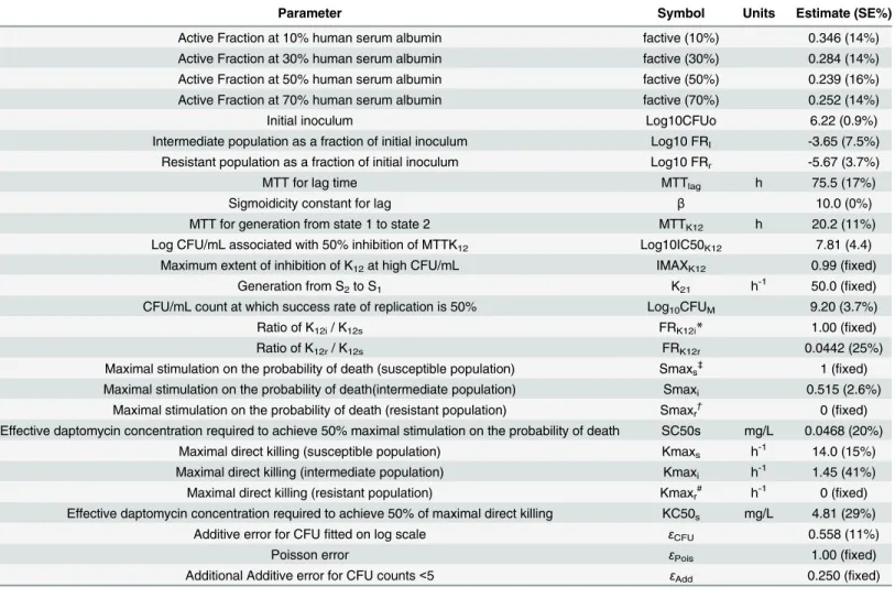
Table 2. Mechanism based mathematical model parameter descriptions, symbols, units, and estimates (standard error (SE)%), characterizing the pharmacodynamics of daptomycin.
Parameter Symbol Units Estimate (SE%)
Active Fraction at 10% human serum albumin factive (10%) 0.346 (14%)
Active Fraction at 30% human serum albumin factive (30%) 0.284 (14%)
Active Fraction at 50% human serum albumin factive (50%) 0.239 (16%)
Active Fraction at 70% human serum albumin factive (70%) 0.252 (14%)
Initial inoculum Log10CFUo 6.22 (0.9%)
Intermediate population as a fraction of initial inoculum Log10 FRI -3.65 (7.5%)
Resistant population as a fraction of initial inoculum Log10 FRr -5.67 (3.7%)
MTT for lag time MTTlag h 75.5 (17%)
Sigmoidicity constant for lag β 10.0 (0%)
MTT for generation from state 1 to state 2 MTTK12 h 20.2 (11%)
Log CFU/mL associated with 50% inhibition of MTTK12 Log10IC50K12 7.81 (4.4)
Maximum extent of inhibition of K12at high CFU/mL IMAXK12 0.99 (fixed)
Generation from S2to S1 K21 h-1 50.0 (fixed)
CFU/mL count at which success rate of replication is 50% Log10CFUM 9.20 (3.7%)
Ratio of K12i/ K12s FRK12i* 1.00 (fixed)
Ratio of K12r/ K12s FRK12r 0.0442 (25%)
Maximal stimulation on the probability of death (susceptible population) Smaxs‡ 1 (fixed)
Maximal stimulation on the probability of death(intermediate population) Smaxi 0.515 (2.6%)
Maximal stimulation on the probability of death (resistant population) Smaxr† 0 (fixed)
Effective daptomycin concentration required to achieve 50% maximal stimulation on the probability of death SC50s mg/L 0.0468 (20%) Maximal direct killing (susceptible population) Kmaxs h-1 14.0 (15%)
Maximal direct killing (intermediate population) Kmaxi h-1 1.45 (41%)
Maximal direct killing (resistant population) Kmaxr# h-1 0 (fixed)
Effective daptomycin concentration required to achieve 50% of maximal direct killing KC50s mg/L 4.81 (29%)
Additive error for CFUfitted on log scale εCFU 0.558 (11%)
Poisson error εPois 1.00 (fixed)
Additional Additive error for CFU counts<5 εAdd 0.250 (fixed)
*FRK12iwas estimated close to 1 so this parameter wasfixed at 1.
‡Smax
swas estimated to be very close to 1 so wasfixed to 0.99.
†Smax
rwas estimated to be close to zero so wasfixed at zero. #Kmax
rwas estimated close to zero and was thusfixed at zero.
which also propose a 28.9 to 51.8% predicted free fraction in varying proportions of human albumin and serum (according to MICs) [8]. This phenomenon may be due to the high relative affinities that daptomycin displays to the site of bactericidal action inS.aureuscompared to plasma protein. For example, daptomycin binds weakly and reversibly to albumin (dissociation constant [Kd] = 90.3μmol/L), while irreversible binding is established with the bacterial cell
membrane. Jung et. al. recently proposed a two step model, where daptomycin undergoes two conformational changes resulting in rapid bactericidal killing in a calcium dependent manner [25]. During the first step, calcium binds to daptomycin that is weakly bound to the bacterial cytoplasmic membrane, thus promoting lipid interaction. Resultantly, the calcium conjugated daptomycin has an increased amphipathicity and a total decreased charge, which allows for peptide oligomerization leading to the second conformational change; this secondary change enables daptomycin to penetrate deep into the cytoplasmic membrane. Based on the mecha-nism of irreversible binding, it was postulated that protein bound daptomycin may continue to be available to bind to the cytoplasmic membrane and display antimicrobial activity indepen-dent of drug concentration [26]. Taken together with the current findings, these findings sug-gest that the low reported free fraction of daptomycin alone is not an optimal predictor of pharmacologic effect.
Lastly, the principle of the‘active fraction’may not be unique to daptomycin, but may potentially apply to other highly protein bound antimicrobials as well. Similar to the discrep-ancy between the activity of daptomycin and the free fraction of drug noted in prior investiga-tions, telavancin has displayed in vitro activity that is disproportionately higher than
predictions based on unbound drug concentrations [8]. It is therefore likely that in vitro inves-tigations that base telavancin concentrations on the free fraction of drug may be
Fig 5. Observed relationship between each concentration of human serum exposure and model estimated active fraction values.
underestimating the activity of telavancin. Further investigations evaluating the performance of highly bound antimicrobials in different concentrations of serum are needed before the model in the present study can be extrapolated to other agents.
An important limitation of the current study is that two daptomycin-susceptible laboratory strains were used in the investigation. While the resistance profiles of the two strains differed for vancomycin, it is unknown whether lipopeptide resistance alters the‘active fraction’of dap-tomycin. EstablishedS.aureuslaboratory strains also possess genotypic differences from con-temporary isolates that may result in a discordance in the‘active fraction’of daptomycin observed clinically in comparison to the present study. Lastly, USA 300 is a CA-MRSA strain and Mu-50 is a VISA strain originally isolated from a sternal abscess [19]. As neither of the investigated strains were obtained from severe nosocomial infections such endocarditis or bac-teremia, caution should be used when extrapolating the results of the current investigation to nosocomial pathogens.
We also acknowledge other potential limitations with this work. Currently, there are no standard methods for incorporating protein intoin vitrostudies to evaluate antimicrobial pharmacodynamics. In the current study,in vitroconditions that were established may fail to account for the dynamic physiological interaction between binding of proteins to antimicrobial
in vivo. Second, daptomycin concentrations were static and the exposure effect relationship, as
it relates to AUC/MIC and protein binding, were not explored. Third, although conditions of 100% human serum would best mimic the in vivo situation, this was not studied, since the growth characteristics of MRSA would be hampered at these higher concentrations of serum. Additional studies are warranted in models which account for dynamically changing in vivo conditions with consideration of metabolism, transport processes, and diffusion between com-partments in humans [2]. Despite such limitations, we believe that the novel mathematical modeling framework provided here describing the active fraction of daptomycin has potential utility to enhance the design ofin vitroexperiments, and to optimize therapeutic regimens of daptomycin in humans.
Supporting Information
S1 Data. The total bacterial counts (log10CFU/mL) for MRSA strain USA300 during
expo-sure to daptomycin in 24 h time-killing experiments performed in the presence of various concentrations of human serum.
(PDF)
S2 Data. The total bacterial counts (log10CFU/mL) for VISA strain Mu50 during exposure
to daptomycin in 24 h time-killing experiments performed in the presence of various con-centrations of human serum.
(PDF)
Acknowledgments
Author Contributions
Conceived and designed the experiments: SG JB BT. Performed the experiments: SG PH. Ana-lyzed the data: SG JB AF. Contributed reagents/materials/analysis tools: BT. Wrote the paper: SG JL.
References
1. Wise R. The clinical relevance of protein binding and tissue concentrations in antimicrobial therapy. Clinical pharmacokinetics. 1986; 11(6):470–82. Epub 1986/11/01. PMID:3542338.
2. Zeitlinger MA, Derendorf H, Mouton JW, Cars O, Craig WA, Andes D, et al. Protein binding: do we ever learn? Antimicrobial agents and chemotherapy. 2011; 55(7):3067–74. Epub 2011/05/04. doi:10.1128/ aac.01433-10PMID:21537013; PubMed Central PMCID: PMC3122431.
3. Craig WA, Welling PG. Protein binding of antimicrobials: clinical pharmacokinetic and therapeutic impli-cations. Clinical pharmacokinetics. 1977; 2(4):252–68. Epub 1977/07/01. PMID:20259.
4. Gould IM, David MZ, Esposito S, Garau J, Lina G, Mazzei T, et al. New insights into meticillin-resistant Staphylococcus aureus (MRSA) pathogenesis, treatment and resistance. International journal of anti-microbial agents. 2012; 39(2):96–104. Epub 2011/12/27. doi:10.1016/j.ijantimicag.2011.09.028PMID:
22196394.
5. Tsuji BT, von Eiff C, Kelchlin PA, Forrest A, Smith PF. Attenuated vancomycin bactericidal activity against Staphylococcus aureus hemB mutants expressing the small-colony-variant phenotype. Antimi-crobial agents and chemotherapy. 2008; 52(4):1533–7. Epub 2008/02/21. doi:10.1128/aac.01254-07
PMID:18285476; PubMed Central PMCID: PMCPmc2292514.
6. Cafini F, Aguilar L, Gonzalez N, Gimenez MJ, Torrico M, Alou L, et al. In vitro effect of the presence of human albumin or human serum on the bactericidal activity of daptomycin against strains with the main resistance phenotypes in Gram-positives. The Journal of antimicrobial chemotherapy. 2007; 59 (6):1185–9. Epub 2007/04/07. doi:10.1093/jac/dkm078PMID:17412725.
7. Garrison MW, Vance-Bryan K, Larson TA, Toscano JP, Rotschafer JC. Assessment of effects of pro-tein binding on daptomycin and vancomycin killing of Staphylococcus aureus by using an in vitro phar-macodynamic model. Antimicrobial agents and chemotherapy. 1990; 34(10):1925–31. Epub 1990/10/ 01. PMID:1963288; PubMed Central PMCID: PMC171966.
8. Tsuji BT, Leonard SN, Rhomberg PR, Jones RN, Rybak MJ. Evaluation of daptomycin, telavancin, tei-coplanin, and vancomycin activity in the presence of albumin or serum. Diagnostic microbiology and infectious disease. 2008; 60(4):441–4. Epub 2008/02/06. doi:10.1016/j.diagmicrobio.2007.11.011
PMID:18248936.
9. Cha R, Rybak MJ. Influence of protein binding under controlled conditions on the bactericidal activity of daptomycin in an in vitro pharmacodynamic model. The Journal of antimicrobial chemotherapy. 2004; 54(1):259–62. Epub 2004/05/20. doi:10.1093/jac/dkh259PMID:15150172.
10. Smith PF, Tsuji B, Booker BM, Forrest A, Bajic S, Kelchlin P, et al. Pharmacodynamics of cefprozil against Haemophilus influenzae in an in vitro pharmacodynamic model. Diagnostic microbiology and infectious disease. 2006; 56(4):379–86. doi:10.1016/j.diagmicrobio.2006.06.019PMID:16930921. 11. Bulitta JB, Yang JC, Yohonn L, Ly NS, Brown SV, D'Hondt RE, et al. Attenuation of colistin bactericidal
activity by high inoculum of Pseudomonas aeruginosa characterized by a new mechanism-based pop-ulation pharmacodynamic model. Antimicrobial agents and chemotherapy. 2010; 54(5):2051–62. doi:
10.1128/AAC.00881-09PMID:20211900; PubMed Central PMCID: PMC2863601.
12. Bulitta JB, Ly NS, Yang JC, Forrest A, Jusko WJ, Tsuji BT. Development and qualification of a pharma-codynamic model for the pronounced inoculum effect of ceftazidime against Pseudomonas aeruginosa. Antimicrobial agents and chemotherapy. 2009; 53(1):46–56. Epub 2008/10/15. doi:10.1128/aac. 00489-08PMID:18852268; PubMed Central PMCID: PMC2612150.
13. Treyaprasert W, Schmidt S, Rand KH, Suvanakoot U, Derendorf H. Pharmacokinetic/pharmacody-namic modeling of in vitro activity of azithromycin against four different bacterial strains. Int J Antimicrob Agents. 2007; 29(3):263–70. Epub 2006/12/30. S0924-8579(06)00428-6 [pii] doi:10.1016/j.
ijantimicag.2006.08.049PMID:17194570.
14. Mouton JW, Vinks AA, Punt NC. Pharmacokinetic-pharmacodynamic modeling of activity of ceftazi-dime during continuous and intermittent infusion. Antimicrobial agents and chemotherapy. 1997; 41 (4):733–8. Epub 1997/04/01. PMID:9087479; PubMed Central PMCID: PMC163784.
16. Jumbe N, Louie A, Leary R, Liu W, Deziel MR, Tam VH, et al. Application of a mathematical model to prevent in vivo amplification of antibiotic-resistant bacterial populations during therapy. The Journal of clinical investigation. 2003; 112(2):275–85. Epub 2003/07/17. doi:10.1172/jci16814PMID:12865415; PubMed Central PMCID: PMC164285.
17. Dudley MN, Mandler HD, Gilbert D, Ericson J, Mayer KH, Zinner SH. Pharmacokinetics and pharmaco-dynamics of intravenous ciprofloxacin. Studies in vivo and in an in vitro dynamic model. The American journal of medicine. 1987; 82(4A):363–8. Epub 1987/04/27. PMID:3555061.
18. Hiramatsu K, Aritaka N, Hanaki H, Kawasaki S, Hosoda Y, Hori S, et al. Dissemination in Japanese hospitals of strains of Staphylococcus aureus heterogeneously resistant to vancomycin. Lancet. 1997; 350(9092):1670–3. Epub 1997/12/24. doi:10.1016/s0140-6736(97)07324-8PMID:9400512. 19. Hiramatsu K, Hanaki H, Ino T, Yabuta K, Oguri T, Tenover FC. Methicillin-resistant Staphylococcus
aureus clinical strain with reduced vancomycin susceptibility. The Journal of antimicrobial chemother-apy. 1997; 40(1):135–6. Epub 1997/07/01. PMID:9249217.
20. Moise PA, North D, Steenbergen JN, Sakoulas G. Susceptibility relationship between vancomycin and daptomycin in Staphylococcus aureus: facts and assumptions. The Lancet Infectious diseases. 2009; 9 (10):617–24. Epub 2009/09/26. doi:10.1016/s1473-3099(09)70200-2PMID:19778764.
21. Wise R. The relevance of pharmacokinetics to in-vitro models: protein binding—does it matter? The Journal of antimicrobial chemotherapy. 1985; 15 Suppl A:77–83. Epub 1985/01/01. PMID:3980339. 22. Wise R, Gillett AP, Cadge B. Protein binding and antibiotic concentrations. Lancet. 1978; 2(8086):431.
Epub 1978/08/19. PMID:79795.
23. Wise R. Protein binding of beta-lactams: the effects on activity and pharmacology particularly tissue penetration. II. Studies in man. The Journal of antimicrobial chemotherapy. 1983; 12(2):105–18. Epub 1983/08/01. PMID:6619052.
24. Wise R. Protein binding of beta-lactams: the effects on activity and pharmacology particularly tissue penetration. I. The Journal of antimicrobial chemotherapy. 1983; 12(1):1–18. Epub 1983/07/01. PMID:
6619044.
25. Jung D, Powers JP, Straus SK, Hancock RE. Lipid-specific binding of the calcium-dependent antibiotic daptomycin leads to changes in lipid polymorphism of model membranes. Chemistry and physics of lip-ids. 2008; 154(2):120–8. Epub 2008/05/21. doi:10.1016/j.chemphyslip.2008.04.004PMID:18489906. 26. Eisenstein BI. Lipopeptides, focusing on daptomycin, for the treatment of Gram-positive infections.