Computational design of ! sheet proteins
Xiaozhen Hu
A dissertation submitted to the faculty of the University of North Carolina at Chapel Hill in partial fulfillment of the requirements for the degree of Doctor of Philosophy in the Department of Biochemistry and Biophysics (Program in Molecular and Cellular Biophysics).
Chapel Hill 2008
Approved by
ABSTRACT
Xiaozhen Hu : Computational design of ! sheet proteins (Under the direction of Brian Kuhlman)
Computational protein design has become a very powerful approach to test our understanding of the forces and energetics of macromolecular systems. The ability to design proteins that have specific structures and functions will be very valuable to future protein drug discovery. Protein design technology has been successfully applied to stabilize proteins, increase protein-protein binding affinity and create new protein structures. However, de novo design remains very challenging, especially for !-sheet proteins. Most de novo designed !-sheet proteins tested to date either misfold or aggregate. In this thesis, we use a hierarchical approach to search for the bottleneck in !-sheet design. First, we tested our ability to redesign the sequence of a naturally occurring !-sheet protein. The molecular modeling program Rosetta was used to design new sequences for the !-sheet protein tenascin. The redesigned proteins are well-folded and have thermal melting temperatures that are 40 °C higher than the wild type. These results indicate that given a designable backbone we can create a well-folded !-sheet protein.
ACKNOWLEDGEMENTS
I would like to thank, first and foremost, Brian for giving me the great opportunity to pursue my doctoral dissertation under his direction. Brian has a beautiful mind and a great sense of scientific insight and intuition. He was always able to find a way to overcome the challenges that seemed to prevent my goals. When I encountered obstacles, he was always patient, understanding, and willing to provide guidance when needed. He also gave me a great degree of freedom to try my own ideas which taught me how to think and work independently. I thankful for all of those years working in his laboratory and none of my work would have been possible without his guidance. I would like to thank the members of the Kuhlman laboratory; we shared challenges, experiences and successes. We had a very comfortable and productive environment to work in — all of which have become very fond memories of UNC. I would like to thank my collaborators, Huancheng Wang and Hengming Ke, for their suggestions and help in the crystal structures. I would like to thank my thesis committee members Nikolay Dokholyan, Marshall Edgell, Jan Hermans and Andrew Lee for their great advice and suggestions. I would like to thank Barry Lents for the opportunity to join the Molecular and Cellular Biophysics Training Program. I would like to thank Ashutosh Tripathy from MacInFac, Greg Young from the NMR facility and Laurie Betts from the X-ray crystallography facility for their kind help and support. Last but certainly not least, I would like to thank my parents, Zhenming and Linai, for believing in me and loving me for whoever I am. They have been always there, supporting and encouraging me. Nothing would have been possible without their support. I would like to thank my little girl Audrey, who let me experience the joy of a being a mother and understand how
TALBE OF CONTENTS
TALBE OF CONTENTS ...VI
LIST OF FIGURES ...IX
LIST OF TABLES ...XI
LIST OF ABBREVIATIONS...XII
CHAPTER 1...1
INTRODUCTION...1
EARLY DEVELOPMENT OF COMPUTATIONAL PROTEIN DESIGN...2
PREVIOUS WORK IN DE NOVO !-SHEET PROTEIN DESIGN...4
MOLECULAR MODELING PROGRAM - ROSETTA...5
TARGET FOLD MODEL SYSTEM...9
DESIGN APPROACH...10
FIGURES...13
REFERENCES...17
CHAPTER 2...22
PROTEIN DESIGN SIMULATIONS SUGGEST THAT SIDE CHAIN CONFORMATIONAL ENTROPY IS NOT A STRONG DETERMINANT OF AMINO ACID ENVIRONMENTAL PREFERENCES ...22
ABSTRACT ...23
INTRODUCTION...24
METHODS ...26
RESULTS AND DISCUSSION ...31
CONCLUSION ...36
TABLES ...39
SUPPLEMENTAL MATERIAL ...44
REFERENCES...46
CHAPTER 3...51
COMPUTER-BASED REDESIGN OF A !-SANDWICH PROTEIN SUGGESTS THAT EXTENSIVE NEGATIVE DESIGN IS NOT REQUIRED FOR DE NOVO !-SHEET DESIGN ...51
ABSTRACT...52
INTRODUCTION...53
RESULTS...55
DISCUSSION...59
EXPERIMENTAL PROCEDURES...60
FIGURES...63
SUPPLEMENTARY MATERIAL...69
REFERENCES...72
CHAPTER 4...76
HIGH RESOLUTION DESIGN OF A PROTEIN LOOP ...76
ABSTRACT...77
INTRODUCTION...78
RESULTS AND DISCUSSION...80
CONCLUSION...86
MATERIALS AND METHODS...86
FIGURES...91
SUPPLEMENTARY MATERIALS...97
REFERENCES...101
CHAPTER 5...105
DE NOVO DESIGN...105
INTRODUCTION...107
MATERIALS AND METHODS...109
EXPERIMENTAL PROCEDURES...115
RESULTS...116
DISCUSSION...118
FIGURES...121
TABLES...131
SUPPLEMENTARY MATERIAL...136
REFERENCES...138
CHAPTER 6...141
LIST OF FIGURES
Figure 1.1 Betabellin 14S... 13
Figure 1.2 Betadoublet ... 14
Figure 1.3 The Metropolis sampling algorithm used in Rosetta... 15
Figure 1.4 Comparison of a monomeric immunoglobulin VH domain(left) with FN3(right)... 16
Figure 2.1 Algorithm for incorporating side chain entropy and free energy into Rosetta ... 37
Figure 2.2 Changes in side-chain conformational entropy and free energy between surface and buried positions... 38
Figure 3.1 Sequences of the wild type and three redesigned proteins... 63
Figure 3.2 One-dimensional 1H spectra of the redesigned proteins... 64
Figure 3.3 CD spectra of the wt and redesigns ... 65
Figure 3.4 Temperature and chemical denaturation ... 66
Figure 3.5 Structure alignment beteen the design model and the crystal structure ... 67
Figure 4.1 Iterative optimization of a loop sequence and conformation... 91
Figure 4.2 Models and sequences of the redesigned proteins... 92
Figure 4.3 Structure prediction with the designed sequences ... 93
Figure 4.4 Thermal unfolding of the designed sequences as monitored with circular dichroism... 94
Figure 4.5 Alignment between the crystal structure and the design model ... 95
Figure 4.6 The crystal structure of LoopA ... 96
Figure 4.7 Representative set of starting structures used for loop design ... 97
Figure 4.8 Alignment between the design models and structure predictions ... 98
Figure 4.9 Energies of design models for different templates ... 98
Figure 4.10 1-dimensional 1H spectra of the designed proteins... 99
Figure 4.11 X-ray diffraction data ... 100
Figure 5.3 Schematic representation of the target fold... 123
Figure 5.4 One example of the starting structures ... 124
Figure 5.5 Schematic representation of the parameters used in Rosetta for hydrogen bonding potential ... 125
Figure 5.6 X angle distribution ... 126
Figure 5.7 Spectra of B002... 127
Figure 5.8 B9 and double mutant... 128
Figure 5.9 Spectra of BN1... 129
LIST OF TABLES
Table 2.1 Average side chain entropy per residue as calculated with a variety of approaches ... 39
Table 2.2 Energies and entropies as a function of environment ... 40
Table 2.3 Entropies as a function of rotamer library size... 41
Table 2.4 Comparison of native sequence recovery rates ... 42
Table 2.5 Environmental preferences of the amino acids ... 43
Table 2.6 PDB codes used in the design simulation... 44
Table 3.1 Sequence features of wild type and redesigned tenascin... 68
Table 3.2 Thermodynamic parameters of wild type and redesigned tenascin ... 69
Table 3.3 Comparison of native sequence recovery rates for design simulations with the standard weight and modified beta sheet weight... 69
Table 3.4 Environmental preferences of the amino acids in design simulations with the standard weight and modified beta sheet weight ... 70
Table 3.5 X-Ray diffraction data collection and refinement statistics ... 71
Table 5.1 Sequence compositions for different generation of designs... 131
LIST OF ABBREVIATIONS
!G: free energy of unfolding
CD: circular dichroism
Cm: midpoint of guanidine chloride unfolding transition
E. Coli: Escherichia coli
FNIII: fibronectin type III domain
GuHCl: guanidine chloride
IPTG: isopropyl "-D-thiogalactoside
m value: slope of !G versus denaturant concentration
NMR: nuclear magnetic resonance
PDB: protein data bank
RMSD: root mean square distance
SASA: solvent accessible surface area
SDS-PAGE: sodium dodecyl sulfate-polyacrylamide gel electrophoresis
ss: secondary structure
Tm: melting temperature
CHAPTER 1
EARLY DEVELOPMENT OF COMPUTATIONAL PROTEIN DESIGN
Computational protein design has become a very powerful approach for testing our understanding of
the forces and energetics of macromolecular systems1,2. Designed proteins with desired structures or
functions have been of special interest to pharmaceutical companies and this computational protein
design method will make a significant impact on the development of new biotechnological
therapeutics3,4. Thirty years ago, computational protein design may have sounded like science fiction
but recently there has been great progress in the development of protein design methodologies and
applications5-9.
The earliest attempts at computational protein design focused on redesigning naturally occurring
proteins while assuming a fixed, native backbone. One remarkable example was the complete
redesign of a zinc finger by Dahiyat et al. with the backbone fixed5. The fixed backbone assumption
greatly reduces computational time and works well when an appropriate backbone scaffold exists;
however, it is incompatible with de novo design because there is no design template available.
Studies have shown that incorporating flexibility can improve sequence prediction and is therefore
better for novel design10. Harbury et al.6 demonstrated the advantage of backbone flexibility by
designing novel right-handed coiled-coil bundles. They created a set of right-handed coiled-coils and
experimentally validated the structures. Another breakthrough in protein design was the creation of a
fold that is not seen in nature so far. Kuhlman et al. used the Rosetta program to iteratively optimize
both sequence and structure and created a protein with a novel fold (TOP7)11. The experimental
results showed that TOP7 is very stable and the crystal structure matched the design model very well
(root mean square deviation RMSD=1.2 Å). This striking result opened a new window to the
exploration of novel proteins.
protein-protein binding affinity, alter binding specificity and redesign a folding pathway12-14. Grand
challenges in the form of de novo design problems, such as creating novel enzymes and biosensors,
have been addressed11,13,15-19. Despite many successes, de novo design remains a very challenging
problem, because it requires the design of a novel sequence that is unrelated to any naturally
occurring protein that can fold into a pre-defined 3-dimensional structure20. In order to fold into a
well-defined structure, the designed sequence should energetically stabilize the desired fold as well as
destabilize the alternative conformations. De novo design is a rigorous test of our understanding of
protein folding energetics, but the underlying principles are not yet understood well enough to ensure
the success of all de novo designs21.
It is fair to say though that we understand relatively well how to make a predominantly helical protein
because some successful de novo designs for # helix bundles and #/" mixed proteins have been
reported11,22-24. However, the de novo design of purely
"-sheet proteins has proven to be more
complicated, as these designed sequences usually misfold or aggregate. One possible reason is that
the relatively slow folding of "-sheet proteins involves long range interactions, and unlike #-helix, "
-sheet formation is determined mostly by tertiary context instead of intrinsic secondary structure
preferences25. Compared with
#-helix proteins, "-sheet proteins are less modular and inherently more
difficult to design due to the fundamental difference in the hydrogen bonding patterns of these two
different secondary structures26. The backbone hydrogen bonds within
#-helix will be satisfied by
nearby residues within the same secondary structure element. In contrast, "-sheet proteins require a
"-strand to interact with a neighboring strand, possibly distant in primary structure, to satisfy
backbone hydrogen bonds27. Side chains will point alternately above and below the "-sheet to
interact with the neighboring residues. The need of a "-sheet to form so many interactions underlies
"-sheet proteins’ great tendency to aggregate. Additionally, residues with good "-sheet-forming
is of great interest.
PREVIOUS WORK IN DE NOVO "-SHEET PROTEIN DESIGN
Recent work includes several groups’ successful designs of some small water-soluble peptides, for
instance "-hairpins30, a 20 residue three-stranded antiparallel "-sheet31 and even a four-stranded "
-sheet32. Since 1981, the Richardson group has been trying to design several generations of betabellins
but the solubility seems a big issue in these designs33. Yan et al. designed a series of betabellins 34,35
but the best one (betabellin 14D) only folded in the presence of a fold-stabilizing interchain disulfide.
It consists of two 32-residue "-sheet packed against each other by a disulfide linkage. The sequence
of each half has a pattern of alternating polar/nonpolar for " strands and statistically favored residues
for " turns as shown in Figure 1.1. D-amino acid residues were used for the turn positions to favor
formation of " turns. The single chain of betabellin 14S is not folded. To make it more globular, a
disulfide bond was introduced to link the two identical subunit. The double-chain form 14D, is
folded into a "-sheet liked structure in the presence of the disulfide linkage suggesting that the folding
is induced by the disulfide bond formation. This disulfide bond strategy was also applied in the
design of betadoublet23(Figure 1.2), which is water soluble only at low pH; however NMR data
suggests that betadoublet does not adopt a single unique conformation, implying that it adopts a
molten globular structure.
Sollazzo et al. designed a small all "-sheet protein that can bind metal zinc upon folding, but again
solubility limited the detailed structural analysis36. Recently, Nanda et al. designed a mimic of the
redox protein rubredoxin, which seems to adopt the target fold37. This is one example of a functional
de novo designed "-sheet protein. To date, attempts at the de novo design of "-sheet proteins are very
validated with a high-resolution structure. De novo design of globular "-sheet proteins still remains
an unsolved problem.
In nature, approximately one quarter of all protein domains are "-sheet folds38. "-sheet proteins form
relatively rigid structures that can serve as good scaffolds for designing molecules with new
functions. "-sheetproteins have also been a focus of considerable attention for medical biologists and
the pharmaceutical industry3,39 because they have proven to be good targets for disrupting unwanted
protein-protein interactions. Protein-protein interactions are essential to many biological processes;
however, uncontrolled interactions may lead to protein misfolding and aggregation which contribute
to many different diseases including Alzheimer’s disease, Huntington’s disease, others40. Protein
misfolding in these diseases involves protein aggregation into "-sheet rich oligomeric structures.
Considerable evidence has shown that these aggregate structures play important roles in disease
pathogenesis41. One strategy to develop therapies for these diseases is to address protein misfolding
and aggregation with rationally designed inhibitors4,42. The computational protein design approach
provides a valuable way for us to better understand how nature “designs” "-sheet proteins that can
avoid misfolding or aggregation, which will be highly useful in future protein therapeutics.
MOLECULAR MODELING PROGRAM - ROSETTA
Rosetta is a molecular modeling software package developed by several research groups. Initially
used for de novo structure prediction, it has since been expanded to contain protocols for
high-resolution structure refinement, loop modeling, molecular docking and protein design43-46. The goal
of protein design, also known as inverse folding, is to identify a compatible low free energy sequence
for describing the interactions in proteins and ranking the fitness of a particular sequence for a given
backbone structure, and a search algorithm for sampling sequence space11.
Energy function
Being able to describe the interactions in proteins accurately is the most difficult problem in protein
design. The energy function used in Rosetta contains physical potentials and knowledge based
potentials derived statistically from the many structures in the Protein Data Bank(PDB)12. The
potentials are combined in the Rosetta energy function as a linear sum of the following main
terms11,47:
!
E
total=
w
atrE
ljatr+
w
repE
ljrep+
w
solE
sol+
w
hbondE
hbond+
w
pairE
pair+
w
rotE
rot+
w
ramaE
rama"
E
refThe main components in the energy function are:
Lennard-Jones potential: A 12-6 Lennard-Jones potential represents van der Waals interactions.
This potential is slightly modified from the standard form with the introduction of a distance cutoff
below which the potential is extrapolated linearly. The attractive and repulsive energies are split into
separate terms, Eljatr and Eljrep, which gives greater flexibility in weighting the terms and improves
sequence recovery. To compensate for the fixed backbone assumption, usually the repulsive term is
softened to implicitly allow for some level of backbone flexibility.
Solvation energy (Esol): The implicit solvation model developed by Lazaridis and Karplus is used to
evaluate the solvation energy for a protein. This is a semiempirical model that is parameterized with
experimental data and does not require surface area calculations48. This term penalizes surface
Hydrogen bonding potential (Ehbond): Hydrogen bonding is very important to stability and
protein-protein interactions. Rosetta uses an orientation-dependent hydrogen bonding term, which is derived
from the distribution of three parameters14 (distance between the hydrogen and acceptor atoms, angle
at the hydrogen atom and angle at the acceptor atom) from PDB database. This term allows buried
polar atoms if they can form hydrogen bond. Together with the solvation energy term, these two
terms balance how many polar residues are placed in the core during a design simulation.
Residue pair potential (Epair): Electrostatic interactions such as salt bridges are very important for
protein function; however, these interactions are very dependent on the local environment, which
makes it very difficult to model. Rosetta uses a knowledge-based term to model electrostatics. This
term is derived from the probability of a pair of polar residues being seen near each other in the PDB
database49.
Rotamer self-energy (Erot): Internal energy of a rotamer is calculated based on the probability of
seeing a particular rotamer for a given phi and psi angle in the PDB database. These probabilities are
taken from Dunbrack library directly50 and their negative log values were used as the rotamer internal
energy as shown in the following equation
!
E
rot=
"
ln(
prob
(
rot
(
i
) |
phi
(
i
),
psi
(
i
))
i residue
#
Torsion potential (Erama): Rosetta uses ideal bond lengths and bond angles for bonded interactions.
The torsion potential is associated with backbone bond torsion angles and is related to Ramachandran
torsion preferences. It is derived from PDB statistics by measuring the probabilities of seeing a
particular amino acid in a secondary structure type (helix, strand and loop) for a particular phi, psi
!
E
rama=
"
ln[
prob
(
phi
(
i
),
psi
(
i
) |
aa
i,
ss
i)]
iresidue
#
aa = amino acid type
ss = secondary structure type
Reference energy (Eref): Calculation of folded state stability requires a reference to the energy of an
unfolded state. Rosetta uses parameterized energies for each residue to represent the free energy of
unfolded state. The reference values and the weights for each energy term are calculated to best
reproduce native sequences for known structures11.
Search function
One major challenge in protein design is determining how to scan through sequence space and
identify the optimum effectively. The size of sequence space is astronomical: for a 50-residue
protein, in which all 20 standard amino acids are allowed at each position, 2050 (1065) sequences are
possible. To make the search computationally feasible, one simplification is to make the search space
discrete. Currently, most computational protein design methods use a discrete set of side chain
conformations (rotamers).
Computational protein design requires an efficient search algorithm that is able to scan an enormous
search space51. The choice of algorithms will influence the accuracy of side chain predictions and the
speed of design simulations. There are two categories of search algorithms, stochastic searches and
deterministic search algorithms51. Deterministic algorithms include self consistent mean field
optimization and dead end elimination; they are semiexhaustive search as which will always converge
to the same solution (if they are able to converge). Stochastic algorithms include Monte Carlo
advantage of these methods is that they can handle complicated problems because they do not require
an exhaustive search, the disadvantage is that they are not guaranteed to find the global energy
minimum.
Rosetta uses a Monte Carlo search algorithm with simulated annealing to identify low energy
sequences for a given structure. This is a simple, fast and widely used stochastic search method. In
the Rosetta search algorithm, the initial conformation is generated randomly. This conformation is
then perturbed by a single rotamer substitution(sequence could be changed). The substitution may or
may not change the sequence identity (Figure 1.3). If the substitution lowers the energy (
!
Enew"Eold = #E <0), it is accepted. Otherwise, the substitution is accepted if
!
e
"#E/kT>
R
(0
$
R
$
1)
, where k is the Boltzman constant, T is the temperature and R is a randomprobability. This condition, called the Metropolis criterion, prevents the simulation from getting
trapped in local energy minima. A trajectory may consist of a few hundred thousand rotamer
substitutions, which is typically for convergence between trajectories.
TARGET FOLD MODEL SYSTEM
Our strategy for de novo design is to use a natural protein fold and design a new sequence that is not
related to any natural protein sequence, but that will fold into the desired structure. To simplify the
design process, we want the template to be just large enough to present true tertiary structure without
requiring disulfide bonds or metal binding sites. One common "-sheet tertiary structure is the
Fibronectin type III domain (FNIII). This domain occurs in many proteins with very diverse
sequences, including cell surface receptors and cell adhesion molecules, which indicates that it is
highly designable and could easily be modified to generate new functions. This small domain (about
exposed loops may be modified to generate novel functions for molecular recognition. The surface
topology is very similar to the immunoglobulin VH structure52 (Figure 1.4, left panel, pdbcode 1ol0),
and this FNIII domain has become one popular scaffold for “monobody” design to date53,54.
Monobodies are antibody-like proteins that bind to specific target proteins. Unlike antibodies,
monobodies are usually easy to express and purify in large quantities and are ideal for inhibiting
protein-protein interactions. Besides, they are small (~10 kDa), monomeric and lack disulfide bonds
so that they are stable in reducing environments. Because of all these excellent characteristics, Huang
et al. used monobodies to generate the affinity resin that binds to a specific conformation of the target
protein so as to purify the desired conformation based on the target protein surface properties55. The
template we used in this study is the third Fibronectin type III domain from tenascin56 ( pdbcode :
1ten, Figure 1.4, right panel ). It is small, cysteine-free and monomeric. It is easy to purify and well
characterized57. Tenascin has been shown to undergo a two-state, thermally reversible unfolding
transition. These properties make it an ideal model system for our study.
De novo designed "-sheet proteins often aggregate in solution. One possible reason is that the
designed sequence favors no folded structure or equally favors many folded structures. Another
reason is that kinetically "-sheet proteins fold slowly so that they easily form aggregated. To design a
well-folded structure, is it enough to only search for a sequence that has low free energy for the target
structure (positive design)? Should we also include some elements that can destabilize the alternative
fold states explicitly in the sequence design process (negative design)58? In this thesis, we address the
importance of positive design and negative design by pursing different design problems.
DESIGN APPROACH
Experience shows that de novo design of all "-sheet proteins is an extremely challenging problem;
The Rosetta energy function is a linear combination of Lennard-Jones interactions, implicit solvation
potential, hydrogen bonding energy and additional knowledge-based energy terms. However, the
program does not explicitly factor in side chain entropy, which is also very important to
thermodynamics. Residues with more degrees of freedom (Lys, Arg, Met, etc) lose more
conformational entropy upon folding and these amino acids are less likely to be buried59. The correct
placement of a given amino acid is likely to partially depend on how much entropy is lost when the
side chain is locally constrained in a folded protein. However, the influence of the side chain entropy
on protein design simulations in Rosetta was not clear. In order to investigate how side chain entropy
influences protein design simulations, in chapter 2 we will explicitly incorporate side chain entropy
into Rosetta and test if it improves recovery of native sequence in design simulations.
To decipher the importance of positive design and negative design, we will pursue a set of different
design problems. In chapter 3, we will try to redesign a naturally occurring all "-sheet protein
(tenascin) with only positive design. The use of tenascin guarantees that the backbone is designable.
This test will determine whether we can use Rosetta to design an all "-sheet protein that is
well-folded and stable using only positive design. In this test case, the backbone is fixed, which will bias
sequence selection. In chapter 4, we will incorporate backbone flexibility into the design simulation
by redesigning a 10 residue loop in tenascin. Being able to explore the backbone degrees of freedom
will increase conformational sampling and design complexity. By only designing part of the
backbone, we will be able to test if we can design a well-structured loop conformation in the context
of a stably folded "-sheet protein.
In chapter 5, we will try to design a whole protein from scratch. From multiple generations of design
and experimental characterization of the results, we will have feedback that may be used to improve
De novo design of "-sheet protein is a rigorous test of our protein design software and our
understanding of the relationship between sequences and structures. By designing a sequence de
novo we would expect to learn new things that we would not have learned by examining or
redesigning naturally occurring proteins. The results of these experiments will give us feedback on
FIGURES
Figure 1.1 Betabellin 14S
Figure 1.2 Betadoublet
Figure 1.4 Comparison of a monomeric immunoglobulin VH domain(left) with FN3(right). The binding loops are colored in magenta on the top.
REFERENCES
1. Pokala N, Handel TM. Review: protein design--where we were, where we are, where we're going. J Struct Biol 2001;134(2-3):269-281.
2. DeGrado WF, Summa CM, Pavone V, Nastri F, Lombardi A. De novo design and structural characterization of proteins and metalloproteins. Annu Rev Biochem 1999;68:779-819.
3. Rosenberg M, Goldblum A. Computational protein design: a novel path to future protein drugs. Curr Pharm Des 2006;12(31):3973-3997.
4. Estrada LD, Soto C. Inhibition of protein misfolding and aggregation by small rationally-designed peptides. Curr Pharm Des 2006;12(20):2557-2567.
5. Dahiyat BI, Mayo SL. De novo protein design: fully automated sequence selection. Science 1997;278(5335):82-87.
6. Harbury PB, Plecs JJ, Tidor B, Alber T, Kim PS. High-resolution protein design with backbone freedom. Science 1998;282(5393):1462-1467.
7. Rohl CA, Strauss CE, Misura KM, Baker D. Protein structure prediction using Rosetta. Methods Enzymol 2004;383:66-93.
8. Rothlisberger D, Khersonsky O, Wollacott AM, Jiang L, DeChancie J, Betker J, Gallaher JL, Althoff EA, Zanghellini A, Dym O, Albeck S, Houk KN, Tawfik DS, Baker D. Kemp elimination catalysts by computational enzyme design. Nature 2008;453(7192):190-195.
9. Lippow SM, Tidor B. Progress in computational protein design. Curr Opin Biotechnol 2007;18(4):305-311.
10. Desjarlais JR, Handel TM. Side-chain and backbone flexibility in protein core design. J Mol Biol 1999;290(1):305-318.
11. Kuhlman B, Dantas G, Ireton GC, Varani G, Stoddard BL, Baker D. Design of a novel globular protein fold with atomic-level accuracy. Science 2003;302(5649):1364-1368.
13. Sammond DW, Eletr ZM, Purbeck C, Kimple RJ, Siderovski DP, Kuhlman B. Structure-based protocol for identifying mutations that enhance protein-protein binding affinities. J Mol Biol 2007;371(5):1392-1404.
14. Kortemme T, Morozov AV, Baker D. An orientation-dependent hydrogen bonding potential improves prediction of specificity and structure for proteins and protein-protein complexes. J Mol Biol 2003;326(4):1239-1259.
15. Hellinga HW, Marvin JS. Protein engineering and the development of generic biosensors. Trends Biotechnol 1998;16(4):183-189.
16. Joachimiak LA, Kortemme T, Stoddard BL, Baker D. Computational design of a new hydrogen bond network and at least a 300-fold specificity switch at a protein-protein interface. J Mol Biol 2006;361(1):195-208.
17. Jiang L, Althoff EA, Clemente FR, Doyle L, Rothlisberger D, Zanghellini A, Gallaher JL, Betker JL, Tanaka F, Barbas CF, 3rd, Hilvert D, Houk KN, Stoddard BL, Baker D. De novo computational design of retro-aldol enzymes. Science 2008;319(5868):1387-1391.
18. Nauli S, Kuhlman B, Baker D. Computer-based redesign of a protein folding pathway. Nat Struct Biol 2001;8(7):602-605.
19. Lippow SM, Wittrup KD, Tidor B. Computational design of antibody-affinity improvement beyond in vivo maturation. Nat Biotechnol 2007;25(10):1171-1176.
20. Butterfoss GL, Kuhlman B. Computer-based design of novel protein structures. Annu Rev Biophys Biomol Struct 2006;35:49-65.
21. Baltzer L, Nilsson H, Nilsson J. De novo design of proteins--what are the rules? Chem Rev 2001;101(10):3153-3163.
22. Regan L, DeGrado WF. Characterization of a helical protein designed from first principles. Science 1988;241(4868):976-978.
23. Quinn TP, Tweedy NB, Williams RW, Richardson JS, Richardson DC. Betadoublet: de novo design, synthesis, and characterization of a beta-sandwich protein. Proc Natl Acad Sci U S A 1994;91(19):8747-8751.
25. Minor DL, Jr., Kim PS. Context is a major determinant of beta-sheet propensity. Nature 1994;371(6494):264-267.
26. Pauling L, Corey RB, Branson HR. The structure of proteins; two hydrogen-bonded helical configurations of the polypeptide chain. Proc Natl Acad Sci U S A 1951;37(4):205-211.
27. Pauling L, Corey RB. The pleated sheet, a new layer configuration of polypeptide chains. Proc Natl Acad Sci U S A 1951;37(5):251-256.
28. Chou PY, Fasman GD. Conformational parameters for amino acids in helical, beta-sheet, and random coil regions calculated from proteins. Biochemistry 1974;13(2):211-222.
29. Koehl P, Levitt M. Structure-based conformational preferences of amino acids. Proc Natl Acad Sci U S A 1999;96(22):12524-12529.
30. Blanco F, Ramirez-Alvarado M, Serrano L. Formation and stability of beta-hairpin structures in polypeptides. Curr Opin Struct Biol 1998;8(1):107-111.
31. Kortemme T, Ramirez-Alvarado M, Serrano L. Design of a 20-amino acid, three-stranded beta-sheet protein. Science 1998;281(5374):253-256.
32. Das C, Nayak V, Raghothama S, Balaram P. Synthetic protein design: construction of a four-stranded beta-sheet structure and evaluation of its integrity in methanol-water systems. J Pept Res 2000;56(5):307-317.
33. Richardson JS, Richardson DC. The de novo design of protein structures. Trends Biochem Sci 1989;14(7):304-309.
34. Yan Y, Erickson BW. Engineering of betabellin 14D: disulfide-induced folding of a beta-sheet protein. Protein Sci 1994;3(7):1069-1073.
35. Lim A, Makhov AM, Bond J, Inouye H, Connors LH, Griffith JD, Erickson BW, Kirschner DA, Costello CE. Betabellins 15D and 16D, de Novo designed beta-sandwich proteins that have amyloidogenic properties. J Struct Biol 2000;130(2-3):363-370.
37. Nanda V, Rosenblatt MM, Osyczka A, Kono H, Getahun Z, Dutton PL, Saven JG, Degrado WF. De novo design of a redox-active minimal rubredoxin mimic. J Am Chem Soc
2005;127(16):5804-5805.
38. Orengo CA, Michie AD, Jones S, Jones DT, Swindells MB, Thornton JM. CATH--a hierarchic classification of protein domain structures. Structure 1997;5(8):1093-1108.
39. Mason JM, Kokkoni N, Stott K, Doig AJ. Design strategies for anti-amyloid agents. Curr Opin Struct Biol 2003;13(4):526-532.
40. Frid P, Anisimov SV, Popovic N. Congo red and protein aggregation in neurodegenerative diseases. Brain Res Rev 2007;53(1):135-160.
41. Kammerer RA, Kostrewa D, Zurdo J, Detken A, Garcia-Echeverria C, Green JD, Muller SA, Meier BH, Winkler FK, Dobson CM, Steinmetz MO. Exploring amyloid formation by a de novo design. Proc Natl Acad Sci U S A 2004;101(13):4435-4440.
42. Kim W, Kim Y, Min J, Kim DJ, Chang YT, Hecht MH. A high-throughput screen for compounds that inhibit aggregation of the Alzheimer's peptide. ACS Chem Biol 2006;1(7):461-469.
43. Qian B, Raman S, Das R, Bradley P, McCoy AJ, Read RJ, Baker D. High-resolution structure prediction and the crystallographic phase problem. Nature 2007;450(7167):259-264.
44. Hu X, Wang H, Ke H, Kuhlman B. High-resolution design of a protein loop. Proc Natl Acad Sci U S A 2007;104(45):17668-17673.
45. Rohl CA, Strauss CE, Chivian D, Baker D. Modeling structurally variable regions in homologous proteins with rosetta. Proteins 2004;55(3):656-677.
46. Gray JJ, Moughon SE, Kortemme T, Schueler-Furman O, Misura KM, Morozov AV, Baker D. Protein-protein docking predictions for the CAPRI experiment. Proteins 2003;52(1):118-122.
47. Kuhlman B, Baker D. Native protein sequences are close to optimal for their structures. Proc Natl Acad Sci U S A 2000;97(19):10383-10388.
49. Simons KT, Ruczinski I, Kooperberg C, Fox BA, Bystroff C, Baker D. Improved recognition of native-like protein structures using a combination of dependent and sequence-independent features of proteins. Proteins 1999;34(1):82-95.
50. Dunbrack RL, Jr., Cohen FE. Bayesian statistical analysis of protein side-chain rotamer preferences. Protein Sci 1997;6(8):1661-1681.
51. Voigt CA, Gordon DB, Mayo SL. Trading accuracy for speed: A quantitative comparison of search algorithms in protein sequence design. J Mol Biol 2000;299(3):789-803.
52. Dottorini T, Vaughan CK, Walsh MA, LoSurdo P, Sollazzo M. Crystal structure of a human VH: requirements for maintaining a monomeric fragment. Biochemistry 2004;43(3):622-628.
53. Koide A, Bailey CW, Huang X, Koide S. The fibronectin type III domain as a scaffold for novel binding proteins. J Mol Biol 1998;284(4):1141-1151.
54. Olson CA, Roberts RW. Design, expression, and stability of a diverse protein library based on the human fibronectin type III domain. Protein Sci 2007;16(3):476-484.
55. Huang J, Koide,A.,Nettle,K.,Greene,G.,Koide,S. Conformation-specific affinity purification of proteins using engineered binding proteins: Application to the estrogen receptor. Protein Expression and Purification 2006(47):348-354.
56. Leahy DJ, Hendrickson WA, Aukhil I, Erickson HP. Structure of a fibronectin type III domain from tenascin phased by MAD analysis of the selenomethionyl protein. Science 1992;258(5084):987-991.
57. Hamill SJ, Cota E, Chothia C, Clarke J. Conservation of folding and stability within a protein family: the tyrosine corner as an evolutionary cul-de-sac. J Mol Biol 2000;295(3):641-649.
58. Richardson JS, Richardson DC. Natural beta-sheet proteins use negative design to avoid edge-to-edge aggregation. Proc Natl Acad Sci U S A 2002;99(5):2754-2759.
CHAPTER 2
PROTEIN DESIGN SIMULATIONS SUGGEST THAT SIDE CHAIN CONFORMATIONAL ENTROPY IS NOT A STRONG DETERMINANT OF AMINO ACID ENVIRONMENTAL
PREFERENCES
Xiaozhen Hu and Brian Kuhlman*
Department of Biochemistry and Biophysics, University of North Carolina, Chapel Hill, NC, 27599
Keywords: Computational Protein Design, Side Chain Entropy, Protein Stability
*corresponding author
This work was published in Proteins: Structure, Function and Bioinformatics(2006)Mar 15;62(3):739-48.
ABSTRACT
Loss of side chain conformational entropy is an important force opposing protein folding and the
relative preferences of the amino acids for being buried or solvent exposed may be partially
determined by which amino acids lose more side chain entropy when placed in the core of a protein.
To investigate these preferences we have incorporated explicit modeling of side chain entropy into
the protein design algorithm, Rosetta. In the standard version of the program the energy of a
particular sequence for a fixed backbone depends only on the lowest energy side chain conformations
that can be identified for that sequence. In the new model, the free energy of a single amino acid
sequence is calculated by evaluating the average energy and entropy of an ensemble of structures
generated by Monte Carlo sampling of amino acid side chain conformations. To evaluate the impact
of including explicit side chain entropy, sequences were designed for 110 native protein backbones
with and without the entropy model. In general, the differences between the two sets of sequences are
modest, with the largest changes being observed for the longer amino acids: methionine and arginine.
Overall, the identity between the designed sequences and the native sequences does not increase with
the addition of entropy, unlike what is observed when other key terms are added to the model
(hydrogen bonding, Lennard-Jones energies and solvation energies). These results suggest that side
chain conformational entropy plays a relatively small role in determining the preferred amino acid at
INTRODUCTION
Protein folding is a competition between the formation of favorable contacts, the loss of
conformational entropy, and added strain. In addition to adopting a relatively fixed backbone
structure, many of the side chains in a folded protein only sample a subset of the rotamers accessible
to them in the unfolded state. A variety of independent methods have been used to estimate the
average change in conformational side chain entropy upon folding 1-17. The consensus from these
studies is that approximately 0.5 kcal·mol-1 of side chain entropy is lost per dihedral angle fixed in the
folded structure. It has also been proposed that the probability of an amino acid being placed in a
buried position in a protein is proportional to how much entropy will be lost when the side chain is
fixed in a single conformation. Amino acids with greater degrees of freedom (Lys, Arg and Met) are
expected to be disfavored in buried positions18,19. Because the polar residues are on average
intrinsically more flexible than the non-polar amino acids, it is not straightforward to determine the
relative role of solvation and entropic effects in the environmental preferences of the amino acids.
Protein design simulations provide one approach for deciphering the relative importance of these two
effects.
Recently, there has been considerable success in the area of computational protein design as a
variety of computer programs have been developed for identifying low energy sequences for target
protein structures20-25. These models generally consist of two primary components: an energy
function for evaluating the favorability of a particular sequence and a search protocol for scanning
through sequence space. The models are frequently tested by redesigning naturally occurring proteins
and comparing the redesigned sequences to the native sequences. Often the redesigned sequences are
noticeably similar to the native sequences, and the usefulness of a specific term in the energy function
can be determined by repeating the comparison without the energy term. In one study, Koehl and
various types of secondary structure is a natural consequence of Lennard-Jones interactions and
hydrophobic burial, thus indicating that a separate term did not need to be added to capture these
preferences.5,26-30
Several approaches have been used to incorporate side chain entropy in protein design simulations.
One common method is to assume that all residues in the protein are fixed in a single side chain
conformation, and therefore the change in side chain entropy upon folding only depends on amino
acid composition and the average side chain entropy of the various amino acids in the unfolded
state31-33. This method is attractive because it is compatible with rotamer optimization protocols such
as Dead End Elimination that require pair wise additive energy functions. However, this approach
does not differentiate between buried and surface residues, and the practical effect is to only perturb
the amino acid composition of the designed sequences. Several laboratories have developed a
mean-field approach in which each residue is simultaneously populated by all possible rotamers at
probabilities related to their energy with neighboring rotamers4,34-36. From this protocol it is
straightforward to calculate side chain entropy at each position and include this in the free energy of
the protein. One limitation of the mean-field method is that it is not entirely physical; it is not
possible for a single residue to simultaneously occupy two conformations.
Farid and co-workers used a two layer Monte Carlo optimization protocol to incorporate explicit side
chain entropy in protein design calculations37. The inner layer used Monte Carlo sampling of side
chain conformational space to calculate the average energy and entropy of fixed sequences, while the
outer layer was used to scan through sequence space. Here, we will use a similar approach in
large-scale protein design simulations to determine if side chain entropy plays a significant role in
determining the environmental preferences of the amino acids. A similar comparison has not been
METHODS
Rosetta. The Rosetta algorithm has been described previously 38,39. It uses a Monte Carlo search
procedure with simulated annealing to identify low energy amino sequences and side chain
conformations for target protein structures. The side chains are modeled using Dunbrack’s backbone
dependent rotamer library 40,41. Starting from a random sequence single amino acid substitutions or
rotamer changes are accepted based on the Metropolis criterion. The energy function is a linear
combination of the following terms: a 12-6 Lennard-Jones potential, the Lazaridis-Karplus implicit
solvation model 42, an explicit orientational dependent hydrogen bonding term 43, the relative free
energy of the various rotamers ( as modeled by -lnP(rot|aa,phi,psi) ) and a statistically based pair term
that gives a weak bonus for putting unlike charges near each other 44.
In addition, each amino acid is assigned a reference energy that controls how often a particular amino
acid is placed during a design simulation and represents to some degree the free energy of that amino
acid in the unfolded state. The reference values and weights on each of the energy terms are
parameterized to best reproduce native sequences. The weights used in this study are the same as
those used previously to design a novel protein structure 39, with the exception that repulsive portion
of the Lennard-Jones potential was dampened to account for the use of fixed backbones. It is
important to note that the amino acid reference energies implicitly account for the various amounts of
side chain entropy that each amino acid has in the unfolded state. This will control how often a
particular amino acid is observed in the designed sequences, but it will not play a significant role in
determining the environmental preferences of the amino acids.
It should also be noted that in addition to the reference energies, other terms in the Rosetta energy
overlap with a side chain entropy term. The Lazaridis-Karplus solvation model is designed to model
desolvation energies, and therefore implicitly accounts for the change in water entropy associated
with the hydrophobic effect. The term representing side chain torsion energies (-lnP(rot|aa,phi,psi))
relates to the relative free energy of each rotamer and therefore may depend in part on vibrational
entropy within each rotamer. It should not incorporate, however, the conformational entropy that we
are modeling in this study that results from switching between rotamers. The pair term is based on
the probability that two amino acids will be found near each other and is related to a free energy. It is
difficult to determine if this term includes any effects from side chain entropy, if it does, there will be
some double counting with our new explicit term for side chain entropy. To insure that this term is
not skewing our results we have repeated the native sequence recovery tests without the pair term.
The effect of including explicit side chain entropy in the Rosetta model is nearly identical with and
without the use of the pair term (data not shown).
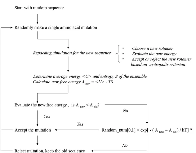
Incorporating explicit side chain entropy into Rosetta. Figure 2.1 outlines our approach for
incorporating side chain entropy into Rosetta. It is a two layer approach; the inner layer is used for
calculating the free energy of a fixed sequence on a fixed backbone, while the outer layer is used to
scan through sequence space. The inner layer uses Monte Carlo sampling to generate an ensemble of
structures with a variety of side chain conformations. Each round of this procedure involves:
1) switching a single residue (chosen at random) to a new Dunbrack rotamer
2) evaluating the new energy of the protein
3) accepting the perturbation if it passes the Metropolis criterion.
At the completion of each round the energy of the protein is added to a running sum that is later
divided by the number of rounds to determine the average energy of the ensemble (U in equation 1).
After each round, it is also determined which rotamer is present at each sequence position and these
each sequence position (p in eq. 2)2. These probabilities are used to calculate the side chain
conformational entropy (S) and Helmholtz free energy (A) of the system:
(2)
)
)
,
(
ln(
)
,
(
(1)
1 1
!
!
= ="
=
"
=
nrot r nres ii
r
p
i
r
p
R
S
TS
U
A
where nres is the number of residues in the protein, nrot is the number of possible rotamers at each
sequence position, and T is the temperature. T was set to a physiologically relevant temperature (310
K) for these calculations. Average energies and rotamer probabilities were calculated after the system
was equilibrated for
( 3 * nrotamers ) rounds, where nrotamers is equal to the number of rotamers being considered at each
sequence position times the number of residues being redesigned. Similar results were obtained if the
system was equilibrated for 5 * nrotamers rounds.
The outer layer uses Monte Carlo sampling to scan through sequence space(Figure 2.1). The
procedure begins with a random sequence. Each round of optimization involves:
1) making a random single amino acid mutation
2) evaluating the free energy of the new sequence with a repacking simulation ( equation 2)
3) accepting the mutation if it passes the Metropolis criterion.
For the outer layer the temperature is set high at the beginning of the simulation and gradually cooled
to 0 K. 100 * number of residues sequence substitutions are used per simulation. Because a
complete repacking simulation is performed after each sequence change, the double layer protocol is
considerably slower than a standard sequence optimization simulation with Rosetta (60 times
slower). To increase computational speed the protocol was modified so that only residues within
residues further away from the mutation site did not change, and therefore it was possible to use
values saved from the previous repacking simulation. The modified protocol was 1.5 times faster,
and gave the same results as when all residues are repacked following each mutation.
The quality of our results will depend in part on how accurately we can pack amino acid side chains
(the inner layer of our protocol). The accuracy of side chain packing algorithms is often evaluated by
removing the side chains from naturally occurring proteins and rebuilding the side chains from
scratch. The procedure is evaluated by determining the fraction of the amino acids that are placed in
the correct side chain conformation. To insure that Rosetta performs satisfactorily on this test we
rebuilt the side chains on 57 high resolution crystal structures. With the standard Dunbrack rotamer
library, 80% of the buried positions had both their chi 1 and chi 2 angles predicted within 40 degrees
of the angles observed in the crystal structure. These results are similar to what has been achieved
with other side chain placement algorithms 12,45-48.
Calculating side chain conformational entropies through complete enumeration. One assumption of
equation 2 is that the rotamer probabilities at the various sequence positions are independent of each
other, or in other words, that there is no covariant motion between side chains. If there is covariant
motion equation 2 will overestimate the side chain entropy of the system. To test the validity of this
assumption we used complete enumeration of rotamer configurations for 6 residue clusters to
calculate the energies of all possible rotamer combinations. These energies were used to generate a
partition function for the cluster and calculate the relative probabilities of each possible packing
combination state (equation 3). These probabilities were then used to calculate the entropy of the
system (equation 4), and compared with results obtained by Monte Carlo sampling as described
(4) ) ln( ) 3 ( p 1 1 / / i
!
!
= = " " " = = nstates i i i nstates i T K E T K E p p R S e e B i B iKB is the Boltzmann constant and T is the temperature (310K). 816 clusters in 110 proteins were
used for this comparison. Side chains outside of the cluster were held fixed during the complete
enumeration protocol and the Monte Carlo sampling protocol.
Calculating side chain conformational entropy from a 80 ns molecular dynamics simulation of eglin
C. To further check if covariant side chain motion reduces the total entropy of a protein we examined
a 80 ns molecular dynamics simulation of eglin C from a previous study.49 Eglin C remains folded
throughout this simulation in a conformation similar to the crystal structure. Previously, Lee and
co-workers used this trajectory to calculate order parameters for the side chains, and there was a good
agreement between the calculated values and order parameters measured with NMR. To calculate
side chain entropy from the simulation dihedral angles for each side chain were extracted from every
0.36 picosecond and binned into rotamers based on Dunbrack’s rotamer definitions41. Rotamer
frequencies were used to calculate side chain entropy using two separate approaches. The first
approach was to treat each site independently, i.e., use the rotamer probabilities from single residues
to calculate entropy (equation 2), and the second was to use probabilities of rotamer pairs to calculate
entropy (equation 5):
(5) 1 ) ln( 1 1 ) , ( 1 ) , ( ! ! =
" "
!where nres is the number of residues in the protein, p(i,j) is the probability of seeing a rotamer pair (i,j)
during the whole simulation. As a control, we also calculated entropy by treating each chi angle in
the protein independently (equation 6).
(6) ) ln( _ 1 _ ) _ ( ) _ (
!
= " = bin nchi bin chi bin chi bin chi p p R SEach chi angle in the protein is divided into bins (chi_bin) based on Dunbrack’s rotamer definitions,
and the probability that a given side chain is in a specific bin (p(chi_bin)) is determined by averaging
over the molecular dynamics simulation.
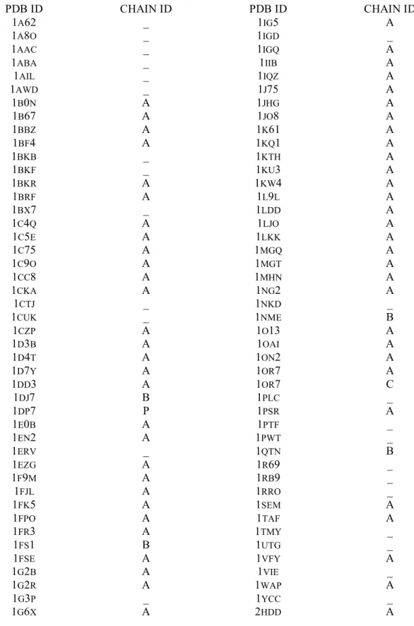
Native sequence recovery tests. Rosetta was used to design sequences for 110 proteins ranging in size
from 50 to 150 residues (see supplementary Table 2.6 for pdb codes), and the designed sequences
were compared to the wild type sequences. Cysteines were held fixed during these simulations
because the Rosetta energy function for disulfide formation still needs to be refined. Residues were
defined as buried if they had greater than 18 neighbors (C# atoms within 10Å), and surface if they had
less than 13 neighbors.
RESULTS AND DISCUSSION
Covariant changes in side chain position do not significantly reduce the side chain conformational
entropy of a protein. Before determining the effects of side chain entropy on sequence design, we
first tested if the total side chain entropy of a protein could be accurately calculated by assuming the
rotamer probabilities at each sequence position were independent of each other (equation 2). Because
number of populated states, and hence entropy, will be lower than if each residue moved
independently of its neighbors.
To test the importance of covariant motion between amino acid side chains we used two alternative
methods for measuring side chain conformational entropy. For the first method we enumerated
through all possible rotamer combinations for 6 residue clusters from naturally occurring proteins and
calculated the energy of each state. Residues outside of the cluster were held fixed. The energies
were then used to derive the probability of each state and the total entropy of the system (equations 3
and 4). For the second method we used Monte Carlo sampling of amino acid rotamers to create an
ensemble of structures. Rotamer probabilities for individual residues were calculated from the
resulting ensembles and total side chain entropy was calculated assuming the rotamer probabilities at
each sequence position were independent of each other (equations 1 and 2). The same 6 residue
clusters were used for this approach as were used for the complete enumeration.
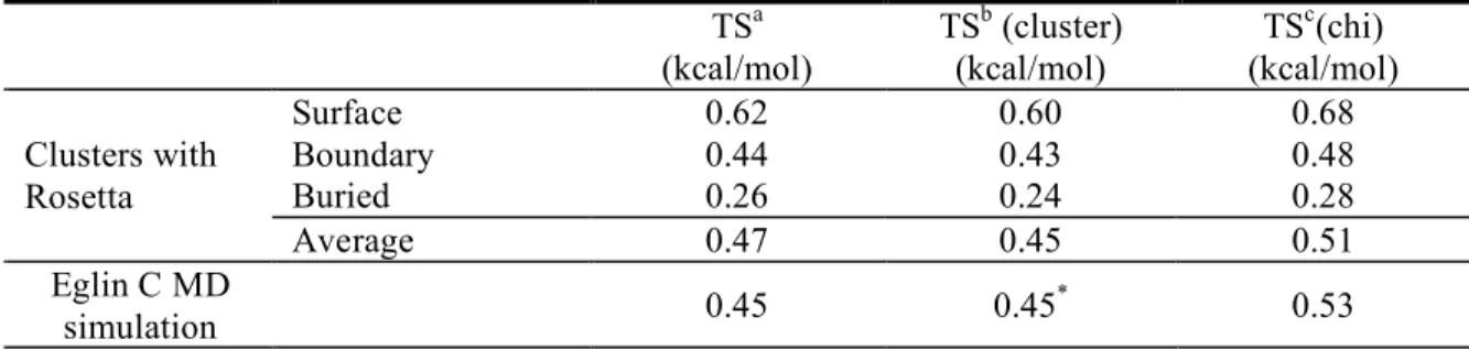
As expected, we measure higher entropies when we assume that the residues behave independently,
but in general the differences are very small ( Table 2.1 ). This suggests that there is not significant
covariant motion between side chains, and that equation 2 is suitable for calculating side chain
entropy during design simulations. A similar conclusion was reached by Leach et al. when using the
A* algorithm to explore rotamer packing proteins50. To further test this result we also examined side
chain motion from an 80 ns molecular dynamics simulation of eglin C. Side chain conformations
were assigned to bins corresponding to the Dunbrack rotamers, and rotamer frequencies were used to
calculate side chain conformational entropy. Entropy was calculated with two approaches, in the first
case the rotamer probabilities for individual residues were used (equation 2) while in the second case
probabilities for rotamer pairs were tabulated (equation 5). The two results were similar, suggesting
again that there is not significant covariant motion between amino acid side chains. In contrast, if we
entropy ( Table 2.1 ). This result indicates, as expected, that the torsion angles within an amino acid
side chain do not behave independently.
Side chain entropy and energy as a function of burial. Introducing the side chain entropy and free
energy model into our energy function should have two competing effects. Flexible side chains will
be rewarded for being able to sample multiple conformations, but at the same time they will be
penalized if those states are not iso-energetic and the average energy of the ensemble is greater than
the energy of the most favorable conformation. To examine the relative strength of these two effects
we performed two sets of repacking simulations on a large set of naturally occurring protein
structures (2832 pdb files). In the first case we used the standard Rosetta model to identify the lowest
energy side chain configuration for each protein and recorded the energy of each type of amino acid
as a function of burial. In the second case we performed repacking simulations at 310 K and recorded
entropies (equation 2) and average energy for each amino acid as a function of burial.
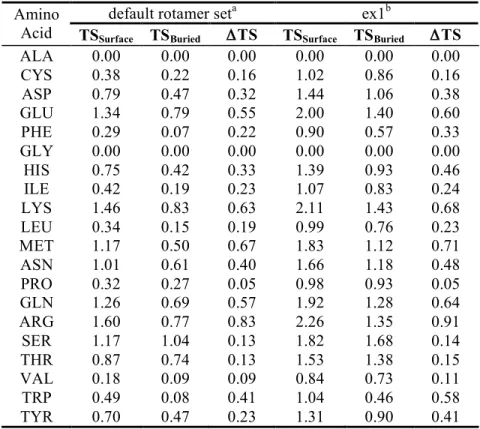
The longer amino acids have the highest average values of conformational entropy and show the
greatest difference between surface and buried positions (Figure 2.2). For instance, the average side
chain entropy (TS) for arginine is 1.60 kcal / mol on the surface of a protein and 0.77 kcal / mol in the
core of a protein, while for a valine the same values are 0.18 and 0.09 kcal / mol (Table 2.2).
However, the longer amino acids also show the greatest differences between average energy and the
energy of the most favorable rotamer. On the surface of proteins the average energy of arginines
when free to sample multiple conformations is on average 0.62 kcal / mol greater then the energy of
the most favorable rotamer. The net result is that the penalty for burying an arginine in the core of a
protein is not as great as would be suggested from just examining the side chain entropy term. In
general, including explicit side chain flexibility in the scoring function does not appear to
difference is smaller for lysine ( 0.26 kcal / mol ) because lysine has a strong intrinsic preference to
be in the extended conformation as evidenced by the Dunbrack rotamer library and high level
quantum mechanics calculations 51.
In these simulations we have been modeling the amino acid side chains using Dunbrack’s backbone
dependent rotamer library. This library does not allow for small perturbations of chi angles within a
rotamer. To test whether increasing the rotamer library would significantly perturb our results we
increased the rotamer library by allowing for rotamers that had their chi 1 angles perturbed +/- one
standard deviation from the most preferred chi 1 angle. These perturbations are generally around 10
degrees and are based on the standard deviations in the Dunbrack library. As other groups have
observed previously 2,50,52, with the expanded rotamer library the absolute side entropy of the amino
acids goes up significantly but the difference between surface exposed and buried positions is largely
unchanged (Table 2.3). This suggests that vibration of torsional angles within rotamers will not be a
key determinant of whether an amino acid prefers to buried or exposed.
Protein Design Simulations with Explicit Side Chain Entropy. To test if side chain entropy plays a
large role in determining the environmental preferences of the amino acids Rosetta was modified to
include explicit side chain entropy and free energy calculations (see methods) and sequences were
designed for 110 naturally occurring protein sequences. Although Rosetta uses a stochastic search
procedure to search for low free energy sequences, independent simulations for a single protein
produce very similar sequences (> 70% identity between simulations), indicating that the protocol
does not get trapped in false minima located far from the global minima. To control the overall
frequency that each amino acid is used during design, Rosetta, assigns a unique reference value to
each amino acid. Because the purpose of these simulations were to determine the role of side chain
reference values for each set of simulations so that the amino acids were designed at native-like
frequencies.
Overall, including side chain entropy as a new energy term did not have a large effect on the recovery
of native sequences (Table 2.4). 32% of the residues in the design set were kept as the native amino
acid in the simulations with and without the entropy term. As anticipated, the largest changes are
observed for the more flexible amino acids (Table 2.5). With the explicit side chain entropy model
49% of methionines are designed in the core while with standard Rosetta 55% of methionines are
placed in the core. Arginine shows the largest changes, 20% are placed in the core without explicit
side chain entropy while only 12% are placed in the core with explicit side chain entropy. For most
amino acids, the environmental preferences were not significantly perturbed by adding the explicit
side chain entropy. Most likely this is because gains in favorable side chain entropy are generally
accompanied by an increase in average energy (Figure 2.2), and the sum of these two effects is
considerably smaller than the magnitude of other terms in the energy function.
Results with and without side chain entropy contrast sharply with simulations performed with and
without the Lazaridis-Karplus solvation model. Without the solvation model there are dramatic
changes in the environmental preferences of the amino acids, and the identity between the designed
sequences and the wild type sequences falls to 19% (Table 2.4). So although the longer polar
residues may be partially disfavored from buried positions because of a loss in conformational
entropy, it is clear that desolvation energies are a much stronger factor than side chain entropy in
determining the environmental preferences of the amino acids.
It is important to point out that the relative importance of side chain entropy does depend on the