Proefschrift
ter verkrijging van
de graad van Doctor aan de Universiteit Leiden,
op gezag van Rector Magnificus prof. mr. P.F. van der Heijden,
volgens besluit van het College voor Promoties
te verdedigen op woensdag 19 mei 2010
klokke 13.45 uur
door
Oliver W. Stockhammer
geboren te Stuttgart, Duitsland
Promotor:
Prof. dr. H.P. Spaink
Co-Promotor:
Dr. A.H. Meijer
Overige leden:
Prof. dr. P.J.J. Hooykaas
Prof. dr. J. den Hertog
Prof. dr. A.J. Durston
Dr. S.A. Renshaw (University of Sheffield)
Charlie Chaplin
Chapter 1 Introduction to the vertebrate innate immune system 9
Chapter 2 MyD88 innate immune function in a zebrafish embryo infection model 27
Chapter 3 Transcriptome profiling and functional analyses of the zebrafish embryonic innate immune response to Salmonella infection 41
Chapter 4 Transcriptome analysis of Traf6 function in the innate immune response of zebrafish embryos 75
Chapter 5 Transcriptome analysis of Traf6 function in early zebrafish embryogenesis 99
Chapter 6 Summary and discussion 121
Samenvatting 129
List of publications 135
system
The immune system of animals is a complex composition of cellular and humoral components that protects the host against infectious diseases and cancer by identi-fying and killing pathogens and detrimental cells. To successfully protect the host, the immune system must be able to distinguish self from non-self and recognize danger signals. The concept of protecting self from non-self is already present in unicellular organisms, demonstrated by a repertoire of mechanisms ranging from the production of antimicrobial peptides to the employment of specific molecular systems protecting against foreign nucleic acids. With the appearance of multicel-lular organisms an increasingly complex immune system evolved. Distinct cells of the organism adopted specialized immune functions showing the ability to detect in vading pathogens, migrate to sites of infection and eventually engulf and eliminate the encountered microorganisms. At the same time, soluble factors such as anti-microbial peptides and acute-phase proteins are used to combat the infection. To be able to recognize a wide variety of pathogens and detrimental cells various classes of receptors have evolved and diversified in different organisms. The most sophisticated immune system today exists in all higher vertebrates and combines a wide range of receptors with the development of immunological memory. This thesis focuses on the use of the zebrafish (Danio rerio) as a model to study the vertebrate immune system.
The vertebrate immune system
recombi-nation of gene segments and clonal selection (3). The high affinity of antibodies to specific antigens is further achieved by somatic hypermutation in the gene segments encoding the variable regions of the antibody (4). Subsets of the activated B- and T-lymphocytes will be retained in lymphoid organs as memory cells. These cells can be reactivated by a recurring infection of a particular pathogen leading to a faster and highly specific immune response, thereby generating a long-lasting immunity against previously encountered pathogens. Although the two systems show obvious differences in the molecular tools used to battle infections, one should keep in mind that the innate and the adaptive immune systems in vertebrates are closely linked in their response to infectious microbes and that the innate immune system is pivotal for an accurate adaptive response. Given that the major focus of this thesis is on in-nate immunity, the components (both humoral and cellular) and the functions of the vertebrate innate immune system will be described in more detail in the following sections.
Humoral components of the vertebrate innate immune system
Acute-phase proteins
Sensing and killing of microbes can be achieved by several molecules of the in-nate immune system. A group of proteins, collectively termed acute-phase proteins (APPs), are greatly increased or decreased in the blood upon infection. Production and secretion of such proteins occurs in hepatocytes activated by cytokines such as TNFα, IL-6 and IL-1 (5). Acute phase proteins are a heterogenic group of proteins with either pro- or anti-inflammatory functions. A well known factor of the acute-phase response is the C-reactive protein (CRP), a member of the pentraxin family. CRP was shown to bind, among others, phosphocholine and phosphoethanolamine, which can be found on the cell surface of bacteria. CRP bound to macromolecules can lead either to opsonisation of bacteria (or apoptotic cells) or to the activation of the classical complement pathway described below (6).
Complement system
is mediated by the spontaneous hydrolysis of complement component 3 (C3) and the subsequent formation of the C3bBb complex without participation of a specific pathogen-recognizing protein. Activation by any one of these pathways will lead to cleavage of C3 to C3a and C3b by a C3 convertase. The C3 convertase has distinct compositions: a C4bC2b complex in the classical and lectin pathways, and a complex of C3b and activated factor B (Bb) in the alternative pathway (3).
The C3a fragment is a potent inflammatory mediator triggering vasodilatation and increasing permeability of small blood vessels. In addition C3a can induce oxi-dative burst in macrophages, neutrophils and eosinophils and leads to degranu lation of mast cells and basophils, thereby sustaining the inflammation. The second pro-duct of C3 cleavage, C3b, can bind to the cell surface of pathogens and either form additional C3bBb complexes that amplify the complement signal in the close prox-imity of the pathogen, or function as opsonin, enhancing phagocytosis. The latter is mediated by complement receptors on the surface of phagocytes (3).
Subsequent to C3 cleavage ,C3b can complex with C4bC2b to form a C5 con-vertase leading to the release of C5a and C5b. The C5a fragment functions as a pro-inflammatory mediator in the same fashion as C3a. On the other hand, C5b leads to the assembly of the membrane attack complex. Essentially, this complex is composed of C9 mole cules that form a pore in the cell membrane of the pathogen, resulting in a loss of cellular homeostasis and free passage of host enzymes such as lysozyme, ultimately causing cell lysis (3).
Antimicrobial peptides
The diverse group of small molecules (<100 amino acids) that are involved in the elimination of pathogenic microbes and enveloped viruses are collectively named antimicrobial peptides (AMPs) (7-9). In vertebrates AMPs can be classified into three groups: defensins, histatins and cathelicidins (10, 11). They are produced in various tissues and cells types such as Paneth cells of the intestine, lung epithelial cells and leukocytes (12-14). The precise mode of action of antimicrobial peptides is not fully understood, but involves membrane permeabilization and/or inhibition of protein and RNA synthesis (15).
Cell-mediated vertebrate innate immunity
Various cell types of the myeloid lineage are responsible for the detection and clear-ance of infectious microorganisms, apoptotic cells and tumour cells. Furthermore, they possess an instructive role towards the adaptive immune system. In addition to the various myeloid cells, the natural killer (NK) cells, derived from a lymphoid precursor, are also considered part of the cellular innate immune system. In the fol-lowing paragraphs, the function of individual cell-types, predominantly studied in rodents, will be discussed in more detail.
cells in mammals and are major effectors of innate immunity. Together with baso-phil and eosinobaso-phil granulocytes they form the polymorphonuclear cell family. Neutrophils can efficiently phagocytose and kill internalized microbes in phago-somes through reactive oxygen species (ROS) and proteolytic enzymes. In addition, by exocytosis of their granules (degranulation), neutrophils can release a multi-tude of antimicrobial proteins and proteases that destroy pathogens extracellularly. Another (phagocytosis-independent) mechanism that is used by neutrophils to kill
pathogens is the activation of neutrophil extracellular traps (NETs) (16). NETs are web-like structures composed of DNA, histone proteins and neutrophil elastase (a serine protease) that can trap and kill microbes. Both anti-bacterial and anti-fungal properties of NETs have been described (16, 17).
Macrophages are the predominant phagocytic cells of mammals. Immature
macro-phages derived from bone marrow circulate as monocytes through the blood. A subpopulation of these monocytes leave blood-circulation and migrate into the sur-rounding tissue were they can develop into resident macrophages such as osteo-clasts (bone), microglia (CNS) or Kupffer cells (liver) depending on the tissue they inhabit. Monocytes and macrophages express various types of pattern recognition receptors on their cell surface and intracellularly, that allow these cells to respond effectively to diverse classes of pathogens. Cytokines produced by NK cells or tissue macrophages upon infection or tissue damage can trigger migration of additional monocytes from the blood to the site of the infection, where they differentiate into mature macrophages. Depending on the mode of activation triggered by diverse combinations of cytokines, macrophages can promote inflammation or even par-ticipate in wound healing (18). During the adaptive immune response macrophages are activated by T-cells, leading to increased production of ROS and activation of the autophagy pathway. Macroautophagy is a major defence mechanism against in-tracellular pathogens (19). Beside their function during inflammation, macrophages also contribute to maintaining homeostasis by removal of cell debris, apoptotic cells and erythrocytes (18).
Dendritic cells form, together with macrophages, the major antigen presenting cells (APC) of the mammalian immune system. Immature DCs are constantly patrolling the tissue, taking up pathogens and apoptotic cell fragments by macro pinocytosis, eventually leading to antigen presentation on the cell surface. Encounter of patho-gen-derived molecules by immature DCs leads to DC activation, maturation and pro-inflammatory cytokine secretion. Mature DCs migrate subsequently to nearby lymph nodes where antigen presentation takes place, inducing primary T-cell medi-ated immune responses (20).
matu-ration after tissue migmatu-ration (21). Several pattern-recognition receptors are expressed on the cell surface of mast cells allowing direct sensing of invading pathogens (22). In addition to this direct activation, mast cells can also be activated indirectly via the complement system, leading to cytokine release and degranulation (22). Mast cell activation leads to a variety of effects, such as degradation of endogenous toxins, bactericidal activity, vasodilatation, T-cell activation and recruitment of neutrophils and DCs to the site of infection (23, 24). On top of their function in innate immune responses, mast cells have been extensively studied in the context of allergies (25).
Natural Killer cells, unlike the aforementioned cell types, derive from a common lymphoid precursor that also generates B- and T- lymphocytes of the cellular adap-tive immune system (26). NK cells are able to target both virally infected and tu-mor cells, and destroy them by releasing cytotoxic molecules such as perforin and granzymes (27). NK cell function is orchestrated by various cell surface receptors that, upon activation, can either lead to inhibition or activation of exocytosis of the cytotoxic granules and lysis of the targeted cell. Inhibitory signals can be mediated via the CD94/NKG2A and CD94/NKG2B receptors that recognize specific major histocompatibility complex (MHC) class I surface proteins (28, 29). Abnormal or virally infected cells tend to down-regulate MHC class I proteins on the cell surface and hence lack an inhibitory signal (30, 31). At the same time these cells present activating signals such as MHC class I chain-related (MIC) molecule MICA that fa-cilitate NK cell activation via NKG2D, a C-type lectin receptor (32, 33). Intercalation of the various signals eventually leads to the formation of the lytic immunological synapse between NK and target cells, cytotoxin release and lysis of the infected cell (34).
Pathogen monitoring by pattern-recognition receptors
Recognizing potentially harmful microorganisms is an essential first step in the initi-ation of an immune response. The innate immune system relies on the recog nition of highly conserved structural components of microbes, often referred to as pathogen-associated molecular patterns (PAMPs) or microbial-pathogen-associated molecular patterns (MAMPs). PAMPs or MAMPs are usually essential for microbial survival, hence a constant factor for the host to detect. Examples are bacterial cell-wall components such as LPS and peptidoglycan, flagellin from bacterial fla gella or viral RNAs. The receptors involved in PAMP recognition, PRRs, are widely expressed on the cells of the innate immune system such as macrophages and DCs, and on non-immune cells that are likely to encounter pathogens, such as epithelial cells. Several families of PRRs have been described in vertebrates, including the Toll-like receptor (TLRs), NOD-like receptor (NLR) and RIG-I-like (RLRs) receptor families (35-37).
The TLRs are the best studied and probably most essential receptors of the ver-tebrate innate immune system. The TLRs are named after the Toll receptor from
dorsal-ventral polarity determination (38). More than a decade later Lemaiter et al.
unravelled a function of Toll in the antifungal immune response of D. melanogaster
and shortly afterwards mammalian TLR4 was identified as the receptor that medi-ates LPS signalling (39, 40).
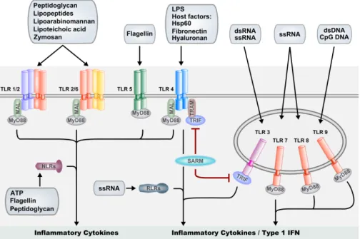
Today, a lot is known about TLR activation and downstream signalling events in mammals. All TLRs are germline-encoded type I transmembrane receptors characterized by a highly variable extracellular leucine-rich repeat (LRR) domain, involved in ligand recognition, and an intracellular tail, containing the conserved Toll/Interleukin-1 receptor (TIR) domain, mediating association of TLRs with down-stream signalling intermediates. A set of 10 TLRs have been described in human so far, showing distinct specificity to various PAMPs. For instance, TLR3, -7, and -8 recognize double- and/or single-stranded RNA, whereas TLR-9 recognizes bacterial DNA. TLR4 has been shown to recognize LPS and TLR5 is specific for bacterial fla-gellin. Heterodimers of TLR2 with TLR1 or TLR6 are able to recognize various lipo-proteins and glycolipids from gram-positive bacteria (Fig.1) (41-45). In accordance with their ligand specificity TLR3, -7, -8 and - 9 are located in the endolysosomal
compartments, whereas the TLR1, -2, -4, -5 and -6 are located on the cell surface (46). Activation of the TLRs initiates distinct signalling pathways that result in the accu-mulation of pro-inflammatory cytokines and type I interferons (IFNs). A group of 5 adaptor proteins (MyD88, MAL, TRIF, TRAM and SARM), binding to the various TLRs upon activation, are differentially used by TLRs to mediate a tailored response. MyD88 is the most commonly used adaptor, and signalling through MyD88 has been reported for all human TLRs with the exception of TLR3, that instead shows a TRIF-dependent signalling route (47, 48). While TLR5, 7, 8 and 9 signal through MyD88 alone, TLR2/TLR1 and TLR2/TLR6 heterodimers require MAL as an addi-tional adaptor to link MyD88 to the receptor complex. TLR4 signalling can lead to activation of pro-inflammatory cytokine genes via a MyD88/MAL-dependent path-way, and can result in type I INF production via a MyD88-independent pathway that utilizes the adaptors TRAM and TRIF (49). The fifth TIR-domain adaptor, SARM, has been proposed to function as a negative regulator of TRIF, but was also shown to positively regulate the response to viral infection in brain cells (50, 51). Downstream of the TLR-adaptors, signals are relayed via TNF-receptor associated factors TRAF6 and TRAF3, activating downstream kinases which eventually lead to gene induction through the nuclear factor kappaB (NF-κB) and interferon response factor (IRF) families of transcription factors, and to MAP kinase (MAPK) signalling pathways activating the AP-1 (JUN/FOS) transcription factor complex (Fig.1) (52, 53).
Whereas the TLRs are located on the cell surface and endolysosomal membranes, the members of the NLR family are predominantly distributed in the cytosol of the cell, where they are primarily involved in bacterial recognition (35). The NLR pro-teins consist of an N-terminal effector domain, a central nucleotide-binding oligo-merization domain (NOD) and a C-terminal LRR domain. The N-terminal domain facilitates signalling through downstream partners, whereas the LRR domain is nec-essary for PAMP detection. Similarly to the TLRs, NLRs can be activated by micro-bial compounds such as flagellin and peptidoglycan. In addition, NLRs are respon-sive to bacterial toxins and crystals as well as to endogenous danger signals. By anal-ogy with PAMPs, these danger signals, of which extracellular ATP is a good example, are referred to as DAMPs (danger associated molecular patterns). As is the case of TLRs, triggering of NLRs can lead to NF-κB and MAPK pathway acti vation that in turn leads to production of cytokines and anti-microbial proteins. Furthermore, NLR signalling can lead to activation of the inflammasome that is required for se-cretion of active IL-1β and IL-18. These potent inflammatory cyto kines are pro-duced as inactive precursors through TLR pathway activation, and caspase-1 medi-ated processing in the inflammasome is required for their activation (35). Thus, a robust inflammatory response is dependent on the cooperative action of TLRs and NLRs (Fig.1).
Similarly to NLRs, RLRs are also located in the cytoplasm. Together with TLR3, -7 and -8 the RLRs provide viral recognition and mount a robust induction of type I
In addition to the above mentioned PRRs, a role in the innate immune defence is also played by the C-type lectin receptor (CLRs) family and the scavenger receptor family. C-type lectin receptors are expressed on such cells as DCs, where they can de-tect fungi, bacteria and viruses through the recognition of mannose, fucose and glu-can carbohydrates. Several CLRs, such as DC-specific ICAM3-grabbing non integrin (DC-SIGN), have been shown to modulate TLR signalling. Dependent on the path-ogen involved, both cooperative and antagonizing interactions between CLR and TLR signalling have been found. CLR signalling has furthermore been implicate d in the tailored activation of T-cell subsets (55). Finally, scavenger receptors have been shown to participate in TLR signalling as TLR co-receptors; these mediate, in addi-tion, non-opsonic phagocytosis of pathogenic microbes (56).
Common factors of the host response to infection
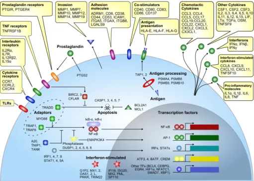
Activation of PRRs leads to the induction of a transcriptional response, priming the host to adequately respond to the encountered pathogen. In a comprehensive review study, Jenner and Young performed a meta-analysis on transcriptome data from vari ous infection studies of different cells types with different pathogens, leading to the identi fication of a gene set that is commonly regulated upon infection. Among
the identified genes were pro-inflammatory mediators (TNF, IL1β, IL6 and IL8), chemotactic and interferon stimulated cytokines (CCL3, CXCL1, CCL8), tissue in-vasion proteins (MMP1, MMP14), cell adhesion proteins (CD6, ICAM1), signalling adaptors (MyD88, TRAF6) and various transcription factors (NF-κB, AP1, STATs and IRFs), as shown in figure 2 (57).
Zebrafish embryos as a model to study vertebrate immunity
In recent years the zebrafish (Danio rerio) embryo system has emerged as a new model to study vertebrate innate immunity, offering several advantages that comple-ment mammalian model systems. The transparent character of the externally ferti-lized zebrafish embryo in combination with fluorescently-labelled immune cells and bacteria facilitate the study of host-microbe interaction and inflammation processes in the living organism (58-64). The efficiency at which infections and chemical treat-ments in zebrafish can be performed at a large-scale allows identification of novel microbial virulence factors and high-throughput compound screens to investigate disease mechanisms (65, 66). Moreover, the zebrafish system is particularly suitable for large-scale forward and reverse genetic screens aimed at the identification of genes with novel functions in the development of the immune system or in the im-mune response (67-69).
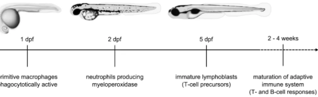
Like other vertebrates the zebrafish has a primitive and definitive wave of hemato-poiesis giving rise to various cell types of the erythroid, lymphoid and myeloid line-age. During development, hematopoiesis occurs at several temporal locations in the embryos, finally shifting to the kidney marrow, which is equivalent to the mam-malian bone marrow. Similarly to other vertebrates, adult zebrafish possess T- and B-cells, macrophages, neutrophils, eosinophils, basophils, mast cells and probably NK cells (70). Cells of the innate immune system are detectable as early as the first day of zebrafish development (71). These primitive macrophages are able to phago-cytose bacteria and foreign material (72, 73). Functional neutrophils, producing the myeloperoxidase enzyme, are present from the second day of embryogenesis. By contrast, a functionally mature adaptive immune system is not active during the first
three weeks of zebrafish development (58, 74, 75). This clear temporal separation in zebrafish embryos provides a convenient system for in vivo study of the vertebrate innate immune response to infection, independently from the adaptive immune re-sponse (Fig.3). In recent years, numerous bacterial and viral infection models have been established for the zebrafish to study host-pathogen interaction, chemotactic responses and inflammation processes (62, 76-78).
Zebrafish pattern-recognition receptors and innate immune response
activation
Genome analysis revealed one or more homologs of the human TLR genes (TLR1,-2, -3,-4,-5-7,-8 and -9) to be present in zebrafish, as well as a group of fish-specific TLRs
(79, 80). The zebrafish genome is also known to contain four of the downstream adaptor protein genes (MyD88, TRIF, MAL and SARM), while the fifth TLR adaptor TRAM remains to be identified (79, 80). Several other genes of the TLR signalling cascade have been identified in the zebrafish genome, as well as members of the NLR family and the downstream adaptor of the RLR family IPS-1 (81-83). TLR and adaptor genes are broadly expressed during zebrafish embryogenesis, even prior to the appearance of the first innate immune cells (84). Challenge of zebrafish embryos with different pathogens activates the expression of a wide range of innate immune response genes strongly conserved with those in mammals (85). In addition to the cell-mediated innate immune response, zebrafish have a well developed complement system and produce acute-phase response proteins such as hepcidin and fibrinogen (78).
A conserved TLR ligand specificity was demonstrated between human and zebra-fish for TLR5 (Chapter 3). Stimulation of zebrazebra-fish embryos with purified flagellin led to transcriptional activation of distinct host defence genes such as interleukin-1β (il1b), matrix metalloproteinase 9 (mmp9) and cxc chemokine ligand C1c (cxcl-C1c)
in vivo that were significantly impaired after knock-down of the tlr5a and tlr5b genes (85). In contrast, the zebrafish counterparts of TLR4 (Tlr4a and Tlr4b) were reported to be non-responsive to LPS, suggesting a rendered ligand specificity or function in zebrafish (86, 87). While the alternative ligand specificities of zebrafish Tlr4a and b remain unknown, Sullivan et al. demonstrated that the intracellular portions of Tlr4a and b have the capacity to activate NF-κB signalling. This is in contrast with data from Sepulcre et al, who reported negative regulation of NF-κB by Tlr4b (87).
One of the best studied TLR adaptors in mammals is MyD88. Myd88 transcript was detected in cells of the myeloid lineage in the head, on the yolk sac, the trunk and the posterior blood island of zebrafish embryos. Myd88-positive leukocytes contribute to inflammatory responses and were able to phagocytose bacteria (88). Knock-down studies in zebrafish embryos revealed an essential function of Myd88 during Salmonella infection, showing a strongly impaired response to an other-wise non-pathogenic Salmonella strain (Chapter 2, 84). Furthermore, challenge of
of Myd88-dependent and -independent signalling pathways, as are also present in mammals (Chapter 3, 85). In addition, like in mammals the zebrafish homolog of TRIF has been shown to play an essential role in antiviral immunity (89). However, the precise mechanisms of NFκB and IFN activation upon viral infection appear to have diverged between fish and mammals. Therefore, while there is a large similarity of TLR receptors and downstream mediators between fish and mammals, further studies should clarify to what extend the TLR ligand specificities and downstream signal transduction mechanisms are conserved.
Outline of this thesis
In the work described in this thesis we make use of the zebrafish embryo to study vertebrate innate immune responses to systemic bacterial infections in general, and to assess the role of the TLR signalling pathway in the innate immune response in particular. To model systemic infections we use the Gram-negative enterobacteria
Salmonella enterica Serovar Typhimurium (S. typhimurium), the cause of human salmonellosis.
In Chapter 2 the fundamental and conserved role of Myd88 in the zebrafish
embryonic innate immune response is demonstrated. Using a morpholino-based knock-down approach we show that Myd88-mediated signalling events are crucial in mounting a sufficiently strong immune response to clear an infection with a non-pathogenic S. typhimurium strain.
Chapter 3 presents a time-resolved transcriptome analysis of the inflammatory and innate immune responses elicited by zebrafish embryos to a systemic infection with a pathogenic and non-pathogenic S. typhimurium strain. The transcriptional response to infection with both strains shows clear conservation with host responses detected in other vertebrate models and human cells, including induction of genes encoding cell surface receptors, signalling intermediates, transcription factors and inflammatory mediators. Extending the work of chapter 2 we show that Salmonella
infection is mediated by Myd88-dependent and -independent signalling events. Additionally, we demonstrate that gene induction by flagellin is mediated by Tlr5 in zebrafish embryos, indicating that ligand specificity for this member of the TLR family is conserved between human and zebrafish.
Chapter 4 is focused on the immune function of the zebrafish homolog of
mam-malian TRAF6, an important downstream mediator of the TLR pathway, demon-strating that traf6 knock-down leads to a strongly decreased transcriptional immune response upon systemic Salmonella infection. Among the Traf6-dependent genes is not only a large set of well known anti-microbial and inflammatory genes but also several genes whose role in the immune system was not previously expected to be Traf6-dependent. One such example is the fertility hormone gene GnRH.
References
1. Kasahara, M., T. Suzuki, and L. D. Pasquier. 2004. On the origins of the adaptive immune system: novel insights from invertebrates and cold-blooded vertebrates. Trends Immunol 25:105-111.
2. Kimbrell, D. A., and B. Beutler. 2001. The evolution and genetics of innate immunity. Nat Rev Genet 2:256-267. 3. Janeway, C. A. 2001. Immunobiology: the immune system in health and disease. Garland Publishing.
4. Li, Z., C. J. Woo, M. D. Iglesias-Ussel, D. Ronai, and M. D. Scharff. 2004. The generation of antibody diversity through somatic hypermutation and class switch recombination. Genes Dev 18:1-11.
5. Gabay, C., and I. Kushner. 1999. Acute-phase proteins and other systemic responses to inflammation. N Engl J Med 340:448-454.
6. Black, S., I. Kushner, and D. Samols. 2004. C-reactive Protein. J Biol Chem 279:48487-48490.
7. Salzman, N. H., D. Ghosh, K. M. Huttner, Y. Paterson, and C. L. Bevins. 2003. Protection against enteric salmo-nellosis in transgenic mice expressing a human intestinal defensin. Nature 422:522-526.
8. Nizet, V., T. Ohtake, X. Lauth, J. Trowbridge, J. Rudisill, R. A. Dorschner, V. Pestonjamasp, J. Piraino, K. Huttner, and R. L. Gallo. 2001. Innate antimicrobial peptide protects the skin from invasive bacterial infection. Nature
414:454-457.
9. Daher, K. A., M. E. Selsted, and R. I. Lehrer. 1986. Direct inactivation of viruses by human granulocyte defensins.
J Virol 60:1068-1074.
10. Yang, D., A. Biragyn, D. M. Hoover, J. Lubkowski, and J. J. Oppenheim. 2004. Multiple roles of antimicrobial de-fensins, cathelicidins, and eosinophil-derived neurotoxin in host defense. Annu Rev Immunol 22:181-215. 11. De Smet, K., and R. Contreras. 2005. Human antimicrobial peptides: defensins, cathelicidins and histatins.
Biotechnol Lett 27:1337-1347.
12. Ayabe, T., D. P. Satchell, C. L. Wilson, W. C. Parks, M. E. Selsted, and A. J. Ouellette. 2000. Secretion of micro-bicidal alpha-defensins by intestinal Paneth cells in response to bacteria. Nat Immunol 1:113-118.
13. Bals, R., X. Wang, M. Zasloff, and J. M. Wilson. 1998. The peptide antibiotic LL-37/hCAP-18 is expressed in epi-thelia of the human lung where it has broad antimicrobial activity at the airway surface. Proc Natl Acad Sci U S A 95:9541-9546.
14. Ganz, T. 1987. Extracellular release of antimicrobial defensins by human polymorphonuclear leukocytes. Infect Immun 55:568-571.
15. Brogden, K. A. 2005. Antimicrobial peptides: pore formers or metabolic inhibitors in bacteria? Nat Rev Microbiol 3:238-250.
16. Brinkmann, V., U. Reichard, C. Goosmann, B. Fauler, Y. Uhlemann, D. S. Weiss, Y. Weinrauch, and A. Zychlinsky. 2004. Neutrophil extracellular traps kill bacteria. Science 303:1532-1535.
17. Urban, C. F., U. Reichard, V. Brinkmann, and A. Zychlinsky. 2006. Neutrophil extracellular traps capture and kill Candida albicans yeast and hyphal forms. Cell Microbiol 8:668-676.
18. Mosser, D. M., and J. P. Edwards. 2008. Exploring the full spectrum of macrophage activation. Nat Rev Immunol
8:958-969.
19. Levine, B., and V. Deretic. 2007. Unveiling the roles of autophagy in innate and adaptive immunity. Nat Rev Immunol 7:767-777.
20. Liu, Y. J. 2001. Dendritic cell subsets and lineages, and their functions in innate and adaptive immunity. Cell
106:259-262.
21. Nakano, T., T. Sonoda, C. Hayashi, A. Yamatodani, Y. Kanayama, T. Yamamura, H. Asai, T. Yonezawa, Y. Kitamura, and S. J. Galli. 1985. Fate of bone marrow-derived cultured mast cells after intracutaneous, intraperi-toneal, and intravenous transfer into genetically mast cell-deficient W/Wv mice. Evidence that cultured mast cells can give rise to both connective tissue type and mucosal mast cells. J Exp Med 162:1025-1043.
22. Marshall, J. S. 2004. Mast-cell responses to pathogens. Nat Rev Immunol 4:787-799.
23. Shelburne, C. P., H. Nakano, A. L. St John, C. Chan, J. B. McLachlan, M. D. Gunn, H. F. Staats, and S. N. Abraham. 2009. Mast cells augment adaptive immunity by orchestrating dendritic cell trafficking through in-fected tissues. Cell Host Microbe 6:331-342.
24. Metz, M., and M. Maurer. 2007. Mast cells--key effector cells in immune responses. Trends Immunol 28:234-241.
25. Gilfillan, A. M., and C. Tkaczyk. 2006. Integrated signalling pathways for mast-cell activation. Nat Rev Immunol
6:218-230.
26. Vosshenrich, C. A., S. I. Samson-Villeger, and J. P. Di Santo. 2005. Distinguishing features of developing natu-ral killer cells. Curr Opin Immunol 17:151-158.
27. Topham, N. J., and E. W. Hewitt. 2009. Natural killer cell cytotoxicity: how do they pull the trigger? Immunology
128:7-15.
28. Braud, V. M., D. S. Allan, C. A. O'Callaghan, K. Soderstrom, A. D'Andrea, G. S. Ogg, S. Lazetic, N. T. Young, J. I. Bell, J. H. Phillips, L. L. Lanier, and A. J. McMichael. 1998. HLA-E binds to natural killer cell receptors CD94/ NKG2A, B and C. Nature 391:795-799.
30. Hewitt, E. W. 2003. The MHC class I antigen presentation pathway: strategies for viral immune evasion.
Immunology 110:163-169.
31. Waldhauer, I., and A. Steinle. 2008. NK cells and cancer immunosurveillance. Oncogene 27:5932-5943. 32. Bahram, S., H. Inoko, T. Shiina, and M. Radosavljevic. 2005. MIC and other NKG2D ligands: from none to too
many. Curr Opin Immunol 17:505-509.
33. Mistry, A. R., and C. A. O'Callaghan. 2007. Regulation of ligands for the activating receptor NKG2D. Immunology
121:439-447.
34. Lanier, L. L. 2005. NK cell recognition. Annu Rev Immunol 23:225-274.
35. Franchi, L., N. Warner, K. Viani, and G. Nunez. 2009. Function of Nod-like receptors in microbial recognition and host defense. Immunol Rev 227:106-128.
36. Kawai, T., and S. Akira. 2008. Toll-like receptor and RIG-I-like receptor signaling. Ann N Y Acad Sci 1143:1-20. 37. Medzhitov, R. 2001. Toll-like receptors and innate immunity. Nat Rev Immunol 1:135-145.
38. Anderson, K. V., G. Jurgens, and C. Nusslein-Volhard. 1985. Establishment of dorsal-ventral polarity in the Drosophila embryo: genetic studies on the role of the Toll gene product. Cell 42:779-789.
39. Lemaitre, B., E. Nicolas, L. Michaut, J. M. Reichhart, and J. A. Hoffmann. 1996. The dorsoventral regulatory gene cassette spatzle/Toll/cactus controls the potent antifungal response in Drosophila adults. Cell 86:973-983. 40. Poltorak, A., X. He, I. Smirnova, M. Y. Liu, C. Van Huffel, X. Du, D. Birdwell, E. Alejos, M. Silva, C. Galanos, M.
Freudenberg, P. Ricciardi-Castagnoli, B. Layton, and B. Beutler. 1998. Defective LPS signaling in C3H/HeJ and C57BL/10ScCr mice: mutations in Tlr4 gene. Science 282:2085-2088.
41. Alexopoulou, L., A. C. Holt, R. Medzhitov, and R. A. Flavell. 2001. Recognition of double-stranded RNA and activation of NF-kappaB by Toll-like receptor 3. Nature 413:732-738.
42. Diebold, S. S., T. Kaisho, H. Hemmi, S. Akira, and C. Reis e Sousa. 2004. Innate antiviral responses by means of TLR7-mediated recognition of single-stranded RNA. Science 303:1529-1531.
43. Hayashi, F., K. D. Smith, A. Ozinsky, T. R. Hawn, E. C. Yi, D. R. Goodlett, J. K. Eng, S. Akira, D. M. Underhill, and A. Aderem. 2001. The innate immune response to bacterial flagellin is mediated by Toll-like receptor 5.
Nature 410:1099-1103.
44. Heil, F., H. Hemmi, H. Hochrein, F. Ampenberger, C. Kirschning, S. Akira, G. Lipford, H. Wagner, and S. Bauer. 2004. Species-specific recognition of single-stranded RNA via toll-like receptor 7 and 8. Science 303:1526-1529. 45. Hemmi, H., O. Takeuchi, T. Kawai, T. Kaisho, S. Sato, H. Sanjo, M. Matsumoto, K. Hoshino, H. Wagner, K.
Takeda, and S. Akira. 2000. A Toll-like receptor recognizes bacterial DNA. Nature 408:740-745.
46. Barton, G. M., and J. C. Kagan. 2009. A cell biological view of Toll-like receptor function: regulation through compartmentalization. Nat Rev Immunol 9:535-542.
47. O'Neill, L. A., and A. G. Bowie. 2007. The family of five: TIR-domain-containing adaptors in Toll-like receptor signalling. Nat Rev Immunol 7:353-364.
48. Oshiumi, H., M. Matsumoto, K. Funami, T. Akazawa, and T. Seya. 2003. TICAM-1, an adaptor molecule that participates in Toll-like receptor 3-mediated interferon-beta induction. Nat Immunol 4:161-167.
49. Yamamoto, M., K. Takeda, and S. Akira. 2004. TIR domain-containing adaptors define the specificity of TLR signaling. Mol Immunol 40:861-868.
50. Carty, M., R. Goodbody, M. Schroder, J. Stack, P. N. Moynagh, and A. G. Bowie. 2006. The human adaptor SARM negatively regulates adaptor protein TRIF-dependent Toll-like receptor signaling. Nat Immunol 7:1074-1081.
51. Szretter, K. J., M. A. Samuel, S. Gilfillan, A. Fuchs, M. Colonna, and M. S. Diamond. 2009. The immune adap-tor molecule SARM modulates tumor necrosis facadap-tor alpha production and microglia activation in the brain-stem and restricts West Nile Virus pathogenesis. J Virol 83:9329-9338.
52. Kawai, T., and S. Akira. 2007. Signaling to NF-kappaB by Toll-like receptors. Trends Mol Med 13:460-469. 53. Oganesyan, G., S. K. Saha, B. Guo, J. Q. He, A. Shahangian, B. Zarnegar, A. Perry, and G. Cheng. 2006. Critical
role of TRAF3 in the Toll-like receptor-dependent and -independent antiviral response. Nature 439:208-211. 54. Mogensen, T. H. 2009. Pathogen recognition and inflammatory signaling in innate immune defenses. Clin
Microbiol Rev 22:240-273, Table of Contents.
55. Geijtenbeek, T. B., and S. I. Gringhuis. 2009. Signalling through C-type lectin receptors: shaping immune re-sponses. Nat Rev Immunol 9:465-479.
56. Areschoug, T., and S. Gordon. 2009. Scavenger receptors: role in innate immunity and microbial pathogenesis.
Cell Microbiol 11:1160-1169.
57. Jenner, R. G., and R. A. Young. 2005. Insights into host responses against pathogens from transcriptional pro-filing. Nat Rev Microbiol 3:281-294.
58. Davis, J. M., H. Clay, J. L. Lewis, N. Ghori, P. Herbomel, and L. Ramakrishnan. 2002. Real-time visualization of mycobacterium-macrophage interactions leading to initiation of granuloma formation in zebrafish embryos.
Immunity 17:693-702.
59. Mathias, J. R., B. J. Perrin, T. X. Liu, J. Kanki, A. T. Look, and A. Huttenlocher. 2006. Resolution of inflamma-tion by retrograde chemotaxis of neutrophils in transgenic zebrafish. J Leukoc Biol 80:1281-1288.
inflammation and mycobacterial granuloma formation in zebrafish. Dev Comp Immunol 32:36-49.
61. Redd, M. J., G. Kelly, G. Dunn, M. Way, and P. Martin. 2006. Imaging macrophage chemotaxis in vivo: studies of microtubule function in zebrafish wound inflammation. Cell Motil Cytoskeleton 63:415-422.
62. Renshaw, S. A., C. A. Loynes, D. M. Trushell, S. Elworthy, P. W. Ingham, and M. K. Whyte. 2006. A transgenic zebrafish model of neutrophilic inflammation. Blood 108:3976-3978.
63. van der Sar, A. M., R. J. Musters, F. J. van Eeden, B. J. Appelmelk, C. M. Vandenbroucke-Grauls, and W. Bitter. 2003. Zebrafish embryos as a model host for the real time analysis of Salmonella typhimurium infections. Cell Microbiol 5:601-611.
64. Ward, A. C., D. O. McPhee, M. M. Condron, S. Varma, S. H. Cody, S. M. Onnebo, B. H. Paw, L. I. Zon, and G. J. Lieschke. 2003. The zebrafish spi1 promoter drives myeloid-specific expression in stable transgenic fish. Blood
102:3238-3240.
65. Miller, J. D., and M. N. Neely. 2005. Large-scale screen highlights the importance of capsule for virulence in the zoonotic pathogen Streptococcus iniae. Infect Immun 73:921-934.
66. Lieschke, G. J., and P. D. Currie. 2007. Animal models of human disease: zebrafish swim into view. Nat Rev Genet 8:353-367.
67. Trede, N. S., T. Ota, H. Kawasaki, B. H. Paw, T. Katz, B. Demarest, S. Hutchinson, Y. Zhou, C. Hersey, A. Zapata, C. T. Amemiya, and L. I. Zon. 2008. Zebrafish mutants with disrupted early T-cell and thymus development identified in early pressure screen. Dev Dyn 237:2575-2584.
68. Schorpp, M., M. Bialecki, D. Diekhoff, B. Walderich, J. Odenthal, H. M. Maischein, A. G. Zapata, and T. Boehm. 2006. Conserved functions of Ikaros in vertebrate lymphocyte development: genetic evidence for distinct lar-val and adult phases of T cell development and two lineages of B cells in zebrafish. J Immunol 177:2463-2476. 69. Pase, L., J. E. Layton, W. P. Kloosterman, D. Carradice, P. M. Waterhouse, and G. J. Lieschke. 2009. miR-451
reg-ulates zebrafish erythroid maturation in vivo via its target gata2. Blood 113:1794-1804.
70. Wang, Y., H. Zong, Y. Chi, Y. Hong, Y. Yang, W. Zou, X. Yun, and J. Gu. 2009. Repression of estrogen receptor alpha by CDK11p58 through promoting its ubiquitin-proteasome degradation. J Biochem 145:331-343. 71. Herbomel, P., B. Thisse, and C. Thisse. 1999. Ontogeny and behaviour of early macrophages in the zebrafish
em-bryo. Development 126:3735-3745.
72. Herbomel, P., B. Thisse, and C. Thisse. 2001. Zebrafish early macrophages colonize cephalic mesenchyme and developing brain, retina, and epidermis through a M-CSF receptor-dependent invasive process. Dev Biol
238:274-288.
73. Lieschke, G. J., A. C. Oates, M. O. Crowhurst, A. C. Ward, and J. E. Layton. 2001. Morphologic and functional characterization of granulocytes and macrophages in embryonic and adult zebrafish. Blood 98:3087-3096. 74. Lam, S. H., H. L. Chua, Z. Gong, T. J. Lam, and Y. M. Sin. 2004. Development and maturation of the immune
system in zebrafish, Danio rerio: a gene expression profiling, in situ hybridization and immunological study.
Dev Comp Immunol 28:9-28.
75. Willett, C. E., A. Cortes, A. Zuasti, and A. G. Zapata. 1999. Early hematopoiesis and developing lymphoid or-gans in the zebrafish. Dev Dyn 214:323-336.
76. Kizy, A. E., and M. N. Neely. 2009. First Streptococcus pyogenes signature-tagged mutagenesis screen identifies novel virulence determinants. Infect Immun 77:1854-1865.
77. Lesley, R., and L. Ramakrishnan. 2008. Insights into early mycobacterial pathogenesis from the zebrafish. Curr Opin Microbiol 11:277-283.
78. Meeker, N. D., and N. S. Trede. 2008. Immunology and zebrafish: spawning new models of human disease. Dev Comp Immunol 32:745-757.
79. Meijer, A. H., S. F. Gabby Krens, I. A. Medina Rodriguez, S. He, W. Bitter, B. Ewa Snaar-Jagalska, and H. P. Spaink. 2004. Expression analysis of the Toll-like receptor and TIR domain adaptor families of zebrafish. Mol Immunol 40:773-783.
80. Jault, C., L. Pichon, and J. Chluba. 2004. Toll-like receptor gene family and TIR-domain adapters in Danio re-rio. Mol Immunol 40:759-771.
81. Stein, C., M. Caccamo, G. Laird, and M. Leptin. 2007. Conservation and divergence of gene families encoding components of innate immune response systems in zebrafish. Genome Biol 8:R251.
82. Laing, K. J., M. K. Purcell, J. R. Winton, and J. D. Hansen. 2008. A genomic view of the NOD-like receptor fam-ily in teleost fish: identification of a novel NLR subfamfam-ily in zebrafish. BMC Evol Biol 8:42.
83. Biacchesi, S., M. LeBerre, A. Lamoureux, Y. Louise, E. Lauret, P. Boudinot, and M. Bremont. 2009. Mitochondrial antiviral signaling protein plays a major role in induction of the fish innate immune response against RNA and DNA viruses. J Virol 83:7815-7827.
84. van der Sar, A. M., O. W. Stockhammer, C. van der Laan, H. P. Spaink, W. Bitter, and A. H. Meijer. 2006. MyD88 innate immune function in a zebrafish embryo infection model. Infect Immun 74:2436-2441.
85. Stockhammer, O. W., A. Zakrzewska, Z. Hegedus, H. P. Spaink, and A. H. Meijer. 2009. Transcriptome profiling and functional analyses of the zebrafish embryonic innate immune response to Salmonella infection. J Immunol
182:5641-5653.
183:5896-Evolution of lipopolysaccharide (LPS) recognition and signaling: fish TLR4 does not recognize LPS and nega-tively regulates NF-kappaB activation. J Immunol 182:1836-1845.
88. Hall, C., M. V. Flores, A. Chien, A. Davidson, K. Crosier, and P. Crosier. 2009. Transgenic zebrafish report-er lines reveal consreport-erved Toll-like receptor signaling potential in embryonic myeloid leukocytes and adult im-mune cell lineages. J Leukoc Biol 85:751-765.
Astrid M. van der Sar1, Oliver W. Stockhammer2, Carina van der Laan1,
Herman P. Spaink2, Wilbert Bitter1 and Annemarie H. Meijer2
1 Department of Medical Microbiology, VU Medical Centre, Amsterdam, The Netherlands 2 Institute of Biology, Leiden University, Leiden, The Netherlands
embryo infection model
Abstract
Innate immunity signalling mechanisms during vertebrate embryogenesis are largely unknown. To study Toll-like receptor (TLR) signalling function in the zebrafish em-bryo model, we designed an experimental setup for antisense morpholino knock-down under conditions of bacterial infection. Clearance of Salmonella typhimurium
Ra bacteria was significantly impaired after knockdown of myeloid differentiation factor 88 (MyD88), a common adaptor protein in TLR and interleukin-1 recep-tor signalling. Thereby, we demonstrate for the first time that the innate immune response of the developing embryo involves MyD88-dependent signalling, which further estab lishes the zebrafish embryo as a model to study vertebrate innate im-munity.
Introduction
Innate immunity relies heavily on signalling by members of the Toll-like receptor (TLR) family (25). TLRs and associated adaptor molecules are highly conserved be-tween zebrafish (Danio rerio) and other vertebrates (13, 18). Bacterial and viral infec-tions were found to induce expression levels of different zebrafish TLR genes (18, 20). However, direct functional evidence to confirm the role of TLR signalling in the innate immune response of zebrafish has not yet been reported.
The exploitation of zebrafish as an animal model to study immunity and infec-tious diseases is attractive for three main reasons (34, 28, 29, 30). First, high-through-put forward genetic screens in zebrafish are a powerful means to uncover novel im-mune functions. Second, the optical transparency of the free-living zebrafish em-bryos makes it possible to examine the early development of the immune system and the progression of microbial infections in real-time (11, 32, 12, 6, 31). Third, the zebrafish embryo is easily accessible to experimental manipulations and efficient in-activation of gene functions can be achieved by injection of antisense morpholino oligonucleotides (19). However, a major obstacle is that many of the immunological details and research tools that are available for more established animal models have not yet been resolved and developed for zebrafish.
the myeloid lineage initially express the transcription factor gene Pu.1 (Spi1), which is essential for their differentiation (21). After 1 dpf Pu.1 expression decreases and two distinct populations of myeloid cells can be distinguished by the expression of two marker genes, L-plastin, which encodes a macrophage-specific actin-bundling protein, and mpx, which encodes a member of the myeloperoxidase family (11, 3, 17). At 2 dpf, the mpx-positive cells show the morphological characteristics of neutrophil granulocytes and are able to migrate to sites of trauma (17, 4). Immature lympho-blasts can first be detected by 3 dpf, but T and B lymphocytes do not mature until 4 to 6 weeks after hatching (5, 16). Therefore, the zebrafish embryo model is useful to determine the role of innate immunity in responses to different infectious agents, as it is uncoupled from adaptive immunity. With this approach, Davis et al. (6) showed that, during the first days of development, innate immunity determinants are suf-ficient for granuloma formation resulting from a mycobacterial infection.
Results and discussion
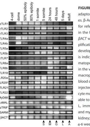
To investigate the potential of the zebrafish embryo as a model to study vertebrate innate immune signalling, we first set out to determine the expression of TLRs and associated adaptor genes during embryo development. Semi-quantitative RT-PCR analysis, using the Superscript II one-step system (Invitrogen) with previously de-scribed conditions and primers (18), showed that at least 15 zebrafish TLR genes are expressed at 1 dpf, when the first functional macrophages and neutrophils enter blood circulation (Fig.1). Most of these TLRs are also maternally present, since ex-pression was already detected at the 4-cell stage, which is prior to the onset of zygotic gene expression. Several TLRs display distinct differential expression patterns during early stages of embryogenesis. For example, zTLR1 expression peaks during blastula and gastrula stages (dome to 80% epiboly) and is high during embryogenesis com-pared to the adult stage. Expression of zTLR3 peaks during gastrulation and segmen-tation (80% epiboly to 5-somite stage), is reduced between 1 to 5 dpf, but returns to higher levels in the adult stage. Diffuse zTLR3 expression in the developing brain of zebrafish embryos was previously reported (20). A peak in the expression of zTLR5a,
zTLR5b, zTLR7, zTLR8a, zTLR8b and zTLR18 coincides with the appearance of em-bryonic macrophages at 1 dpf. Expression of the zMyD88 adaptor gene is highest in adults. In the embryo, maternal zMyD88 transcript levels are reduced during blastula and gastrula stages and return to higher levels during segmentation and later stages (Fig.1). The other MyD88-like adaptor genes zMAL, zTRIF and zSARM are also ma-ternally present and expressed throughout embryogenesis (Fig.1).
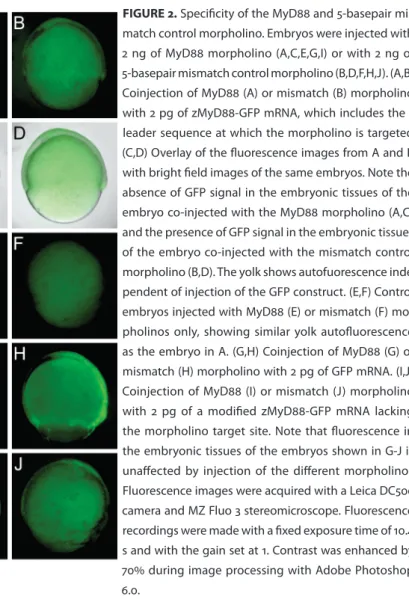
morpholino knockdown approach (19) was used to interfere with MyD88 function by inhibition of its mRNA translation. An antisense morpholino (GeneTools) (5’-TAGCAAAACCTCTGTTATCCAGCGA-3’) was designed, which targets the leader sequence of the zMyD88 mRNA (DQ100359) at positions -30 to -7 with respect to the ATG. A 5-basepair mismatch control morpholino was used with the sequence 5’-TAcCAtAACCTgTGTTATCgAGgGA-3’ (mismatches in lower case). For micro-injection morpholinos were diluted to different concentrations in Danieu’s buffer (19) and approximately 1 nl was injected into the blastomere of the 1-2 cell stage embryo. To test the specificity of the MyD88 morpholino and its 5-mismatch control sequence, each of these morpholinos was first coinjected with a zMyD88-EGFP fusion mRNA including the 5’ leader sequence. Embryos coinjected with the 5-mismatch control morpholino and zMyD88-GFP mRNA showed clear fluorescence at the 70-90% epi-boly stage (Fig.2B,D). In contrast, embryos coinjected with the MyD88 morpholino and zMyD88-GFP mRNA showed only autofluorescence of the yolk (Fig.2A,C), simi-lar as in embryos injected with morpholinos only (Fig.2E,F). Therefore, the MyD88 morpholino effectively blocks translation of zMyD88-GFP mRNA. Coinjection of each of the morpholinos with GFP mRNA (Fig.2G,H) or with a modified zMyD88-GFP mRNA lacking the 5’ leader sequence (Fig.2I,J), resulted in similar fluorescence levels in embryo tissues, confirming that the MyD88 morpholino specifically targets the zMyD88 leader sequence.
FIGURE 1. RT-PCR analysis of zebrafish TLR and adaptor genes at different developmental stag-es. β-Actin (βACT) expression was determined for reference. 100 ng of total RNA was used in the RT-PCR reactions, except for zTRIF and
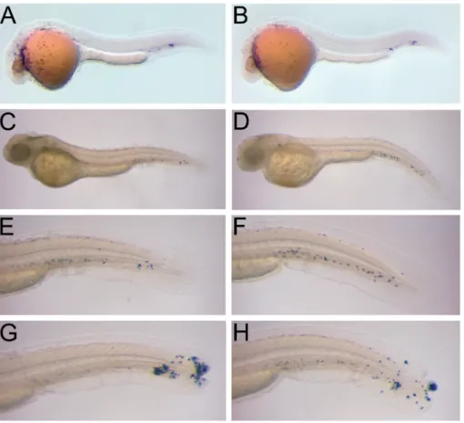
Embryos injected with 1.7 to 4 ng of MyD88 or control morpholinos showed no apparent morphological differences with wild type embryos. To determine if myelo-poiesis was affected in MyD88 morphants, we analysed the expression of myeloid cell markers. From 1 dpf onwards all myeloid cells of zebrafish embryos express either one of the two markers, L-plastin or mpx (11, 3, 17, 4). Expression of the macrophage marker gene, L-plastin, was examined at 1 dpf, after the onset of blood circulation. To this extent a digoxigenin-labeled antisense riboprobe was synthesized with T7 RNA polymerase from an EcoRI-linearized l-plastin cDNA clone (AF157110) and whole-mount in situ hybridization was carried out according to the protocol of Thisse et al. (27). In MyD88 morphants, L-plastin-positive macrophages were dispersed over the yolk sac and some had accumulated in the ventral venous plexus (Fig.3A). This
tern was similar to that observed in mismatch control morphants (Fig.3B) and to the reported L-plastin expression pattern of wild type zebrafish embryos (11).
A histochemical staining for myeloperoxidase (MPX) activity (17) was performed to check for the presence of granulocytes in embryos at 2 dpf.Peroxidase-positive cells in both the MyD88 and control morphants were abundantly present in the ven-tral venous plexus and some were scattered over the yolk surface or had invaded the head region (Fig.3C-F). The same distribution pattern of granulocytes was observed in non-injected control embryos (data not shown) and has been previously reported, based on both peroxidase staining and mpx gene expression (3, 17). Next, we took
vantage of an acute inflammation assay devised by Lieschke et al. (17) to determine if the granulocytes of MyD88 morphants were functional. After wounding of the caudal fin with a sharp forceps, peroxidase-positive granulocytes of both MyD88 and control morphants accumulated at the site of trauma within 6 hours, indicating their functional involvement in acute inflammation (Fig.3G,H). In conclusion, based on
L-plastin and MPX marker analyses, MyD88 morphants showed no apparent myelo-poietic defects.
To demonstrate a function for MyD88 in the innate immune response of the zebrafish embryo, we made use of a Salmonella typhimurium infection modelthat was previously established (31). In this infection model, a low dose of DsRed-labeled bacteria is injected into the embryo’s bloodstream just after the onset of
tion at 1 dpf. While injection of the wild type S. typhimurium strain SL1027 resulted in a rapid lethal infection, its isogenic lipopolysaccharide (LPS) derivative SF1592 (Ra-type LPS mutant) proved to be non-pathogenic (31). In the present study, wild-type embryos, embryos injected with 1.7 ng of MyD88 morpholino, and embryos injected with the control morpholino were challenged in an infection experiment with S. typhi murium Ra mutant bacteria containing the DsRED plasmid pGMDs3 (31). Embryos were staged at 28 hpf (15) and individually infected by microinjection of approximately 100 colony forming units (cfu) into the axial vein near the blood island and the urogenital opening as previously described (31). As a control, a simi-lar dose as used in the infection experiment was spotted onto LB agar plates for cfu counting.
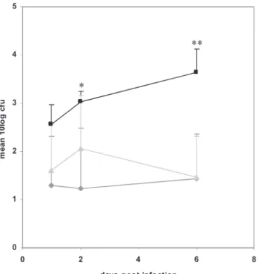
Embryos were monitored daily until 6 days after infection with S. typhimuri-um Ra. No differences in survival rate were found between the infected wild-type embryos and MyD88 morphants. However, when embryos were examined for the presence of fluorescent bacteria, the MyD88 morphants showed more red spots, repre senting bacteria, than the wild-type embryos (data not shown). Therefore, total cfu counts were analysed from groups of five embryos that were sampled at 1 day post infection (dpi), 2 and 6 dpi. The pooled embryos were disintegrated (31) and the mixture was plated on LB agar plates. Four independent infection experiments were performed. At 1 dpi the average number of total cfu was approximately 5-fold higher in the MyD88 morphants as compared to wild type and mismatch control embryos (Fig.4). At 2 dpi, the difference between MyD88 morphants and wild type embryos was 10-fold and significant at p<0.05. Although it is not likely that mor-pholino knockdown is completely penetrant after 3 days of embryo development (19), a further increase of total cfu was still observed in MyD88 morphants at 6 dpi. At this stage the difference with total cfu in wild type and mismatch control embryos was significant at p<0.01. Between different experiments MyD88 morphant embryos harboured 100- to 1000- fold more bacteria than wild-type and mismatch control embryos, which had either completely cleared the infection or contained only low amounts of bacteria not higher than the inoculum size (Fig.4). Therefore we con-clude that MyD88 morphants are not able to clear an infection with S. typhimurium
Ra effectively.
Although there was a clear increase in cfu counts, it is interesting that infection with the normally non-pathogenic LPS Ra mutant of S. typhimurium was not lethal for MyD88 morphant embryos, indicating that multiplication of S. typhimurium
Ra is not completely uncontrolled in MyD88 morphant embryos. Future analysis of a stable MyD88 knockout line should clarify if this was due to incomplete loss of MyD88 function in morphant embryos or due to MyD88-independent innate im-munity mechanisms.
inside macrophages of MyD88 morphants (Fig.5), similar as in wild type embryos or in embryos injected with the mismatch control morpholino. Although we cannot yet exclude differences in phagocytosis efficiency or phagosome maturation, our present observations suggest that MyD88 morphants are primarily affected in activation of the bacterial killing mechanisms.
In conclusion, we have shown that TLRs are broadly expressed during zebra-fish embryo development and that MyD88 is required for a wild-type response of zebrafish embryos to S. typhimurium Ra infection. These results indicate that MyD88-dependent signalling functions and is important in the early innate im-mune responses of embryonic zebrafish, independent from coupling to adaptive signalling responses. Furthermore, we have shown that zebrafish embryos express other MyD88-like adaptor molecules, such as Mal, TRIF and SARM, suggesting that MyD88-independent signalling pathways also exist in zebrafish, similar as in other vertebrates (14, 33, 8). The critical role of zebrafish MyD88 is consistent with many infection studies in MyD88-/- adult mice, which showed increased susceptibility to a variety of pathogens (26, 9, 23, 10, 22, 24). Therefore, the present study validates the zebrafish embryo as a useful model for analysis of the vertebrate innate immune system, which creates exciting possibilities for future studies in zebrafish embryo infection models.
Acknowledgments
References
Adachi, O., T. Kawai, K. Takeda, M. Matsumoto, H. Tsutsui, M. Sakagami, K. Nakanishi, and S. Akira.
1. 1998.
Targeted disruption of the MyD88 gene results in loss of IL-1- and IL-18-mediated function. Immunity 9: 143– 150.
Akira, S., and K. Takeda.
2. 2004. Toll-like receptor signalling. Nat. Rev. Immunol. 4:499-511.
Bennett, C. M., J. P. Kanki, J. Rhodes, T. X. Liu, B. H. Paw, M. W. Kieran, D. M. Langenau, A. Delahaye-Brown, 3.
L. I. Zon, M. D. Fleming, and A. T. Look.2001. Myelopoiesis in the zebrafish, Danio rerio. Blood 98:643-651. Crowhurst, M. O., J. E. Layton, and G. J. Lieschke.
4. 2002. Developmental biology of zebrafish myeloid cells. Int.
J. Dev. Biol. 46:483-492. Davidson, A. J., and L. I. Zon.
5. 2004. The 'definitive' (and 'primitive') guide to zebrafish hematopoiesis.
Oncogene 23:7233-7246.
Davis, J. M., H. Clay, J. L. Lewis, N. Ghori, P. Herbomel, and L. Ramakrishnan.
6. 2002. Real-time visualization
of mycobacterium-macrophage interactions leading to initiation of granuloma formation in zebrafish embry-os. Immunity 17:693-702.
Dunne, A., and L. A. O'Neill.
7. 2003. The interleukin-1 receptor/Toll-like receptor superfamily: signal transduc-tion during inflammatransduc-tion and host defense. Sci. STKE. 2003:re3.
Dunne, A., and L. A. O'Neill.
8. 2005. Adaptor usage and Toll-like receptor signaling specificity. FEBS Lett.
579:3330-3335.
Edelson, B. T., and E. R. Unanue.
9. 2002. MyD88-dependent but Toll-like receptor 2-independent innate
immu-nity to Listeria: no role for either in macrophage listericidal activity. J. Immunol. 169:3869-3875. Feng, C. G., C. A. Scanga, C. M. Collazo-Custodio, A. W. Cheever, S. Hieny, P. Caspar, and A. Sher.
10. 2003. Mice
lacking myeloid differentiation factor 88 display profound defects in host resistance and immune responses to Mycobacterium avium infection not exhibited by Toll-like receptor 2 (TLR2)- and TLR4-deficient animals. J. Immunol. 171:4758-4764.
Herbomel, P., B. Thisse, and C. Thisse.
11. 1999. Ontogeny and behaviour of early macrophages in the zebrafish
embryo. Development 126:3735-3745. Herbomel, P., B. Thisse, and C. Thisse.
12. 2001. Zebrafish early macrophages colonize cephalic mesenchyme and
developing brain, retina, and epidermis through a M-CSF receptor-dependent invasive process. Dev. Biol. 238:274-288.
Jault, C., L. Pichon, and J. Chluba.
13. 2004. Toll-like receptor gene family and TIR-domain adapters in Danio
re-rio. Mol. Immunol. 40:759-771.
Kaisho, T., O. Takeuchi, T. Kawai, K. Hoshino, and S. Akira.
14. 2001. Endotoxin-induced maturation of
MyD88-deficient dendritic cells. J. Immunol. 166:5688-5694.
Kimmel, C. B., W. W. Ballard, S. R. Kimmel, B. Ullmann, and T. F. Schilling.
15. 1995. Stages of embryonic
devel-opment of the zebrafish. Dev. Dyn. 203:253-310. Lam, S. H., H. L. Chua, Z. Gong, T. J. Lam, and Y. M. Sin.
16. 2004. Development and maturation of the immune
system in zebrafish, Danio rerio: a gene expression profiling, in situ hybridization and immunological study. Dev. Comp. Immunol. 28:9-28.
Lieschke, G. J., A. C. Oates, M. O. Crowhurst, A. C. Ward, and J. E. Layton.
17. 2001. Morphologic and functional
characterization of granulocytes and macrophages in embryonic and adult zebrafish. Blood 98:3087-3096. Meijer, A. H., S. F. G. Krens, I. A. Medina Rodriguez, S. He, W. Bitter, B. E. Snaar-Jagalska, and H. P. Spaink. 18.
2004. Expression analysis of the Toll-like receptor and TIR domain adaptor families of zebrafish. Mol. Immunol. 40:773-783.
Nasevicius, A., and S. C. Ekker.
19. 2000. Effective targeted gene 'knockdown' in zebrafish. Nat. Genet.
26:216-220.
Phelan, P. E., M. T. Mellon, and C. H. Kim.
20. 2005. Functional characterization of full-length TLR3, IRAK-4,
and TRAF6 in zebrafish (Danio rerio). Mol. Immunol. 42:1057-1071. Rhodes, J., A.
21. Hagen, K. Hsu, M. Deng, T.X. Liu, A.T. Look, and J. P. Kanki.2005. Interplay of Pu.1 and Gata1
determines myelo-erythroid progenitor cell fate in zebrafish. Dev. Cell, 8:97–108. Scanga, C. A., A. Bafica, C. G. Feng, A. W. Cheever, S. Hieny, and A. Sher.
22. 2004. MyD88-deficient mice display
a profound loss in resistance to Mycobacterium tuberculosis associated with partially impaired Th1 cytokine and nitric oxide synthase 2 expression. Infect. Immun. 72:2400-2404.
Seki, E., H. Tsutsui, N. M. Tsuji, N. Hayashi, K. Adachi, H. Nakano, S. Futatsugi-Yumikura, O. Takeuchi, K. 23.
Hoshino, S. Akira, J. Fujimoto, and K. Nakanishi.2002. Critical roles of myeloid differentiation factor 88-dependent proinflammatory cytokine release in early phase clearance of Listeria monocytogenes in mice. J. Immunol. 169:3863-3868.
Skerrett, S. J., H. D. Liggitt, A. M. Hajjar, and C. B. Wilson.
24. 2004. Cutting edge: myeloid differentiation
fac-tor 88 is essential for pulmonary host defense against Pseudomonas aeruginosa but not Staphylococcus aureus. J. Immunol. 172:3377-3381.
Takeda, K., T. Kaisho, and S. Akira.
25. 2003. Toll-like receptors. Annu. Rev. Immunol. 21:335-376.
Takeuchi, O., K. Hoshino, and S. Akira.
highly susceptible to Staphylococcus aureus infection. J. Immunol. 165:5392-5396. Thisse, C., B. Thisse, T. F. Schilling, and J. H. Postlethwait.
27. 1993. Structure of the zebrafish snail1 gene and its
expression in wild-type, spadetail and no tail mutant embryos. Development 119:1203-1215.
Traver, D., P. Herbomel, E. E. Patton, R. D. Murphey, J. A. Yoder, G. W. Litman, A. Catic, C. T. Amemiya, L. I. 28.
Zon, and N. S. Trede.2003. The zebrafish as a model organism to study development of the immune system. Adv. Immunol. 81:253-330.
Trede, N. S., D. M. Langenau, D. Traver, A. T. Look, and L. I. Zon.
29. 2004. The use of zebrafish to understand
immunity. Immunity 20:367-379.
Van der Sar, A. M., B. J. Appelmelk, C. M. J. E. Vandenbroucke-Grauls, and W. Bitter.
30. 2004. A star with stripes:
zebrafish as an infection model. Trends Microbiol. 12:451-457.
Van der Sar, A. M., R. J. P. Musters, F. J. M. Van Eeden, B. J. Appelmelk, C. M. J. E. Vandenbroucke-Grauls, and 31.
W. Bitter.2003. Zebrafish embryos as a model host for the real time analysis of Salmonella typhimurium infec-tions. Cell. Microbiol. 5:601-611.
Willett, C. E., A. Cortes, A. Zuasti, and A. G. Zapata.
32. 1999. Early hematopoiesis and developing lymphoid
or-gans in the zebrafish. Dev. Dyn. 214:323-336. Yamamoto, M., K. Takeda, and S. Akira.
33. 2004. TIR domain-containing adaptors define the specificity of TLR
signaling. Mol. Immunol. 40:861-868.
Yoder, J. A., M. E. Nielsen, C. T. Amemiya, and G. W. Litman.
34. 2002. Zebrafish as an immunological model
Oliver W. Stockhammer1, Anna Zakrzewska1, Zoltán Hegedûs2, 3,
Herman P. Spaink1 and Annemarie H. Meijer1
1 Institute of Biology, Leiden University, Leiden, The Netherlands 2 ZenonBio Ltd., Szeged, Hungary 3 Bioinformatics Laboratory, Biological Research Center,
Hungarian Academy of Sciences, Szeged, Hungary
published in The Journal of Immunology, 2009, 182, 5641 -5653 Copyright © 2009 by The American Association of Immunologists, Inc.
of the zebrafish embryonic innate immune
response to
Salmonella
infection
Abstract
Due to the clear separation of innate immunity from adaptive responses, the ex-ternally developing zebrafish embryo represents a useful in vivo model for identi-fication of innate host determinants of the response to bacterial infection. Here we performed a time-course transcriptome profiling study and gene ontology analysis of the embryonic innate immune response to infection with two model Salmonella
strains that elicit either a lethal infection or an attenuated response. The transcrip-tional response to infection with both the lethal strain and the avirulent LPS O-antigen mutant strain showed clear conservation with host responses detected in other vertebrate models and human cells, including induction of genes encoding cell surface receptors, signalling intermediates, transcription factors and inflammatory mediators. Furthermore, our study led to the identification of a large set of novel immune response genes and infection markers, the future functional characteriza-tion of which will support vertebrate genome annotacharacteriza-tion. From the time series and bacterial strain comparisons, matrix metalloproteinase genes, including mmp9, were among the most consistent infection-responsive genes. Purified Salmonella flagellin also strongly induced mmp9 expression. Using knockdown analysis we showed that this genewas downstream of the zebrafish homologs of the flagellin receptor TLR5 and the adaptor MyD88. In addition, flagellin-mediated induction of other inflam-mation markers, including il1b, il8 and cxcl-C1c, was reduced upon TLR5 knockdown as well as expression of irak3, a putative negative TLR pathway regulator. Finally, we showed that induction of il1b, mmp9 and irak3 requires MyD88-dependent signalling, while ifn1 and il8 were induced MyD88-independently during Salmonella infection.
Introduction
The innate immune system represents the evolutionary ancient part of vertebrate im-munity and relies on germline encoded receptors, commonly referred to as pattern recognition receptors (PRRs), to mediate immune responses to pathogenic micro-organisms. Triggering of these receptors activates a variety of signal transduction pathways ultimately resulting in large alterations of the host transcriptome profile, of which the dynamic complexity and underlying mechanisms are still poorly under-stood, especially at the whole organism level.
pro-teins to the receptors. Differential utilization of the adaptor molecules by the TLRs causes specific activation of a range of transcription factors such as nuclear factor-κB (NF-κB), activator protein 1 (AP-1) and interferon regulatory factor (IRF) 3, 5 and 7 through distinct signalling pathways eventually leading to the downstream activa-tion of pro-inflammatory cytokines (4). To date five adaptor proteins involved in TLR signalling have been described in human: myeloid differentiation factor-88 (MyD88), MyD88 adaptor-like protein (Mal/TIRAP), Toll/IL1 receptor (TIR) domain- contain-ing adaptor protein induccontain-ing IFNβ (TRIF/TICAM1), TRIF-related adaptor molecule (TRAM/TICAM2) and sterile α- and armadillo motif-containing protein (SARM) (4). Among all adaptors, MyD88 is the most commonly used adaptor and signalling through MyD88 has been implicated for all human TLRs with the exception of TLR3 that was shown to signal in a TRIF-dependent and MyD88-independent fashion (4, 5). Furthermore, MyD88 is involved in signal transduction of the interleukin 1 recep-tor (IL-1R) and is associated with interferon-γ receprecep-tor signalling (IFN-γR) leading to p38 activation (6, 7).
In recent years the zebrafish (Danio rerio) embryo system has emerged as a new model to study vertebrate innate immunity, offering several advantages that comple-ment mammalian model systems. The transparent character of the externally ferti-lized zebrafish embryo in combination with fluorescently labelled immune cells and bacteria facilitate the study of host microbe interaction and inflammation processes in the living organism (8-14). The efficiency at which infections and chemical treat-ments in zebrafish can be performed at a large-scale allows identification of novel microbial virulence factors and high throughput compound screens to investigate disease mechanisms (15, 16). Moreover, the zebrafish system is particularly suitable for large-scale forward and reverse genetic screens aimed at the identification of genes with novel functions in development of the immune system or in the immune response (17-19).