TISSUE RESERVOIRS OF HIV-1: INSIGHTS FROM THE CENTRAL NERVOUS SYSTEM
Lauren A. Rackoff Tompkins
A thesis submitted to the faculty at the University of North Carolina at Chapel Hill in partial fulfillment of the requirements for the degree of Master of Science in the Microbiology and Immunology Department in
the School of Medicine.
Chapel Hill 2015
Approved by:
Ronald Swanstrom
Mark Heise
Nathaniel Moorman
ii © 2015
iii ABSTRACT
Lauren A. Rackoff Tompkins: Tissue Reservoirs of HIV-1: Insights from the Central Nervous System (Under the direction of Ronald Swanstrom)
Cellular and anatomical reservoirs of HIV-1 preclude a cure to infection. Efforts to characterize
these reservoirs are an important part of developing a strategy to eradicate all forms of HIV-1. The central
nervous system (CNS) is a unique bodily compartment that can support viral replication independent of
that in the blood (compartmentalization) and may be an anatomical reservoir of unique viruses. In this
‘proof of principle’ study, we characterized viral sequences (RNA and provirus) from the blood,
cerebrospinal fluid, brain, and liver of two infected donors who died with HIV-1-associated dementia and
disparate states of viral replication (compartmentalized versus equilibrated). We show that selective
pressures exist within CNS and liver tissue to drive expansion of particular viral species. We found
macrophage-tropic viral lineages archived in the brain, implicating macrophages as a potential cellular
reservoir. By including more donors and tissues, our study provides insight towards HIV-1 reservoirs and
iv
ACKNOWLEDGEMENTS
I would like to thank my mentor, Ron, for his valuable guidance, unwavering support, and
friendship. He stuck by me through thick-and-thin, was always inspiring, and I am a better scientist for
knowing him. The Swanstrom Lab is not only my scientific home base, but also a place where I’ve met
lifelong friends who I cherish greatly. We share laughs and beers, prank each other mercilessly, and
always seem to make light of even the toughest parts of being a scientist. To my closest friends in the lab,
you know who you are, thank you from the bottom of my heart for being the incredible friends you are.
You made work fun, which is such a gift, especially when you’re in the throws of grad school. More so,
you’ve been nothing but supportive, encouraging, and wonderful people - I am truly blessed for having
known you.
To my committee, I cannot express how grateful I am to have had you with me throughout this
journey. Your guidance has been invaluable to me and has shaped my journey as a scientist. I’ve learned
so much from each of you and could always come to you with questions or to ask for advice. You’ve been
my cheerleaders when I needed them most and never lost faith in me. I couldn’t have done it without you.
I would also like to thank UNC and all the people who have helped me along the way, especially
Dixie Flannery, Bob Bourret, and Bill Goldman of the Microbiology & Immunology Department. Thank you
for all of your help in navigating grad school and taking a personal interest in my graduate career as well
as those of each and every one of my classmates. I am overcome with gratitude for the immense support
I’ve received from every person I’ve met at UNC – this is truly a school that cares about its students.
My greatest appreciation is reserved for my family and friends: thank you for being in my life. You
are the people who have always been there and will always be there, whose love in unconditional and so
essential. My parents are truly great people; their selflessness and generosity inspires me every day. To
Matt, my husband and soul mate: you are my rock and my world, I love you more than anything. You
v
TABLE OF CONTENTS
LIST OF TABLES ... vii
LIST OF FIGURES ... viii
CHAPTER 1: ASSESSING TISSUE RESERVOIRS OF HIV-1 IN DONORS WHO DIED WITH HIV-ASSOCIATED DEMENTIA ... 1
INTRODUCTION ... 1
1.1 HIV-1 Disease Burden and Molecular Biology ... 1
1.1.1 The HIV-1 pandemic: treatable, but not curable ... 1
1.1.2 HIV-1 particle structure and genome organization ... 2
1.1.3 The viral life cycle of HIV-1 ... 3
1.2 Tissue Reservoirs of HIV-1 ... 4
1.2.1 HIV-1 persistence in viral reservoirs ... 4
1.2.2 The CNS is a unique bodily compartment and a potential anatomical reservoir of HIV-1 ... 6
1.2.3 Potential cellular reservoirs in the CNS ... 10
1.2.4 Logistics and complications in evaluating the CNS reservoir ... 13
1.3 Examining HIV-1 Proviruses Archived in Tissues on a Whole Body Scale ... 15
METHODS ... 15
Study Design ... 15
PCR Amplification of HIV-1 Genes and Sequencing ... 15
Phylogenetic Analysis of Viral Genes ... 16
Cloning HIV-1 env for Protein Expression ... 17
Co-transfection for Generating Env-pseudotyped Virus ... 17
Single-cycle Infection of Reporter Cells for Determining Cell tropism ... 17
RESULTS ... 18
vi
Method Development: Full-Length Provirus PCR ... 20
Assessing Compartmentalized Viral Replication ... 22
HIV-1 env Sequences from Brain Tissue Resemble Those in the CSF
and Distinct Viral Lineages Exist within Different Brain Regions ... 23
Individual Viral Lineages Also Exist within the Liver, a Peripheral
Tissue Exposed to Blood Virus ... 25
Macrophage-Tropic Virus is Archived as Provirus in Brain Tissue ... 26
DISCUSSION AND FUTURE DIRECTIONS ... 28
APPENDIX: MODIFIED PROTOCOL FOR PROVIRUS CHARACTERIZATION
IN TISSUES ... 38
vii
LIST OF TABLES
Table 1 – Study Population Characteristics ... 1
viii
LIST OF FIGURES
Figure 1 – Method workflow for characterizing proviruses present in extracted
total DNA from cells or tissue ... 1
Figure 2 – Compartmentalized viral replication in the CSF/CNS is associated
with macrophage-tropic virus in the CSF ... 1
Figure 3 – Tissue-specific viral lineages exist throughout the brain and within the liver
in the presence of compartmentalized viral replication in the CNS ... 1
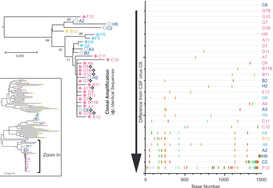
Figure 4 – Clonal amplification of viral species through local replication diversifies the
population of virus throughout the CNS and in the peripheral liver tissue ... 1
Figure 5 – Clonal amplification occurs in the liver irrespective of an otherwise
1
CHAPTER I. ASSESSING TISSUE RESERVIORS OF 1 IN DONORS WHO DIED WITH HIV-ASSOCIATED DEMENTIA
INTRODUCTION
1.1 HIV-1 Disease Burden And Molecular Biology
1.1.1 The HIV-1 pandemic: treatable, but not curable
Human immunodeficiency virus type 1 (HIV-1) is the cause of acquired immunodeficiency
syndrome (AIDS), where uncontrolled infection in the blood and lymphoid organs depletes CD4+ T cells
and thus diminishes immune competence over time. HIV-1 primarily infects CD4+ T cells and productive
infection usually results in cell death via toxic effects of viral replication or immune attack. When a
sufficient number of CD4+ T cells are lost, HIV-infected people become susceptible to opportunistic
infections, cancers, and other co-morbidities. Without treatment, HIV-1 disease progresses to AIDS in
about 10-15 years, and most people die within a few years of AIDS diagnosis. Roughly 37 million people
are currently living with HIV-1, with an estimated 5,600 new infections occurring per day. The disease
burden is greatest in low- and middle-income countries: 70% of people with HIV-1 live in Africa and 65%
of AIDS-related deaths also occur in Africa [1].
The introduction of antiretroviral therapy (ART) has drastically changed the landscape of HIV-1
morbidity and mortality. ART controls viral replication and thus disease progression, thereby extending
longevity in HIV-infected people. At the end of 2013, 12.9 million people globally were receiving ART,
11.7 million of them in low- and middle-income countries [1, 2]. An additional 1.9 million people were
newly enrolled in ART in 2014, which is one of the largest annual increases in treatment initiation to date.
Still, only 14.8 of the 37 million HIV-1+ people are receiving ART, which means that the majority of people
are untreated and capable of transmitting the virus (60% or 22.2 of 37 million) [1, 2]. Efforts are underway
2
recommending a “treat-all” approach where all populations and age groups are recommended to receive
ART as soon after diagnosis as possible [1].
HIV-1 infection is indeed treatable, but there is currently no cure. Although ART is successful at
achieving viral suppression (i.e. very low or undetectable viral load), these drugs must be taken
throughout life to control infection. A major focus of current research is to eradicate HIV-1 and cure
infected patients. Viral persistence in cellular and anatomical reservoirs precludes a cure, thus efforts to
characterize these reservoirs are an important part of developing a strategy for eradicating all forms of
HIV-1.
1.1.2 HIV-1 particle structure and genome organization
HIV-1 is a single-stranded RNA (ssRNA), enveloped virus that belongs to the Lentivirus genus,
Retroviridae family. The HIV-1 genome is ~9.7 kilobases in length, has a 5’ cap and 3’ polyA tail, and
encodes for nine genes that are flanked by long terminal repeats (LTR): gag, pol, env, vif, vpr, tat, rev,
vpu, and nef. Structural proteins are encoded in the env gene (viral envelope, “Env protein”) and the gag
gene (the Gag polyprotein precursor, which is processed into the matrix [MA], capsid [CA], and
nucleocapsid [NC] structural proteins). The pro and pol genes encodes the viral enzymes protease (Pro),
integrase (IN), and reverse transcriptase (RT). The remainder of genes encode accessory proteins (Vif,
Vpr, Vpu, and Nef) or regulatory proteins (Tat and Rev) [3].
Two identical copies of the ssRNA viral genome are packaged within a conical capsid core. The
dimeric ssRNA genome associates with NC protein to form a NC-RNA complex that is surrounded by CA
protein to form the conical capsid core [4]. A spherical shell composed of MA protein surrounds the core.
MA protein is embedded in the viral envelope, a lipid bilayer derived from the infected host cell
membrane. Trimeric Env proteins are also embedded in the viral envelope and are exposed as spikes on
the outer surface of the virion. A mature virion contains all of the components required for infectivity: the
dimeric ssRNA genome, cellular tRNALys3 molecules to prime cDNA synthesis, the viral structural
proteins that form a functional virion and permit attachment to viral receptors expressed on permissive
cells, and the three viral enzymes. Vpr, Vif, and Nef are also packaged in the virion, as well as cellular
3 1.1.3 The viral life cycle of HIV-1
The first step of the HIV-1 replication cycle is attachment to the surface of a permissive cell via
interactions between HIV-1 Env protein and the cell-surface viral receptor, CD4, and co-receptors, CCR5
or CXCR4 [5]. Viral Env is a heavily glycosylated trimeric protein composed of the gp120 surface
glycoprotein, which is exposed on the outer face of the viral particle, and the gp41 transmembrane
glycoprotein, which is embedded in the viral membrane (viral envelope). These proteins remain tethered
to one another noncovalently in the viral envelope. Both gp120 and gp41 originate from the same
precursor protein, gp160, which is proteolytically cleaved by host furin-like proteases in an infected cell.
Cell attachment begins with gp120 binding CD4 on the cell surface. This interaction initiates a
cascade of conformational changes in viral Env gp120 and gp41 eventually leading to fusion with the
cellular membrane [5]. First, gp120-CD4 association induces conformational changes in gp120 that allow
interaction with the viral co-receptor, CCR5 or CXCR4. Co-receptor binding causes exposure of the
hydrophobic gp41 fusion peptide, which inserts into the host cell membrane thereby tethering the viral
and cell membranes. Each gp41 monomer within the trimer bends at a hinge region to form a stable
six-helix bundle, which is the driving force in forming a fusion pore. Contents of the viral particle are then
delivered into the cell cytoplasm through the fusion pore.
Upon release of the viral core into the cell cytoplasm, the viral capsid is uncoated and viral
genomic RNA and proteins are released [5]. In the cytoplasm, viral RNA is reverse transcribed to DNA by
viral RT. Viral RT has an RNA-dependent DNA polymerase for synthesizing a DNA copy of the viral
genome and RNase H activity for degrading RNA in the RNA:DNA replication intermediate. The
pre-integration complex (PIC), composed of viral genomic DNA and viral and cellular proteins, is then formed
and translocates into the cell nucleus through the intact nuclear envelope, allowing HIV-1 to replicate in
non-dividing cells [6]. With the help of viral IN, viral DNA is integrated into a host cell chromosome
(provirus), which is an essential step of the viral life cycle [7]. Integration can happen anywhere in the
host genome, but tends to favor transcriptionally active genes. A recent study has highlighted the
importance of the nuclear architecture in integration site selection, as integration occurs near the nuclear
4
nucleus [8]. Viral integration allows the virus to persist in the form of an infected cell that can become a
reservoir when dormant (latent infection).
The HIV-1 provirus can then be transcribed using cellular machinery, namely RNA polymerase II,
with the help of the viral Tat protein [9]. Viral transcripts, spliced or unspliced, are then transported out of
the nucleus, a process mediated in part by the viral Rev protein, for translation of viral proteins or
packaging into a nascent virion (viral assembly) in the cell cytoplasm [10]. Viral gag, pro, pol, and env are
translated into polyprotein precursors, which are then further processed/cleaved by the viral Pro or a host
protease during and after viral assembly and budding [11]. Viral assembly occurs at the cell surface,
where two copies of an unspliced, full-length viral RNA genome, viral proteins, and components of the cell
cytosol are packaged into an immature virion that buds from the cell surface. In a highly ordered process
of maturation, viral Pro cleaves the structural viral polyprotein precursors (Gag and Gag-Pro-Pol),
ultimately leading to the formation of a mature, infectious viral particle.
1.2 Tissue Reservoirs Of HIV-1
1.2.1 HIV-1 persistence in viral reservoirs
HIV-1 persistence in cellular and anatomical reservoirs precludes a cure to infection. Two
essential criteria exist to define a viral reservoir of HIV-1 [12]. First, a reservoir must preserve
replication-competent virus in some form (i.e. viral particles or viral genomes) so that the virus can reestablish
productive infection in the future. Second, a reservoir must have mechanisms of longevity. For example,
reservoirs composed of virions would require escape from biochemical decay, as seen with virion
particles that become trapped extracellularly in dendritic cell processes. Cell-associated viral reservoirs,
however, require cell survival and escape from immune control including cytotoxic T cell (CTL) activity.
Latent infection, a state with no active replication, is the best characterized cellular reservoir of HIV-1, but
another type of reservoir could be composed of productively infected cells with slow turnover. The
concept of a reservoir is further complicated by the detection of cells that clonally expand in vivo through
5
A reservoir will most likely occur in cells that are normally infected with HIV-1. This virus infects
cells that express the viral receptor CD4 and co-receptor (CCR5 or CXCR4). Activated CD4+ T cells are
the most permissive cell type for HIV-1. Latently infected resting memory CD4+ T cells are the hallmark
reservoir of HIV-1 infection and are thought to predominantly arise from productive infection of activated
CD4+ T cells as they are transitioning back into a resting state [14]. Latently infected T cells contain
replication-competent HIV-1 genomic DNA (provirus) integrated within the human genome in the absence
of ongoing viral replication. Although dormant, latently infected resting T cells can be induced to become
activated and thereby transcribe integrated DNA to generate new progeny virions capable of productive
infection. It is important to note that integrated DNA can be either intact or defective, thus many infected T
cells contain HIV-1 DNA that is incapable of producing functional virions [15]. Latently infected cells are
established early after a person becomes infected and these cells persist even in the presence of
ART-mediated viral suppression. Persistence of the latent reservoir is due in part to the virus escaping immune
surveillance, as integrated viral DNA in latently infected cells is likely transcriptionally silent and does not
produce antigens that signal immune attack [16].
A second type of HIV-1 reservoir could be a productively infected cell with slow turnover, which is
illustrated in HIV-1 infection of the central nervous system (CNS). The CNS is a bodily compartment
separated from the periphery by the blood-brain barrier. HIV-1 enters the CNS and can establish
productive infection with features that are distinct from those in the blood [17-22]. One such feature
involves comparing disparate viral decay kinetics in the blood and cerebrospinal fluid (CSF), the latter of
which bathes the CNS and is an indirect measure of CNS infection. After the onset of suppressive ART,
which prevents new infections without affecting previously infected cells, the blood viral load decreases
rapidly (1-2 weeks) due to rapid turnover of short-lived infected T cells [23-25]. For most people, the same
pattern of rapid viral decay is observed in the CSF. However, occasionally the viral load decays much
more slowly in the CSF than in the blood, which suggests that cells with a longer half-life than T cells can
support HIV-1 replication in the CNS [20, 26].
The concept of anatomical reservoirs is a current topic of interest, and the CNS is a unique
anatomical compartment capable of sustaining viral replication and potentially harboring viral reservoirs
6
reservoirs, as tissue microenvironments are highly structured and functionally specialized. Importantly,
only 1-2% of lymphocytes in the body are present in the blood at any given time [27, 28], whereas the
vast majority of lymphocytes are tissue-resident, indicating that a greater frequency of permissive cells
are present in the tissues compared to the blood [29]. Consistent with the idea of tissue reservoirs
differing from the blood, reservoir establishment in at least some tissues appears to precede that in the
blood, as demonstrated by a recent rhesus macaque animal model of HIV-1 infection [30]. In macaques
treated with suppressive ART at day three post-infection with simian immunodeficiency virus (SIV), viral
DNA was found in lymph node and gut tissue before detectable viremia and in the absence of SIV DNA in
PBMCs. Importantly, treatment interruption was associated with rebound virus, which indicates that the
reservoir was indeed established in these macaques. Understanding how tissues act as anatomical
compartments for viral replication and reservoir establishment will be important for disease treatment
methods and advancement towards finding a cure for HIV-1.
1.2.2 The CNS is a unique bodily compartment and a potential anatomical reservoir of HIV-1
HIV-1 is detectable in the CSF early in acute infection, indicating that virus enters the CNS and
may replicate there [22]. HIV-1 infection of the CNS can cause neurocognitive dysfunction ranging in
severity from mild forms of impairment to full-blown dementia (HIV-associated dementia, HAD), diseases
collectively termed HIV Associated Neurocognitive Dysfunction (HAND). Incidence of HAD in HIV-infected
people has decreased dramatically with the use of antiretroviral therapy (ART), yet milder forms of HAND
have increased in prevalence in people undergoing ART [31]. Up to 50% of patients being treated for
HIV-1 have some neurocognitive impairment, and the mechanisms of mild HAND pathogenesis in
ART-mediated, virally suppressed people are unknown. Studies have highlighted the likely contribution of
chronic low-grade CNS inflammation to neurocognitive disease [32-35]. Indeed, people with
ART-suppressed plasma viremia can have higher viral load in the CSF, which correlates with increased CNS
inflammation and immune activation. Whether persistent CNS viral replication or release of virus from
cellular reservoirs is responsible for chronic inflammation is yet to be determined. Understanding the
complexity of CNS infection and mechanisms of pathology requires knowledge of CNS anatomy and
7
The CNS, consisting of the brain and spinal cord, is an anatomical compartment isolated from the
rest of the body by the blood-brain barrier (BBB) and blood-cerebrospinal fluid barrier (BCSFB) [36]. The
BBB and BCSFB are semipermeable barriers that limit exchange of substances between the CNS and
peripheral blood. These physiologically distinct barriers influence the composition of CNS fluid, including
the CSF and CNS interstitial fluid, which differs greatly from the blood. For example, concentrations of
white blood cells, albumin, and immunoglobulin G (IgG) in the CSF are all <1% of that in the blood in spite
of the fact that the water portion of the CSF is derived from the blood plasma. Key to barrier function of
the BBB and BCSFB are intercellular tight junctions, which connect cerebrovascular endothelial cells
(BBB) or choroid plexus epithelial cells (BCSFB). Otherwise, the BBB and BCSFB differ in physical
composition and function.
The BBB is present along CNS blood vessels throughout the brain. Tight junction-connected BBB
endothelial cells are separated from the brain parenchyma (the brain tissue proper) by two basement
membranes: the endothelial and parenchymal. At the capillary level, these membranes are fused, but at
all other vascular levels, these membranes separate to delineate the CSF-filled perivascular space [37].
The BBB exhibits specialized function based on its position within the overall CNS vasculature: nutrient
transport occurs primarily at capillaries that lie in close proximity to neurons, whereas immune modulation
occurs at the postcapillary venule (a small vessel that blood flows through after leaving the capillaries)
where the perivascular space can accommodate the presence and movement of cells [38].
The BCSFB resides in the choroid plexus located in each of the four brain ventricles. The choroid
plexus is composed of fenestrated, permeable blood vessels (vessels containing endothelial pores)
surrounded by tight junction-connected epithelial cells that are directly exposed to the CSF [39].
Ependymal cells of the choroid plexus produce CSF from arterial blood. Newly produced CSF fills the
brain ventricles, circulates around the exterior surfaces and perivascular spaces of the brain, and
ultimately is reabsorbed into venous blood at the meninges. CSF flow is mediated by pulsation of the
choroid plexus and action of ependymal cell cilia. Interstitial fluid of the brain parenchyma is also drained
into the CSF, a fluid that acts as a surrogate for lymph by mediating immune surveillance throughout the
8
The CSF is considered to be an immunologically active fluid as it houses T cells, B cells, and
monocytes, cells which have limited access to the CNS [36]. During physiologic conditions, T cells are
primarily restricted to the CSF. However, monocytes can exit blood vessels, enter the brain, and
differentiate into macrophages that primarily concentrate around CNS vasculature (perivascular
macrophages), but can also be found in the meninges (meningeal macrophages) and choroid plexus
(choroid plexus macrophages). CNS myeloid cells, as well as microglia (resident macrophages of the
brain), have antigen-presenting capability and are considered important for immune surveillance and
interaction with circulating central memory T cells.
HIV-1 is found in the CNS very early after transmission, although the associated mechanisms of
entry are not well understood [22]. One theory, which is the most favored, proposes trafficking of
HIV-infected CD4+ T cells into the CNS as part of routine immune surveillance or in the context of
neuroinflammation. Alternatively, HIV-1 virions could cross the BBB/BCSFB, likely in the setting of high
blood viral load. Consistent with this hypothesis, some studies suggest that HIV-1 can specifically
interfere with the production of proteins involved in the maintenance of tight junctions thereby disrupting
the integrity of the BBB [34]. In contrast, the work of Price and colleagues has shown that, in the absence
of neurocognitive symptoms, the CSF viral load decreases late in infection while the viral load in the blood
is on average increasing. This disparity in viral load occurs as CD4+ T cells are being depleted in the
blood and white blood cell (WBC) count drops in the CSF, the latter of which suggests that T cells in the
CSF are also reducing in number. These data suggest that the reduction in CSF viral load is due to a loss
of CD4+ T cells that come from the blood, and thus virus enters the CNS in the form of trafficking T cells
[35].
Brain pathology affects barrier function of the BBB/BCSFB, and as an associated immune
response is mounted, this increases the number of CNS-infiltrating immune cells. During inflammation,
CNS inflammatory cells secrete leukocyte-attracting chemokines and endothelial cells upregulate
expression of adhesion molecule receptors, thereby aiding immune cell recruitment and extravasation
into the CNS [38]. Neuroinflammation can be a protective immune response to CNS tissue damage or
infection, but immune pathology can also compromise the BBB/BCSFB, which alters CNS homeostasis
9
HIV-1 infection is associated with increased inflammation and immune activation both
systemically and in the CNS. Neuroinflammation and BBB/BCSFB integrity both appear to affect the
population of HIV-1 present in an infected CNS in a multifaceted, dynamic manner. Theories behind some
of this complexity have been proposed based on studies comparing viral populations in the CSF, a
surrogate for examining the CNS in living people, with those in the peripheral blood of the same
individuals [22]. First, in people who have very low or undetectable CSF viral load, it is likely that virus is
not replicating, at least not appreciably, in the CSF or CNS, but perhaps gains transient entry to the CNS.
CSF virus likely comes from infected CD4+ T cells in the blood that cross the BBB/BCSFB and release
viral progeny in the CNS. Second, in people who have elevated levels of HIV-1 in the CSF that is
genetically similar to virus in the peripheral blood, it is likely that CSF virus also comes from migrating
infected T cells, but now in higher levels as part of an immune response associated with increased white
blood cell count in the CSF (pleocytosis). Low-level, focal replication in the CSF or CNS could also
account for increased viral load with or without pleocytosis, although without establishment of a persistent
CNS infection. Third, in people who have detectable CSF virus that is genetically distinct from that in the
peripheral blood, CSF virus could come from transient, clonal amplification of certain viral species in the
CSF or CNS that may or may not establish persistent infection over time. The term “compartmentalized”
viral replication is used to describe independent replication of HIV-1 within a given bodily compartment,
and is illustrated by genetic differences in the viral population between compartments, such as the
CSF/CNS and blood. “Equilibrated” viral replication defines a state where HIV-1 populations are
genetically similar between two compartments due to ongoing or recent intercompartmental movement of
viruses.
Roughly 30% of acutely HIV-1 infected people have pleocytosis, which generally correlates with
higher viral load in the CSF [22, 40]. Thus, increased viral burden in the CSF could result from an influx of
infected cells in response to neuroinflammation. Consistent with this hypothesis, BBB dysfunction,
indicated by an increased CSF/blood albumin ratio, often accompanies pleocytosis. A loss of barrier
integrity in the context of enhanced immune cell trafficking to the CNS could allow more infected cells
and/or cell-free virus to enter the CNS. Viral replication in the CSF/CNS also increases viral load in the
10
CNS replication, and in an even greater proportion of people with equilibrated replication. How
pleocytosis affects viral replication in the CNS is unclear, and it is plausible that pleocytosis occurs as a
consequence of CNS HIV-1 infection. On the other hand, an influx of permissive cell types for infection
could also promote viral replication in the CNS (Sturdevant 2015; Spudich 2005).
1.2.3 Potential cellular reservoirs in the CNS
CD4+ T cells
The primary target of HIV-1 infection is the CD4+ T cell, however, there are relatively few T cells
in the healthy CNS. The concentration of T cells found in the CSF is less than 1% of that found in blood
and even fewer, if any, are seen in the brain parenchyma. Despite the low absolute number of T cells
present, the CSF has a relatively large proportion of permissive T cells; the CSF cellular composition
includes primarily T cells (90% of total CSF cells), which are mostly of memory phenotype (central and
effector) and recently activated (CD69+) [36]. As noted above, pro-inflammatory conditions promote
immune cell influx into the CSF/CNS, thus increasing the number of potential target cells for HIV-1
replication.
The traditionally described cellular reservoir of HIV-1 is the latently infected T cell. For such a cell
to contribute to a CNS reservoir of HIV-1, the cell must reside over time in the CNS. CD8+ T cells have
been shown to persist in the CNS of mice infected with vesicular stomatitis virus (VSV) [41]. These cells
are CD103+, which is an integrin found on tissue-resident CD8+ T cells, and expression of CD103 follows
antigen recognition in the brain. Furthermore, CD103 appears to be important for retention of CD8+ T
cells in the CNS, as knockdown of this molecule resulted in reduced accumulation of CNS T cells.
Interestingly, CNS-resident CD8+ T cells in the brain parenchyma were shown to form clusters, some of
which contained CD4+ T cells. Although CNS CD4+ T cells were not analyzed thoroughly in this study,
another study showed that tissue-resident CD4+ T cells in the skin are antigen-experienced and express
CD103 [42]. Taken together, these data suggest that a population of CNS-resident CD4+CD103+ T cells
could exist in the CNS and harbor HIV-1.
Some evidence exists in support of HIV-1 replication in CNS T cells. In some cases there is
11
in the CSF upon ART initiation, suggesting that this virus was replicating in a short-lived cell, such as a T
cell [26]. Some people with rapid CSF viral decay have compartmentalized replication of HIV-1 in the
CSF, and CSF virus is T cell-tropic, meaning that the virus replicates best in T cells compared to other
cell types. Such individuals likely have a CNS-derived population of HIV-1 arising from infected T cells, a
population that differs from T cell-tropic virus in the blood. Alternatively, people with relatively high CSF
viral load, equilibrated viral population, and evidence of pleocytosis may also have HIV-1 replicating in
CNS T cells due to an increase in CSF/CNS T cell concentration, but in a manner that does not result in a
distinct population of virus in the CSF compared to the blood [22].
Macrophages
Slow decay of CSF virus with ART suggests that, in this case, HIV-1 is being produced from a
longer-lived cell type than a T cell. HIV-1 cell tropism depends at least in part on CD4 receptor expression
density on the surface of a cell. R5 T cell-tropic virus replicates robustly in cells that express high levels of
CD4 (T cells), but poorly in cells that express low levels of CD4, including macrophages, which have a
similar number of cell surface CD4 molecules to T cells, but the molecules are less densely packed due
to the larger surface area of macrophages [26].
The ability to use low levels of CD4 for cell entry is an evolved feature of the viral Env protein that
cannot be attributed to a single mutation [43]. Rather, macrophage tropism likely evolves as an
adaptation to the lack of CD4-rich target cells in the CNS, and the evolution of macrophage tropism
appears to involve multiple genetic changes that differ between people. Pleocytosis further complicates
macrophage-tropic evolution as it may alter the relative proportion of permissive cell types in the CNS
thereby supporting viral replication from either T cell- or macrophage-tropic lineages. Indeed, a rhesus
macaque animal model of HIV-1 infection showed that infection causes activation of bone marrow-derived
monocytes and increased traffic of activated monocytes to the CNS with subsequent differentiation in to
CNS macrophages [44].
The CNS is rich in macrophages that could serve as a reservoir for HIV-1. Perivascular
macrophages, choroid plexus macrophages, and meningeal macrophages are all bone marrow-derived
12
macrophage-tropic virus or with much lower efficiency by an R5 T cell-tropic virus. Perivascular
macrophages are likely exposed to cell-free or cell-associated virus that crosses the BBB. Such virus
could come from either the blood or CSF, depending on barrier physiology at the point of entry. Indeed,
immunohistochemical staining of autopsied brain shows the presence of HIV-1 nucleic acid and protein in
perivascular macrophages. Similarly, meningeal macrophages, located at the superficial brain meninges,
are likely also exposed to blood or CSF virus that crosses the leptomeningeal BBB. Choroid plexus
macrophages, on the other hand, are likely exposed to predominantly blood virus, as these macrophages
are located in the choroid plexus stroma, which harbors fenestrated capillaries that provide blood for the
production of CSF.
A rhesus macaque animal model of HIV-1 infection suggested that the virus could migrate
between the CNS meningeal and parenchymal regions or replicate autonomously in each of them [45].
The rapid migration of genetically homogeneous virus throughout the brain was associated with faster
disease progression and widespread encephalitis. Furthermore, compartmentalized replication in the
meninges versus parenchyma was associated with localized detrimental inflammation within these
respective brain regions. One macaque with compartmentalized replication in the meninges versus
parenchyma was suggested to have macrophage-tropic virus present in both regions, an observation that
exemplifies how regional macrophages may contribute to CNS infection and disease. Collectively, these
data suggest that the uncontrolled replication of HIV-1 in different brain regions, and potentially in regional
macrophages, may be detrimental for local physiology through induction of pathological inflammation.
Microglia
Microglia are resident macrophages of the CNS and the predominant immune cell type in the
brain parenchyma. Unlike macrophages, microglia are not bone marrow-derived, rather they arise during
embryonic development and are maintained throughout adulthood via local proliferation [46]. Microglia
have immune functions including phagocytic ability, inflammatory cytokine secretion, and weak
antigen-presentation. Studies using HIV-infected human brain tissue at autopsy show that microglia can contain
HIV-1 nucleic acid and protein. Furthermore, as with monocyte-derived macrophages, HIV-1 can infect
13
microglia have low surface densities of CD4, so virus capable of successfully infecting these cells is likely
macrophage-tropic [26]. Microglia are thought to have very long life spans, even longer than CNS bone
marrow-derived macrophages, thus persistent infection of these cells could constitute a CNS reservoir of
HIV-1. Alternatively, persistent replication in this cell compartment deep in the brain parenchyma could
maintain an active reservoir even in the face of poorly penetrating anti-HIV-1 therapy.
Astrocytes
Astrocytes provide mechanical and metabolic support for neurons and are the most abundant cell
type in the brain [47]. Viral DNA has been detected in astrocytes of HIV-infected people, and astrocytes
can be infected at low levels in vitro [48]. However, it is unclear whether astrocytes are productively
infected in vivo, as they express no CD4 [49], the viral receptor. Indeed, an analysis of macrophage-tropic
HIV-1 env genes from individuals with HIV-associated dementia failed to detect CD4-independent
infection by their encoded Env proteins [50]. Yet, CD4-independent infection was characterized in another
study where rhesus macaques were infected with chimeric human/simian immunodeficiency virus (SHIV)
that contains an R5 T cell-tropic HIV-1 env in the context of an SIV backbone. Still, only one HIV-1 env
clone isolated from the CNS of a single infected macaque was able to infect CD4- cells in vitro, so it is
difficult to draw definitive conclusions from a single observation [51]. An alternative explanation for the
presence of HIV-1 nucleic acid in astrocytes is that these cells have phagocytic ability and could ingest
infected T cells [52]. The extent to which HIV-1 can enter cells in the absence of receptor and/or
co-receptor is a poorly studied issue that deserves more attention given the number of cells without viral
receptors present in a person and the concern that these alternative cell types could contribute to the
reservoir.
1.2.4 Logistics and complications in evaluating the CNS reservoir
The defining criterion of a reservoir is that the virus is preserved in some form that allows for
reestablishment of productive infection. In the case of the blood reservoir, latently infected CD4+ T cells
can be isolated from the blood and used in a viral outgrowth assay to directly assess the prototypical
14
logistics of collecting viable CNS cells postmortem in a timely manner. An alternative method of
determining whether HIV-1 in the CNS can reestablish infection is to characterize virus in the CSF that
emerges following ART treatment interruption (rebound virus).
HIV-1 rebound virus appears in the CSF roughly two weeks after the detection of virus in the
blood [53]. Phylogenetic analysis of rebound viral populations in the CSF versus blood could be used to
determine if populations in these two compartments differ, indicating that “compartmentalized” CSF
rebound virus comes from the CNS and thus illustrates the presence of a CNS reservoir of HIV-1.
Furthermore, if viral species previously confined to the CSF arise in the blood during rebound, then
re-establishment of systemic infection would be influenced by the CNS reservoir. Although it would be
difficult to prove what cell type recrudesced CNS virus originates from, the use of in vitro infectivity assays
would illustrate cell tropism of rebound virus. The phenotype of CSF rebound virus likely depends on the
state of viral replication in the CNS prior to initiation of ART, therefore, studies are required to
characterize viral populations throughout the brain of ART-naïve as well as experienced individuals.
Although evidence greatly supports the concept of HIV-1 persistence being attributable to latency,
the immune privileged CNS may represent a unique anatomical reservoir, as crosstalk with the periphery
is limited due to the BBB, and many ART drugs are relatively poor at penetrating the CSF/CNS.
Treatment intensification using ART drugs with optimal CNS penetrance (relative to others) does not
reduce levels of HIV-1 RNA in the CSF [54]. These data suggest that low level viral replication does not
account for the presence of residual CSF virus. However, treatment intensification studies are limited in
informing our understanding of latency in the CNS. Examination of CSF rebound virus could help fill this
gap in knowledge. CNS viral persistence is further complicated by the fact that the cellular composition of
the CNS is macrophage-rich with limited exposure to T cells, which reside primarily within the CSF. Thus
it is possible that mechanisms of HIV-1 persistence differ between the CSF/meninges and the
CNS/parenchyma and these mechanisms are affected by the presence of neuroinflammation, which
alters the interaction between these bodily compartments. Finally, we do not understand the extent to
15
1.3 Examining HIV-1 Proviruses Archived in Tissues on a Whole Body Scale
We hypothesize that at least some tissues, likely the CNS, are unique environments where HIV-1
replicates and diversifies, thereby seeding anatomical reservoirs that differ from the blood reservoir. To
address our hypothesis, we analyzed free virus from bodily fluids (blood and CSF) and
cell-associated viral sequences obtained from autopsied tissues of HIV-1-infected donors using phylogenetic
methods. Our initial analysis focused on donors who died with HAD, as they are more likely to exhibit
compartmentalized replication in the CNS and thus harbor distinct viruses potentially capable of infecting
multiple cell types. Since we utilized samples from ART-naïve patients, cells containing HIV-1 DNA
represent active infectious events (either productive or nonproductive) as well as persistent, dormant
infections constituting cellular reservoirs. Furthermore, we characterized potential cell types supporting
recent viral replication using a cell entry assay that illustrates viral infectivity of different permissive cell
types based on receptor expression. Future directions include expanding our analysis to additional
tissues of HAD donors, as well as including a greater number of HAD donors and a group of ART-treated
donors.
METHODS
Study Design
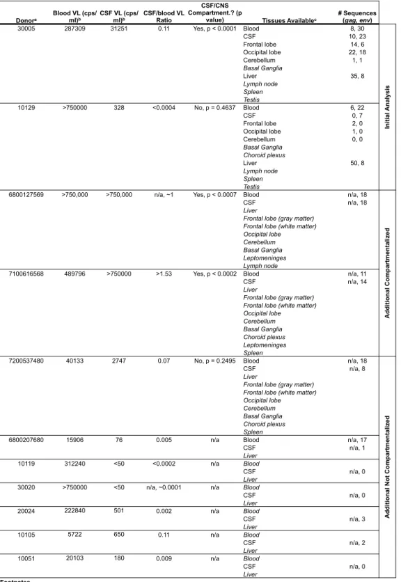
We obtained repository specimens from the National NeuroAIDS Tissue Consortium (NNTC),
which stores tissues generously donated for research by people who died with HIV-1. We obtained blood,
CSF, and at least one type of tissue from 11 donors who died with HAD (Table 1). Brain tissue was
available for five of 11 donors; only liver tissue was available for the remaining six. The majority of donors
had a final CD4 count below 400 cell/mm3 (82%; n = 9/11) and all were viremic at death.
PCR Amplification of HIV-1 Genes and Sequencing
Viral RNA was extracted from bodily fluids (blood and CSF) using the QIAmp Viral RNA Mini Kit
16
hours at 4°C. Purified viral RNA was reverse transcribed using Superscript III Reverse Transcriptase
(Invitrogen) and oligo-d(T)20 according to the manufacturer’s instructions. cDNA was subjected to single genome amplification (also known as end-point dilution PCR) for analysis of individual viral genomes.
cDNA was serially diluted to end-point and the full-length env gene was obtained using Platinum Taq
High Fidelity polymerase (Invitrogen) and primers that anneal within vif (F5010;
TGCCAAGAAAAGCAAAGATCATTAG-3’) and 3’ LTR U3 (LTRDN1;
5’-GACTCTCGAGAAGCACTCAAGGCAAGCTTTATTGAG-3’), followed by semi-nested PCR using primers
LTRDN1 and B5957UP1 (5’-GATCAAGCTTTAGGCATCTCCTATGGCAGGAAGAAG-3’). Full-length env
(~2.5 kb) was sequenced from ~3.7 kb PCR amplicons at the UNC-CH Genome Analysis Facility.
Chromatograms were analyzed for quality in Sequencher and chromatograms with double peaks,
indicating amplification from more than one cDNA template, were excluded from analysis.
For analysis of proviruses in tissues, total DNA was extracted using DNeasy Blood and Tissue Kit
(Qiagen) according to the manufacturer’s protocol. Briefly, ~2-3mm3 pieces of tissue were cut,
mechanically disrupted, and digested overnight in the presence of proteinase K, prior to DNA extraction
using a column-based method. DNA was quantified using UV-Vis spectrophotometry (Eppendorf). DNA
was end-point diluted and subjected to multiple rounds of PCR amplification of various HIV-1 genes as
described previously [15]. A thorough description of this protocol is included below in the “Method
Development” portion of the results section.
Phylogenetic Analysis of Viral Genes
DNA sequence alignments of viral env or gag obtained from bodily fluids or tissues (i.e. viral RNA
or DNA, respectively) were performed using ClustalΩ. Phylogenetic trees were inferred using the
neighbor-joining method and 500 bootstrap replicates (MEGA 5.2.2). Compartmentalized viral replication
was assessed using the Slatkin & Maddison statistical test of population structure with 10,000
permutations (HyPhy). Highlighter plots were generated using the tool available through the Los Alamos
17 Cloning HIV-1 env for Protein Expression
Viral env amplicons were chosen for cloning based on the phylogenetic tree structure, so that at
least one amplicon was used to represent each viral lineage. First-round PCR products were used as
template in a nested PCR reaction using Phusion Hot Start High Fidelity DNA Polymerase (Finnzymes)
and cloning primers. These primers were identical to those used in nested amplification of viral env
except for the addition of 5’-CACC-3’ at the 5’ end of the forward primer for the purpose of topoisomerase
cloning. PCR amplicons were gel-purified using the QIAquick Gel Extraction Kit (Qiagen) and 50ng of
purified amplicons were cloned into the pcDNA3.1D/V5-His-TOPO expression vector (Invitrogen) using
the pcDNA 3.1 Directional TOPO Expression Kit (Invitrogen). The entire cloning reaction was transformed
into MAX Efficiency Stbl2 competent cells (50ul) per the manufacturer’s instructions. Bacterial colonies
were screened for unidirectional insertion of viral env using colony PCR (Platinum Taq DNA Polymerase,
Invitrogen). DNA was extracted from 3-6 colonies using QIAprep Spin Miniprep Kit (Qiagen).
Co-transfection for Generating Env-pseudotyped Virus
For transfection,293T cells were seeded at a density of 4.8 x 10⁵ cells/well in 6-well tissue culture plates (DMEM, 10% FBS, 100 mg/ml penicillin and streptomycin culture medium). A 1:1 w/w ratio of env
clone and backbone (pNL4-3.LucR-E-; NIH AIDS Research and Reference Reagent Program, Division of
AIDS, NIAID, NIH) was used for co-transfection of 293T cells in serum-free DMEM using Fugene 6
Transfection Reagent (Roche). Transfection medium was replaced five hours later with culture medium
and cells were incubated at 37°C, 5% CO2 for 48 hours. Supernatants containing pseudotyped virus were harvested, passed through a 0.45 uM filter (Millipore), and stored at -80°C.
Single-cycle Infection of Reporter Cells for Determining Cell tropism
Affinofile cells are HEK293 cell derivatives that constitutively express viral co-receptor CXCR4
and can be differentially induced to express variable levels of viral receptor CD4 and viral co-receptor
CCR5 using doxycycline and ponasterone A (Invitrogen), respectively. Titration of Env-pseudotyped
luciferase reporter viruses was performed in triplicate on Affinofile cells with maximum induction levels for
18
that subsequent experiments were performed within the linear range of cell infectivity, a volume of virus
stock equivalent to 800,000 relative light units (RLUs) of luciferase expression was calculated for use in
cell tropism experiments.
For all Affinofile cell experiments, 96-well black bottom tissue culture plates were treated with
10% poly-L lysine and then seeded with 1.85 x 10⁴ cells/well (DMEM, 10% 12-14 kD dialyzed FBS, 50 mg/ml blasticidin culture medium). 18 to 24 hours later, expression of CD4 and CCR5 was induced at two
conditions in triplicate: CD4hi/CCR5hi (6 ng/ml doxycycline and 5 uM ponasterone A, respectively) and CD4lo/CCR5hi (5 uM ponasterone A only). Induction medium was removed 18 to 24 hours later and replaced with 100 ul/well of fresh, warmed culture medium containing 800,000 RLUs of Env-pseudotyped
luciferase reporter virus. Plates were spinoculated at 2,000 rpm for 2 hours at 37°C, and then incubated
at 37°C, 5% CO2 for 48 hours. Infection medium was removed prior to cell lysis for luciferase expression analysis (Firefly Luciferase Assay System, Promega). Lysates were stored at -80°C prior to thawing and
analysis using a luminometer. Between any medium change and before cell lysis, cells were washed
twice with 1X PBS. The ability to utilize low levels of CD4 expression for cell entry (macrophage tropism)
was defined by percent infectivity of CD4lo/CCR5hi relative to CD4hi/CCR5hi cells in terms of RLUs with a 12% cutoff for defining macrophage tropism.
RESULTS
Method Development: Probe Enrichment of Provirus
Our primary goal was to analyze viral DNA sequences from both productively and latently
infected cells present in tissues from HIV-infected donors. To this end, we attempted to develop a
sensitive polymerase chain reaction (PCR)-based method for amplifying HIV-1 DNA extracted from
infected tissues. Total DNA (cellular and viral DNA) was extracted from in vitro infected cells (control
experiments) or ~3 mm3 pieces of infected tissue (Qiagen DNeasy). Extracted total DNA contains cellular DNA in excess of viral DNA (perhaps by 1 billion-fold by mass), which increases the likelihood of
off-target amplification. In order to improve the specificity of HIV-1 DNA amplification, we attempted to enrich
19
we designed a nucleic acid probe for hybridization to HIV-1 sequence present in total DNA, followed by
streptavidin-biotin purification of nucleic acid hybrids on magnetic beads. The DNA fraction enriched for
viral DNA was then subjected to single genome amplification (SGA), also known as end-point dilution
PCR, where a nucleic acid template is diluted for use in subsequent PCR steps to ensure that a single
viral genome is amplified. This technique allows for phylogenetic analysis of viral populations within an
individual.
We designed two ~50 base pair (bp)RNA probes complementary to conserved regions in env
and nef of the HIV-1 genome. These probes are composed of “Locked Nucleic Acids” (LNAs), for optimal
hybridization efficiency. LNAs have a methylene bridge that locks the ribose ring of each nucleotide in the
optimal confirmation for base pairing, thereby increasing binding affinity and stability for specific, low
abundance sequences [55]. The 5’ end of each LNA probe is conjugated to biotin via a triethyleneglycol
(TEG) spacer 15 atoms in length. The TEG spacer is more than twice the length of the standard
six-carbon spacer, which increases the distance between the hybridized molecule and the surface of the
magnetic sphere for reduced steric hindrance. The 5’ biotin-TEG modification allows for purification of
nucleic acid hybrids on streptavidin-coated magnetic beads. The 3’ end of each LNA probe is modified
with hexanediol, a six-carbon glycol spacer that blocks extension by DNA polymerase. This modification
prevents the hybridized probe from interfering with downstream PCRs by blocking extension of DNAs at
the probe-binding site.
We used a control cell line called “8E5” to optimize our method. 8E5 cells are a derivative of
A3.01 cells, which is a continuous human CD4+ T cell line that is permissive to HIV-1 infection [56]. The
majority of A3.01 cells die from infection, but some CD4-downregulated cells survive and contain
provirus. 8E5 cells were selected from a pool of A3.01 survivor cells infected with LAV (1% nt pairwise
distance from HXB2)[57]. 8E5 cells contain a single copy of defective provirus. In the region of pol that
encodes RT, a single nucleotide insertion introduces a frameshift in the open reading frame (ORF) 3,
which is corrected by a compensatory mutation in vpr to maintain expression of viral env and other genes
in ORF3. This mutation results in the translation of an enzymatically inactive, truncated RT protein [58].
8E5 cells support provirus transcription and virus production, but RT-defective progeny virions are not
20
equate cell number with provirus copy number. In method development, we controlled for provirus copy
number in experiments using 8E5 provirus as template DNA. We also added controlled amounts of 293T
cell DNA relative to 8E5 DNA in order to reflect a putative range of viral-to-cellular DNA ratios in natural
infection. Thus, our control system better represents naturally infected cells as opposed to using a
plasmid control. Our immediate goal was to amplify full-length env sequences from infected tissues in
order to characterize viral populations and replication, as well as cell tropism.
Although our protocol was successful at purifying provirus from background DNA, it was
unsuccessful at purifying all or even a majority of input proviruses: we could only purify ~1% of input
proviruses with this method. We spent a great deal of time attempting to optimize our method, but were
not able to improve our efficiency (Table 2). It is likely that the addition of more probes spanning the
HIV-1 genome would improve provirus recovery as another lab developed such a method for Plasmodium
falciparum (malaria) sequencing, and we are currently investigating this possibility [59]. We then moved
on to alternative methods of provirus amplification.
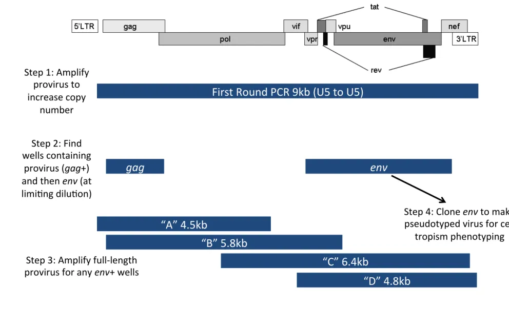
Method Development: Full-Length Provirus PCR
Robert Siliciano and colleagues (Johns Hopkins U.) published a promising method for PCR
amplification of HIV-1 DNA in latently infected T cells [15]. In short, resting CD4+ T cells were purified
from PBMCs of patients on suppressive ART and total DNA was extracted for PCR amplification of
proviruses. Proviruses were amplified in limiting dilution PCR and then a series of second-round nested
PCRs. First-round limiting dilution PCR amplifies essentially the entire viral genome using primers that
anneal within the U5 regions of the 5’ and 3’ LTRs (9.1 kb; Figure 1). Replicate PCR wells in the
first-round reaction were then screened for the presence of provirus with a nested second-first-round PCR. In
similar additional PCRs, overlapping fragments spanning the viral genome are obtained for sequence
analysis (Figure 1).
We used the same mixed DNA controls as before (mixtures of 8E5 and 293T cell DNA) in
reproducing the method published by Siliciano and colleagues. We first assessed the efficiency of this
method using known quantities of proviral 8E5 DNA. Total DNA was extracted from 8E5 cells and
21
replicates. In order to screen for the presence of provirus in round PCR replicate wells, 1 ul of
first-round product was used as template in the second-first-round nested PCR targeting gag. We found that gag
amplification consistently resulted in a single band of approximately the correct size (~1.5 kb) and almost
always was the expected HIV-1 sequence.
All first-round PCR wells that were positive for gag were then subjected to env amplification in a
second nested PCR. Unfortunately, we found that only ~30% of the time we could obtain both gag and
env from the same first-round PCR well. Furthermore, env PCR resulted in non-specific amplification of
the human genome, resulting in a band of incorrect size or multiple bands that were sequence-verified as
human genomic sequence. Since obtaining env is important for cell tropism analysis, we sought to
determine why the gag PCR appeared relatively efficient compared to env. We found that the same
first-round PCR wells were consistently positive for any subsequent PCRs, whereas those that are gag+/env-
were always negative in subsequent PCRs. We also designed primers to amplify a small ~500 bp
fragment of the LTR, and found that additional wells (gag-) were positive for LTR, indicating the presence
of provirus missed by the gag PCR. PCR efficiency was usually best for LTR (~500 bp), then for gag
(~1500 bp), then for env (~3kb), and finally for overlapping fragments spanning the whole genome (~5 kb
each) indicating that PCR of smaller fragments is more efficient than larger ones. Our experiments show
that first-round PCR of the essentially full viral genome is inefficient, resulting in too few template copies
for subsequent PCRs.
After thorough analysis and speaking directly with the Siliciano lab, we found that increasing the
primer concentration was important for improving env amplification efficiency. In a side-by-side
comparison of 1 uM versus 0.4 uM primer in all PCRs using 8E5 template DNA, we found that only gag+
reactions obtained with 1 uM primer were also positive for env+ in every replicate well. However,
increasing primer concentration does not increase the total number of LTR+ wells. These data collectively
indicate that the effect of increasing primer concentration in first-round PCR increased the copy number
of proviruses rather than our ability to detect them. We think that this improvement in first-round PCR
efficiency is due to a greater probability of correctly priming the HIV-1 genome in the initial cycles of
first-round PCR. When too little primer is present, the human genome, which is in vast excess to the single
22
the provirus. Indeed, when HIV-1 sequence was not obtained from a given PCR amplicons, the amplicons
almost always corresponded to human genomic sequence. We reformatted our general workflow for the
remainder of this study moving forward and an associated protocol is attached (Supplemental 1).
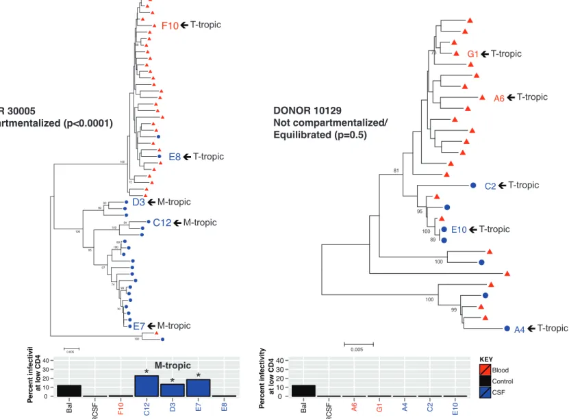
Assessing Compartmentalized Viral Replication
We conducted a preliminary phylogenetic analysis of two individuals with disparate states of viral
replication (donors 30005 and 10129). Blood and CSF were available from roughly 11 months prior to the
date of death for each donor. We first compared viral env sequences from circulating virus (viral RNA) in
the blood plasma and CSF. In donor 30005, the population of virus in the blood is distinct from that in the
CSF, indicating compartmentalized replication within the CSF/CNS (Figure 2). The Slatkin & Maddison
statistical test of population structure was used to confirm compartmentalization (p = <0.0001). Regions in
env with the greatest sequence variability between the blood and CSF viral populations are in V1/V2 and
V4/V5 with the majority of differences in the V1 and V4 hypervariable regions.
A similar analysis was completed for donor 10129. In this donor, blood and CSF virus are mixed
within the overall population, which indicates that virus is similar between compartments and thus is in an
equilibrated state (Figure 2). A lack of compartmentalized replication was confirmed with the Slatkin &
Maddison statistical test of population structure (p = 0.4637). Due to a relatively low CSF viral load in this
donor (328 cp/ml), few env sequences were obtained from the entirety of the sample, thereby sacrificing
some statistical power. Low CSF viral load in this donor suggests that virus predominantly replicates in
the peripheral blood and tissues of this donor, but not appreciably in the CNS.
Eight additional donors will eventually be included in this study, two of which have
compartmentalized viral replication in the CNS as determined by viral env sequences in the CSF and
blood (Table 1, work completed by Laura Kincer and Sarah Joseph). Multiple brain and peripheral tissues
23
HIV-1 env Sequences from Brain Tissue Resemble Those in the CSF and Distinct Viral Lineages Exist within Different Brain Regions
Our analysis of proviral sequences was initially based on viral env, since the viral Env protein
determines cell tropism based on the efficiency of CD4 receptor utilization. Assessing cell tropism of an
archived provirus reveals which cell type(s) the virus was capable of replicating in. To analyze proviruses
archived in brain tissue (viral DNA), 42 viral env sequences were obtained from total DNA extracted from
donor brain tissues (Table 1). 31 of 42 env sequences came from donor 30005, 21 of 31 env genes from
this donor are defective as they contained deletions and 10 env genes are putatively functional. Donor
30005 had highly compartmentalized viral replication in the CSF (viral RNA) 11 months prior to death,
and five full-length env sequences (four functional and one defective) were obtained from provirus in the
frontal and occipital lobes for incorporation into the phylogenetic tree of cell-free virus described above.
Proviral sequences in the brain most closely resemble virus in the CSF compared to the blood,
suggesting compartmentalized replication within the overall CNS at death as well as 11 months prior.
Three sequences obtained from the frontal lobe suggest the presence of a brain region-specific viral
lineage (100% bootstrap), which appears to be distinct from CSF virus, although the statistical
significance is low (<70% bootstrap).
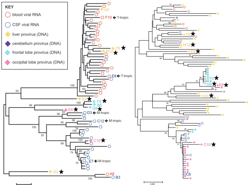
PCR amplification of env in tissue DNA is relatively inefficient compared to gag, at least with our
protocol. Therefore, we had a greater number of gag sequences to analyze with phylogenetics. We
compared gag sequences obtained from virus in the blood plasma and CSF (viral RNA) and virus in
tissues (viral DNA; Figure 3) of donor 30005. As predicted, virus obtained form brain tissue more closely
resembles CSF virus as opposed to that in the blood. Furthermore, compartmentalized viral replication in
the CNS at death was verified using predominantly proviral sequences. The presence of brain
region-specific viral lineages was also confirmed with our gag data as the frontal and occipital lobes contain
lineages that are distinct from one another. Furthermore, multiple lineages exist within each lobe: three in
the frontal lobe and two in the occipital lobe.
Clonal amplification of particular viral species occurred in both lobes, which, in the absence of
ART, suggests that selective pressure exists within these brain regions. Clonal viral sequences could
24
The presence of two CSF viruses, which came from a sample collected 11 months prior to death, within
the clonal lineage in the occipital lobe suggests that clonally expanded cells arose from an infectious
event, likely from viral clones of CSF virus C6, that occurred at least 11 months prior to death.
A highlighter plot of each clonal viral lineage within the tree for donor 30005 was used to find
evidence of viral replication as opposed to clonally expanded cells (Figure 4 and data not shown). We
analyzed highly similar occipital lobe sequences using a representative of eight identical gag sequences
(seven from brain tissue and one from the CSF) as the master sequence with which every other
sequence is compared: the CSF virus (C6) was chosen, as it represents actively replicating virus 11
months prior to tissue collection at death. This analysis compares sequence variation relative to the
seven clonal sequences within a lineage composed of highly similar sequences. If clonal expansion of
cells occurred, then we would expect to see identical nucleotide differences in the remaining sequences
when grouped by branch length (i.e. the five sequences with medium branch lengths and the three with
longer). However, this lineage appears to have come from a single infectious event followed by local
replication in neighboring cells, as nucleotide differences at any given position in the remaining
sequences are randomly distributed. The same was observed in a similar analysis of the frontal lobe
(data not shown).
11 env genes were obtained for donor 10129, and all but one (occipital lobe) came from the liver.
7 of 11 env genes are putatively functional (1 sequence needs to be verified) and 4 of 11 are defective (3
with deletions and 1 with stop codons). In donor 10129, the CSF viral load was very low compared with a
high blood viral load (328 v. >750,000 cps/ml), suggesting that no appreciable viral replication occurred in
the CNS. Similarly to the CSF, few HIV-1 proviral sequences (env or gag) were obtained from the brain of
this donor (occipital and frontal lobes and cerebellum were sampled). Indeed, only one env sequence was
obtained from the brain, particularly the occipital lobe, and this sequence corresponded to a defective
provirus due to a deletion from pol to env (data not shown). Thus, no functional env genes (viral DNA)
were obtained from the brain of donor 10129 and only 7 functional env genes (viral RNA) were obtained
from 1 mL of CSF. Furthermore, there was no evidence of compartmentalized or clonal replication in the
CSF/CNS. These data collectively suggest that virus did not replicate in the CNS of this donor,
25
The CSF we received from donor 10129 was exhausted in the initial analysis of viral env due to
low CSF viral load, therefore CSF gag sequences could not be obtained for this donor. Few proviral gag
sequences were obtained from the brain, 2 from the frontal and 1 from the occipital lobe, again
suggesting a lack of appreciable viral replication in the brain. The equilibrated state of virus throughout
the body in this donor was also verified with gag sequences (Figure 5).
Individual Viral Lineages Also Exist within the Liver, a Peripheral Tissue Exposed to Blood Virus In donor 30005, 3 of 5 full-length proviral env sequences obtained from the liver (viral DNA)
grouped with those in the blood (viral RNA), indicating that virus can travel between these compartments,
as predicted. There are two liver viruses distinct from those that grouped with the blood: E4 and D3. Liver
virus E4 is more similar to blood virus than CSF virus, but is still a statistically significant distinct entity
(93% bootstrap). Liver virus D3 is positioned near a unique viral lineage (blood virus A8 and CSF virus
B3, 100% bootstrap) in the phylogenetic tree, but with a poor bootstrap value of 51%. These distinct liver
sequences may represent minor viral lineages that are sampled poorly with env PCR. Therefore, we
analyzed gag sequences, as a greater number were available than env.
Comparison of HIV-1 proviral gag sequences from donor 30005 revealed individual viral lineages
within a given tissue, including the liver (Figure 3). At least two viral lineages were found within the liver:
one that resembles virus in the blood and another that resembles CSF virus. Since sequences obtained
from the frontal lobe as well as the liver resemble the clonal lineage within the occipital lobe, we created a
highlighter plot to determine if any sequence features are shared between compartments. Indeed, when
designating CSF virus H2 as the master sequence, two additional CSF sequences (A3 and B2), four
frontal lobe sequences (G8, H5, F9, and H9), and one liver sequence (B12) all contain the same two
mutations observed in the occipital lobe clonal lineage (Figure 4). These data suggest that virus produced
in the occipital lobe can travel to the frontal lobe, likely via CSF, and replicate there. It is important to note
that CSF flow directionality favors this hypothesis. CSF virus that seeded the brain and locally replicated
may have then exited the CNS, entered the periphery, and replicated in liver tissue.
The same first-round PCR products (amplified provirus) were used as template in subsequent,