P2Y Receptor Trafficking in Polarized Epithelial Cells
Darrell Ross DuBose Jr.
A dissertation submitted to the faculty of the University of North Carolina at Chapel Hill in partial fulfillment of the requirements for the degree of Doctor of Philosophy in the
Department of Pharmacology.
Chapel Hill 2013
Approved by, Robert Nicholas Kendall Harden Martina Gentzsch Klaus Hahn
ii © 2013
iii
Abstract
DARRELL ROSS DUBOSE JR: P2Y Receptor Trafficking in Polarized Epithelial Cells (Under the direction of Dr. Robert Nicholas)
iv
Table of Contents
List of Figures... vi
List of Abbreviations ... vii
Chapter 1: Introduction ... 1
G protein coupled receptors and G protein signaling ... 1
Nucleotide Signaling ... 7
P2Y Receptors ... 8
P2Y1 Receptor... 10
P2Y2 Receptor ... 11
P2Y4 Receptor... 12
P2Y6 Receptor... 14
P2Y11 Receptor ... 16
P2Y12 Receptor ... 18
P2Y13 Receptor ... 19
P2Y14 Receptor ... 20
Epithelial Cells and Cell Polarization ... 22
Sorting Signals in Transmembrane Proteins ... 25
Apical-targeting signals ... 25
Basolateral-targeting signals ... 28
Polarized Expression of P2Y Receptors ... 31
The Apical-sorting signal of the P2Y2 Receptor ... 34
The Basolateral-sorting signal of the P2Y1 Receptor ... 35
v
Trafficking Itineraries and Technological Limitations ... 37
In Vivo Covalent Fluorophore Attachment ... 39
Chapter 2: Apical Targeting of the P2Y4 Receptor is Directed by Hydrophobic and Basic Residues in the Cytoplasmic Tail ... 41
Overview ... 41
Introduction ... 42
Methods ... 46
Results ... 51
Discussion ... 61
Chapter 3: Distinct Trafficking Itineraries of the P2Y1, P2Y2, and P2Y4 Receptors in Polarized Madin-Darby Canine Kidney Epithelial Cells ... 69
Overview ... 69
Introduction ... 70
Materials and Methods ... 73
Results ... 82
Discussion ... 96
Chapter 4: Conclusions and Future Directions ... 102
Conclusions ... 102
Future Directions... 104
vi
List of Figures
Figure 1.1. G proteins are tightly regulated molecular switches. ... 3
Figure 1.2. GPCR signaling is dependent on the Gα subtype. ... 5
Figure 1.3. Endogenous agonists and signaling pathways of the eight cloned P2Y receptors. ... 9
Figure 1.4. Epithelial cells have two distinct membrane domains. ... 23
Figure 1.5. Confocal microscopy of wild-type (WT) MDCK(II) cells and MDCK(II) cells expressing HA-tagged human P2Y receptors. ... 32
Figure 1.6. P2Y receptor-targeting signals are contained within the main body and C-terminal tail of the receptors. ... 33
Figure 2.1. The P2Y4 apical-targeting signal ends before Asp343. ... 52
Figure 2.2. The P2Y4 apical-targeting signal begins after Cys321. ... 53
Figure 2.3. Subcellular localization of receptor constructs with mutant P2Y4 C-tails. ... 55
Figure 2.4. Surface expression of receptor constructs with mutant P2Y4 C-tails. ... 56
Figure 2.5. The P2Y4 apical-targeting signal is dominant over a basolateral signal. ... 60
Figure 2.6. Conservation of the apical-targeting sequence of the P2Y4 receptor. ... 62
Figure 3.1. Design and Labeling of SNAP-HA-P2Y Receptors. ... 83
Figure 3.2. Activity of SNAP-HA-P2Y Receptors. ... 85
Figure 3.3. SNAP-tag Kinetics, Stability, and Membrane Leak. ... 87
Figure 3.4. Cell Surface Delivery of SNAP-HA-P2Y1. ... 89
Figure 3.5. Cell Surface Delivery of SNAP-HA-P2Y2. ... 90
Figure 3.6. Cell Surface Delivery of SNAP-HA-P2Y4. ... 91
vii
List of Abbreviations
5’UTR – Five prime untranslated region AC – Adenylate cyclase
ADP – Adenosine diphosphate AMP – Adenosine monophosphate AP - Apical
ATP – Adenosine triphosphate BK2 – Bradykinin 2
BL – Basolateral
BSA – Bovine serum Albumin
cAMP – Cyclic adenosine monophosphate
CFTR – Cystic fibrosis transmembrane conductance regulator CHO – Chinese hamster ovary
CNG – Cyclic nucleotide gated DAG – Diacylglycerol
DMEM/F12 - Dulbecco's modified eagle medium/nutrient mixture F-12 DMSO - Dimethylsulfoxide
DNA – Deoxyribonucleic acid DTT – Dithiothreitol
EGF – Epithelial growth factor ENaC – Epithelial sodium channel eNOS – Epithelial nitric oxide synthase FLIPR – Fluorometric imaging plate reader
viii GDP – Guanosine diphosphate
GEF – Guanine nucleotide exchange factor GFP – Green fluorescent protein
GMP – Guanosine monophosphate GPCR – G protein-coupled receptor GPI – Glycophosphatidylinositol GST – Glutathione S-transferase GTP – Guanosine triphosphate IP3 – Inositol 1,4,5-trisphosphate IP3R – IP3 receptor
IPTG - Isopropylthio-β-galactoside LDL – Low density lipoprotein
MAPK – Mitogen-activated protein kinase MDCK – Madin-Darby canine kidney NFDM – Non-fat dry milk
PBS – Phosphate buffered saline
PDZ – Post synaptic density protein/drosophila disc large tumor suppressor/zonula occludens-1
pen/strep – Penicillin/streptomycin PFA – Paraformaldehyde
PI3K – Phosphatidylinositide 3-kinases pIgR – Polymeric immunoglobulin receptor PIP2 - Phosphatidylinositol 4,5-bisphosphate PKA – Protein kinase A
PKC – Protein kinase C
ix PLC – Phospholipase C
PVDF – Polyvinylidene difluoride RGS – Regulator of G protein signaling RIA – Radio-immunosorbant assay RIPA – Radio-immunoprecipitation assay RNA – Ribonucleic acid
SDS-PAGE – Sodium dodecyl sulfate polyacrylamide gel electrophoresis TM7 – Transmembrane region 7
TNFα – Tumor necrosis factor alpha UDP – Uridine diphosphate
UTP – Uridine triphosphate WT – Wild type
Chapter 1: Introduction
G protein coupled receptors and G protein signaling
G protein coupled receptors (GPCRs) are metabotropic transmembrane receptors – they mediate slow (second to minute responses) changes in cell physiology in response to extracellular ligands. GPCRs comprise about 750 proteins (in humans) that share a common seven-transmembrane structure. Of those, about half are olfactory or chemosensory receptors, 180 have known endogenous ligands, and 187 are known as orphan receptors (Vassilatis et al., 2003). An orphan receptor is a GPCR that is expected to be a receptor with an endogenous cognate ligand based on sequence homology with known receptors, but that ligand has not yet been discovered. GPCRs are of particular interest because they affect nearly every aspect of cell biology and are often expressed in a restricted, tissue-dependent manner. This, combined with extracellular binding sites, makes GPCRs highly attractive as potential drug targets. Indeed, almost half of the drugs on the market today target GPCRs (Drews, 2000).
2
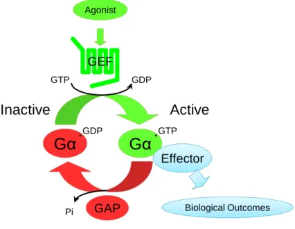
G proteins act as molecular switches that regulate intracellular signaling. There are two types of G proteins: small (or Ras-like) G proteins and heterotrimeric G proteins. Small G proteins consist of a single catalytic subunit and are not directly associated with GPCRs. Heterotrimeric G proteins have three subunits: the catalytic alpha subunit (Gα), as well as beta and gamma subunits that form obligate heterodimers and are referred to simply as Gβγ or βγ. At rest, Gα is bound to GDP and exists as a heterotrimer with Gβγ and
cannot interact with downstream effectors. When stimulated by an agonist-bound GPCR, GDP is released from Gα and GTP, which is present at high concentrations in the cell, binds to the G protein and causes a conformational change that releases Gα•GTP from Gβγ and allows it to bind effectors. Gβγ can also interact with effectors and trigger
downstream signaling events. GTP is hydrolyzed to GDP by the inherent GTPase activity of Gα, which promotes rebinding to Gβγ, and signaling ceases.
The nucleotide cycling of G proteins is a tightly controlled process; the two slowest steps, GDP release and GTP hydrolysis, are regulated by other proteins. The first step, release of GDP, is catalyzed by guanine nucleotide exchange factors (GEFs). There is a large class of specific GEFs for small G proteins, but for heterotrimeric G proteins the GEF is normally a GPCR. The second step of the cycle is the hydrolysis of GTP. Although G proteins have the inherent ability to hydrolyze GTP, it is rather slow; the reaction proceeds much faster with the association of a second class of enzymes – GTPase-activating proteins (GAPs). There are a variety of GAPs for both small and heterotrimeric G proteins, but some of the most common are known as regulators of G protein signaling (RGS) proteins.
3
G
α
G
α
GDP
.
GTP GDP
Pi GAP
Agonist
Biological Outcomes
Inactive
Active
Effector
GTP
.
GEF
Figure 1.1. G proteins are tightly regulated molecular switches. G proteins cycle
4
These subunits have variable affinities for different GPCRs, and often a GPCR will signal through only one of the four classes.
GTP-bound Gq adopts a conformation that allows it to interact with its primary effector, phospholipase C. While bound to Gq-GTP, phospholipase C catalyzes the hydrolysis of phosphatidylinositol 4,5-bisphosphate (PIP2) into diacylglycerol (DAG) and inositol 1,4,5-trisphosphate (IP3). PIP2 in the cell membrane normally stabilizes the opening of ion channels, including the epithelial sodium channel, ENaC. The reduction in PIP2 concentration by PLC is sufficient to reduce ion flux through these channels. Upon cleavage, IP3 is released into the cytoplasm, where it is free to bind its cognate receptor on the surface of the endoplasmic reticulum, releasing stored calcium ions. This spike in calcium activates a variety of effectors, including cell surface chloride and potassium channels, calmodulin, and protein kinase C. The DAG fragment remains membrane bound, where it recruits and activates protein kinase C. In addition to PLC, Gq can bind RhoGEFs (similar to G12/13, described below), beginning signaling cascades that affect the cytoskeleton.
Adenylate cyclase (AC) is the primary effector of both Gs and Gi. GTP-bound Gs binds AC to stimulate the production of cAMP. GTP-bound Gi binds AC at an allosteric site, blocking cAMP production. As a potent second messenger, cAMP activates many effectors, including cyclic-nucleotide gated ion channels and protein kinase A(which phosphorylates and activates CFTR as well as many other targets). Note that some of these ion channels are Ca2+ permeable, and as such can replicate some aspects of Gq signaling.
5 PLCβ
Gαq Gαs Gαi Gα12/13
AC
cAMP RhoGEF
Rho PKA
DAG
IP3
ER
IP3R
Ca2+
Ca2+
CNG channels PKC
Figure 1.2. GPCR signaling is dependent on the Gα subtype. GPCRs signal through
6
7
Nucleotide Signaling
Nucleotides are ubiquitous small molecules in nature involved in a wide variety of biological processes. They are the building blocks of DNA, RNA, and many enzymatic co-factors. They are also energy “currency”, used to drive forward reactions that would otherwise be unfavorable. They are cycled into potent second-messengers that transduce intracellular signals (e.g. cyclic AMP and cyclic GMP). Lastly, they are extracellular signaling molecules that bind transmembrane receptors, often in an autocrine/paracrine manner, to convey a signal across the plasma membrane. It is this receptor signaling role that is pertinent to this work.
Nucleotide receptors have been found in all cell types, where they mediate a plurality of cell activities. There are two distinct families of nucleotide receptors: P2X and P2Y. P2X receptors are transmembrane channels that open in response to binding extracellular ATP. Once open, Ca2+ and Na+ ions pass through the pore and depolarize the cell membrane. P2X receptors are primarily involved in synaptic and neuromuscular signaling. A functional P2X receptor is composed of three P2X subunits. There are seven known P2X subunits which can combine to form a variety of homo- and heteromeric receptors. The P2X subunits are designated P2X1 through P2X7.
8
P2Y Receptors
P2Y receptors are a family of GPCRs that respond to extracellular nucleotides. There have been eight P2Y receptors identified to date—P2Y1, P2Y2, P2Y4, P2Y6, P2Y11, P2Y12, P2Y13, and P2Y14. The numbering is not sequential because several receptors have been named as a P2Y receptor, only to be determined later either to not to be activated by nucleotides at all, or to be homologues of a mammalian P2Y receptor. For example, the p2y3 receptor was later determined to be the avian homologue of the human P2Y6 receptor (Li et al., 1998). P2Y receptors can be divided into two subclasses based on their downstream signaling properties. The P2Y1–like receptors (P2Y1, P2Y2, P2Y4, P2Y6, and P2Y11) signal primarily through Gq, while the P2Y12-like receptors (P2Y12, P2Y13, and P2Y14) signal primarily through Gi. The P2Y11 receptor, while most efficiently coupling to Gq, also signals through Gs to stimulate AC. The receptors can also be characterized by their activating ligands, as shown in Figure 1.3.
9
UTP=
UDP-Ligand ADP ATP UTP UDP ATP ADP ADP glucose
Receptor P2Y1 P2Y2 P2Y4 P2Y6 P2Y11 P2Y12 P2Y13 P2Y14
Signaling Gq Gq Gq Gq Gq Gs Gi Gi Gi
Response PLC PLC PLC PLC PLC AC AC AC AC
Figure 1.3. Endogenous agonists and signaling pathways of the eight cloned P2Y
10
P2Y1 Receptor
The P2Y1 receptor is a 373-amino acid protein encoded by a single exon on the forward strand of chromosome 3 (Genbank Accession Number S81950). For endogenous nucleotides, the P2Y1 receptor responds primarily to ADP (Ki = 0.92 µM), but also to ATP (Ki = 17.7 µM), albeit with lesser efficacy (Palmer et al., 1998;Waldo and Harden, 2004). Synthetic agonists include 2-MeSADP (Ki = 0.0099 µM), ATPγS (Ki = 1.33 µM), 2-MeSATP (Ki = 1.87 µM), ADPβS (Ki = 2.42 µM), and (N)-methanocarba-2MeSADP (MRS2365, EC50 = 34 nM) (Bourdon et al., 2006;Waldo and Harden, 2004). Of these, only MRS2365 is highly selective for the P2Y1 receptor. Selective antagonists (a rarity for P2Y receptors) have also been developed for the P2Y1 receptor: MRS2179 (pKB = 6.75 (Moro et al., 1998), MRS2279 (pKB = 8.10) (Boyer et al., 2002), and MRS2500 (Ki = 0.79 nM and KB = 1.74 nM) (Kim et al., 2003). These compounds have been instrumental in the study of P2Y1 receptor physiology.
As mentioned above, the P2Y1 receptor is involved in a wide variety of physiological processes depending on the tissue in which it is expressed. Of specific interest to this work, the Gq coupled P2Y receptors (including P2Y1) induce luminal ion flux in epithelial cells, thus modulating osmotic balance between the body and external fluids (Christofi et al., 2004;Fang et al., 2006;Lee et al., 2007;Matos et al., 2005;Rajagopal et al., 2011). Interestingly, the P2Y1 receptor is expressed only on the basolateral surface of all the epithelial cells in which its location has been investigated, meaning that it only responds to ADP released within the organ (i.e. those tissues with access to the basolateral membrane) and not from the lumen (Wolff et al., 2005). In contrast, P2Y2, P2Y4, and P2Y6 receptors are primarily expressed at the apical membrane and therefore respond to nucleotides released at the luminal compartment.
11
and Burnstock, 1998). In platelets, the P2Y1 receptor induces shape change that, along with P2Y12 receptor activation, leads to aggregation (Hechler et al., 1998). P2Y1 receptor antagonists are currently being investigated as potential antithrombotic agents (Bird et al., 2012). In bone, the P2Y1 receptor functions in osteoclastic bone resorption (Hoebertz et al., 2001). The P2Y1 receptor regulates a variety of functions in the nervous system as well. It inhibits N-type calcium channels (Filippov et al., 2000), modulates synaptic transmission (von Kugelgen and Wetter, 2000), and has a role in astrocyte calcium signaling (Fumagalli et al., 2003).
P2Y2 Receptor
12
Like the P2Y1 receptor, the P2Y2 receptor stimulates ion flux in epithelial cells via Gq activation (Ghanem et al., 2005;Hosoya et al., 1999;Hwang et al., 1996;Parr et al., 1994;Rajagopal et al., 2011). In contrast to the P2Y1 receptor, the P2Y2 receptor is expressed primarily at the apical surface and thus responds to luminal signals (Wolff et al., 2005). This physiological activity is pronounced in epithelium of both the lung and the lacrimal duct. As such, agonists of the P2Y2 receptor are under investigation as potential therapeutic agents for cystic fibrosis and dry eye disease (Hosoya et al., 1999;Pintor et al., 2002;Yerxa et al., 2002).
Additionally, the P2Y2 receptor has been linked with a long list of physiological activities, including both vasodilatation and vasoconstriction (da Silva et al., 2009;Ralevic and Burnstock, 1998), apoptosis in colorectal carcinoma cells (Burnstock and Knight, 2004), bone remodeling (Hoebertz et al., 2002), monocyte recruitment (Seye et al., 2003), cell proliferation (Burrell et al., 2003;Greig et al., 2003;Muscella et al., 2003;Schafer et al., 2003); neutrophil degranulation and infiltration (Ayata et al., 2012;Meshki et al., 2004), inflammation (Kruse et al., 2012;Schuchardt et al., 2011), HIV-1 infection (Seror et al., 2011), amyloid precursor processing (Leon-Otegui et al., 2011), wound healing (Boucher et al., 2011), metastasis (Schumacher et al., 2013), and neuroprotection (Chorna et al., 2004;Weisman et al., 2012).
P2Y4 Receptor
13
Interestingly, the rat P2Y4 receptor responds to a wider variety of ligands. Both UTP (EC50 = 0.20 µM) and ATP (EC50 = 0. 51 µM) are agonists, as well as Ap4A (EC50 = 1.24 µM), ITP (EC50 = 1.82 µM), GTP (EC50 = 2. 28 µM), CTP (EC50 = 7.24 µM), and XTP (EC50 = 22.9 µM) (Kennedy et al., 2000). These differences in agonist activation have been traced to three amino acids (Asn-177, Ile-183, and Leu-190) in the second extracellular loop of the rat receptor (Herold et al., 2004). Until recently, no selective ligands of any type were known for the P2Y4 receptor, making pharmacological differentiation between the P2Y2 receptor and P2Y4 receptor difficult. Recently, a few moderately selective P2Y4 receptor agonists have been synthesized. First, iso-CMP (EC50 = 4.98 µM) is >20-fold selective for the P2Y4 receptor versus P2Y2 and P2Y6 receptors (El-Tayeb et al., 2011). A second group synthesized N4-(phenylpropoxy)-CTP (MRS4062, EC50 = 23 nM), Up4-[1]3’-deoxy-3’-fluoroglucose (MRS2927, EC50 = 62 nM), and N4-(phenylethoxy)-CTP (EC50 = 73 nM), each of which has 10-fold or greater selectivity for the P2Y4 receptor versus P2Y2 and P2Y6 receptors (Maruoka et al., 2011).
High expression levels of the P2Y4 receptor have been detected in the placenta, intestine, pituitary, and brain, with lower levels in liver, bone marrow, monocytes and lymphocytes (Communi et al., 1995;Jin et al., 1998;Moore et al., 2001), but relatively few physiological functions are known.
14
also be of therapeutic value. Bacterial infection of the gut is known to cause nucleotide release and chloride secretion. As excess luminal chloride secretion is a central mediator of diarrhea symptoms (Field et al., 1989;Kunzelmann and Mall, 2002), pharmacological inhibition of the P2Y4 receptor (and likely the P2Y2 receptor as well) may attenuate infection symptoms.
In addition to the intestine, the P2Y4 receptor has a role in auditory neurotransmission. It is expressed in vestibular dark cell epithelium and strial marginal cells, where it controls K+ secretion (Hur et al., 2007;Lee et al., 2006;Marcus and Scofield, 2001). The P2Y4 receptor has also been implicated in vasodilation (Burnstock, 2002;McMillan et al., 1999), cell proliferation (Burnstock, 2002), and cardiac development (Horckmans et al., 2012).
P2Y6 Receptor
The P2Y6 receptor is a 328-amino acid protein encoded by a single exon on the forward strand of chromosome 11. There are eight splice variants, seven of which encode the same protein; they differ only in the 5’UTR (Genbank Accession Numbers NM_176797, NM_176798, NM_176796, NM_001277204, NM_001277205, NM_001277206, and NM_001277207). A single splice variant begins translation at an alternate start site and encodes additional amino acids at the N-terminus of the protein (Genbank Accession Number NM_001277208).
15
recently, 5-OMe-UDP (EC50 = 0.08 µM) and N3-phenacyl-β,γ-dichloromethylene-UTP (EC50 = 0.142 µM) were synthesized and shown to be selective for the P2Y6 receptor versus the P2Y4 receptor but not the P2Y2 receptor (El-Tayeb et al., 2011;Ginsburg-Shmuel et al., 2010). Additionally, three compounds have been synthesized that are insurmountable, selective antagonists of the P2Y6 receptor; diisothiocyanate derivatives of 1,2-diphenylethane (MRS2567, IC50 = 126 nM), 1,4-di-(phenylthioureido)butane (MRS2578, IC50 = 37 nM), and 1,4-phenylendiisothiocyanate (MRS2575, IC50 = 155 nM, human only) (Mamedova et al., 2004).
The P2Y6 receptor is expressed in particularly high levels in the spleen, and has also been detected in placenta, thymus, intestine, vascular smooth muscle, lung, kidney, bone, adipose, heart, and parts of the brain (Communi et al., 1996;Moore et al., 2001;Ralevic and Burnstock, 1998).
Like the receptors above, the P2Y6 receptor also regulates ion flux in epithelial cells (Burnstock and Knight, 2004). Of particular interest, it seems to play a primary role in gallbladder epithelia, where it may be a therapeutic target to correct Cl- secretion in cystic fibrosis patients (Lazarowski et al., 2001). Some evidence has been presented indicating that the P2Y6 receptor also stimulates Cl- secretion in various epithelial cells through a second mechanism involving the CFTR channel (Dulong et al., 2007;Kottgen et al., 2003;Schreiber and Kunzelmann, 2005;Wong et al., 2009). However, none of these studies directly demonstrates that the effects observed are dependent on P2Y6 receptor activation, and each uses 100 µM UDP, which is more than 300-fold higher than the EC50 in other assays. Also, in early experiments with the P2Y6 receptor, no direct coupling of P2Y6 to Gs was observed (Chang et al., 1995).
16
respectively, and suppresses T-cell activation during allergic inflammation (Bar et al., 2008;Giannattasio et al., 2011;Warny et al., 2001). UDP, acting through the P2Y6 receptor, has also been shown to induce chemokine production in microglia and astrocytes (Kim et al., 2011). In bone, the P2Y6 receptor stimulates differentiation into osteoclasts and increases their resorptive activity (Orriss et al., 2011). In the vasculature, activation of the P2Y6 receptor causes relaxation of endothelial cells and contraction of nitric-oxide-blocked vascular smooth muscle cells (Bar et al., 2008). Lastly, it has a cytoprotective role, preventing TNFα-induced apoptosis (Kim et al., 2003;Kim et al., 2003;Mamedova et al., 2008).
P2Y11 Receptor
The P2Y11 receptor is a 374-amino acid protein encoded by two exons on the forward strand of chromosome 19 (Genbank Accession Number AJ298334). Transcription of the P2Y11 receptor has an uncommon feature. Through alternate splicing a chimeric protein can be formed between the P2Y11 receptor and Ssf1, the neighboring gene on chromosome 19. This alters the extracellular N-terminus of the receptor, removing the first 5 amino acids and instead fusing the Ssf1 protein. Chimeric mRNA was detected in a variety of human tissues and the presence of a 90kDa protein product was identified from transfected CHO cells. ATP and other agonists have reduced potency and/or efficacy at the chimeric receptor, but the function of this fusion protein is unknown (Communi et al., 2001).
17
al., 2003) regarding UTP), with a preference for ATP over ADP. In contrast, ADP is more potent than ATP at the canine receptor (Qi et al., 2001). Interestingly, agonist potency at the P2Y11 receptor varies between the Gαq and Gαs pathways. Originally, it was reported that all agonists were more potent at stimulating cAMP production than IP (EC50 = 17.4 and 65 µM, respectively for ATP) (Communi et al., 1999). However, these two assays were performed in two different cell lines, CHO and 1321N1. It was later discovered that, when expressed in the same cell line, P2Y11 agonists are more potent stimulating IP3 production than cAMP production (EC50 = 3.6 and 62.4 µM, respectively for ATP in CHO cells, 8.5 and 130 µM in 1321N1 cells) (Qi et al., 2001). The P2Y11 receptor also responds to the synthetic agonists, ATPγS, BzATP, dATP, ADPβS, 2MeSATP, and 2MeSADP in roughly that rank order, but again the exact EC50 values vary between assays (Communi et al., 1999;Qi et al., 2001). More recently, β-NAD+, and NAADP+ were shown to be P2Y11 agonists (Moreschi et al., 2006;Moreschi et al., 2008). Also, a non-nucleotide agonist, NF546 (4,4'-(carbonylbis(imino-3,1-phenylene-carbonylimino-3,1-(4-methyl-phenylene)-carbonylimino))-bis(1,3-xylene-alpha,alpha'-diphosphonic acid) tetrasodium salt) (EC50 = 0.54 µM, Ca2+ assay) (Meis et al., 2010), and two antagonists, NF340 (4,4′-(carbonylbis(imino-3,1-(4-methyl-phenylene)carbonylimino)) bis(naphthalene-2,6-disulfonic acid) tetrasodium salt) (IC50 = 0.37 µM, Ca2+ assay) and NF157 (8,8′ -[carbonylbis[imino-3,1-phenylenecarbonylimino(4-fluoro-3,1-phenylene)carbonylimino]]bis-1,3,5-naphthalene trisulfonic acid hexasodium salt) (IC50 = 0.46 µM, Ca2+ assay) were synthesized and shown to be antagonists at the P2Y11 receptor, although the selectivity of these compounds is not remarkable (Meis et al., 2010;Ullmann et al., 2005).
18
involves the cAMP pathway (Nguyen et al., 2001). Similarly, the P2Y11 receptor has been shown to function in nasal epithelial cells and in MDCK cells (Kim et al., 2004;Torres et al., 2002). Outside of epithelial physiology, the P2Y11 receptor is involved in immune responses, through modulation of cytokine production (Alkayed et al., 2012;Communi et al., 2000;Meis et al., 2010;Sakaki et al., 2013;Schnurr et al., 2003;Wilkin et al., 2001), it inhibits the proliferation of certain cells (Schafer et al., 2006;Xiao et al., 2011), and may have a cardioprotective role (Djerada et al., 2013).
P2Y12 Receptor
The P2Y12 receptor is a 342 amino acid protein encoded by a single exon on the reverse strand of chromosome 3. There are two splice variants, but they differ only in the 5’UTR; the encoded proteins are identical (Genbank Accession Numbers NM_022788 and NM_176876).
The P2Y12 receptor was the first nucleotide receptor discovered that signals through Gi rather than Gq (Cooper and Rodbell, 1979). Interestingly, this activity was discovered more than twenty years before the molecular identity of the receptor was known (Hollopeter et al., 2001;Takasaki et al., 2001;Zhang et al., 2001). Expression of the P2Y12 receptor has been detected in platelets and their precursors, spinal cord, brain (especially glia), differentiating osteoclasts, and nasal epithelial and inferior turbinate cells (Sasaki et al., 2003;Shirasaki et al., 2013;Su et al., 2012;Zhang et al., 2001).
19
receptor are administered clinically as antithrombotics. Clopidogrel and prasugrel, as well as their clinical predecessor ticlopidine, are prodrugs, the active metabolites of which irreversibly bind and inactivate the P2Y12 receptor (Gachet et al., 1992). Ticagrelor (AZD6140) is a reversible, non-competitive antagonist (Van Giezen et al., 2009) and cangrelor (AR-C69931MX, IC50 = 0.4 nM) is a competitive antagonist (Norgard, 2009), though the latter is also an antagonist of the P2Y13 receptor (Marteau et al., 2003).
In addition to the well documented role in platelet aggregation, the P2Y12 receptor may have a role in osteoclast function, as P2Y12 knockout mice and mice treated with clopidogrel are protected from multiple conditions that trigger pathologic bone loss (Su et al., 2012). The full role of the P2Y12 receptor in the nervous system has not been well characterized, but it has been implicated in microglial chemotaxis and the development of neuropathic pain (Ohsawa et al., 2007;Tozaki-Saitoh et al., 2008). Despite its striking basolateral polarity (Wolff et al., 2005) and expression in nasal epithelium (Shirasaki et al., 2013), no role has yet been established for the P2Y12 receptor in epithelial cells.
P2Y13 Receptor
The P2Y13 receptor is a 354 amino acid protein encoded by two exons on the reverse strand of chromosome 3 (Genbank Accession Number NM_176894). This is an area where several P2Y receptor genes are encoded (including the P2Y1 receptor), and the P2Y13 receptor was discovered due to its homology with the nearby P2Y12 receptor (Communi et al., 2001).
20
mentioned above, the clinical drug cangrelor (AR-C69931MX, IC50 = 4.6 nM) is a potent antagonist of the P2Y13 receptor (Marteau et al., 2003). A moderately selective (>20-fold vs. P2Y1 or P2Y12) antagonist of the P2Y13 receptor was recently synthesized, MRS2211 (IC50 = 1.1 µM) (Kim et al., 2005).
The physiological roles of the P2Y13 receptor are not well characterized. Various reports have suggested that the P2Y13 receptor has a role in N-type calcium channel regulation (Wirkner et al., 2004), high-density lipoprotein and cholesterol transport (Fabre et al., 2010;Jacquet et al., 2005), neuroprotection (Espada et al., 2010), regulation of insulin secretion (Amisten et al., 2010), mast cell degranulation (Gao et al., 2010), inhibition of neuronal differentiation (Yano et al., 2012), and osteogenesis regulation (Wang et al., 2013).
P2Y14 Receptor
The P2Y14 receptor is a 338 amino acid protein encoded by a single exon on the reverse strand of chromosome 3 adjacent to P2Y12 and P2Y13 receptors. There are two splice variants, but they differ only in the 5’UTR; the encoded proteins are identical (Genbank Accession Numbers NM_001081455 and NM_014879).
21
receptor over the P2Y6 receptor (Carter et al., 2009). A very potent, selective, competitive antagonist of the P2Y14 receptor was also synthesized (4-((piperidin-4-yl)-phenyl)-7-(4-(trifluoromethyl)-phenyl)-2-naphthoic acid (PPTN, IC50 = 8 nM) (Barrett et al., 2013;Gauthier et al., 2011).
22
Epithelial Cells and Cell Polarization
Monolayers of epithelial cells line the lumens of essentially all of the body’s organs, including those of the digestive, respiratory, urinary, and reproductive systems. They have three basic functions – absorption of nutrients, secretion of wastes, and protection from external pathogens. Depending on the subtype of epithelial cell, they may serve one, two, or all three of these functions.
Cell polarization – an asymmetry in shape, structure, or function - is an essential characteristic of many cell types, including neurons, endothelial, and epithelial cells. Cells create and maintain this polarity by specific sorting of lipids and proteins to different regions of the cell membrane (Giepmans and van Ijzendoorn, 2009). This involves a complex system of tightly regulated processes, the mechanisms of which are only beginning to be understood (Brown et al., 2009;Weisz and Rodriguez-Boulan, 2009).
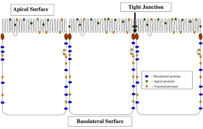
Polarization in epithelial cells takes the form of two distinct membrane regions separated by a specialized protein structure known as the tight junction. The separation of the two regions formed by the tight junction is complete, preventing free diffusion of both water and ions across the monolayer. This is often called the “fence” function of the tight junction. As the tight junction exists at the apical end of the lateral membrane, the two membrane regions formed by the tight junctions are referred to as apical and basolateral. The apical membrane is the portion of the cell membrane that faces the organ lumen and is in contact with the external milieu. The basolateral membrane faces the underlying cells and basement membrane of the organ and is in direct contact with the interstitial fluid that fills the spaces between the cells.
23
Apical Surface Tight Junction
Basolateral Surface
= Basolateral proteins = Apical proteins = Unsorted proteins
Figure 1.4. Epithelial cells have two distinct membrane domains. All epithelial cells
24
cells. The protein meshwork between the cells is water- and ion-tight, preventing paracellular diffusion between the lumen and the underlying tissues. This also helps protect the body from external pathogens, and is referred to as the “gate” or “barrier” function.
25
Sorting Signals in Transmembrane Proteins
The intracellular machinery of epithelial cells has evolved mechanisms to detect signals within many proteins that direct their expression to only one side of the tight junction. These signals take many forms; they can be post-translational modifications or primary sequences, they can be presented on either the cytoplasmic/extravesicular face or the extracellular/intravesicular face of vesicles, and they can be conserved among many different proteins, or unique to only one protein.
Apical-targeting signals
Apical-targeting signals fall into four categories: 1) Association with lipid rafts, 2) glycosylation, 3) GPI anchors, and 4) amino acid sequences (Folsch, 2008). As the mechanisms involved are poorly understood, these signals are not necessarily mutually exclusive. For example, a GPI anchor often makes a protein more likely to be associated with a lipid raft (Brown and Rose, 1992).
Cholesterol-rich and detergent-resistant lipid rafts are separated out of the trans-golgi network and targeted to the apical membrane of epithelial cells (Schuck and Simons, 2004). Some proteins associate with rafts by partitioning into these detergent-resistant membrane fractions and thus are delivered to the apical membrane (Schuck et al., 2003). This was the original theory of apical sorting in epithelial cells (van Meer and Simons, 1988). Only recently, however, has the formation of these lipid rafts in the trans-Golgi network been demonstrated, albeit in yeast (Klemm et al., 2009).
26
no consensus sequence for the attachment. The role of glycosylation in polarized trafficking was first demonstrated for the soluble protein clusterin (referred to then only as an 80 kD glycoprotein). Normally, clusterin is secreted only into the apical media, but following tunicamycin treatment (which inhibits N-glycosylation) it is secreted equally into the apical and basolateral compartments (Urban et al., 1987). It was later determined that many transmembrane glycoproteins were also sorted apically (Lisanti et al., 1989). Yeaman et al. found that O-linked glycosylation is necessary for the apical sorting of the p75 neurotrophin receptor tyrosine kinase (Yeaman et al., 1997). Both forms of glycosylation have been implicated in the sorting of several other proteins, including endolyn (Potter et al., 2004) and lactase-phlorizin hydrolase (Delacour et al., 2006).
A GPI anchor is a glycolipid modification that is added to the N-terminus of an otherwise soluble protein. GPI anchored proteins are generally expressed only on the apical membrane of epithelial cells, including MDCK cells (Lisanti et al., 1989), but this is not the case for all epithelial cells (Zurzolo et al., 1993). The apical targeting of GPI-anchored proteins was originally thought to be a product of their integration into lipid rafts (Brown and Rose, 1992). While this is likely the underlying mechanism for many proteins, more recent data has demonstrated apical targeting of a GPI-anchored protein independent of its raft association (Castillon et al., 2013). Furthermore, in the same cell line, the prion protein PrP(C), a GPI-anchored protein that is N-glycosylated and partitions into detergent-resistant lipid rafts, is targeted to the basolateral membrane (Puig et al., 2011;Sarnataro et al., 2002).
27
2008). The closest example of a conserved apical targeting motif was described for three sodium-dependent acid transporter proteins. A peptide sequence from the rat sodium-dependent bile acid transporter containing the sequence NKGF was shown by NMR to adopt a β–turn conformation that was critical for apical delivery (Sun et al., 2003). Apical targeting of the related excitatory amino acid transporter-3 and the human sodium-dependent vitamin C transporter were shown to depend on similar sequences (NGGF and FKGF, respectively) and computer modeling suggested that they too form
β–turn conformations (Cheng et al., 2002;Subramanian et al., 2004).
Amino acid-based sorting signals are the only type that has been shown to be involved in the polarized targeting of GPCRs, although this does not mean the other types of signals play no role in targeting. In addition to the P2Y receptors, apical-targeting signals have been described for the A1 adenosine receptor, two metabotropic glutamate receptors, mGluR1b and mGluR7, a serotonin receptor 5HT1B, a muscarinic acetylcholine receptor M2AchR, and rhodopsin (Chmelar and Nathanson, 2006;Chuang and Sung, 1998;Francesconi and Duvoisin, 2002;Jolimay et al., 2000;Wang et al., 2004). The rhodopsin targeting signal has the most well-defined mechanism for apical targeting. Portions of the rhodopsin C-tail interact directly with the microtubule motor dynein light chain Tctex-1, which directs it along the cytoskeleton to the apical surface (Tai et al., 2001).
28
when stimulated by immunoglobulins (Schaerer et al., 1990). Ligand binding stimulates transcytosis by inducing phosphorylation of a residue within the basolateral-targeting signal (Casanova et al., 1991), after which a separate signal drives the receptor to the apical surface (Luton et al., 2009).
Basolateral-targeting signals
Unlike apical signals, all known basolateral-targeting signals are amino acid sequences. There are three defined basolateral-targeting motifs and a large assortment of other signal sequences that are either unique or so poorly conserved that their similarities are not readily apparent.
The first basolateral-targeting signal described was within the pIgR C-tail, mentioned above, which is apparently unique to that protein (Casanova et al., 1991). This was shortly followed by the discovery of an NPVY basolateral-targeting signal in the cytoplasmic domain of the LDL receptor (as well as a second, less efficient signal) (Matter et al., 1992). This sequence overlaps the previously determined clathrin-mediated internalization signal of the receptor, which by species comparison was shown to depend on the same NPXY motif (where X is any amino acid) (Chen et al., 1990). Not surprisingly, the NPXY motif was shown to direct basolateral targeting by interacting with the epithelial-specific subunit (µ1B) of the AP-1 clathrin adaptor protein (Folsch et al., 1999). LLC-PK1, a porcine epithelial cell line, does not express µ1B and proteins containing NPXY-dependent signals are mis-sorted to the apical membrane. Exogenous expression of µ1B in LLC-PK1 cells corrects the sorting defect (Folsch et al., 1999).
29
phosphatase (Prill et al., 1993) established the consensus sequence YXXΦ, where X is any amino acid and Φ is an amino acid with a bulky hydrophobic side chain (Thomas and Roth, 1994). As with the NPXY motif, YXXΦ signals also direct proteins to the basolateral membrane of polarized epithelial cells by interaction with the µ1B clathrin adaptor protein (Ohno et al., 1995;Ohno et al., 1999). YXXΦ-containing signals have also been identified as determinates for other types of vesicular sorting, including lysosomal (Guarnieri et al., 1993), endoplasmic reticulum (Mallabiabarrena et al., 1992), and trans-golgi network targeting (Bos et al., 1993;Humphrey et al., 1993;Wong and Hong, 1993), as well as endocytosis (Chang et al., 1991;Girones et al., 1991). Therefore, basolateral targeting cannot be assumed from the presence of a YXXΦ motif alone. These signals have been shown to interact with the µ-subunits of AP-1, AP-2, and AP-3, with some specificity derived from the amino acids surrounding the tyrosine residue (Ohno et al., 1998), which likely determines their intracellular functions.
The last class of conserved basolateral-targeting signals is based on a di-hydrophobic (often, but not always, a di-leucine) pair. This motif was first identified in the IgG Fc Receptor FcRII-B2 (Hunziker and Fumey, 1994;Matter et al., 1994). However, like the previous motifs, di-leucine motifs have also been shown to direct endocytosis and lysosomal targeting (Letourneur and Klausner, 1992;Pond et al., 1995). As with NPXY and YXXΦ motifs, di-leucine motifs were also shown to interact with clathrin adaptor proteins, the γ/σ1 subunits of AP-1 or δ/σ3 of AP-3 (Janvier et al., 2003).
30
31
Polarized Expression of P2Y Receptors
32
Figure 1.5. Confocal microscopy of wild-type (WT) MDCK(II) cells and MDCK(II)
cells expressing HA-tagged human P2Y receptors. MDCK(II) epithelial cells
33
Receptor Distribution Nterm-TM7(Body) C-tail
P2Y1 Basolateral Apical (1stECL) Basolateral
P2Y2 Apical Apical (1stECL) No Signal
P2Y4 Apical No Signal Apical
P2Y6 Apical Apical (1stECL) Not Tested
P2Y11 Basolateral Not Expressed Basolateral
P2Y12 Basolateral Basolateral Basolateral
P2Y13 Unsorted N/A N/A
P2Y14 Basolateral Not Expressed Basolateral
Figure 1.6. P2Y receptor-targeting signals are contained within the main body and
C-terminal tail of the receptors. Polarized sorting of three constructs are shown. The
34
The Apical-sorting signal of the P2Y2 Receptor
Perhaps the most striking finding of these initial receptor constructs was the apparent difference in the mechanism of apical targeting of P2Y2 and P2Y4 receptors, despite their overall high homology (52% amino acid identity). This difference, coupled with the high homology of the two receptors, provided a useful approach to further delineate the P2Y2 receptor apical-targeting signal. A series of chimeric constructs were created that contained the N-terminal region of the P2Y2 receptor fused at various points to the corresponding region of the P2Y4 receptor lacking its C-tail and thus its targeting signal. In this way, the location of the apical targeting sequence of the P2Y2 receptor was narrowed down to the first extracellular loop (Qi et al., 2005).
Sequence alignment of the first extracellular loops of the P2Y2 and P2Y4 receptors narrowed the list of possible critical residues to just nine amino acids. These amino acids were mutated in the P2Y2 receptor to the corresponding residue in the P2Y4 receptor, allowing identification of four critical amino acids: Arg95, Gly96, Asp97, and Leu108. Interestingly, the RGD sequence is a well-characterized integrin-binding motif, suggesting that perhaps interactions with integrins are involved in apical targeting. However, further analysis was not consistent with this possibility. RGE and QGE mutations, both of which would not be expected to bind integrins, did not disrupt apical targeting (Qi et al., 2005).
35
The Basolateral-sorting signal of the P2Y1 Receptor
Initial studies as described above demonstrated that the P2Y1 receptor contained two signals: a cryptic apical signal in the main body of the receptor (later shown to be in the first extracellular loop; see above) and a dominant basolateral signal in the C-terminal tail. The C-C-terminal tail, when used to replace the endogenous C-C-terminal tails of a variety of GPCRs, was capable of moving these receptors completely to the basolateral surface. While the extracellular apical-targeting signal appears to play no role in polarized sorting of the P2Y1 receptor (See Chapter 3; DuBose et al., in preparation), it did provide a useful tool for characterization of the dominant basolateral-targeting signal in the C-terminal tail (Wolff et al., 2010). As demonstrated by the P2Y1 receptor ∆CT truncation construct, disruption of the basolateral targeting sequence uncovers the cryptic apical signal, and thus mutation of key amino acids required for basolateral sorting resulted in a receptor that not only was no longer basolateral, or even unsorted, but expressed entirely at the apical domain of epithelial monolayers.
36
Polarized Sorting of Other P2Y Receptors
The apical-sorting signal of the P2Y4 receptor is described herein in Chapter 2. Similar to the basolateral signal in the P2Y1 receptor, the apical signal in the P2Y4 receptor is unlike any previously described apical-targeting signal and appears to be unique to the P2Y4 receptor (DuBose et al., 2013). The P2Y12 receptor has two basolateral-targeting signals: one within its C-tail that appears to be dependent on the PDZ ligand (unpublished results), and one within the main body of the receptor that has yet to be explored further (DuBose et al., 2013). The P2Y14 receptor C-tail contains a basolateral-targeting signal that shares many of the properties of the P2Y1 receptor signal, although it bears little sequence homology. This signal, like that of the P2Y1 receptor, is long (~23 amino acids), dependent on the number of charged residues, but it is also dependent on the type of charge (positive) and several hydrophobic residues (unpublished results). The P2Y11 receptor basolateral-targeting signal has yet to be explored beyond its general location within the C-tail of the receptor.
37
Trafficking Itineraries and Technological Limitations
A second area of interest for our lab, in addition to identifying and characterizing the targeting signals of P2Y receptors, has been to define the physical pathway that a receptor takes through the vesicular trafficking machinery to reach its final membrane destination. Ideally, this would include every subset of endosomal compartments that a protein traverses as well as every molecular interaction that drives it along its way. This type of experiment is, of course, quite technically challenging. In the broadest sense, all of these individual interactions can be summed up into one of two categories: direct and indirect.
Direct delivery refers to a mechanism in which a protein undergoes all of its polarized sorting during its biosynthesis before reaching the plasma membrane for the first time. In this pathway, all (or nearly all) of the newly synthesized protein appears on the same side of the tight junction where the protein is found at steady-state. With indirect targeting, newly synthesized proteins appear at both apical and basolateral membrane domains and are only later sorted to their polarized steady-state locations. There are many subdivisions and possible mechanisms that can underlie either of these mechanisms, but until recent technological advances even this distinction was difficult to make.
38
streptavidin for biotin), and the released material was then re-precipitated with a receptor-specific antibody to separate the receptor from the remaining biotinylated proteins. This 2nd precipitation was then separated by SDS-PAGE and detected by autoradiography. Western blotting could not be used as it would detect all of the proteins that were already present at the membrane of interest instead of just those that were labeled during the pulse.
While this technique has been used to study the delivery mechanisms of several polarized proteins (Anderson et al., 2005;Chmelar and Nathanson, 2006;Keefer and Limbird, 1993;Wozniak and Limbird, 1996), it suffers from three important drawbacks. First, the method relies on high expression of the protein of interest into sorting machinery that has been shown to be saturable (Marmorstein et al., 2000;Matter et al., 1992). Second, the method requires that the rates of transcytosis be slower than the rates of initial delivery, such that an accumulation of protein can be detected before moving to its final polarized location. Third, there is no direct demonstration that the trafficking itineraries observed are due to selective delivery or transcytosis rather than selective internalization and degradation.
39
In Vivo Covalent Fluorophore Attachment
Several technologies have recently been developed for the covalent labeling of proteins in live cells. The first and simplest approach relies on the specific labeling of a tetra-cysteine motif within an alpha-helix by 4′,5′-bis(1,3,2-dithioarsolan-2-yl)fluorescein in the presence of AsCl3 (Griffin et al., 1998). The benefit of this method is the relatively small genetic alteration necessary to create a target protein. The downside is that it is limited to a few fluorophore choices, the fluorophores are prohibitively expensive, and each site is capable of binding either one or two fluorophore molecules, complicating quantification.
Two methods, BioEase™ and AviTag™, rely on the specific biotinylation of relatively small (72 and 15 amino acids, respectively) tags by biotin protein ligases, followed by recognition by (strept)avidin probes (Ashraf et al., 2004;de Boer et al., 2003). While an improvement in many ways over previous methods, this technology is more suited to protein purification than fluorescence labeling.
The last two methods involve the attachment of a larger, enzyme-based “epitope” to the protein of interest. The enzymes react covalently with specific types of small molecules at a 1:1 stoichiometry, allowing the attachment of a wide variety of probes. The obvious downside to these techniques is that the presence of a large protein domain connected to the protein of interest may cause steric hindrance or otherwise impede its normal function.
40
room temperature and are suitable for both microscopy and biochemical applications (Los et al., 2008).
Chapter 2: Apical Targeting of the P2Y4 Receptor is Directed by
Hydrophobic and Basic Residues in the Cytoplasmic Tail
Overview
42
Introduction
Nucleotides are ubiquitous small molecules involved in a wide variety of biological processes. In addition to playing essential roles in phosphorylation, energy utilization and metabolism, and synthesis of nucleic acids and enzymatic co-factors, nucleotides are also released from cells where they serve as extracellular ligands for transmembrane receptors involved in signal transduction (Lazarowski et al., 2003;Lazarowski, 2012). Nucleotide receptors have been found in all cell types, where they mediate a broad range of cell activities. There are two distinct families of nucleotide receptors, P2X and P2Y. P2X receptors are ion channels that open in response to extracellular ATP, while the P2Y receptors are a family of G protein coupled receptors (GPCRs) that respond to extracellular nucleotides (Coddou et al., 2011;von Kugelgen and Harden, 2011). Eight P2Y receptors have been identified to date—P2Y1, P2Y2, P2Y4, P2Y6, P2Y11, P2Y12, P2Y13, and P2Y14. The numbering is not sequential because several receptors were reported to be P2Y receptors but later determined to either not respond to nucleotides or to be orthologs of existing mammalian P2Y receptors (Herold et al., 1997;Janssens et al., 1997;Li et al., 1998;Qi et al., 2004).
43
P2Y receptors are expressed in a variety of tissue types, and the subtype and density of the receptors varies significantly. P2Y4 receptor mRNA is widely distributed and most abundant in the intestine (Moore et al., 2001), where it has been shown to play a role in luminal Cl- secretion (Robaye et al., 2003). Since bacterial invasion can induce nucleotide release (Crane et al., 2002;McNamara et al., 2001;Tran Van Nhieu et al., 2003) and Cl- secretion is the known mediator of diarrhea symptoms (Field et al., 1989;Field et al., 1989;Kunzelmann and Mall, 2002), antagonists of the P2Y4 receptor may have therapeutic value for the treatment of infectious diarrhea. Agonists of the P2Y4 receptor may also be useful as it has been hypothesized that stimulation of the Cl -secretory pathway may alleviate intestinal abnormalities associated with cystic fibrosis (Robaye et al., 2003). Unfortunately, there are currently no selective ligands for the P2Y4 receptor available. The P2Y4 receptor has also been implicated in the control of K+ secretion in vestibular dark cell epithelium and in mouse colon (Marcus and Scofield, 2001;Matos et al., 2005).
44
family of P2Y receptors, and shown that seven of the eight P2Y receptors are strongly polarized when expressed in epithelial cell lines (Wolff et al., 2005).
Despite the fact that aberrant protein sorting is often associated with disease states, the mechanisms by which epithelial cells establish and maintain these polarized distributions are poorly understood (Keitel et al., 2003;Kleizen et al., 2000;Marr et al., 2002;Marr et al., 2002;Rotin et al., 2001). It is well established that many proteins contain amino acid sequences that act as trafficking signals to direct the protein to the appropriate membrane (Folsch, 2008;Rodriguez-Boulan et al., 2004;Weisz and Rodriguez-Boulan, 2009). Tyrosine- and di-hydrophobic-based signals have been shown to interact with clathrin adaptor complex proteins and to direct basolateral targeting (Folsch et al., 1999;Hunziker et al., 1991;Hunziker and Fumey, 1994;Matter et al., 1994;Ohno et al., 1995). In contrast, post-translational modification, such as glycosylation or glypiation, is often sufficient to confer apical targeting (Lisanti et al., 1989;Scheiffele et al., 1995;Vagin et al., 2009;Wilson et al., 1990;Yeaman et al., 1997). Oligomerization and lipid raft association have also been suggested to direct apical trafficking (Lingwood and Simons, 2010;Paladino et al., 2004;Paladino et al., 2006;Schuck and Simons, 2004).
45
and Leu108 (Qi et al., 2005). This signal is highly unusual because, following receptor synthesis, it is located on the inside of vesicles and therefore inaccessible to intracellular sorting machinery. This is the only protein-based extracellular apical-targeting signal identified in any protein to date.
46
Methods
Construction of Mutant and Chimeric Receptors—Construction and cloning of
HA-tagged P2Y4 and BK2/Y4 receptors into the retroviral vector pLXSN was accomplished as described previously (Qi et al., 2005;Wolff et al., 2005). Targeted mutations were introduced into the P2Y4 receptor C-tail of these constructs by overlap-extension PCR (Ho et al., 1989), whereas P2Y4 receptor truncation constructs were made using PCR with a 5’ vector primer and 3’ primers containing a stop codon at the appropriate location followed by a XhoI site to facilitate cloning.
The P2Y12/P2Y4 receptor chimera was constructed using overlap extension PCR. One set of primers amplified the P2Y12 receptor coding sequence from the second codon (with an MluI site to facilitate cloning) through the codon for Ser304, the start of the C-tail, and also included the first seven codons of the P2Y4 receptor C-tail starting at Asp311. The second set of primers amplified the C-tail of the P2Y4 receptor starting at Asp311 through the end of the gene and contained a XhoI site at the end of the downstream primer. After the initial amplification, the two PCR products were isolated, then combined and amplified with only the outside primers. The resulting product was digested with MluI and XhoI and ligated into a similarly digested modified pLXSN vector that added an HA-tag to the N-terminus of the chimera.
47
and SbfI sites were introduced by incorporating silent mutations into the codons for Arg314/Arg315 and Ser345, respectively.
We initially created receptor mutants in the context of either the BK2-P2Y4 C-tail chimeric receptor or the P2Y4 receptor. While confocal microscopy revealed essentially identical targeting as observed with modified P2Y4 receptor constructs described below, cell surface expression was often too low for accurate quantification of apical versus basolateral receptor distribution (data not shown). Therefore, we introduced a cleavable signal sequence (MKTIIALSYIFCLVPA) and FLAG epitope tag (DYKDDDDA) immediately upstream of the HA-tag to increase receptor expression (Guan et al., 1992). All of the constructs shown in Figure 2.3A, with the exception of the BK2/P2Y4-C321S receptor, contained this addition. Inclusion of the signal sequence increased steady-state receptor levels, which facilitated imaging and quantification, without having an appreciable effect on localization in polarized monolayers.
Cell culture—All cells were grown in a humidified incubator at 37°C in a 5%
CO2/95% air atmosphere. Type II Madin-Darby canine kidney cells (MDCK(II); ATCC, Rockville, MD) were maintained in 1:1 DMEM/F12 medium containing 5% fetal bovine serum and 1X pen/strep. PA317 cells were maintained in DMEM containing 10% fetal bovine serum and 1X pen/strep.
Retroviral infection—Recombinant retroviruses were produced by calcium
48
Confocal Fluorescence Microscopy—MDCK(II) cells were seeded at 6 x 10⁵
cells/well in 12 mm Transwell inserts (Corning Life Sciences, Acton, MA) and grown for 5-7 days with daily medium changes to allow the cells to form polarized monolayers. Cells were prepared for confocal microscopy as described previously (Wolff et al., 2005). Briefly, cells were washed and fixed in 2% PFA in PBS with 2 mM CaCl2 and 2 mM MgCl2 for 30 minutes at 4 ºC. After fixation, cells were permeabilized with cold (-20 ºC) methanol for 30 seconds. Cells were then quenched by three washes of 150 mM sodium acetate in 1% non-fat dry milk (NFDM) and blocked by three more washes in 1% NFDM. Cells were incubated with a 1:1000 dilution of mouse monoclonal anti-HA antibody (HA.11; Covance, Berkeley, CA) and a 1:500 dilution of rabbit anti-ZO-1 antibody (Zymed Laboratories Inc., South San Francisco, CA) in 1% NFDM overnight at 4 ºC. Cells were then washed three times and incubated with both goat anti-mouse Alexa-488 and goat anti-rabbit Alexa-594 (Molecular Probes, Eugene, OR), each diluted 1:500 in 1% NFDM, for one hr at room temperature. After washing five times in PBS and once in Molecular Probes Equilibration Buffer, the polyester membranes were removed from the transwell inserts with a scalpel and mounted under cover slips in Slow Fade A mounting media (Invitrogen, Carlsbad, CA).
49
background fluorescence. In most cases, auto-fluorescence of the polyester membrane has been removed for clarity. Representative images are shown for each receptor construct.
Polarized Cell-surface Biotinylation—MDCK(II) cells were seeded on 12 mm (6 x
105 cells/well) or 24 mm (1.2 x 10⁶ cells/well) Transwell inserts and grown for 5-7 days with daily medium changes to allow the cells to form a polarized monolayer. A polarized biotinylation assay was used to quantify cell-surface expression of HA-tagged receptors essentially as described previously (Wolff et al., 2005). Briefly, cells were carefully cooled to 4ºC and kept cold for the entire assay to avoid potential nucleotide release and redistribution due to receptor activation. Cells were washed twice in PBS++ (phosphate-buffered saline, pH 8.0, plus 2 mM CaCl2 and 2 mM MgCl2), then PBS++ containing 2 mg/mL Sulfo-NHS-SS-Biotin (Pierce, Rockford, IL) was applied to either the apical or basolateral surface, and the reaction was allowed to proceed for 20 min. Following aspiration of the biotinylation solution, the cells were incubated with PBS++ containing 100 mM glycine (pH 8.0) for 10 minutes to quench the reaction, and then washed three times with PBS++.
Proteins extracts were prepared by adding RIPA lysis buffer (50 mM Tris HCl pH 8.0, 100 mM NaCl, 5 mM EDTA, 1% Triton X-100, 0.5% deoxycholate, and 0.1% SDS) to washed cells and passing the lysate through a 25-gauge needle 7-10 times to ensure complete disaggregation. Insoluble materials were removed by centrifugation at 13,000 x
50
51
Results
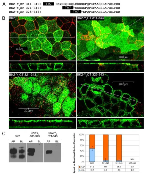
Delimitation of the apical-targeting signal of the P2Y4 receptor—We showed
previously that the human P2Y4 receptor is sorted to the apical membrane of three different epithelial cell types (MDCK(II), 16HBE14o-, and Caco-2 cells), and that the apical targeting sequence is located within the C-tail of the receptor (Qi et al., 2005;Wolff et al., 2005). To delimit the C-terminal end of the apical-targeting signal, we constructed a series of HA-tagged human P2Y4 receptors in which increasing amounts of the C-terminus were truncated. The truncated receptors were expressed in MDCK(II) cells and their steady-state localization was determined by confocal microscopy (Fig. 2.1). XZ scans of the full-length human P2Y4 receptor and P2Y4 receptors truncated at amino acids 355 or 343 revealed that these receptors were localized to the apical membrane at steady state, indicating that the signal remained intact (Fig. 2.1). In contrast, the P2Y4 receptor truncated at amino acid 332 resulted in an unsorted phenotype. These data indicate that the C-terminal end of the apical targeting sequence is no further downstream than amino acid 343.
52
Figure 2.1. The P2Y4 apical-targeting signal ends before Asp343. A, HA-tagged wild
53
Figure 2.2. The P2Y4 apical-targeting signal begins after Cys321. A, HA-tagged
54
amino acids 311-343 or 321-343 of the P2Y4 C-tail was sufficient to confer polarized sorting of the BK2 receptor to the apical surface of MDCK(II) cells (Fig. 2.2B), and the apical localization of these chimeric receptors revealed by confocal microscopy was confirmed by an established polarized cell-surface biotinylation assay (Fig. 2.2C) (Keefer and Limbird, 1993;Qi et al., 2005;Wolff et al., 2005). In contrast to the other chimeras, the BK2 receptor with the fewest number of amino acids (325-343) of the P2Y4 C-tail was not sorted to the apical membrane. This receptor construct was presumably unstable and failed to reach the cell surface, as we were unable to pull down sufficient quantities of receptor to produce a visible band in our biotinylation assay. These data indicate that the N-terminal end of the apical-targeting signal of the P2Y4 receptor is no farther upstream than amino acid 321.
Taken together, these experiments defined a 23-amino-acid sequence (Cys321 to Asp343) that is both necessary and sufficient to target the P2Y4 or the BK2 receptor to the apical surface. This sequence is unique to the P2Y4 receptor, bears no similarity to any known sorting signal, and does not contain any known binding motifs or conserved domains.
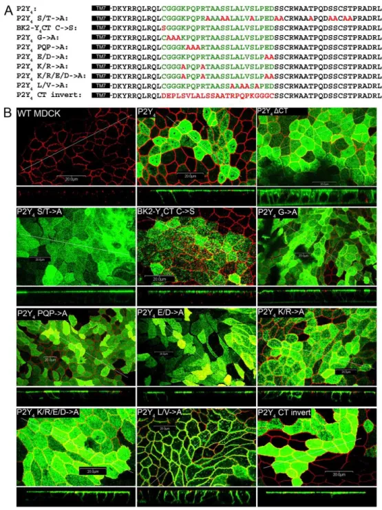
Identification of key amino acids in the P2Y4 apical targeting sequence—Because
55
Figure 2.3. Subcellular localization of receptor constructs with mutant P2Y4
C-tails. A, mutant receptors with the C-tail sequences shown were stably expressed in
56
Figure 2.4. Surface expression of receptor constructs with mutant P2Y4 C-tails.
57
potentially could affect steady-state receptor distribution. However, these mutations had no effect on the polarized expression of the P2Y4 receptor (Figs. 2.3 & 2.4).
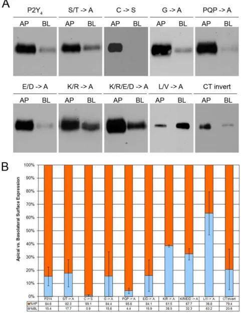
We next targeted the single cysteine residue (Cys321) in the apical targeting sequence, as palmitoylation of cysteines has been implicated in protein stability as well as trafficking (Huang and El-Husseini, 2005;Linder and Deschenes, 2007). We found that a C321S mutation had no effect on receptor localization of the BK2/P2Y4 C-tail chimeric receptor (Figs. 2.3 & 2.4). Likewise, mutation of three consecutive glycines or the PQP triad near the beginning of the sequence to alanines in the context of the P2Y4 receptor had no effect on apical targeting (Figs. 2.3 & 2.4).
Charged residues within the basolateral targeting sequence of the P2Y1 receptor are critical to its function (Wolff et al., 2010). To address the role of charged residues in the P2Y4 receptor apical-targeting signal, we mutated the acidic and/or basic residues of the apical targeting sequence to alanine. Mutation of the glutamate and aspartate residues to alanine had no effect on targeting, whereas mutation of lysine and arginine residues to alanine reduced apical polarization by 24% (61% apical versus 85% apical for wild-type P2Y4). Mutation of both the basic and acidic amino acids to alanine closely matched the results of mutating the basic residues alone.
We also made mutations to a small hydrophobic region in the latter half of the apical targeting sequence (one valine and three leucine residues within six residues). Mutation of these four amino acids to alanine markedly disrupted the apical targeting of the P2Y4 receptor and in fact promoted pronounced (but not complete) basolateral targeting (63% basolateral). These data highlight the importance of these four hydrophobic residues for proper apical targeting of the P2Y4 receptor.
The apical-targeting signal of the P2Y4 receptor is sequence-independent-We