The transformation in biomarker detection and management of
drug-induced liver injury
Rachel J. Church1,2 and Paul B. Watkins1,2
1Institute for Drug Safety Sciences, University of North Carolina at Chapel Hill, Research Triangle
Park, North Carolina
2Division of Pharmacotherapy and Experimental Therapeutics, UNC Eshelman School of
Pharmacy, Chapel Hill, North Carolina, USA
Abstract
Drug-induced liver injury (DILI) is a major concern for patients, care givers and the pharmaceutical industry. Interpretation of the serum biomarkers routinely used to detect and monitor DILI, which have not changed in almost 50 years, can be improved with recently proposed models employing quantitative systems pharmacology. In addition, several newer serum biomarkers are showing great promise. Studies in rodents indicate that the ratio of the caspase cleaved fragment of cytokeratin 18 to total K18 in serum (termed the “apoptotic index”) estimates the relative proportions of apoptosis vs necrosis during drug-induced liver injury. Glutamate dehydrogenase can reliably differentiate liver from muscle injury and, when serum is properly prepared, may also detect mitochondrial toxicity as a mechanism of liver injury. MicroRNA-122 is liver-specific, but recent data suggests it can be actively released from hepatocytes in the absence of overt toxicity limiting enthusiasm for it as a DILI biomarker. Finally, damage associated molecular patterns, particularly high mobility group box 1 and its various modified forms, are promising biomarkers of innate immune activation, which may be useful in distinguishing benign elevations in aminotransferases from those that portend clinically important liver injury. These new biomarkers are already being measured in early clinical trials, but broad acceptance will require widespread archiving of serum from diverse clinical trials and probably pre-competitive analysis efforts. We believe that utilization of a panel of traditional and newer biomarkers in conjunction with quantitative systems pharmacology modeling approaches will transform DILI detection and risk management.
Keywords
drug-induced liver injury; idiosyncratic DILI; biomarkers; hepatotoxicity
HHS Public Access
Author manuscript
Liver Int
. Author manuscript; available in PMC 2017 November 01. Published in final edited form as:Liver Int. 2017 November ; 37(11): 1582–1590. doi:10.1111/liv.13441.
A
uthor Man
uscr
ipt
A
uthor Man
uscr
ipt
A
uthor Man
uscr
ipt
A
uthor Man
uscr
Drug Induced Liver Injury (DILI)
Drug induced liver injury (DILI) accounts for greater than half of all acute liver failure (ALF) cases occurring in the United States.1 The majority of DILI cases that lead to ALF are due to overdose of acetaminophen which is considered an “intrinsic” liver toxin. Intrinsic hepatotoxicity due to drugs is typified by direct chemical damage to liver cells, a short latency period, and a clear dose-dependent response.2 Intrinsic hepatotoxins would cause liver injury in virtually all patients if they received sufficient overdoses, although the threshold for toxic response can show marked inter-individual differences. Intrinsic hepatoxins are generally identified as such in preclinical animal testing and in Phase 1 dose escalation clinical trials.
Intrinsic hepatoxins are to be distinguished from idiosyncratic hepatoxins, which cause clinically significant DILI in only a very small minority of treated individuals, typically less than 1 in 10,000.3 While rare, idiosyncratic DILI (iDILI) events carry a significant risk for liver failure. 4, 5 Although there appears to be a dose threshold required to elicit iDILI, overdosing generally does not produce liver toxicity in most treated patients.6 There is often a prolonged latency between initiation of treatment and the onset of iDILI. iDILI is
particularly problematic in drug development because preclinical testing and early clinical trials generally fail to identify iDILI liabilities, which may only become evident late in Phase 3 clinical development or post-marketing use, after thousands of patients have received prolonged treatment with the drug.
Traditional DILI Biomarkers and Hy’s Law
For about the last half century, the serum biomarkers routinely utilized to detect and manage DILI in clinical trials and in the clinic are alanine aminotransferase (ALT), aspartate aminotransferase (AST), alkaline phosphatase (ALP), and total bilirubin (TBIL). ALT and AST are present within hepatocytes and during an acute DILI event, increased serum levels of ALT and AST should generally correlate well with the rate at which hepatocytes are dying and releasing their contents. Elevation in serum ALP generally indicates injury to the canalicular membrane or biliary epithelial cells. Elevated levels of TBIL may reflect hepatic functional impairment, or alterations in bilirubin production (hemolysis) or processing. However, these traditional serum biomarkers suffer from several shortcomings. None of these markers are completely specific to liver injury or provide mechanistic insight into the mode of injury. Additionally, release of cellular injury biomarkers such as ALT and AST into the circulation takes place when hepatocyte injury has already occurred and therefore these biomarkers cannot be utilized to identify a potential for DILI prior to the appearance of overt liver injury.7, 8 Moreover, elevations in serum ALT/AST can occur during treatment with drugs that do not pose much or any risk of progressive liver injury (eg. statins, tacrine, heparins and cholestyramine).9–11 Even when drugs are capable of causing progressive and clinically important liver injury, most drug-induced elevations in serum ALT and AST resolve despite continued treatment with the offending drug – a process termed “adaptation.”12
A
uthor Man
uscr
ipt
A
uthor Man
uscr
ipt
A
uthor Man
uscr
ipt
A
uthor Man
uscr
Recognizing the limitations of serum ALT and AST in assessing liver safety in clinical trials of new drug candidates, a 2009 FDA guidance defined a “Hy’s Law” case as the event that most accurately predicts the potential for a drug to cause acute liver failure.13 This is based on observations by Dr. Hyman Zimmerman, and confirmed in large international DILI registries, that patients with hepatocellular jaundice due to a medication have at least a 10% chance of developing liver failure.4, 14, 15 According to the FDA guidance, a Hy’s Law case is a patient with a healthy liver at the start of a clinical trial who experiences hepatocellular injury characterized by a rise in serum ALT >3X above an established upper limit of normal (ULN) and also a rise in serum TBIL >2X ULN with no more likely explanation for the event than the study drug. The assumption is that the rise in serum TBIL has resulted from global loss of liver function due a substantial loss of viable hepatocytes. It should be noted that in a true Hy’s Law case, the combined elevations in serum ALT and TBIL are not biomarkers that predict the potential for severe liver injury, but are biomarkers documenting that severe and potentially life-threatening liver injury has occurred in this subject.
A challenge to interpreting a Hy’s Law case is that drugs may cause elevations in serum TBIL for reasons other than impairment of global liver dysfunction. Reasons include hemolysis (increased heme load), inhibition by the drug of the uptake or efflux proteins that transport bilirubin, or inhibition of the major hepatocyte enzyme that conjugates bilirubin (UGT1A1). Quantitative systems pharmacology has been recently applied to integrate estimates of liver exposure to a drug with the quantified effects of the drug on inhibiting the transporters and UGT1A1 as assessed in in vitro systems.15 This approach should advance understanding and prediction of drug-induced elevations in serum TBIL not due to global liver dysfunction.
It has also been recently proposed that the percent of hepatocytes dying in an acute DILI event can be assessed from serial measurements of serum ALT.16 This is based on the idea that each hepatocyte contains a finite amount of ALT which is passively released during cell death. The area under the serum ALT vs. time curve should therefore correspond to the number of hepatocytes lost. Furthermore, it has been proposed that the estimate of hepatocyte loss obtained in this way can be used to predict the magnitude in rise of serum bilirubin expected based on the implied loss of aggregate liver function. Hepatectomy studies in animals suggest that over 70% of hepatocytes must be lost before there is a rise in serum TBIL sufficient to cause jaundice.17 In an acute DILI event, however, function would be expected to be impaired in a proportion of hepatocytes that may not undergo cell death. Supportive data to incorporate this phenomenon into the ALT derived estimate of hepatocyte loss may be a liver biopsy study conducted several decades ago in patients experiencing liver injury due to acetaminophen overdose.18 From data provided by this study, it can be
concluded that more that 40% of hepatocytes must die before serum TBIL will rise to greater than 2X ULN. This estimate has been used together with the AUC of serum ALT in two subjects who experienced elevations in serum ALT >3X ULN and serum TBIL >2X ULN believed due to study drug, i.e. both were Hy’s Law cases by current definition. The serial serum ALT values resulted in estimates predicting the loss of hepatocytes as far less than 40% in each subject.19 The conclusion from the modeling was that the rise in serum TBIL was not due solely to hepatocellular toxicity. This conclusion would be further supported if the systems pharmacology approach indicated that transporter and/or enzyme
A
uthor Man
uscr
ipt
A
uthor Man
uscr
ipt
A
uthor Man
uscr
ipt
A
uthor Man
uscr
inhibition by the drug likely contributed to the observed rise in serum TBIL.15 These approaches hold the potential to refine interpretation of potential Hy’s Law cases.
Newer DILI biomarkers
The use of serial serum ALT values to assess percent hepatocyte loss assumes that the majority of ALT entering circulation is the result of hepatocytes dying with release of their content of ALT. This is likely to be the case in serious DILI events, but substantial elevations in serum ALT may also result from muscle disease or injury.20, 21 MicroRNA-122
(miR-122) and glutamate dehydrogenase (GLDH) have been proposed as more sensitive and specific biomarkers of liver injury than ALT. The mode of hepatocyte death, apoptosis or necrosis, is also likely to influence the calculations of percent hepatocyte loss based on serial measurement of ALT (or other intracellular hepatocyte proteins) in serum. This is because in apoptosis, hepatocyte proteins and RNA species may be largely degraded prior to release into circulation. In this regard, an “apoptotic index” (AI) based on serum biomarkers has been recently proposed to estimate the relative contributions of apoptosis and necrosis to liver injury. As is the case with serum ALT, optimal interpretation of these newer biomarkers will benefit from modeling that incorporates the release and clearance kinetics of each.
The “Apoptotic Index”—The AI is the ratio of caspase cleaved keratin 18 (ccK18) to total keratin 18 (K18; comprising both full length K18 and caspase cleaved fragments). K18 is a type I intermediate filament found in epithelial cells that provides structural support for cells. Early during apoptosis, K18 is cleaved by caspases, resulting in a stable fragmented form of the protein (ccK18) that is released into circulation (Figure 1).22 During necrosis, full-length K18 is released passively into circulation but little ccK18 should be released. Mass spectrometry (for mice) or immunoassays (for humans) that detect K18 or ccK18 concentrations in blood have been investigated in acetaminophen-induced liver injury (AILI) to determine whether they correlate with extent of necrosis vs. apoptosis, respectively, occurring in the liver.23 In mice, elevated levels of ccK18 correlated strongly with procaspase processing, DNA fragmentation, and the appearance of apoptotic hepatocytes that expressed caspase 3.24 Additionally, the ratio of ccK18 to K18 mirrored the time-dependent shift in the liver from mixed apoptosis/necrosis to primarily necrotic injury. It has also been noted that, in AILI, there is a significant elevation of both K18 and ccK18 levels prior to a significant elevation in ALT levels.24 These data suggest that, at least in preclinical models, quantifying K18 and ccK18 may provide both an early detection of hepatocyte cell death and a quantitative estimate of the relative amounts of apoptosis vs. necrosis occurring in the liver. Since K18 is not limited to epithelial cells of hepatic origin, utilizing it on its own as a DILI biomarker may be problematic because it assumes that liver is the only organ experiencing cell death. The ratio of miR-122 or GLDH to K18 may permit such a
conclusion, but this has not yet been investigated.
Quantification of serum K18 and ccK18 has also been conducted in the setting of clinical DILI.25–27 Both have demonstrated enhanced sensitivity for the detection of liver injury as compared to ALT.
A
uthor Man
uscr
ipt
A
uthor Man
uscr
ipt
A
uthor Man
uscr
ipt
A
uthor Man
uscr
Serum K18 and ccK18 have also shown promise as prognostic biomarkers for prediction of liver failure and death.28, 29 In these investigations, both K18 and ccK18 levels were significantly elevated in patients who met King’s College Criteria (KCC), a model for predicting liver failure in AILI, compared to those that did not meet these criteria.
Estimation of the AI revealed that patients meeting KCC had a lower values (more necrosis) compared to those that did not meet KCC. These results are consistent with the idea that necrotic liver injury, as opposed to apoptotic injury, is more severe and dangerous.26
It should be noted, however, that calculating an AI can be challenging when utilizing the commercially available ELISAs. Experimental evidence conducted in serum from patients with cancer suggests that this ratio may not be useful when K18 and ccK18 levels are low, near background levels. It is therefore recommended that an AI should be conducted only when values of both K18 and ccK18 exceed certain background thresholds.30 Additionally, two versions of the ELISA that quantifies total K18, M65® and M65 EpiDeath® are
available. The newer M65 EpiDeath® kit has been optimized to reduce background antibody binding; however, the corresponding ccK18 assay, M30 Apoptosense® ELISA, has currently not undergone a similar optimization. Therefore when K18 levels are low, measured ccK18 levels may exceed measured total K18 levels (unpublished observations) due to increased levels of non-specific background binding. Again, this supports the view that an AI should be calculated only when values are elevated over a specific background level and when K18 levels exceed ccK18 levels. The ELISA manufacturer’s now recommend utilizing the older version of the K18 ELISA (M65®) when calculation of an AI is desired.31 Finally, these ELISAs are not currently available for rodents, making translational studies challenging.
GLDH—GLDH, an enzyme located in the mitochondrial matrix, is involved in amino acid oxidation and urea production.32 This protein is primarily expressed in the pericentral region of the liver although low levels of GLDH are also observed in the kidney and brain.33 In a recent AILI study conducted in rodents, GLDH slightly outperformed ALT for identification of hepatocellular necrosis and demonstrated improved correlation with injury severity.34 Similarly, in a canine study, serum GLDH levels, when compared to ALT levels, were found to more accurately reflect the absence of injury histologically observed liver injury at study termination.35 A clinical study demonstrated a strong correlation between serum GLDH and ALT levels and showed that GLDH displayed high diagnostic power for predicting hepatic injury in a large cohort of patients with liver damage resulting from various etiologies.36 It is suggested that because the half-life of GLDH is shorter than that of ALT (~16h vs. ~47h, respectively), serum levels of GLDH more accurately reflect ongoing liver injury. In muscle diseases, such as Muscular Dystrophy serum ALT and AST can exceed 10X ULN.21 Creatine-kinase, a muscle specific enzyme, can be quantified to determine whether the origins of altered ALT are related to muscle injury.37 A problem arises however, when patients with underlying muscular disorders are suspected to have liver injury. Unlike ALT, serum GLDH is typically within normal limits in patients with Duchenne’s Muscular Dystrophy and may therefore be valuable in allowing clinicians to estimate the extent of liver damage occurring in this patient population.38 Recently, the Predictive Safety Testing Consortium (PSTC) formally proposed to the FDA biomarker qualification program that GLDH be considered a DILI biomarker in patients with muscle disease.39 When this
A
uthor Man
uscr
ipt
A
uthor Man
uscr
ipt
A
uthor Man
uscr
ipt
A
uthor Man
uscr
manuscript was submitted for publication, the status was listed as “Consultation and Advice.”
It has also been proposed that GLDH release into serum may signal mitochondrial toxicity as a mechanism underlying the liver injury. This view derives from the fact that if necrosis occurs that does not lead to mitochondrial toxicity, intact mitochondria that are released can be removed from fresh serum by centrifugation (the “post-mitochondrial supernatant”). Using this technique the evidence that GLDH functions as a mitochondrial injury biomarker appears supported by research in mice examining GLDH content in post-mitochondrial supernatant obtained from AILI or furosemide-induced liver toxicity.40 Although both toxicants cause a similar pattern of pericentral necrosis and peak serum ALT elevations, significantly elevated levels of GLDH were observed only in post-mitochondrial serum supernatant from AILI but not furosemide-treated mice. This is consistent with the known mitochondrial toxicity of acetaminophen and the lack of mitochondrial toxicity due to furosemide, and suggests that analyzing GLDH in serum post-mitochondrial supernatant may provide mechanistic insight during DILI due to many different drugs.
miR-122—MicroRNAs are small, non-coding RNAs that contribute to post-transcriptional gene regulation. One attractive feature of these species is their high stability in biofluids. MicroRNA-122 is specifically expressed by hepatocytes where it accounts for 70% of the total miRNA content found in the liver, making it potentially an ideal candidate as a DILI biomarker, given that traditional biomarkers such as ALT are not entirely liver-specific.41
In mice with AILI, miR-122 was found at very high levels in the plasma while being concurrently reduced in liver tissue.42 Further, when compared to ALT, circulatory miR-122 levels became elevated both earlier and following lower doses of APAP. In the clinic, multiple DILI studies have demonstrated that circulatory miR-122 levels become elevated prior to ALT elevations.25, 27, 43 In another study, serum miR-122 levels were elevated in patients with acute liver injury, regardless of etiology, but not in healthy controls or patients who took overdoses of acetaminophen but did not develop liver injury.44 Further, miR-122 levels in samples collected at presentation in AILI patients were enhanced in those who experienced a poor outcome (death or liver transplant) compared to those who
spontaneously recovered. In both this clinical study and in a preclinical study utilizing dogs with DILI unrelated to acetaminophen, miR-122 appeared to return to baseline faster than ALT, suggesting miR-122 has a shorter serum half-life than ALT and could therefore more accurately reflect the extent of ongoing liver injury.35, 44
Large miRNA profiling studies have also been conducted in clinical AILI datasets obtained from both adults and children.45–48 In all studies, miR-122 was among the highest elevated miRNAs in circulation. Interestingly, in one study that involved evaluation of both AILI and ischemic hepatic injury, there was a distinct difference in the profiles of miRNAs between the two injury types.45 In the future, miRNA profiling may provide interesting insights into mechanistic differences between DILI compounds.
Although there has been much enthusiasm for miR-122 as a sensitive and specific biomarker for liver injury, several recent observations have suggested that hepatocyte release of
A
uthor Man
uscr
ipt
A
uthor Man
uscr
ipt
A
uthor Man
uscr
ipt
A
uthor Man
uscr
miR-122 may be regulated and may not merely reflect passive release during hepatocyte death. For instance, a recent study conducted in rats demonstrated that 2h following administration of acetaminophen, treated rats had significantly reduced plasma levels of miR-122, compared to control rats.49 Elevated serum levels of miR-122 were observed later, coincident with the onset of hepatic necrosis. In addition, it has recently been reported that miR-122 is released during an acute phase liver response and may travel to the kidney as a cause of anemia often observed in chronic inflammatory conditions.50 miR-122 has also been found to have a significantly large degree of both inter- and intra-variability in healthy volunteers (manuscript in preparation). This variability, in part, has prompted the PSTC to deprioritize miR-122 as a DILI biomarker in favor of GLDH which has lower inter and intra subject variability (personal communications).
Biomarkers to predict idiosyncratic DILI
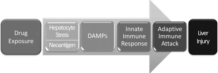
It now appears that many and perhaps most iDILI reactions result from an adaptive immune attack on the liver (Figure 2). This requires the body’s immune system to view the liver as foreign, presumably as a result of drug-induced creation of novel antigens presented by Class I HLA molecules on the surface of liver cells. However, presentation of new antigens alone is not thought to be sufficient to elicit an adaptive immune attack on the liver. This is believed to require activation of innate immune cells, particularly Kupffer cells within the liver. This calls for drug-induced release of damage associated molecular patterns, or DAMPs, that initiate activation of the innate immune cells.51 If this is the case, elevations in serum ALT in the absence of release of DAMPs, and/or the absence of activation of innate immune cells should mean that an adaptive immune attack on the liver will not occur. There has therefore been an intense search for the appropriate DAMPs released from liver cells, and for biomarkers that can detect activation of innate immune cells in the liver. To date, high mobility group box 1 (HMGB1) and its various post-translationally modified forms have received most attention in this regard.
HMGB1
This ubiquitous nuclear protein functions primarily in DNA-binding and transcription regulation; however it can also be actively secreted into the extracellular milieu.52 Evidence suggests that HMGB1 is released passively into circulation from injured or dying liver cells and can act as a DAMP, initiating an immune response.53 This function appears to be mediated by the redox state of specific residues within HMGB1. HMGB1 isoforms that are fully or partially reduced at these residues are characteristically released during necrosis and facilitate chemotaxis and cytokine release from innate immune cells. Conversely, HMGB1 that is fully oxidized at these key sites is characteristically released by apoptotic cells and does not elicit an innate immune response.54
It may be important that HMGB1 can also be actively secreted in a process that requires hyper-acetylation of critical lysine residues. When detected in serum during a DILI event, most hyper-acetylated HMGB1 is believed to have been released by innate immune cells, signifying activation by DAMPs; however, hepatocytes have also been shown to secrete this isoform under certain conditions (Figure 1).55, 56
A
uthor Man
uscr
ipt
A
uthor Man
uscr
ipt
A
uthor Man
uscr
ipt
A
uthor Man
uscr
Both non-clinical and clinical studies have demonstrated HMGB1 to be a sensitive
biomarker of AILI. In mice, total serum HMGB1 became significantly increased earlier than ALT and returned to baseline more rapidly, correlating well with serial histopathological assessments of liver necrosis.24 Consistent with the idea that leakage of HMGB1 from necrotic cells stimulates inflammation and active release of HMGB1 from immune cells, elevation of hyper-acetylated HMGB1 in serum occurred concurrently with the appearance of inflammatory cells in the liver. Moreover, treatment with a chimeric humanized anti-HMGB1 antibody reduced liver injury due to acetaminophen in a recent mouse study, supporting a role of this protein in the progression of DILI due to this drug.57
In clinical AILI studies, HMGB1 displayed enhanced sensitivity for hepatotoxicity detection, compared to ALT.25, 26, 43 In one study, measurements were collected at presentation from patients whose ALT levels were still within the normal range. In these samples, total HMGB1 levels were significantly increased only in patients who later developed hepatic injury.25 In a second study, hyper-acetylated HMGB1 was observed only in AILI patients who died or needed a liver transplant, compared to those who
spontaneously survived, suggesting that this form of HMGB1 may be prognostic for a poor outcome in AILI.26
While total HMGB1 can be quantified by immunoassay, there are currently no antibodies that can decipher different isoforms, necessitating mass spectrometry to determine the post-translational states of HMGB1. Nevertheless, the measurement of total HMGB1 was also demonstrated to be a sensitive biomarker for DILI in the above described AILI studies.
Exosomes
As discussed above, DAMPs are released during hepatocyte necrosis and initiate activation of innate immune cells in the liver which is proposed to be necessary but not sufficient to initiate an adaptive immune attack on the liver. However, there is growing evidence that hepatocyte necrosis is not necessarily a prerequisite for the release of DAMPs and ultimate initiation of an adaptive immune attack on the liver. For example, strong HLA associations have been observed with relatively minor elevations in serum ALT in clinical trials of ximelagatran, lumiracoxib and lapatinib.58–60 In addition, in the case of iDILI due the antibiotic isoniazid, drug reactive T-cells have been identified in patients’ blood prior to the onset of elevations in serum ALT.61 These observations suggest that mild elevations in serum ALT do not necessarily reflect direct drug-induced hepatocyte necrosis that could then initiate an adaptive immune attack, but rather that the initial hepatocyte death is mediated by an adaptive immune attack. That an adaptive immune attack on the liver can be triggered in the absence of hepatocyte death is consistent with hepatitis B infection. The hepatitis B virus is not cytolytic yet generates a liver-specific adaptive immune attack resulting in hepatocyte necrosis and the clinical disease.62 Recent data suggest that the DAMPs may travel in hepatocyte-derived exosomes and be released by drug-induced hepatocyte stress in the absence of cell death.49 Unpublished observations also suggest that exosomes released from hepatocytes treated with sub- toxic doses of acetaminophen produce greater activation of monocytes than exosomes released from control hepatocytes
(manuscript in preparation). It is plausible that stressed but not dying hepatocytes release
A
uthor Man
uscr
ipt
A
uthor Man
uscr
ipt
A
uthor Man
uscr
ipt
A
uthor Man
uscr
DAMPs (and possibly the neoantigens) in exosomes which then travel to and activate innate immune cells. This is an important area of investigation because, due to the porous
fenestrations in liver endothelia, liver-derived exosomes can enter circulation and potentially serve as blood biomarkers. It may therefore be that isolation of exosomes from peripheral blood, and profiling them for DAMPs such as HMGB1, may provide the earliest biomarker predictive of progressive and clinically important DILI potential.
The future
Although they are unlikely to replace traditional biomarkers in general practice, the addition of new candidate biomarkers to standard measurements could soon transform DILI
prediction, detection, and risk management. For instance, data has demonstrated that quantification of candidate biomarkers would have enabled early administration of n-acetyl cysteine to a patient who developed significant AILI following a self-reported drug over dose.43 While this patient had no ALT elevation at presentation (4.5 hours post-over dose) and was sent home without treatment, he eventually developed significant liver injury. Retrospective analysis revealed substantial elevation of K18, HMGB1, and miR-122 in the blood sample collected at presentation.
Further, some of these biomarkers are already being used in early clinical trials with the hope of distinguishing benign elevations in serum ALT from those that portend potential for progressive liver injury, perhaps even to predict potential for very rare iDILI events. Take for example elevations in serum ALT/AST and bilirubin attributed to a new drug candidate in a clinical trial. The bilirubin elevations might be completely accounted for by the quantitative systems pharmacology modeling based on the drugs ability, determined in in vitro
experimental systems, to inhibit bilirubin transporters and/or UGT 1A1, together with estimates of the serum ALT based estimated of percent hepatocyte described above. The serum ALT elevations might further be viewed as benign “transaminitis,” even if the GLDH/ miR-122 assessments indicate hepatic origin, if a) the serum AI suggested hepatocyte death is primarily due to apoptosis, b) only inactive DAMPs are detected concomitantly in serum and c) serum biomarkers of innate immune activation are not detected. The modified forms of HMGB1 may be helpful here, but more work is necessary to define their roles, as hyper-acetylated HMGB1 was detected in healthy volunteers during serum ALT elevations due to heparins, which are entirely safe for the liver.63
One major challenge is that most of our current knowledge regarding investigational biomarkers of DILI has been gained from relatively few patient populations, primarily those experiencing AILI. In order to establish both the context of use and the limitations for each of these candidate biomarkers, it is necessary to study their performance in many different patient populations, especially patients experiencing DILI unrelated to AILI. In
collaboration with the Safer Faster Evidence-based Translation (SAFE-T) and PSTC, the Drug-Induced Liver Injury Network (DILIN) is exploring a number of candidate DILI biomarkers, including several that were included in this review (manuscript in
preparation).64–66 However, the majority of serum specimens available to investigators are from patients once they are already found to be experiencing DILI and it is therefore not possible to assess the ability of the biomarkers to predict DILI before it occurs. The ability
A
uthor Man
uscr
ipt
A
uthor Man
uscr
ipt
A
uthor Man
uscr
ipt
A
uthor Man
uscr
to predict DILI before it occurs would best be assessed in serum samples prospectively collected and archived during clinical trials. Unfortunately, serial serum samples collected during trials for the purpose of monitoring and evaluating perceived alterations in safety signals are not routinely collected, and companies are often not eager to subject what samples have been archived to exploratory studies. Perhaps a “safe haven” needs to be created by regulatory agencies to house such data, as exemplified by the FDA’s Guidance on Voluntary Genomic Data Submissions.67
An additional caveat is that current candidate biomarkers are not specific to DILI, i.e. hepatic injury of various etiologies results in increased serum levels of these markers. For instance, elevated levels of K18 and ccK18 are reported with hepatitis, fibrosis, and non-alcoholic fatty liver disease.68 GLDH, HMGB1, and miR-122 elevations are also associated with liver injuries unrelated to DILI.36, 69, 70 As noted earlier, microRNA profiling identified distinct miRNA changes between AILI and ischemic liver injury.45 “Omics” type
approaches, in which large numbers of molecules are measured concurrently, such as microRNA profiling, metabolomics, or proteomics, may in the future be useful for identifying unique DILI signatures and/or for identifying novel candidate biomarkers with improved performance for DILI detection. Omics technologies for biomarker discovery, however, have been described elsewhere and are outside the scope of the current review.
In conclusion, while preliminary data surrounding the performance of candidate biomarkers in DILI is very promising and is likely to transform DILI assessment and management in the future, further work is necessary before there is universal acceptance of these biomarkers. In reality, it is unlikely that a single candidate biomarker will emerge as the new “gold
standard.” It is more likely that a panel of candidate biomarkers, perhaps in partnership with both traditional biomarkers and quantitative systems pharmacology modeling approaches may give the most complete picture of DILI risk and guide improved risk management strategies.
Acknowledgments
Financial Support: Funding for P.B.W. was provided by the National Institute of Diabetes and Digestive and
Kidney Diseases Project Number 5U01DK065201-13. Funding for R.J.C. was from an internal source.
Abbreviations
DILI drug-induced liver injury
ALF acute liver failure
iDILI idiosyncratic DILI
ALT alanine aminotransferase
AST aspartate aminotransferase
ALP alkaline phosphatase
TBIL total bilirubin
A
uthor Man
uscr
ipt
A
uthor Man
uscr
ipt
A
uthor Man
uscr
ipt
A
uthor Man
uscr
ULN upper limit of normal
miR-122 microRNA-122
GLDH glutamate dehydrogenase
AI apoptotic index
ccK18 caspase cleaved keratin 18
K18 keratin 18
AILI acetaminophen-induced liver injury
KCC King’s College Criteria
PSTC Predictive Safety Testing Consortium
DAMPs damage associated molecular patterns
HMGB1 high mobility group box 1
SAFE-T Safer Faster Evidence-based Translation
DILIN Drug-Induced Liver Injury Network
References
1. Lee WM. Acute liver failure. Semin Respir Crit Care Med. 2012; 33(1):36–45. [PubMed: 22447259]
2. Corsini A, Ganey P, Ju C, et al. Current challenges and controversies in drug-induced liver injury. Drug Saf. 2012; 35(12):1099–1117. [PubMed: 23137150]
3. Uetrecht J. Idiosyncratic drug reactions: past, present, and future. Chem Res Toxicol. 2008; 21(1): 84–92. [PubMed: 18052104]
4. Fontana RJ, Hayashi PH, Gu J, et al. Idiosyncratic drug-induced liver injury is associated with substantial morbidity and mortality within 6 months from onset. Gastroenterology. 2014; 147(1): 96–108 e104. [PubMed: 24681128]
5. Chalasani N, Bonkovsky HL, Fontana R, et al. Features and Outcomes of 889 Patients with Drug-Induced Liver Injury: The DILIN Prospective Study. Gastroenterology. 2015
6. Lammert C, Einarsson S, Saha C, Niklasson A, Bjornsson E, Chalasani N. Relationship between daily dose of oral medications and idiosyncratic drug-induced liver injury: search for signals. Hepatology. 2008; 47(6):2003–2009. [PubMed: 18454504]
7. Nathwani RA, Pais S, Reynolds TB, Kaplowitz N. Serum alanine aminotransferase in skeletal muscle diseases. Hepatology. 2005; 41(2):380–382. [PubMed: 15660433]
8. Magnusson P, Larsson L, Englund G, Larsson B, Strang P, Selin-Sjogren L. Differences of bone alkaline phosphatase isoforms in metastatic bone disease and discrepant effects of clodronate on different skeletal sites indicated by the location of pain. Clin Chem. 1998; 44(8 Pt 1):1621–1628. [PubMed: 9702948]
9. Dukes GE Jr, Sanders SW, Russo J Jr, et al. Transaminase elevations in patients receiving bovine or porcine heparin. Ann Intern Med. 1984; 100(5):646–650. [PubMed: 6712030]
10. Comparative efficacy and safety of pravastatin and cholestyramine alone and combined in patients with hypercholesterolemia. Pravastatin Multicenter Study Group II. Arch Intern Med. 1993; 153(11):1321–1329. [PubMed: 8507122]
A
uthor Man
uscr
ipt
A
uthor Man
uscr
ipt
A
uthor Man
uscr
ipt
A
uthor Man
uscr
11. Watkins PB, Zimmerman HJ, Knapp MJ, Gracon SI, Lewis KW. Hepatotoxic effects of tacrine administration in patients with Alzheimer’s disease. JAMA. 1994; 271(13):992–998. [PubMed: 8139084]
12. Watkins PB. Idiosyncratic liver injury: challenges and approaches. Toxicol Pathol. 2005; 33(1):1– 5. [PubMed: 15805049]
13. Temple R. Hy’s law: predicting serious hepatotoxicity. Pharmacoepidemiol Drug Saf. 2006; 15(4): 241–243. [PubMed: 16552790]
14. Andrade RJ, Lucena MI, Fernandez MC, et al. Drug-induced liver injury: an analysis of 461 incidences submitted to the Spanish registry over a 10-year period. Gastroenterology. 2005; 129(2):512–521. [PubMed: 16083708]
15. Yang K, Battista C, Woodhead JL, et al. Systems Pharmacology Modeling of Drug-Induced Hyperbilirubinemia: Differentiating Hepatotoxicity and Inhibition of Enzymes/Transporters. Clin Pharmacol Ther. 2017 [Epub ahead of print].
16. Howell BA, Siler SQ, Shoda LK, Yang Y, Woodhead JL, Watkins PB. A mechanistic model of drug-induced liver injury AIDS the interpretation of elevated liver transaminase levels in a phase I clinical trial. CPT Pharmacometrics Syst Pharmacol. 2014; 3:e98. [PubMed: 24500662]
17. Sekas G, Cook RT. The evaluation of liver function after partial hepatectomy in the rat: serum changes. Br J Exp Pathol. 1979; 60(5):447–452. [PubMed: 518814]
18. Portmann B, Talbot IC, Day DW, Davidson AR, Murray-Lyon IM, Williams R. Histopathological changes in the liver following a paracetamol overdose: correlation with clinical and biochemical parameters. J Pathol. 1975; 117(3):169–181. [PubMed: 1214189]
19. Longo DM, Generaux GT, Howell BA, et al. Refining Liver Safety Risk Assessment: Application of Mechanistic Modeling and Serum Biomarkers to GGF2 (Cimaglermin alfa) Phase 1 Clinical Trials. Clin Pharmacol Ther. 2017 [submitted].
20. Pettersson J, Hindorf U, Persson P, et al. Muscular exercise can cause highly pathological liver function tests in healthy men. Br J Clin Pharmacol. 2008; 65(2):253–259. [PubMed: 17764474] 21. McMillan HJ, Gregas M, Darras BT, Kang PB. Serum transaminase levels in boys with Duchenne
and Becker muscular dystrophy. Pediatrics. 2011; 127(1):e132–136. [PubMed: 21149430] 22. Caulin C, Salvesen GS, Oshima RG. Caspase cleavage of keratin 18 and reorganization of
intermediate filaments during epithelial cell apoptosis. J Cell Biol. 1997; 138(6):1379–1394. [PubMed: 9298992]
23. Kramer G, Erdal H, Mertens HJ, et al. Differentiation between cell death modes using
measurements of different soluble forms of extracellular cytokeratin 18. Cancer Res. 2004; 64(5): 1751–1756. [PubMed: 14996736]
24. Antoine DJ, Williams DP, Kipar A, et al. High-mobility group box-1 protein and keratin-18, circulating serum proteins informative of acetaminophen-induced necrosis and apoptosis in vivo. Toxicol Sci. 2009; 112(2):521–531. [PubMed: 19783637]
25. Antoine DJ, Dear JW, Lewis PS, et al. Mechanistic biomarkers provide early and sensitive detection of acetaminophen-induced acute liver injury at first presentation to hospital. Hepatology. 2013; 58(2):777–787. [PubMed: 23390034]
26. Antoine DJ, Jenkins RE, Dear JW, et al. Molecular forms of HMGB1 and keratin-18 as mechanistic biomarkers for mode of cell death and prognosis during clinical acetaminophen hepatotoxicity. J Hepatol. 2012; 56(5):1070–1079. [PubMed: 22266604]
27. Thulin P, Nordahl G, Gry M, et al. Keratin-18 and microRNA-122 complement alanine
aminotransferase as novel safety biomarkers for drug-induced liver injury in two human cohorts. Liver Int. 2014; 34(3):367–378. [PubMed: 24118944]
28. Bechmann LP, Jochum C, Kocabayoglu P, et al. Cytokeratin 18-based modification of the MELD score improves prediction of spontaneous survival after acute liver injury. J Hepatol. 2010; 53(4): 639–647. [PubMed: 20630612]
29. Rutherford A, King LY, Hynan LS, et al. Development of an accurate index for predicting outcomes of patients with acute liver failure. Gastroenterology. 2012; 143(5):1237–1243. [PubMed: 22885329]
30. Linder S, Olofsson MH, Herrmann R, Ulukaya E. Utilization of cytokeratin-based biomarkers for pharmacodynamic studies. Expert Rev Mol Diagn. 2010; 10(3):353–359. [PubMed: 20370591]
A
uthor Man
uscr
ipt
A
uthor Man
uscr
ipt
A
uthor Man
uscr
ipt
A
uthor Man
uscr
31. M65 EpiDeath(R) CK18 Kit. Available from: http://diapharma.com/product/apoptosis/m65-epideath-elisa/
32. Schmidt ES, Schmidt FW. Glutamate dehydrogenase: biochemical and clinical aspects of an interesting enzyme. Clin Chim Acta. 1988; 173(1):43–55. [PubMed: 3289795]
33. Lindena J, Sommerfeld U, Hopfel C, Trautschold I. Catalytic enzyme activity concentration in tissues of man, dog, rabbit, guinea pig, rat and mouse. Approach to a quantitative diagnostic enzymology, III. Communication. J Clin Chem Clin Biochem. 1986; 24(1):35–47. [PubMed: 3701270]
34. Thulin P, Hornby RJ, Auli M, et al. A longitudinal assessment of miR-122 and GLDH as biomarkers of drug-induced liver injury in the rat. Biomarkers. 2016 [Epub ahead of print]. 35. Harrill AH, Eaddy JS, Rose K, et al. Liver biomarker and in vitro assessment confirm the hepatic
origin of aminotransferase elevations lacking histopathological correlate in beagle dogs treated with GABAA receptor antagonist NP260. Toxicol Appl Pharmacol. 2014; 277(2):131–137. [PubMed: 24699182]
36. Schomaker S, Warner R, Bock J, et al. Assessment of emerging biomarkers of liver injury in human subjects. Toxicol Sci. 2013; 132(2):276–283. [PubMed: 23339181]
37. Wright MA, Yang ML, Parsons JA, Westfall JM, Yee AS. Consider muscle disease in children with elevated transaminase. J Am Board Fam Med. 2012; 25(4):536–540. [PubMed: 22773723] 38. Flanigan KM, Voit T, Rosales XQ, et al. Pharmacokinetics and safety of single doses of drisapersen
in non-ambulant subjects with Duchenne muscular dystrophy: results of a double-blind randomized clinical trial. Neuromuscul Disord. 2014; 24(1):16–24. [PubMed: 24321374] 39. Current Biomarker Qualification Submissions. Jan 24. 2017 Available from: http://www.fda.gov/
Drugs/DevelopmentApprovalProcess/DrugDevelopmentToolsQualificationProgram/ BiomarkerQualificationProgram/ucm535881.htm
40. McGill MR, Sharpe MR, Williams CD, Taha M, Curry SC, Jaeschke H. The mechanism underlying acetaminophen-induced hepatotoxicity in humans and mice involves mitochondrial damage and nuclear DNA fragmentation. J Clin Invest. 2012; 122(4):1574–1583. [PubMed: 22378043]
41. Lagos-Quintana M, Rauhut R, Yalcin A, Meyer J, Lendeckel W, Tuschl T. Identification of tissue-specific microRNAs from mouse. Curr Biol. 2002; 12(9):735–739. [PubMed: 12007417]
42. Wang K, Zhang S, Marzolf B, et al. Circulating microRNAs, potential biomarkers for drug-induced liver injury. Proc Natl Acad Sci U S A. 2009; 106(11):4402–4407. [PubMed: 19246379]
43. Dear JW, Antoine DJ, Starkey-Lewis P, Goldring CE, Park BK. Early detection of paracetamol toxicity using circulating liver microRNA and markers of cell necrosis. Br J Clin Pharmacol. 2014; 77(5):904–905. [PubMed: 23879521]
44. Starkey Lewis PJ, Dear J, Platt V, et al. Circulating microRNAs as potential markers of human drug-induced liver injury. Hepatology. 2011; 54(5):1767–1776. [PubMed: 22045675]
45. Ward J, Kanchagar C, Veksler-Lublinsky I, et al. Circulating microRNA profiles in human patients with acetaminophen hepatotoxicity or ischemic hepatitis. Proc Natl Acad Sci U S A. 2014; 111(33):12169–12174. [PubMed: 25092309]
46. Yang X, Salminen WF, Shi Q, et al. Potential of extracellular microRNAs as biomarkers of acetaminophen toxicity in children. Toxicol Appl Pharmacol. 2015; 284(2):180–187. [PubMed: 25708609]
47. Krauskopf J, Caiment F, Claessen SM, et al. Application of high-throughput sequencing to circulating microRNAs reveals novel biomarkers for drug-induced liver injury. Toxicol Sci. 2015; 143(2):268–276. [PubMed: 25359176]
48. Vliegenthart AD, Shaffer JM, Clarke JI, et al. Comprehensive microRNA profiling in
acetaminophen toxicity identifies novel circulating biomarkers for human liver and kidney injury. Sci Rep. 2015; 5:15501. [PubMed: 26489516]
49. Holman NS, Mosedale M, Wolf KK, LeCluyse EL, Watkins PB. Subtoxic Alterations in Hepatocyte-Derived Exosomes: An Early Step in Drug-Induced Liver Injury? Toxicol Sci. 2016; 151(2):365–375. [PubMed: 26962055]
A
uthor Man
uscr
ipt
A
uthor Man
uscr
ipt
A
uthor Man
uscr
ipt
A
uthor Man
uscr
50. Rivkin M, Simerzin A, Zorde-Khvalevsky E, et al. Inflammation-Induced Expression and Secretion of MicroRNA 122 Leads to Reduced Blood Levels of Kidney-Derived Erythropoietin and Anemia. Gastroenterology. 2016; 151(5):999–1010 e1013. [PubMed: 27477940]
51. Mosedale M, Watkins PB. Drug-induced liver injury: Advances in mechanistic understanding that will inform risk management. Clin Pharmacol Ther. 2016 [Epub ahead of print].
52. Andersson U, Tracey KJ. HMGB1 is a therapeutic target for sterile inflammation and infection. Annu Rev Immunol. 2011; 29:139–162. [PubMed: 21219181]
53. Huebener P, Pradere JP, Hernandez C, et al. The HMGB1/RAGE axis triggers neutrophil-mediated injury amplification following necrosis. J Clin Invest. 2015; 125(2):539–550. [PubMed: 25562324] 54. Yang H, Antoine DJ, Andersson U, Tracey KJ. The many faces of HMGB1: molecular
structure-functional activity in inflammation, apoptosis, and chemotaxis. J Leukoc Biol. 2013; 93(6):865– 873. [PubMed: 23446148]
55. Bonaldi T, Talamo F, Scaffidi P, et al. Monocytic cells hyperacetylate chromatin protein HMGB1 to redirect it towards secretion. EMBO J. 2003; 22(20):5551–5560. [PubMed: 14532127]
56. Ge X, Antoine DJ, Lu Y, et al. High mobility group box-1 (HMGB1) participates in the pathogenesis of alcoholic liver disease (ALD). J Biol Chem. 2014; 289(33):22672–22691. [PubMed: 24928512]
57. Lundback P, Lea JD, Sowinska A, et al. A novel high mobility group box 1 neutralizing chimeric antibody attenuates drug-induced liver injury and postinjury inflammation in mice. Hepatology. 2016; 64(5):1699–1710. [PubMed: 27474782]
58. Spraggs CF, Budde LR, Briley LP, et al. HLA-DQA1*02:01 is a major risk factor for lapatinib-induced hepatotoxicity in women with advanced breast cancer. J Clin Oncol. 2011; 29(6):667–673. [PubMed: 21245432]
59. Singer JB, Lewitzky S, Leroy E, et al. A genome-wide study identifies HLA alleles associated with lumiracoxib-related liver injury. Nat Genet. 2010; 42(8):711–714. [PubMed: 20639878]
60. Kindmark A, Jawaid A, Harbron CG, et al. Genome-wide pharmacogenetic investigation of a hepatic adverse event without clinical signs of immunopathology suggests an underlying immune pathogenesis. Pharmacogenomics J. 2008; 8(3):186–195. [PubMed: 17505501]
61. Warrington RJ, McPhilips-Feener S, Rutherford WJ. The predictive value of the lymphocyte transformation test in isoniazid-associated hepatitis. Clin Allergy. 1982; 12(3):217–222. [PubMed: 7105389]
62. Chisari FV, Isogawa M, Wieland SF. Pathogenesis of Hepatitis B Virus Infection. Pathologie-biologie. 2010; 58(4):258–266. [PubMed: 20116937]
63. Harrill AH, Roach J, Fier I, et al. The effects of heparins on the liver: application of mechanistic serum biomarkers in a randomized study in healthy volunteers. Clin Pharmacol Ther. 2012; 92(2): 214–220. [PubMed: 22739141]
64. Predictive Safety Testing Consortium. Available from: https://c-path.org/programs/pstc/
65. Drug-Induced Liver Injury Network. Available from: http://www.dilin.org/
66. Safer and Faster evidence-based Translation. Available from: http://www.imi-safe-t.eu/
67. Orr MS, Goodsaid F, Amur S, Rudman A, Frueh FW. The experience with voluntary genomic data submissions at the FDA and a vision for the future of the voluntary data submission program. Clin Pharmacol Ther. 2007; 81(2):294–297. [PubMed: 17259954]
68. Joka D, Wahl K, Moeller S, et al. Prospective biopsy-controlled evaluation of cell death biomarkers for prediction of liver fibrosis and nonalcoholic steatohepatitis. Hepatology. 2012; 55(2):455–464. [PubMed: 21993925]
69. Albayrak A, Uyanik MH, Cerrah S, et al. Is HMGB1 a new indirect marker for revealing fibrosis in chronic hepatitis and a new therapeutic target in treatment? Viral Immunol. 2010; 23(6):633–638. [PubMed: 21142449]
70. Pirola CJ, Fernandez Gianotti T, Castano GO, et al. Circulating microRNA signature in non-alcoholic fatty liver disease: from serum non-coding RNAs to liver histology and disease pathogenesis. Gut. 2015; 64(5):800–812. [PubMed: 24973316]
A
uthor Man
uscr
ipt
A
uthor Man
uscr
ipt
A
uthor Man
uscr
ipt
A
uthor Man
uscr
Key Points
• Traditional DILI biomarkers are not liver-specific, mechanistically informative, or sufficiently predictive of a serious DILI liability.
• Quantitative systems pharmacology can aid in the interpretation of traditional liver chemistry elevations.
• An “apoptotic index” may inform on DAMP leakage, innate immune activation, and severity of DILI.
• miR-122 and GLDH are more liver-specific than ALT and may become important measurements when the origin of ALT release is unclear.
• Quantification of DAMPs, released passively during necrosis or actively as exosomal cargo, and biomarkers of innate immune activation together with systems pharmacology modeling show promise early prediction of serious DILI events.
A
uthor Man
uscr
ipt
A
uthor Man
uscr
ipt
A
uthor Man
uscr
ipt
A
uthor Man
uscr
Figure 1. The origin of candidate biomarkers for DILI
MicroRNA-122 and GLDH are candidate biomarkers that appear to be specific to the liver (red stars). Necrotic cell death results in the release of HMGB1 that is fully or partially reduced at key cysteine residues and therefore acts as a “Damage Associated Molecular Pattern,” or DAMP. Necrotic hepatocytes also release full length K18 (FL-K18), and GLDH. In contrast, hepatocytes undergoing apoptotic cell death release HMGB1 fully oxidized at key cysteine residues, which is not a DAMP, and caspase cleaved K18 (ccK18). The “apoptotic index” is the ratio of ccK18/K18 and is proposed to estimate the relative proportion of apoptosis vs, necrosis occurring during a DILI event. DAMPs activate innate immune cells such as Kupffer cells which then release hyperacetylated HMGB1.
A
uthor Man
uscr
ipt
A
uthor Man
uscr
ipt
A
uthor Man
uscr
ipt
A
uthor Man
uscr
Figure 2. Proposed sequence of events in idiosyncratic DILI involving adaptive immunity
In order to initiate an adaptive immune attack on the liver, it is proposed that drug must cause hepatocyte stress or death leading to release of Damage Associated Molecular Patterns (DAMPs) which in turn activate innate immune cells, particularly Kupffer cells in the liver. The activated innate immune cells release cytokines and chemokines that draw inflammatory cells into the liver, a process thought to be a prerequisite for a targeted adaptive immune attack on the liver. The biomarkers in Figure 1 may be useful in detecting the initial steps and thereby identify potential for severe DILI events before they occur.