Introduction
Immune cells harboring transcriptionally silent HIV-1 (HIV) are not detected by the host immune system in HIV-infected indi-viduals with suppressive antiretroviral therapy (ART) and pose a major barrier to HIV eradication (1–6). HIV transcription is high-ly regulated and is responsive to several cell signaling pathways. Transcriptional activation of latent HIV has been reported using latency reversal agents (LRAs) that activate protein kinase C/ NF-κB and pTEFb signaling (7). It has been well recognized that epigenetic regulation of histone tails at the HIV long-terminal repeat (LTR) is critical for the establishment of latent reservoirs (8). Recent studies showed that inhibition of histone deacetylase (HDAC) or histone methyltransferase can reactivate HIV, and some of these inhibitors have been investigated as LRAs in human clinical trials (9–14). While HDAC inhibitors can reactivate latent HIV, their potency is low compared with those of other LRAs (7, 15, 16). The landscape of epigenetic modifications at the HIV LTR reg-ulating viral transcription or latency is not fully defined. Addition-al unknown epigenetic modifications may exist that limit efficient HIV reactivation. Elucidation of these novel epigenetic modifica-tions will provide a better understanding of viral transcriptional regulation and identify novel targets for drug discovery.
A recently discovered histone posttranslational modification by lysine crotonylation is involved in regulating host gene
expres-sion (17, 18). Histone crotonylation at the gene promoter can be induced by increasing of intracellular levels of crotonyl-CoA through the addition of sodium crotonate (Na-Cro) to cells (18, 19). However, it is not known whether histone crotonylation is involved in HIV transcription and whether it interacts with or influences other histone modifications at the HIV LTR that may be important for efficient HIV transcription. Similar to histone acetyl-ation marks, several “writers” of histone crotonylacetyl-ation have been reported (20–22), including the crotonyl-CoA–producing enzyme acyl-CoA synthetase short-chain family member 2 (ACSS2).
The gastrointestinal tract, where the ACSS2 enzyme is an essential component of fatty acid metabolism, plays an import-ant role in lipid homeostasis (23). It has been reported that HIV infection leads to lipid dysregulation (24–26), and we and others have reported that lipid metabolism was altered or dysregulated in HIV-infected primary CD4+ T cells as well as in intestinal tis-sues from individuals with early HIV infection (27–29). Interest-ingly, aberrant fatty acid metabolism is associated with immune dysregulation and nutritional complications in HIV patients with advanced disease (30–32). However, linkage between the changes in the fatty acid metabolism encompassing ACSS2 expression and HIV transcription and latency during the course of HIV disease has not been well investigated.
In this report, we demonstrate that histone crotonylation at the HIV LTR regulates HIV transcription and is involved in the establishment of HIV latency. The ACSS2 enzyme of fatty acid metabolism promotes histone crotonylation at the HIV LTR, lead-ing to the reactivation of latent HIV and viral transcription in vitro and ex vivo, while suppression of ACSS2 inhibits HIV replication
Eradication of HIV-1 (HIV) is hindered by stable viral reservoirs. Viral latency is epigenetically regulated. While the effects of histone acetylation and methylation at the HIV long-terminal repeat (LTR) have been described, our knowledge of the proviral epigenetic landscape is incomplete. We report that a previously unrecognized epigenetic modification of the HIV LTR, histone crotonylation, is a regulator of HIV latency. Reactivation of latent HIV was achieved following the induction of histone crotonylation through increased expression of the crotonyl-CoA–producing enzyme acyl-CoA synthetase short-chain family member 2 (ACSS2). This reprogrammed the local chromatin at the HIV LTR through increased histone acetylation and reduced histone methylation. Pharmacologic inhibition or siRNA knockdown of ACSS2 diminished histone crotonylation–induced HIV replication and reactivation. ACSS2 induction was highly synergistic in combination with either a protein kinase C agonist (PEP005) or a histone deacetylase inhibitor (vorinostat) in reactivating latent HIV. In the SIV-infected nonhuman primate model of AIDS, the expression of ACSS2 was significantly induced in intestinal mucosa in vivo, which correlated with altered fatty acid metabolism. Our study links the HIV/SIV infection–induced fatty acid enzyme ACSS2 to HIV latency and identifies histone lysine crotonylation as a novel epigenetic regulator for HIV transcription that can be targeted for HIV eradication.
HIV latency is reversed by ACSS2-driven histone
crotonylation
Guochun Jiang,1 Don Nguyen,1 Nancie M. Archin,2 Steven A. Yukl,2 Gema Méndez-Lagares,1 Yuyang Tang,1 Maher M. Elsheikh,1 George R. Thompson III,1 Dennis J. Hartigan-O’Connor,1 David M. Margolis,2 Joseph K. Wong,3 and Satya Dandekar1
1Department of Medical Microbiology and Immunology, UCD, Davis, California, USA. 2Department of Medicine, University of North Carolina at Chapel Hill, Chapel Hill, North Carolina, USA.
3Department of Medicine, UCSF, and San Francisco Veterans Affairs Medical Center, San Francisco, California, USA.
Conflict of interest: The authors have declared that no conflict of interest exists. Submitted: October 13, 2017; Accepted: January 9, 2018.
ported the reversal of HIV latency ex vivo (Figure 1B). Interesting-ly, cells from the first donor (UNC-1) were not responsive to either SAHA (vorinostat) or Na-Cro, maybe because of a relatively lower proviral reservoir in comparison with the other patients.
Since the analysis of epigenetic marks from relatively a very small number of latently HIV-infected cells in vivo is challeng-ing, we expanded the analysis of histone crotonylation for latent HIV reactivation to 2 well-characterized cell line models of HIV latency, J-Lat A1 cells (derived from Jurkat cells harboring 1 copy of HIV LTR-Tat-GFP gene) and U1 cells (a U937 promonocytic cell line harboring HIV genome with defective Tat gene). Simi-lar to our findings in primary CD4+ T cells, addition of Na-Cro to J-Lat A1 cells resulted in 70-fold increased expression of ACSS2 (Figure 1C). HIV LTR–driven viral transcription was induced in a dose-dependent manner, and Na-Cro increased HIV transcrip-tion 9- to 12-fold in comparison with untreated controls (Figure 1D). Minimal cellular toxicity was observed with concentrations of Na-Cro ranging from 10 to 40 mM (Figure 1E). Similar effects of crotonylation were observed in the U1 latent HIV cell line model (Supplemental Figure 1). To determine the role of ACSS2 in the replication of HIV, we used ACSS2-specific siRNA to suppress the and reactivation of latent HIV. Our data suggest that ACSS2-
induced histone crotonylation is a novel epigenetic mark regulat-ing viral transcription and may be an attractive target for develop-ing new strategies for HIV eradication.
Results
Induction of histone crotonylation reactivates HIV from latency.
Inducing crotonylation at histone tails in target genes by the addi-tion of Na-Cro resulted in transcripaddi-tional activaaddi-tion (18). We sought to examine the effects of histone crotonylation in primary human CD4+ T cells. Expression of the crotonyl-CoA–converting enzyme ACSS2, at both the protein and the mRNA level, was induced in the primary CD4+ T cells from healthy HIV-negative donors following the addition of Na-Cro to the cultures (Figure 1A). To examine the effect of ACSS2 induction on HIV reservoirs, resting CD4+ T cells were isolated from peripheral blood of HIV-infected individuals (n = 5) with undetectable plasma viral loads (Supplemental Table 1; supplemental material available online with this article; https:// doi.org/10.1172/JCI98071DS1). Replication-competent HIV was recovered from the cells of 4 of the 5 HIV-infected donors follow-ing exposure to Na-Cro, suggestfollow-ing that induction of ACSS2
sup-Figure 1. Induction of ACSS2 reactivates HIV from latency in vitro and ex vivo. (A) Expression of ACSS2 was induced in Na-Cro-treated primary CD4+ T cells
from HIV-negative healthy donors (n = 5). Levels of ACSS2 protein (upper panel) or mRNA (lower panel) were induced in the primary CD4+ T cells after
incu-bation with Na-Cro for 6 hours. **P < 0.01 vs. control treatment. (B) Induction of ACSS2 reactivated latent HIV in the resting CD4+ T cells from HIV-infected
individuals (n = 5) under suppressive ART. Virus expression was compared using quantitative viral outgrowth assay in patients after the resting CD4+ T cells
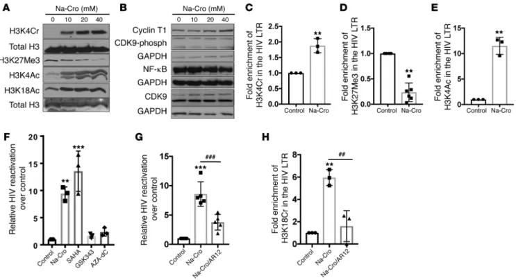
even at low Na-Cro concentrations (Figure 2A). These data sug-gested that induction of ACSS2 following addition of Na-Cro leads to global reprogramming of histone tails in CD4+ T cells. However, it did not alter expression of transcription factors essential for HIV transcription except for a minimal change in the CDK9 protein levels (Figure 2B). Evaluation of chromatin remodeling in the HIV LTR by chromatin immunoprecipitation (ChIP) assay showed that induction of ACSS2 increased the crotonylation and acetylation of histone tail at H3K4 but decreased trimethylation of histone tail at H3K27 in the HIV LTR (Figure 2, C–E).
In agreement with our previous findings and other reports, we found that inhibition of histone methyltransferase (EZH2) by GSK343 or inhibition of DNA methyltransferase by 5-aza-2′ deoxycytidine (AZA-dC) minimally reactivated latent HIV (7, 33). Induction of ACSS2 by Na-Cro or suppression of HDAC by vori-nostat had similar effects in reactivating latent HIV in J-Lat A1 cells, and these findings were comparable to data from the resting CD4+ T cells from HIV-positive patients under ART (Figure 2F). To further verify the role of histone crotonylation in the maintenance of HIV latency, we pharmacologically suppressed the activity of ACSS2 using AR12, a known ACSS2 inhibitor, and investigated the expression of ACSS2 in the HIV LTR reporter cell line TZM-bl (a
CXCR4-positive HeLa cell line engineered to express CD4 and CCR5 constitutively and firefly luciferase under the control of the HIV LTR). A significant reduction was detected in the HIV LTR– driven luciferase expression in the ACSS2 siRNA–treated TZM-bl cells as compared with the cells treated with control siRNA, with or without Na-Cro treatment (Figure 1, F–H). Collectively, our data demonstrate that induction of ACSS2 is effective in disrupt-ing HIV latency across multiple models of HIV latency ex vivo and in vitro and crotonylation-mediated transcription of HIV was pre-dominantly driven by ACSS2.
ACSS2 enzyme leads to chromatin reprogramming through his-tone crotonylation. To determine the molecular mechanism of
ACSS2-induced reactivation of latent HIV, we examined sever-al histone modifications in Na-Cro–treated T cells and assessed expression of major components of transcription factors involv-ing HIV transcription and viral latency. Interestinvolv-ingly, induction of ACSS2 increased not only H3K4 crotonylation (H3K4cr) but also H3K4 acetylation (H3K4Ac) and H3K18 acetylation (H3K18Ac). In contrast, it markedly decreased H3K27 trimethylation (H3K27Me). Changes in histone crotonylation and acetylation were detectable
Figure 2. Histone crotonylation by ACSS2 induction reprograms histone tails at the HIV LTR, and suppression of ACSS2 dampens reactivation of latent HIV. (A) Induction of ACSS2 increased global expression of H3K4 crotonylation (H3K4Cr), H3K4 acetylation (H3K4Ac), and H3K18 acetylation (H3K18Ac) while decreasing expression of H3K27 methylation (H3K27Me) after Na-Cro treatment for 4 hours in J-Lat A1 cells. Immunoblots are representative of 3 indepen-dent experiments. (B) Protein expression after Na-Cro treatment for 4 hours in J-Lat A1 cells. Immunoblots are representative of 3 independent experiments. (C–E) Na-Cro addition induced histone crotonylation at H3K4 (C) and histone acetylation at H3K4 (E) while suppressing histone trimethylation at H3K27 (D) at the HIV LTR. ChIP assay was performed using anti-H3K4Cr (n = 3), anti-H3K4Ac (n = 3), and anti-H3K27Me3 (n = 6) antibodies after J-Lat A1 cells were treated with 30 mM Na-Cro for 4 hours. PCR primers were specific for the HIV LTR region. **P < 0.01 versus control treatment. (F) Reactivation of latent HIV was examined using different epigenetic modifiers. J-Lat A1 cells were treated with 30 mM Na-Cro, 500 nM vorinostat, 2 μM GSK343, or 5 μM AZA-dC. HIV expression was measured by RT-qPCR. **P < 0.01 and ***P < 0.001 versus control treatment (n = 3). (G) Pretreatment with the ACSS2 inhibitor AR12 (5 μM for 30 minutes) dampened histone crotonylation–induced reactivation of latent HIV. ***P < 0.001 versus control treatment; ###P < 0.001 versus
matin remodeling by inhibiting histone deacetylation (12). A com-bination of Na-Cro with the bromodomain protein BRD4 inhibitor JQ1, the histone methyltransferase inhibitor GSK343, or the DNA methyltransferase inhibitor AZA-dC (7, 36) did not show any syner-gistic effect on HIV reactivation in J-Lat A1 cell lines. It is intriguing to note that pretreatment with Na-Cro followed by vorinostat treat-ment demonstrated a synergistic effect on HIV reactivation (Figure 3A). In contrast, pretreatment with vorinostat followed by Na-Cro failed to show any combination effect in either J-Lat A1 cells or U1 cell models of HIV latency (Figure 3B and Supplemental Figure 2B). Since a combination of Na-Cro and PEP005 displayed the most potent effect on reactivation of latent HIV, we further sought to measure the magnitude of HIV LTR reactivation. A significant level of viral reactivation was detected by flow cytometric analy-sis (Figure 3C). Similarly, a synergistic increase in HIV reactivation was also identified in U1 monocytic cells (Figure 3D).
Induction of ACSS2 has no impact on immune activation. We
examined whether induction of histone crotonylation in T cells impacted the level of immune activation in these cells. Peripheral blood mononuclear cells from HIV-negative healthy donors (n = 5) were treated with PMA/ionomycin or Na-Cro for 24 or 72 hours. Changes in the immune cell status were examined by measure-ment of the levels of HLA-DR, CD69, and PD-1 in the T cell sub-sets by flow cytometric analysis (Supplemental Figure 3). Among CD3+, CD4+, or CD8+ T cells, Na-Cro–induced histone crotonyla-tion had no effect on the expression of those immune activacrotonyla-tion or checkpoint markers. In contrast, PMA/ionomycin treatment significantly induced CD69, HLA-DR, and/or PD-1 expression (Supplemental Figure 3, A–C). These findings suggest that histone crotonylation by induction of ACSS2 does not cause changes in T cell activation or suppression.
downstream effects on HIV transcription (34). Induction of ACSS2 by Na-Cro had 8-fold higher HIV reactivation compared with the untreated control. In contrast, addition of the ACSS2 inhibitor AR12 resulted in a substantial decrease in latent HIV reactivation (>60%). Thus, pretreatment with AR12 diminished the effects of ACSS2 induction in disrupting HIV latency (Figure 2G). Impor-tantly, pharmacologic inhibition of the ACSS2 enzyme markedly dampened histone crotonylation at HIV LTR (Figure 2H). Col-lectively, our data show that ACSS2-driven histone crotonylation at HIV LTR remodeled the histone tails to reactivate HIV from latency. These data identify a new mechanism that maintains HIV latency through limitation of ACSS2, thereby reducing histone crotonylation at the HIV LTR.
ACSS2 reactivates HIV synergistically with other LRAs. The
estab-lishment and maintenance of HIV latency involve several molecu-lar signaling pathways. Therefore, an efficient reactivation of latent HIV in the “shock-and-kill” approach may require the use of a com-bination of LRAs targeting different viral latency mechanisms. We sought to determine whether an intervention to induce histone crotonylation to reactivate latent HIV was synergistic with other LRAs that have different mechanisms of action. In combination treatments, J-Lat A1 cells were pretreated with Na-Cro, followed by treatment with other LRAs. Our data showed that crotonylation by Na-Cro addition was synergistic with PEP005, bryostatin-1, or vorinostat in reactivating latent HIV. Combination with Na-Cro increased reactivation of latent HIV 8-fold, 3.3-fold, and 2.0-fold compared with single treatment of PEP005, bryostatin-1, or vori-nostat (Figure 3A and Supplemental Figure 2A). The protein kinase C (PKC) agonists PEP005 and bryostatin-1 have been shown to reactivate HIV through the PKC/NF-κB pathway (7, 35). Vorinostat is an HDAC inhibitor that activates HIV expression through
chro-Figure 3. A synergistic reactivation of latent HIV expression by the histone crotonylation in combi-nation with other LRAs. (A) The J-Lat A1 cells were treated overnight with 30 mM Na-Cro (n = 3), 6 nM PEP005 (n = 3), 2 μM JQ1 (n = 4), 10 nM bryostatin (n = 4), 250 nM vorinostat (n = 3), 2 μM GSK343 (n = 4), 5
μM AZA-dC (n = 4), or 5 ng/ml PMA (n = 4) individually or in combination as indicated in the panel and eval-uated for HIV reactivation by RT-qPCR. *P < 0.05 and ***P < 0.001 versus LRA alone. (B) The J-Lat A1 cells were pretreated with 250 nM of vorinostat; then 40 mM of Na-Cro was added overnight. The HIV reactiva-tion was measured by real-time PCR. **P < 0.01 and ***P < 0.001 versus control treatment (n = 3). NS, not significant compared with Na-Cro treatment alone. (C) Effect of 30 mM Na-Cro, individually or in combina-tion with PEP005, on reactivacombina-tion of latent HIV was tested. The cells were treated overnight, and J-Lat A1 cells with HIV reactivation were measured by detection of GFP-expressing cells using flow cytometry. ***P < 0.001 versus control treatment; ###P < 0.001 versus
PEP005 alone (n = 3). (D) U1 cells were treated similarly as in C; RT-qPCR was performed to measure viral expression. ***P < 0.001 vs. untreated control; ###P <
initiation of HIV transcripts in primary CD4+ T cells from 5 of 6 patients and elongation of HIV transcripts in 4 of 6 patients (Fig-ure 4, A–C). Treatment with PEP005 induced the initiation of HIV transcription in CD4+ T cells from all 6 patients, while the elon-gation of long viral transcripts was detected in 5 of 6 patient sam-ples. In agreement with our previous reports, PEP005 alone also induced full-length transcription of HIV in most patient samples (5 of 6 samples) (7). These data demonstrate the ability of histone crotonylation by ACSS2 induction to reactivate HIV from latency in primary CD4+ T cells from HIV-infected patients under sup-pressive ART. A combination of ACSS2 induction with PEP005 in primary CD4+ T cells from HIV-infected patients induced greater reactivation of latent HIV than either agent alone (Figure 4, A–C) and exceeded levels induced even by PMA/ionomycin treatment in most of the patient samples, indicating a significant potency of this combination treatment (Supplemental Figure 4). An analysis of synergy using the Bliss independence model confirmed that the combination of PEP005 and Na-Cro synergistically reactivates latent HIV ex vivo (Figure 4D, Supplemental Figure 5, and ref. 39). These findings are in agreement with our data from HIV latency
Induction of histone crotonylation in combination with other LRAs disrupts HIV latency in primary CD4+ T cells ex vivo. In HIV latency cell culture models in vitro, we found that Na-Cro dis-played the greatest synergy for HIV reactivation in combination with the PKC agonist PEP005 as compared with other LRAs (Fig-ure 3A and Supplemental Fig(Fig-ure 2A). We sought to validate this finding in the ex vivo primary CD4+ T cells from HIV-infected patients. Peripheral blood samples were obtained from 6 HIV- infected patients receiving suppressive ART. They had undetect-able plasma viral loads (<20 copies/ml plasma) and greater than 400 CD4+ T cell number per microliter of blood (465–885 cells/μl) (Supplemental Table 1, patients 1–6). Primary CD4+ T cells were treated with PMA/ionomycin, PEP005, Na-Cro, or PEP005 in combination with Na-Cro for 6 hours. HIV transcription following viral reactivation was measured by digital droplet PCR targeting the TAR region (initiation), long LTR region (proximal elongation), or poly(A) region (completed transcription) of the HIV genome (7, 37, 38). In concordance with the findings from resting CD4+ T cells using quantitative viral outgrowth assay ex vivo and from HIV latency cell models in vitro, histone crotonylation induced
Figure 4. Histone crotonylation modifier and PEP005 synergistically reactivate latent HIV in CD4+ T cells from HIV-infected individuals under sup-pressive ART. (A–C) Primary CD4+ T cells isolated from HIV-positive patients (n = 6) under ART were treated with 100 ng/ml PMA plus 2 μM ionomycin,
12 nM PEP005, 40 mM Na-Cro, or 12 nM PEP005 combined with 40 mM Na-Cro for 6 hours. Viral transcription from total RNA was analyzed by reverse transcriptase digital droplet PCR with primers targeting initiation (TAR region) (A), elongation (long LTR) (B), or full transcription [poly(A) region] (C) of the HIV genome. (D) Na-Cro synergistically reactivates the latent HIV with PEP005 in primary CD4+ T cells isolated from patients under suppressive ART.
cell culture models in vitro (Figure 3). While the combination of Na-Cro with vorinostat (SAHA) increased reactivation of latent HIV compared with vorinostat alone in 4 of 6 primary CD4+ T cell samples isolated from patients under suppressive ART, synergy was not observed (Supplemental Figure 6). Therefore, the impact of crotonylation on acetylation-induced latent HIV reactivation warrants further investigation. Collectively, our data suggest that histone crotonylation by ACSS2 induction is not only able to reac-tivate the latent HIV in primary CD4+ T cells from HIV-infected patients under ART but demonstrates synergism with PEP005, an LRA targeting PKC/NF-κB signaling, in its reactivation activity.
ACSS2 is induced during acute SIV infection in the rhesus macaque model of AIDS in vivo. As shown above, the induction of ACSS2
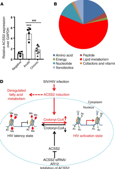
results in chromatin remodeling via crotonylation of histone tails at the HIV LTR; the latter drives the replication of HIV or reacti-vates HIV from latency in vitro and ex vivo. To determine whether viral infection affects ACSS2 expression in vivo, we examined the expression of ACSS2 in intestinal tissues during viral infection in the SIV-infected rhesus macaque model of AIDS (Supplemental Table 2). We found that ACSS2 expression was highly induced in the intestine during the primary acute SIV infection in vivo.
How-ever, the ACSS2 levels decreased during the chronic stage of viral infection (Figure 5A). These findings suggest that ACSS2 may influence viral replication in vivo. Interestingly, ACSS2 is one of the essential enzymes for controlling lipid or fatty acid metabo-lism (19, 40). To determine whether lipid or fatty acid metabometabo-lism was altered during viral infection, we analyzed the metabolic pro-files of the luminal contents from SIV-infected rhesus macaques. We found that approximately 60% of the changes in the metabolic products during SIV infection were related to lipid metabolism. Within lipid metabolism, approximately 45% of the altered meta-bolic products belonged to fatty acid metabolism (Figure 5, B and C). These data indicate that SIV/HIV infection–modulated fatty acid metabolism may be potentially involved in the regulation of HIV replication through induction of ACSS2 in the gut during viral infection in vivo.
Discussion
Latent HIV reservoirs in the host are established very early in viral infection (41–45). Since HIV provirus preferentially integrates in transcriptionally active regions of the host genome (8), it is nec-essary to establish a quiescent chromatin microenvironment and Figure 5. Early SIV infection induces expression of the fatty acid metabolic gene ACSS2. (A) Expression of ACSS2 was induced in the intestinal tissues during early SIV infection. RNA from control (n = 4), acutely SIV-infected (1–2 weeks,
n = 4), and chronically SIV-infected (26–28 weeks, n = 6)
rhesus macaques was extracted, and expression of ACSS2 was determined by RT-qPCR. ***P < 0.001 versus negative infection; ###P < 0.001 versus acute viral infection. The data
interactions among cellular and viral transcriptional regulators for inducing and maintaining HIV latency (9, 46). Latent HIV is reactivated by interference with chromatin modifications as evidenced by efficacy of HDAC inhibitors or EZH2 inhibitors in reactivation of latent HIV (16). However, the level of latent HIV reactivation is relatively modest in CD4+ T cell cultures in vitro and in ex vivo primary CD4+ T cells from HIV-infected patients (7, 15, 47–49). It is possible that HIV latency is regulated by addi-tional mechanisms of histone modifications that have yet to be discovered. After inducing ACSS2 and histone crotonylation, the ability of the HDAC inhibitor vorinostat to reactivate latent HIV was significantly enhanced, indicating that an efficient reacti-vation of latent HIV by HDAC inhibitors may require the croto-nylation of histone tails at the HIV LTR. Our findings point to the need to further characterize the epigenetic landscape of the HIV LTR and investigate multiple epigenetic modifications to achieve optimal viral transcription or silencing. Previous studies report-ed the ability of SIRT1 and SIRT3 proteins from the sirtuin (SIRT) family to erase crotonylation marks from lysine residues in HeLa S3 cell lines (22). However, our data did not support an essential role of SIRT1 or SIRT3 as a decrotonylase at the HIV LTR and suggest that regulation of HIV latency is independent of sirtuins (data not shown).
We and others have previously reported that the lipid metab-olism was altered in HIV-infected primary CD4+ T cells in vitro or in the intestine during early stages of HIV infection in vivo (27–29). We also found that expression of the fatty acid enzyme ACSS2 was significantly induced in HIV-infected individuals following therapy interruption (50). In the current study, we have identified the modulation of fatty acid metabolism and the induction of ACSS2 expression in intestinal tissues during early stages of SIV infection in vivo. Aberrant fatty acid metabolism may activate proinflammatory signaling and disrupt mucosal integrity (51). It is not known whether host metabolites produced during HIV/SIV infection are responsible for gut mucosal dam-age in vivo, which is under investigation. Further analysis showed that ACSS2 can also influence HIV replication and viral latency by modulating histone crotonylation at HIV LTR. Disruption of HIV latency was observed by histone crotonylation following the induction of ACSS2 in vitro and ex vivo. Therefore, our find-ings favor a model whereby suppression of histone crotonylation inhibits reactivation of latent HIV and supports the maintenance of viral latency (Figure 5D). Previously, we found that expression of the ACSS2 gene was significantly induced in the intestinal biopsies of HIV-infected patients following interruption of ART in vivo (50). Our data, for the first time to our knowledge, have linked fatty acid metabolism to the epigenetic regulation of HIV transcription and maintenance of viral latency through crotonyla-tion of histone tails by ACSS2 at the HIV LTR.
HIV RNA transcription can be detected in the absence of the production of virus and viral antigens and therefore may be insuf-ficient to trigger viral cytolytic effects or immune recognition and clearance of reservoir cells (1, 52). Recently, Pollack et al. reported that even defective viruses can produce viral proteins that can be recognized by cytotoxic T lymphocytes (CTLs) mediating immune clearance (53). Detection of polyadenylated HIV RNA is consid-ered to be the best correlate of viral production (39). In this study,
we assessed HIV reactivation by measuring HIV transcripts rep-resenting the viral initiation (TAR), elongation (long LTR), and complete transcription (polyadenylated RNA). In addition, we also measured HIV p24 levels in culture supernatants as a correlate of viral production. When induction of ACSS2 was used alone to reverse HIV latency, the magnitude of effects on initiation of tran-scription was greater than that of effects on the elongation or gen-eration of fully transcribed HIV RNA. However, detection of HIV p24 outgrowth from the resting CD4+ T cells in quantitative viral outgrowth assay indicated that the net effects of ACSS2-induced HIV transcription were sufficient to elicit production of viral p24 and virus. Importantly, our data show a remarkable synergistic reactivation of latent HIV when histone crotonylation is combined with the PKC agonist PEP005, vorinostat, or JQ1 in T cell cultures in vitro and/or CD4+ T cells from HIV-infected patients ex vivo. A robust reactivation of latent HIV following the combination treat-ment of cells from HIV-infected patients indicated that increased levels of viral particles could be induced that are recognized by CTLs for immune clearance when an effective killing strategy is applied. On the other hand, inhibition of histone crotonylation by suppression of the crotonyl-CoA–converting enzyme ACSS2 dampened latent HIV reactivation, indicating that the loss of ACSS2 leads to suppression of HIV transcription and a potential role of histone decrotonylation in establishment of HIV latency. SIRT proteins were characterized as decrotonylases (22, 54). How-ever, our data did not support their role as histone decrotonylases in the HIV LTR. In contrast to many if not most other LRAs, induc-tion of ACSS2 by Na-Cro did not induce immune activainduc-tion or modulate levels of the immune checkpoint protein PD-1 on CD4+ T cells or CD8+ T cells.
In summary, our data uncovered an important role for the fatty acid metabolic enzyme ACSS2 in the regulation of HIV tran-scription by crotonylation of histone tails at the HIV LTR. We have identified decrotonylation of histone tails at the HIV LTR as a potential novel histone mark for establishment and maintenance of HIV latency. This epigenetic modification mechanism and its reversal open new avenues for HIV cure approaches.
Methods
Further information can be found in Supplemental Methods, available online with this article.
Cell culture. J-Lat A1 cells (derived from Jurkat cells harboring HIV
LTR-Tat-GFP gene) and U1 cells (promonocytic cell line harboring HIV proviruses with defective Tat gene) were obtained from the NIH AIDS Reagent Program and cultured at 37°C with 5% CO2 in RPMI 1640 medium containing 10% FBS and 1% penicillin/streptomycin as previously described (7).
Latency reversal agents. For reactivation of latent HIV, cells were
at day 15 and was increased in concentration at day 19. The number of resting CD4+ T cells in infected units per million was estimated by a maximum likelihood method (55).
Statistics. Data represent the mean ± SEM, calculated using all
data points from at least 3 independent experiments. Statistical signif-icance was determined using a 2-way Student’s t test for samples with only 2 groups. For multiple-comparison analysis of samples from 3 or more groups, we applied 1-way ANOVA analysis followed by a post-hoc Tukey’s test, and a P value of less than 0.05 was considered significant.
Study approval. This study was carried out under the
recommen-dations of the Public Health Service Policy on Humane Care and Use of Laboratory Animals. Human peripheral blood samples (n = 17) were obtained under informed written consent and a protocol approved by the Institutional Review Boards at UCD and the University of North Carolina at Chapel Hill. Animals were housed at the California Nation-al Primate Research Center at UCD, and procedures were approved by the Institutional Animal Care and Use Committee of UCD.
Author contributions
GJ and SD conceived and designed the experiments. GJ, DN, YT, MME, NMA, and GML performed the experiments. GJ, SAY, JKW, NMA, DMM, DJHO, and SD analyzed the data. NMA and GRT coordinated patient samples. GJ and SD wrote the manuscript.
Acknowledgments
We thank Lauren A. Hirao and Clarissa Santos Rocha for the initia-tion of the metabolomic study and Peggy Kim for technical support. We are very grateful to all patients for participating in this study. This work was supported by NIH grants AI123105 and AI43274 and a UC Davis Research Investments in Science and Engineer-ing grant to SD; NIH grants U19-AI096113, UM1-AI126619, and UNC CFAR P30-AI504100 to DMM; and NIH grants AI116342, AI116218, DK108349, and AI132128 to JKW and SAY.
Address correspondence to: Satya Dandekar, Department of Med-ical Microbiology and Immunology, UCD, One Shields Drive, 3146 Tupper Hall, Davis, California 95616, USA. Phone: 530.752.3409; Email: sdandekar@ucdavis.edu.
Gene knockdown by ACSS2 siRNA. TZM-bl HIV transcription/
replication reporter cells (1 × 10⁵) were seeded in the 12-well plate, and then the cells were transfected with ACSS2 siRNA (M-010396) or non-targeting control siRNA (M-006526) (Dharmacon) twice. The cells were treated with or without 40 mM Na-Cro overnight and collected for luciferase assay of HIV transcription and/or lysed for quantitative reverse transcriptase PCR (RT-qPCR) of ACSS2 gene expression.
Primary CD4+ T cell isolation, treatment, and digital droplet PCR
assays. Peripheral blood samples were obtained from HIV-infected
individuals (age ranging from 22 to 62 years) on suppressive ART for more than 2 years (n = 12, average age 10.6 ± 5.7 years). The plasma viral loads were below the detection level (<20 copies/ml plasma), and the average CD4+ T cell number was 680.2 ± 225.6 cells/μl. The pri-mary CD4+ T cells were isolated using the EasySep kit (STEMCELL Technologies) as previously described (7, 15). The purified CD4+ T cells were plated at a density of 0.5 × 10⁶ to 1 × 10⁶ cells and treated with DMSO, 200 ng/ml PMA plus 2 μM ionomycin, 12 nM PEP005, 40 mM Na-Cro, or 12 nM PEP005 plus 40 mM Na-Cro for 6 hours. Cell pellets were collected for RNA isolation. Initiation, elongation, or full transcription of HIV was analyzed with digital droplet PCR assays as reported before (7, 37, 38).
Quantitative viral outgrowth assay. Peripheral blood mononuclear
cells (PBMCs) were obtained from HIV-infected individuals on sup-pressive ART (n = 5) by continuous-flow leukapheresis. Isolation of resting CD4+ T cells and quantification of replication-competent virus were performed as previously described (41). Briefly, approximately 34 million to 50 million resting CD4+ T cells per each treatment con-dition were plated in replicate limiting dilutions of 2.5 million (18 cul-tures), 0.5 million (6 culcul-tures), and 0.1 million (6 cultures) cells per well and stimulated for 24 hours with either (a) PHA (Remel, Thermo Fisher Scientific), a 5-fold excess of allogeneic irradiated PBMCs from a seronegative donor, and IL-2; (b) 40 mM Na-Cro plus IL-2 or 350 nM SAHA plus IL-2; or (c) IL-2 as unstimulated control. Cultures were washed and cocultivated with CD8+ T cell–depleted PBMCs that were obtained from selected HIV-seronegative donors previously screened for adequate CCR5 expression. Culture supernatants were harvested on days 15 and 19 and assayed for virus production by p24 antigen cap-ture ELISA (ABL). Culcap-tures were scored as positive if p24 was detected
1. Bruner KM, Hosmane NN, Siliciano RF. Towards an HIV-1 cure: measuring the latent reservoir. Trends Microbiol. 2015;23(4):192–203. 2. Margolis DM. How might we cure HIV? Curr
Infect Dis Rep. 2014;16(3):392.
3. Siliciano JD, et al. Long-term follow-up studies confirm the stability of the latent reservoir for HIV-1 in resting CD4+ T cells. Nat Med.
2003;9(6):727–728.
4. Finzi D, et al. Identification of a reservoir for HIV-1 in patients on highly active antiretroviral therapy. Science. 1997;278(5341):1295–1300. 5. Wong JK, et al. Recovery of replication-competent
HIV despite prolonged suppression of plasma viremia. Science. 1997;278(5341):1291–1295. 6. Chun TW, et al. Presence of an inducible
HIV-1 latent reservoir during highly active antiretroviral therapy. Proc Natl Acad Sci U S A. 1997;94(24):13193–13197.
7. Jiang G, et al. Synergistic reactivation of latent
HIV expression by ingenol-3-angelate, PEP005, targeted NF-kB signaling in combination with JQ1 induced p-TEFb activation. PLoS Pathog. 2015;11(7):e1005066.
8. Hakre S, Chavez L, Shirakawa K, Verdin E. Epi-genetic regulation of HIV latency. Curr Opin HIV AIDS. 2011;6(1):19–24.
9. Jiang G, Espeseth A, Hazuda DJ, Margolis DM. c-Myc and Sp1 contribute to proviral latency by recruiting histone deacetylase 1 to the human immunodeficiency virus type 1 promoter. J Virol. 2007;81(20):10914–10923.
10. du Chéné I, et al. Suv39H1 and HP1γ are respon-sible for chromatin-mediated HIV-1 transcrip-tional silencing and post-integration latency. EMBO J. 2007;26(2):424–435.
11. Friedman J, et al. Epigenetic silencing of HIV-1 by the histone H3 lysine 27 methyltransferase enhancer of Zeste 2. J Virol. 2011;
85(17):9078–9089.
12. Archin NM, et al. Administration of vorinostat disrupts HIV-1 latency in patients on antiretrovi-ral therapy. Nature. 2012;487(7408):482–485. 13. Rasmussen TA, et al. Panobinostat, a histone
deacetylase inhibitor, for latent-virus reacti-vation in HIV-infected patients on suppressive antiretroviral therapy: a phase 1/2, single group, clinical trial. Lancet HIV. 2014;1(1):e13–e21. 14. Søgaard OS, et al. The depsipeptide romidepsin
reverses HIV-1 latency in vivo. PLoS Pathog. 2015;11(9):e1005142.
15. Jiang G, et al. Reactivation of HIV latency by a newly modified Ingenol derivative via protein kinase Cδ-NF-κB signaling. AIDS. 2014;28(11):1555–1566.
16. Jiang G, Dandekar S. Targeting NF-κB signaling with protein kinase C agonists as an emerging strategy for combating HIV latency. AIDS Res Hum Retroviruses. 2015;31(1):4–12.
histone lysine crotonylation as a new type of his-tone modification. Cell. 2011;146(6):1016–1028. 18. Sabari BR, et al. Intracellular crotonyl-CoA
stim-ulates transcription through p300-catalyzed his-tone crotonylation. Mol Cell. 2015;58(2):203–215. 19. Sabari BR, Zhang D, Allis CD, Zhao Y.
Meta-bolic regulation of gene expression through histone acylations. Nat Rev Mol Cell Biol. 2017;18(2):90–101.
20. Li Y, Zhao D, Chen Z, Li H. YEATS domain: linking histone crotonylation to gene regulation. Transcription. 2017;8(1):9–14.
21. Andrews FH, et al. The Taf14 YEATS domain is a reader of histone crotonylation. Nat Chem Biol. 2016;12(6):396–398.
22. Bao X, et al. Identification of ‘erasers’ for lysine crotonylated histone marks using a chemical pro-teomics approach. Elife. 2014;3:e02999. 23. Abumrad NA, Davidson NO. Role of the gut in lipid
homeostasis. Physiol Rev. 2012;92(3):1061–1085. 24. Stanley TL, Grinspoon SK. Body composition and
metabolic changes in HIV-infected patients. J Infect Dis. 2012;205(suppl 3):S383–S390. 25. Bociąga-Jasik M, et al. Metabolic complications
and selected cytokines in HIV-infected individu-als. Pol Arch Med Wewn. 2014;124(1–2):27–35. 26. Koethe JR, Hulgan T, Niswender K. Adipose tissue and immune function: a review of evi-dence relevant to HIV infection. J Infect Dis. 2013;208(8):1194–1201.
27. Sankaran S, et al. Rapid onset of intestinal epi-thelial barrier dysfunction in primary human immunodeficiency virus infection is driven by an imbalance between immune response and mucosal repair and regeneration. J Virol. 2008;82(1):538–545.
28. Wu JQ, Dwyer DE, Dyer WB, Yang YH, Wang B, Saksena NK. Genome-wide analysis of primary CD4+ and CD8+ T cell transcriptomes shows
evi-dence for a network of enriched pathways associ-ated with HIV disease. Retrovirology. 2011;8:18. 29. Guadalupe M, et al. Viral suppression and immune
restoration in the gastrointestinal mucosa of human immunodeficiency virus type 1-infected patients initiating therapy during primary or chronic infection. J Virol. 2006;80(16):8236–8247. 30. Waagsbø B, et al. Low levels of short- and
medium-chain acylcarnitines in HIV-infected patients. Eur J Clin Invest. 2016;46(5):408–417. 31. Babirekere-Iriso E, et al. Essential fatty acid
composition and correlates in children with severe acute malnutrition. Clin Nutr ESPEN. 2016;11:e40–e46.
32. Nyirenda CK, et al. Plasma fatty acids in Zambian adults with HIV/AIDS: relation to dietary intake and cardiovascular risk factors. J Nutr Metab. 2015;2015:635817.
33. Kauder SE, Bosque A, Lindqvist A, Planelles V, Verdin E. Epigenetic regulation of HIV-1 latency by cytosine methylation. PLoS Pathog. 2009;5(6):e1000495.
34. Koselny K, et al. Antitumor/antifungal celecoxib derivative AR-12 is a non-nucleoside inhibitor of the ANL-family adenylating enzyme acetyl CoA synthetase. ACS Infect Dis. 2016;2(4):268–280. 35. Díaz L, et al. Bryostatin activates HIV-1 latent
expression in human astrocytes through a PKC and NF-ĸB-dependent mechanism. Sci Rep. 2015;5:12442.
36. Banerjee C, et al. BET bromodomain inhibition as a novel strategy for reactivation of HIV-1. J Leukoc Biol. 2012;92(6):1147–1154.
37. Kaiser P, et al. Assays for precise quantification of total (including short) and elongated HIV-1 tran-scripts. J Virol Methods. 2017;242:1–8.
38. Shan L, et al. A novel PCR assay for quantification of HIV-1 RNA. J Virol. 2013;87(11):6521–6525. 39. Laird GM, et al. Ex vivo analysis identifies
effec-tive HIV-1 latency-reversing drug combinations. J Clin Invest. 2015;125(5):1901–1912.
40. Xu H, et al. Acyl-CoA synthetase short-chain family member 2 (ACSS2) is regulated by SREBP-1 and plays a role in fatty acid synthesis in caprine mammary epithelial cells. J Cell Physiol. 2018;233(2):1005–1016.
41. Dandekar S. Pathogenesis of HIV in the gastrointestinal tract. Curr HIV/AIDS Rep. 2007;4(1):10–15.
42. Lackner AA, Lederman MM, Rodriguez B. HIV pathogenesis: the host. Cold Spring Harb Perspect Med. 2012;2(9):a007005.
43. Somsouk M, et al. Gut epithelial barrier and systemic inflammation during chronic HIV infec-tion. AIDS. 2015;29(1):43–51.
44. Hirao LA, et al. Early mucosal sensing of SIV infection by paneth cells induces IL-1β produc-tion and initiates gut epithelial disrupproduc-tion. PLoS Pathog. 2014;10(8):e1004311.
45. Whitney JB, et al. Rapid seeding of the viral res-ervoir prior to SIV viraemia in rhesus monkeys. Nature. 2014;512(7512):74–77.
46. Tyagi M, Karn J. CBF-1 promotes transcriptional silencing during the establishment of HIV-1 latency. EMBO J. 2007;26(24):4985–4995. 47. Klase Z, et al. Activation of HIV-1 from
latent infection via synergy of RUNX1 inhibitor Ro5-3335 and SAHA. PLoS Pathog. 2014;10(3):e1003997.
48. Pandeló José D, et al. Reactivation of latent HIV-1 by new semi-synthetic ingenol esters. Virology. 2014;462–463:328–339.
49. Bouchat S, et al. Sequential treatment with 5-aza-2′-deoxycytidine and deacetylase inhibitors reactivates HIV-1. EMBO Mol Med. 2016;8(2):117–138.
50. Lerner P, et al. The gut mucosal viral reservoir in HIV-infected patients is not the major source of rebound plasma viremia following interruption of highly active antiretroviral therapy. J Virol. 2011;85(10):4772–4782.
51. Rutkowsky JM, et al. Acylcarnitines activate proinflammatory signaling pathways. Am J Physi-ol EndocrinPhysi-ol Metab. 2014;306(12):E1378–E1387. 52. Bullen CK, Laird GM, Durand CM, Siliciano
JD, Siliciano RF. New ex vivo approaches dis-tinguish effective and ineffective single agents for reversing HIV-1 latency in vivo. Nat Med. 2014;20(4):425–429.
53. Pollack RA, et al. Defective HIV-1 proviruses are expressed and can be recognized by cytotoxic T lymphocytes, which shape the proviral land-scape. Cell Host Microbe. 2017;21(4):494–506.e4. 54. Feldman JL, Baeza J, Denu JM. Activation of the
protein deacetylase SIRT6 by long-chain fatty acids and widespread deacylation by mammalian sirtuins. J Biol Chem. 2013;288(43):31350–31356. 55. Archin NM, Keedy KS, Espeseth A, Dang H,