Original Research Article
Peripheral lymphadenopathy and FNA: a two-year evaluation at a
tertiary care centre
Jiby Soosen Ninan
1*, De Souza Johanna Alba
2, Thoppil Reba Philipose
1INTRODUCTION
FNA provides a simple and inexpensive test for diagnosis of common lymphadenopathies like reactive hyperplasia, infections, granulomatous lymphadenopathies and metastatic diseases.1 In cases of reactive lymphoid
hyperplasia's and infectious lymphadenopathies fine needle aspiration can significantly reduce the number of open biopsies. In infectious and granulomatous lymphadenopathies, if an infectious agent is identified, not only the cause of lymphadenopathy is determined, but
also microbiological studies can be performed on the aspirated material which can help the clinicians to choose appropriate antimicrobial therapy. FNA is a well-established method for the diagnosis of metastatic malignancies in lymph nodes. It confirms the presence of metastatic disease and also gives clues about the nature and origin of the primary tumour. In patients with previously documented malignancy presenting with enlarged lymph nodes, FNA can obviate further surgery performed merely to confirm the presence of metastasis. FNA of lymph nodes is very useful in confirmation of
ABSTRACT
Background: Lymphadenopathy is an age old affliction of mankind and a very common presentation in clinical practice. The main purpose of an FNA biopsy of abnormal peripheral lymph nodes is to determine whether further surgical excision of the lymph node is indicated for histopathological examination. The aim of the present study was to evaluate the role of fine needle aspiration cytology in patients with superficial lymphadenopathy and to correlate with histopathology wherever possible.
Methods: A two-year study was undertaken at the Central Diagnostic Laboratory at A.J. Institute of Medical Sciences, Mangalore. Patients of all age groups referred to the Central Diagnostic Laboratory for FNA of superficial lymph nodes were included in the study. All the slides of the cases were reviewed and impression recorded.
Results: Out of 200 cases, 73% were non neoplastic, 27% were neoplastic. Cases occurred most commonly in age group of 21-30 years. The male to female ratio was 1.7:1 and most common site of lymph node aspiration was cervical lymph node in (n =107) 53.5% cases. Reactive hyperplasia was the most common non- neoplastic cause of lymphadenopathy seen in 34.5% cases and metastasis to lymph node was the most common cause of neoplastic lymphadenopathy seen in 22% of the cases. The sensitivity was 90%, specificity was 100% and accuracy was 96.2%.
Conclusions: FNA is a very efficient, simple, safe, inexpensive and economical test for detecting the various causes of lymphadenopathy.
Keywords: FNA, Histopathology, Lymphadenopathy, Metastasis
1Department of Pathology, A.J Institute of Medical Sciences and Research Centre, Mangalore, Karnataka, India 2Consultant Pathologist, Aster Hospital, Margao, Goa, India
Received: 02 January 2019
Accepted: 08 January 2019
*Correspondence:
Dr. Jiby Soosen Ninan,
E-mail: jibysoosen@gmail.com
Copyright: © the author(s), publisher and licensee Medip Academy. This is an open-access article distributed under the terms of the Creative Commons Attribution Non-Commercial License, which permits unrestricted non-commercial use, distribution, and reproduction in any medium, provided the original work is properly cited.
diagnosis in advanced inoperable cancers.2 Although the
role of FNA in initial diagnosis, sub classification and management of patients with lymphomas may be limited, it aids in the initial suspicion of the disease; detection of residual disease, recurrences and progression of low grade to high grade lymphoma and helps in staging the disease. Availability of prior FNA report facilitates the subsequent histological diagnosis and classification of lymphomas.3 Ancillary techniques, including cell block
preparations, immunocytochemistry, quantitative immunophenotypying using flow cytometry, and molecular techniques to determine clonality and cell lineage are applied to FNA material, all enhance the accuracy of diagnosis.4 The lymphomas that are diffuse
by nature and are composed of one cell type with characteristic flow cytometric pattern are best amenable to unequivocal diagnosis by FNA.3 Although FNA is not
a replacement for open biopsy, in each and every case of lymphadenopathy it can help to establish a workable diagnosis and reduce the number of total biopsies. In the present study we have tried to make a scientific study and analysis through FNA, of the incidence, nature and types of lymphadenopathy with respect to age, sex, and site of distribution
.
METHODS
The material for this study was obtained from FNA’s done at the Central Diagnostic Laboratory at A.J Institute of Medical Sciences, Mangalore on 200 patients who presented with superficial lymphadenopathy between July 2013 and July 2015.
Inclusion criteria
Patients of all age groups that were referred to the Central Diagnostic Laboratory at A. J. Institute of Medical Sciences for FNA of superficial lymph nodes during the study period.
FNAC that was provisionally thought to be non- lymphoid in origin, that later turned out to be that from a lymph node.
Exclusion criteria
• FNA of deep lymph nodes.
• USG/CT scan guided FNA of lymph node.
• FNAC that was provisionally thought to be of lymph node origin that later turned out to be non- lymphoid origin.
• Inadequate aspirate.
In each case the clinical history, the clinical examination findings and relevant investigation results were noted. FNA smears were air dried and wet fixed in 95% ethyl alcohol and stained with May Grunwald Giemsa and Papanicolaou stain respectively. The slides were reviewed and the impression recorded.
Lymph nodes of the patients who underwent subsequent surgical biopsy were fixed in 10% formalin and subjected to gross examination. Biopsy specimens were routinely processed to obtain 3 micrometre paraffin sections, which were stained with Haematoxylin and Eosin stains. Special stains like Ziehl Neelsen stain, PAS were done whenever indicated. Histopathological study was done separately and then results of cytological and histopathological study were correlated to evaluate efficacy of the procedure.
RESULTS
The youngest patient in this study was 4 years old and the oldest was 78 years old. 19.5% of the patients belonged to 21-30 years age group, which was the age group in which most patients were involved. There were 126 males (63 %) and 74 females (37%) in the present study. The male to female ratio was 1.7:1.
Majority of the patients presented with localized lymphadenopathy accounting for 196 cases and the remaining 4 cases presented with generalized lymphadenopathy.
The most common site of lymph node aspiration was cervical lymph node in 53.5% of the cases (n =107 cases) followed by supraclavicular lymph nodes in 11.5% of the cases (n = 23 cases). Submandibular lymph node was affected in 10.5% of the cases (n= 21), inguinal lymph node in 10.5% (n= 21), axillary lymph node in 7% (n= 14), submental lymph node in 4.5 % (n = 9), pre-auricular lymph node in 1.5% (n= 3) and post auricular lymph node in 1% (n= 2).
Of the 200 cases, 146 cases (73%) were diagnosed as non-neoplastic lesions, 54 cases (27%) were diagnosed as neoplastic lesions.
Figure 1: FNA smear of reactive hyperplasia. (MGG x100). Note polymorphous population of lymphocytes
and tingible body macrophages.
% of the cases, 8.5% cases were of granulomatous lymphadenitis, 4.5 % cases were of suppurative lymphadenitis (Figure 3), 0.5 % cases were of Kikuchi’ s disease (Figure 4) and 0.5% were of Kimura’s disease.
Figure 2: A) FNA smear of tuberculous lymphadenitis. (MGG x 400) granuloma consisting of
epithelioid cells. 2B) Smear showing numerous acid fast bacilli (oil immersion x1000).
Figure 3: FNA smear of suppurative lymphadenitis: aspirate shows acute inflammatory cells, cellular debris and tingible body macrophages (MGG x 100).
Figure 4: FNA smear: Kikuchi’s disease (MGG x 100). Note karyorrhectic debris and histiocytes with
crescent-shaped nuclei.
The 54 (27%) neoplastic lesions comprising of 10 (5%) cases of lymphoma included 4% cases of Non-Hodgkin’ s lymphoma and 1% case of Hodgkin’ s lymphoma (Figure 5) and 44 (22%) cases of metastases to lymph node.
Figure 5: FNA smear of Hodgkin’s lymphoma. Note Reed Sternberg cell (MGGx400).
Histopathological correlation was available in 27 cases of which, 17 cases which were benign on cytology and also turned out to be benign on histopathology. 1 case which was benign on cytology turned out to be malignant on histopathology. 9 cases which were malignant on cytology were also proved to be malignant on histopathology.
The overall sensitivity, specificity and accuracy of fine needle aspiration cytology in the diagnosis of superficial lymphadenopathy in the present study were 90%, 100% and 96.2% respectively (Table 1)
.
Table 1: Comparative analysis of cytological diagnosis by histopathological diagnosis.
Cytological Diagnosis
Histopathological
diagnosis Total
Benign Malignant
Benign 17 (TN) 1(FN) 17 Malignant 0 (FP) 9 (TP) 9
TN-True Negative; FN-False negative; FP-False positive; TP-True positive
DISCUSSION
The male to female ratio was 1.7:1 and this was comparable to studies done by Haque MA, Talukdar SI et al, Rakshan M, Rakshan A et aland Hirachand S et al, all of which showed a male preponderance.5-7
The most common age of presentation with lymphadenopathy was the 3rd to 4th decades as in other
studies done by Rajasekaran et al.8
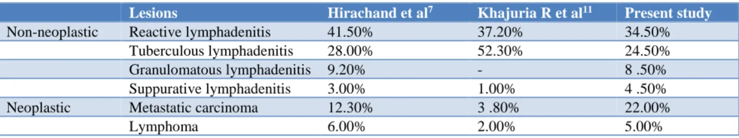
Non-neoplastic lesions were more prevalent than neoplastic lesions in the present study and this was
similar to studies done by Haque MA, Talukdar SI et al and Hirachand S et al.5-7 The most common non
neoplastic lesion was reactive hyperplasia followed by tuberculous lymphadenitis.7
One case was misdiagnosed as reactive hyperplasia on cytology. On histopathology, this case was diagnosed as Non-Hodgkin’s lymphoma-Follicular subtype. Brandao et al,reported that the follicular lymphoma might present a particular difficulty in FNA specimens because neoplastic element itself was polymorphous (centrocytes and centroblasts), and there might be a significant
population of T lymphocytes and, less commonly, histiocytes.9 Almost the same observations were reported
by Dong HY et al,who concluded that the difficulty to distinguish follicular lymphoma from reactive hyperplasia was largely due to the fact that the interfollicular areas in follicular lymphoma might contain large number of small lymphocytes as well as histiocytes that aspirated with the neoplastic cells.10 Most common
neoplastic lesion was metastatic carcinoma in the present study which was comparable with other studies7,11 (Table
2).
Table 2: Comparison of frequency of cytological diagnosis in the present study and other studies.
Lesions Hirachand et al7 Khajuria R et al11 Present study
Non-neoplastic Reactive lymphadenitis 41.50% 37.20% 34.50% Tuberculous lymphadenitis 28.00% 52.30% 24.50% Granulomatous lymphadenitis 9.20% - 8 .50% Suppurative lymphadenitis 3.00% 1.00% 4 .50% Neoplastic Metastatic carcinoma 12.30% 3 .80% 22.00%
Lymphoma 6.00% 2.00% 5.00%
Table 3: Site of FNA and type of metastasis to lymph node.
Site
Types of metastasis Cervical Supra
clavicular Axillary Inguinal Submandibular Submental Total
Mets from papillary carcinoma
thyroid 1 1
Mets from SCC 14 4 2 8 28
Mets from adenocarcinoma 3 3 1 7
Mets from poorly differentiated
carcinoma 1 1 2
Mets from IDC 1 1
Mets from small cell carcinoma 1 1 2
Mets from malignant melanoma 1 2 3
20 9 2 4 8 1 44
mets: metastasis; SCC-Squamous cell carcinoma; IDC-infiltrating ductal carcinoma
Around 22% had metastasis to lymph node. Males (70.4%) were most commonly affected than females (29.5%). 31.8% of the patients were in the age group of 41-50 years which was the most common age group to be affected, followed by 27.2% of patients in the age group of 61-70 years. This was consistent with the studies conducted by Pandav et al.12 Most of the patients in the
present study had metastasis from squamous cell carcinoma (64%, n= 28) arising from upper aero-digestive tract, followed by adenocarcinoma (15%, n= 7), metastasis from malignant melanoma (7%, n=3), metastasis from small cell carcinoma (n=2, 5%), metastasis from poorly differentiated carcinoma (n=2,5%), metastasis from infiltrating ductal carcinoma (n=1, 2%), metastasis from papillary carcinoma of thyroid (n=1, 2%). Cervical lymph node was the most
commonly affected in 45% of patients followed by axillary lymph node which was affected in 20.4% of patients. Similar results obtained in study by Wilkinson et al.13 Biopsy was available in 6 cases and
Table 4: Comparison of the site distribution of lymph node FNA’s in various studies.
Site Present study Haque and Talukder Pandit et al Nasuti et al Van de Schoot L et al
Cervical 53.5% 87.18% 65.03% 30.13%
Supraclavicular 11.5% 7.69% 13.69%
Submandibular 10.5% 8.39% 20.54%
Submental 4.5% 1.36%
Preauricular 1.5% 5.47%
Postauricular 1% 2.79% 4.10%
Axillary 7% 12.82% 11.53% 15.34% 8.21%
Inguinal 10.5% 4.54% 10.68% 13.69%
CONCLUSION
In conclusion FNA of lymph node is a useful diagnostic tool in patients with peripheral lymphadenopathy thereby avoiding unnecessary surgery.
ACKNOWLEDGEMENTS
Authors would like to thank Dr. Umaru N, former HOD, Department of Pathology, AJ Institute of Medical Sciences and Research Centre for his guidance and support.
Funding: No funding sources Conflict of interest: None declared Ethical approval: Not required
REFERENCES
1. Saboorian MH, Ashafaq R. The use of fine needle aspiration biopsy in the evaluation of lymphadenopathy. Semin Diagn Pathol. 2001;18(2):110-23.
2. Bagwan N, Kane SV, Chino y RF. Cytologic evaluation of the enlarged neck node: FNAC utility in metastatic neck disease. The Int J Pathol. 2007;6(2):13-7.
3. Das DK. Value and limitations of fine needle aspiration cytology in diagnosis and classification of lymphomas: A review. Diagn Cytopathol. 1999;21(4):240-9.
4. Buley ID. Fine needle aspiration cytology of lymph nodes. J Clin Pathol. 1998;51:881-5.
5. Haque MA, Talukder SI. Evaluation of Fine needle aspiration cytology (FNAC) of lymph nodes in Mymensingh. Mymensingh Med J. 2003;12(1):33-5. 6. Rakshan M, Rakshan A. The diagnostic accuracy of
fine needle aspiration cytology in neck lymphoid masses. Iranian J Pathol. 2009;4(4):147-50.
7. Hirachand S, Lakhey M, Akhter J, Thapa B. Evaluation of fine needle aspiration cytology of lymph
nodes in Kathmandu Medical College, Teaching hospital. Kathmandu Univ Med J. 2009;7(26):139-42. 8. Rajasekaran S, Gunasekaran M, Jayakumar DD,
Jeyaganesh D, Bhanumati V. Tuberculous cervical lymphadenitis in HIV positive and negative patients. Indian J Tuber. 2001;48(4):201-4.
9. Brandao GD, Rose R, McKenzie F. Grading of follicular lymphoma in fine needle aspiration biopsies: The role of thin preparation of slide and FCM. Cancer. 2006;108(5):319-23.
10. Dong HY, Harris NL, Preffer FI, Pitman MB. Fine-needle aspiration biopsy in the diagnosis and classification of primary and recurrent lymphoma: a retrospective analysis of the utility of cytomorphology and flow cytometry. Modern Pathol. 2001 May;14(5):472-81.
11. Khajuria R, Goswami KC, Singh K, Dubey VK. Pattern of lymphadenopathy on fine needle aspiration cytology in Jammu. JK Sci. 2006 Jul;8(3):145-9. 12. Pandav AB, Patil PP, Lanjewar DN. Cervical
lymphadenopathy-diagnosis by FNAC, a study of 219 cases. Asian J Med Res. 2012 Jun 15;1(3):10-4. 13. Wilkinson AR, Sadhana DM, Sabiha AM. FNAC in
the diagnosis of lymph node malignancies: A simple and sensitive tool. Ind J Med Paediatr Oncol. 2012;33(1):21-5.
14. Pandit AA, Candes FP, Khubchandani SR. Fine needle aspiration cytology of lymph nodes. J Postgrad Med. 1987;33(3):134-6.
15. Nasuti JF, Mehrotra R, Gupta PK. Diagnostic value of fine needle aspiration in supraclavicular lymphadenopathy: A study of 106 patients and review of literature. Diagn Cytopathol. 2001;25(6):351-5. 16. Van de Schoot L, Aronson DC, Behrendt H, Bras J.
The role of fine-needle aspiration cytology in children with persistent or suspicious lymphadenopathy. J Pediatr Surg. 2001;36(1):7-11.