REVIEW
Possibility for reverse zoonotic transmission
of SARS-CoV-2 to free-ranging wildlife: A case
study of bats
Kevin J. OlivalID1☯*, Paul M. CryanID2☯*, Brian R. AmmanID3, Ralph S. BaricID4, David
S. BlehertID5, Cara E. BrookID6, Charles H. Calisher7, Kevin T. CastleID8, Jeremy T.
H. ColemanID9, Peter DaszakID1, Jonathan H. EpsteinID1, Hume FieldID1,11, Winifred
F. FrickID10,12, Amy T. GilbertID13, David T. S. HaymanID14, Hon S. IpID5, William
B. KareshID1, Christine K. Johnson15, Rebekah C. KadingID7, Tigga KingstonID16, Jeffrey
M. Lorch5, Ian H. MendenhallID17, Alison J. PeelID18, Kendra L. PhelpsID1, Raina
K. Plowright19, DeeAnn M. ReederID20, Jonathan D. ReichardID9, Jonathan M. SleemanID5,
Daniel G. StreickerID21,22, Jonathan S. TownerID3, Lin-Fa WangID17
1 EcoHealth Alliance, New York, New York, United States of America, 2 US Geological Survey, Fort Collins Science Center, Ft. Collins, Colorado, United States of America, 3 US Centers for Disease Control and Prevention, Atlanta, Georgia, United States of America, 4 Department of Epidemiology, University of North Carolina, Chapel Hill, North Carolina, United States of America, 5 US Geological Survey, National Wildlife Health Center, Madison, Wisconsin, United States of America, 6 Department of Integrative Biology, University of California Berkeley, Berkeley, California, United States of America, 7 Arthropod-borne and Infectious Diseases Laboratory, Department of Microbiology, Immunology & Pathology, College of Veterinary Medicine & Biomedical Sciences, Colorado State University, Ft. Collins, Colorado, United States of America, 8 Wildlife Veterinary Consulting, Livermore, Colorado, United States of America, 9 US Fish and Wildlife Service, Hadley, Massachusetts, United States of America, 10 School of Veterinary Science, University of Queensland, Gatton, Queensland, Australia, 11 Bat Conservation International, Austin, Texas, United States of America, 12 Department of Ecology & Evolutionary Biology, University of California Santa Cruz, Santa Cruz, California, United States of America, 13 US Department of Agriculture, National Wildlife Research Center, Ft. Collins, Colorado, United States of America, 14 School of Veterinary Science, Massey University, Palmerston North, New Zealand, 15 One Health Institute, School of Veterinary Medicine, University of California Davis, Davis, California, United States of America, 16 Department of Biological Sciences, Texas Tech University, Lubbock, Texas, United States of America, 17 Programme in Emerging Infectious Diseases, Duke-National University of Singapore Medical School, Singapore, 18 Environmental Futures Research Institute, Griffith University, Nathan, Australia, 19 Department of Microbiology & Immunology, Montana State University, Bozeman, Montana, United States of America, 20 Department of Biology, Bucknell University, Lewisburg, Pennsylvania, United States of America, 21 Institute of Biodiversity, Animal Health &
Comparative Medicine, University of Glasgow, Scotland, United Kingdom, 22 MRC-University of Glasgow Centre for Virus Research, Glasgow, Scotland, United Kingdom
☯These authors contributed equally to this work.
*[email protected](KJO);[email protected](PMC)
Abstract
The COVID-19 pandemic highlights the substantial public health, economic, and societal consequences of virus spillover from a wildlife reservoir. Widespread human transmission of severe acute respiratory syndrome coronavirus 2 (SARS-CoV-2) also presents a new set of challenges when considering viral spillover from people to naïve wildlife and other animal populations. The establishment of new wildlife reservoirs for SARS-CoV-2 would further complicate public health control measures and could lead to wildlife health and conservation impacts. Given the likely bat origin of SARS-CoV-2 and related beta-coronaviruses (β-CoVs), free-ranging bats are a key group of concern for spillover from humans back to a1111111111 a1111111111 a1111111111 a1111111111 a1111111111 OPEN ACCESS
Citation: Olival KJ, Cryan PM, Amman BR, Baric RS, Blehert DS, Brook CE, et al. (2020) Possibility for reverse zoonotic transmission of SARS-CoV-2 to free-ranging wildlife: A case study of bats. PLoS Pathog 16(9): e1008758.https://doi.org/10.1371/ journal.ppat.1008758
Editor: Seema Lakdawala, University of Pittsburgh, UNITED STATES
Published: September 3, 2020
Copyright: This is an open access article, free of all copyright, and may be freely reproduced, distributed, transmitted, modified, built upon, or otherwise used by anyone for any lawful purpose. The work is made available under theCreative Commons CC0public domain dedication.
wildlife. Here, we review the diversity and natural host range ofβ-CoVs in bats and examine the risk of humans inadvertently infecting free-ranging bats with SARS-CoV-2. Our review of the global distribution and host range ofβ-CoV evolutionary lineages suggests that 40+ species of temperate-zone North American bats could be immunologically naïve and sus-ceptible to infection by SARS-CoV-2. We highlight an urgent need to proactively connect the wellbeing of human and wildlife health during the current pandemic and to implement new tools to continue wildlife research while avoiding potentially severe health and conservation impacts of SARS-CoV-2 "spilling back" into free-ranging bat populations.
Spillover of pandemic viruses
The threat of emerging infectious diseases (EIDs) to wildlife health and biodiversity conserva-tion is recognized [1], but cross-species transmission of novel pathogens, or spillover, is typi-cally viewed in the specific context of originating in a wildlife reservoir and transmitting to humans [2]. Research assessing EID risk has typically focused on identifying geographic regions [3,4] and wildlife species [5–7] whereby spillover of zoonotic diseases into humans is most likely. Among recent pandemic zoonotic viruses, some have no evidence of transmission back to wildlife or domestic animal populations after establishment in people (e.g., human immunodeficiency virus, which causes acquired immunodeficiency syndrome), while others have repeatedly crossed species boundaries (e.g., pandemic H1N1 influenza A virus) [8,9]. Evidence of “reverse zoonotic” transmission, sometime referred to as “spillback,” from people to wildlife and domestic animals is widespread [9]; however, systematic surveys to determine the proportion of EIDs that spill back into novel wildlife hosts are lacking. Infection of bats by viruses of probable human origin has been recorded only twice [10,11], and further transmis-sion [12], or spread to a wider bat population, has not been recorded.
In December 2019, a novel coronavirus was detected from a cluster of 41 atypical pneumo-nia cases in Wuhan, China, and has since spread to cause a pandemic with significant global morbidity, mortality, and economic impact [13]. Phylogenetic evidence suggests that this virus, severe acute respiratory syndrome coronavirus 2 (CoV-2), and the clade of SARS-related coronaviruses (SARSr-CoVs) that it belongs in evolved in Old-World bats of the family Rhinolophidae [14–16]. There is no epidemiological evidence of direct or indirect transmis-sion of SARS-CoV-2 from bats to people, but a full genome of its closest known relative (with 96.2% sequence similarity) was reported from an Intermediate Horseshoe Bat (Rhinolophus affinis) sampled from Yunnan province, China, in 2013 [17]. The timing of SARS-CoV-2 spill-over from bats and any involvement of intermediate host species remain undetermined [18,
19]. The United States currently has the highest number of confirmed human cases of
COVID-19, the disease caused by SARS-CoV-2. The consequences of this pandemic are many and include the possibility of SARS-CoV-2 transmission from humans to free-ranging wildlife populations. Given the likely bat origin of SARS-CoV-2, free-ranging bats are a key group of concern for spillover from humans. Humans frequently handle and come into close contact with North American temperate-zone bats during the course of ecological research, wildlife rehabilitation, wildlife/pest control, and disease investigations. Anticipating the need for simi-lar risk assessments across many potentially vulnerable species of wildlife and domesticated mammals globally, we here examine the possibility of humans inadvertently infecting free-ranging North American bats with SARS-CoV-2. We further discuss the possible public health and wildlife conservation consequences of SARS-CoV-2 becoming endemic in bats outside its natural host range.
Threats of SARS-CoV-2 to North American bats
The pandemic spread of SARS-CoV-2 may directly or indirectly threaten North American bat populations in at least three different ways. First, SARS-CoV-2 might infect any of the diverse and historically isolated 40+ endemic species of temperate-zone North American bats, with or without causing disease, morbidity, and mortality. Second, SARS-CoV-2 might infect and become established in one or more North American bat species, creating novel reservoirs capable of causing human infections (e.g., bat rabies lyssaviruses in the New World [20]). Third, if SARS-CoV-2 infection persists in North American bats of one or more species, it could potentially evolve or recombine with endemic viruses [19,21] to become more patho-genic or infectious to humans or other animals. In addition to new public health challenges, the latter outcomes could quickly shift public perception of bats from mostly beneficial wildlife with associated disease risks that are manageable to bats posing unacceptable disease risks to human and animal health. Such a shift could increase the likelihood of negative human–bat interactions and conflicts, as well as undermine decades of concerted science, conservation, and education efforts aimed at conserving these valuable animals [22–24]. The potential threat of SARS-CoV-2 transmission from humans to other animals applies to many species of wildlife and domesticated mammals, but the likely bat origin of SARS-CoV-2 and the current threats to bat populations due to another disease in North America influenced us to focus this review on bats.
Lessons from an epizootic—Susceptibility of North American bats
to an introduced pathogen
SARS-CoV-2 is not the first pathogen with the potential for inadvertent spread from people to North American bats. The COVID-19 pandemic follows the arrival of a fungal pathogen (Pseu-dogymnoascus destructans) that as early as 2006 began infecting hibernating bat populations in North America, spreading within and among species to alter the evolutionary trajectory of the continent’s bats [25–28]. Genetic analyses indicate thatP.destructanswas introduced to North America [29], in our opinion likely by movement of humans or materials contaminated with fungal spores. White-nose syndrome (WNS), the disease caused byP.destructans, remains the only documented bat epizootic to cause multiyear, widespread mass mortality [30], although short-term bat die-offs have been also linked to Lloviu virus in Europe [31]. WNS has killed millions of North American bats, affected populations of at least 12 species of 3 genera, and has already spread across half of the US and Canada (whitenosesyndrome.org, accessed 11 May 2020). Effective methods to mitigate WNS spread and impacts remain elusive despite sub-stantial research effort, and targeted mitigation actions have had limited success against its impacts [32]. It took years of concerted international scientific effort to identify the cold-grow-ing fungus, determine that it likely originated somewhere in the temperate zones of Europe or Asia, understand its mechanisms of infection and pathogenicity, develop strategies to limit accidental translocation, and track its rapid spread through an immunologically naïve conti-nental assemblage of hibernating bats [33–35].
the Americas also occurs outside of the Americas [40,41], and no bats migrate across the Pacific or Atlantic Oceans [42,43]. The WNS epizootic demonstrates that a large proportion of these historically isolated bats can be vulnerable to a pathogen introduced from another continent during a single event. Additionally, bats already in a physiologically stressed condi-tion due to WNS or other pressures may be more susceptible to viral infeccondi-tion, experience exacerbated disease outcomes, and/or experience increased viral shedding [44,45]. The COVID-19 pandemic resembles WNS with respect to potential spread of a pathogen from another continent through interconnected, multispecies assemblages of North American bats that might be immunologically naïve and highlights deficits in our understanding of temper-ate-zone bat pathogens in North America.
Gaps in understanding global patterns of Bat–CoV diversity,
evolution, and host range
Bats are among the world’s most diverse mammals (comprising approximately 1,400 species [46]), and the global distribution and diversity of CoVs in bats proportionally reflects that of their hosts [47,48]. Available evidence indicates that bats are natural reservoirs of CoVs, some of which have the potential to cause diseases in humans, domesticated animals, and wildlife [17,47,49–59]. Coronaviruses appear to have ancient and ancestral relationships with bats, diversifying globally through a process of within-host evolution and cross-taxonomic host-switching events [47,59–61]. Bats are the likely mammalian progenitor hosts of all alpha (α-) and beta (β-) CoVs [58,59,62,63] and potentially all coronaviruses [60]. Alpha-CoVs of likely bat origin include the causative agent of swine acute diarrhea syndrome (SADS), which caused mass mortality of over 25,000 piglets on farms in Guangdong province, China [57], and a vari-ant strain of porcine epidemic diarrhea virus (PEDV) that spread rapidly from China in recent decades and caused mass piglet mortality in multiple US states [64]. Human CoVs NL63 and 229E also likely had their evolutionary origins in bats [59,65]. Two recent human disease epi-demics (severe acute respiratory syndrome [SARS] and Middle East respiratory syndrome [MERS]) and now the current COVID-19 pandemic are caused by viruses that probably origi-nated fromβ-CoVs circulating in bat populations in regions where outbreaks occurred [17,19,
50–54,58,66–68].
The emergence of diseases like SADS, PEDV, SARS, MERS, and now COVID-19 strongly indicates a close association between CoVs that become pathogenic in humans and the wildlife reservoirs from which they originate [17,50–54,67]. The evolutionary relationships of CoVs within bats are consistent with geographically structured transmission cycles, with occasional transmission among related bat species [47,58,69]. These phylogeographic factors are also universal determinants of viral sharing among all mammals [70]. However, bat–virus associa-tion patterns can be particularly difficult to discern because bats often roost together in multi-species aggregations that can facilitate viral sharing, with each multi-species capable of harboring multiple CoV lineages [47,58,68,71]. Host shifts from bats to more divergent taxa are more difficult to predict—firstly, because the potential host breadth for many CoVs is broad [55,56,
60,72], and secondly, because host susceptibility and onward transmission involve complex, multistage processes [2,12]. Bat–CoV associations likely remain substantially undersampled and understudied in temperate-zone North America [47,71,73,74].
Are viruses like SARS-CoV-2 already present in North American
bats?
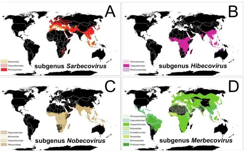
immunologically naïve to infection by viruses like SARS-CoV-2. Alpha andβ-CoVs have been detected in bats on most continents, sometimes with both types occurring in bats of the same species [58,68]. However, an exception to this pattern is the lack of published evidence thatβ -CoVs infect bats of temperate-zone North America, despite several search efforts which used methods suitable to detect bothα- andβ-CoVs [59,71,74,75]. Multiple novelα-CoVs have been detected and described in vespertilionid bats of the US and Canada, infecting species both living in close contact with humans and in remote wild areas [59,71,74–76]. However, SARSr-CoVs andβ-CoVs of the viral subgenusSarbecovirushave thus far been detected almost exclusively in species of the Old-World Chiropteran suborder Yinpterochiroptera (Fig 1A) [47,58,69]. The few exceptions to this pattern are the detection of novel Clade 3 and Clade 1 Sarbecovirus(sensu[53]) viruses in the wrinkle-lipped free-tailed bat (Mops plicatus, family Molossidae) in China [77] and the vespertilionid Leisler’s noctule (Nyctalus leisleri) cohabiting a Bulgarian cave during autumn with several species of rhinolophids in which other SARSrβ -CoVs were concurrently detected, suggesting cross-species infections (Fig 1A) [78]. Putative detections of a Clade 1Sarbecoviruswere also reported from guano samples of the
Fig 1. Global patterns of bats and associatedβ-CoVs. (A) Red-shaded distributions of bat species in which SARS-relatedβ-CoVs of the subgenusSarbecovirushave been detected; (B) pink-shaded distributions of bat species known to hostβ-CoVs of the subgenusHibecovirus; (C) brown-shaded distributions of bats in whichβ-CoVs of theNobecoviruslineage have been detected; and (D) green-shaded distributions of bats known to host MERS-relatedβ-CoVs of the subgenusMerbecovirus. Different colors and shade styles within each panel represent different families of bats. A data table that includes all known bat species associations for eachβ-CoVs subgenus and peer-reviewed citations is available at US Geological Survey data release https://doi.org/10.5066/P9U461PJ.Maps created using ArcMap (ESRI,Redlands,California, United States of America) and bat ranges derived from spatial data on terrestrial mammals from the International Union for the Conservation of Nature (IUCN 2020.The IUCN Red List of Threatened Species.January 2019 [version 6.2].https://www.iucnredlist.org; Downloaded on 11 April 2020).β-CoV, beta-coronavirus; MERS, Middle East respiratory syndrome; SARS, severe acute respiratory syndrome.
vespertilionid brown long-eared bat (Plecotus auritus) and the molossid European free-tailed bat (Tadarida teniotis) on Sardinia, where the same novelβ-CoV was described in the greater horseshoe bat (R.ferrumequinum) [79].
Viruses in theβ-CoV subgeneraHibecovirusandNobecovirusalso have been reported mostly from Old-World bat families Rhinolophidae, Hipposideridae, Rhinonycteridae, and Pteropodidae, except for novel viruses of the latter subgenus detected in four species of the ves-pertilionid genusScotophilusin Asia and Africa (Fig 1B and 1C) [47,58,69].
Batβ-CoVs of the subgenusMerbecovirus(MERS-related lineages) occur in a greater diver-sity of bat families and across more global regions than the other subgenera (Fig 1D) [47,58,
69]. These widely distributed MERS-like viruses can cause disease in humans (e.g., MERS) and notably appear to be the only batβ-CoVs to diversify among several families of the globally dis-tributed suborder Yangochiroptera (Fig 1D) [47,58,69].
Lack of evidence for
β
-CoVs in temperate-zone North American
bats
The several hundred species of extant bats spanning the Americas all belong to the suborder Yangochiroptera, which likely diverged from the Old-World suborder Yinpterochiroptera more than 50 million years ago (Fig 2) [80]. The onlyβ-CoVs detected in the Americas to date
Fig 2. Old-World and New-World bats. Overlapping species distribution outlines of bats in the globally distributed suborder Yangochiroptera (blue) and Old-World
Yinpterochiroptera (yellow).Maps created using ArcMap (ESRI,Redlands,California,USA) and bat ranges derived from spatial data on terrestrial mammals from the International Union for the Conservation of Nature Red List of Threatened Species,January 2019 [version 6.2].https://www.iucnredlist.org; Downloaded on 11 April 2020.
belong to the subgenusMerbecovirusand appear restricted to two exclusively Neotropical bat families (Phyllostomidae and Mormoopidae) and one that is globally distributed (Molossidae). Distinct CoV lineages in the subgenusMerbecoviruswere described from three species of Pter-onotus(family Mormoopidae), four species ofArtibeus, and Seba’s short-tailed bat (Carollia perspicillata; family Phyllostomidae) from tropical regions of Mexico [47,81]. Novelβ-CoVs of the subgenusMerbecoviruswere detected in two neotropical bat species of the family Molos-sidae: Wagner’s bonneted bat (Eumops glaucinus) in southern Brazil and the broad-eared free-tailed bat (Nyctinomops laticaudatus) in southern Mexico [81,82]. In vitro infections have shown that primary kidney cells from the Jamaican fruit-eating bat (Artibeus jamaicensis) can be infected with MERS-CoV, and virus replication and shedding was reported in experimen-tally infected bats of this species but without obvious clinical signs of disease [83]. Similar to the evidence for natural invasion of bat rabies viruses among New World bats [84], available evidence suggestsβ-CoVs may have arrived through South America and have long been evolv-ing in Neotropical bats. Although some bat hosts ofMerbecovirusesoverlap geographically with species of temperate-zone North American bats, none occur outside of the Neotropics. Sampling has been limited, but we are not aware of any published detections ofMerbecoviruses or any otherβ-CoVs in temperate-zone North American vespertilionid bats.
Our inference of true patterns of CoV occurrence and distribution in bat populations is limited by uneven global sampling. Yet SARSr-CoVs (Sarbecovirusspp.), a focus of many sur-veillance efforts, have been almost exclusively documented in Old-World Yinpterochiroptera. SARSr-CoVs were only found in the ultra-diverse and globally distributed bat suborder Yan-gochiroptera under conditions with plausible transmission from co-roostingRhinolophussp. bats [53,85]. This absence of evidence for SARS-likeβ-CoVs in yangochiropteran bats in gen-eral, and in temperate-zone vespertilionid bats of North America in particular, likely repre-sents a unique biogeographic pattern driven by underlying factors of host susceptibility or life history. These observations also point to the susceptibility of vespertilionid bats under circum-stances of SARSr-CoV environmental exposure and that they may not be naturally immune to these viruses.
infecting the more than two dozen species of bats in the US and Canada that hibernate to sur-vive harsh temperate-zone winters.
Proactively connecting the wellbeing of human and bat
populations
Scientists have long recognized the risk of pathogen spillover from humans to bats [94–96], but bat researchers in North America have not systematically addressed this risk prior to WNS. Outside of reservoir host studies, few bat researchers studied infectious diseases in bats before WNS emerged in 2007 [73] nor studied bat viruses (other than rabies) before bats were retrospectively connected to the SARS epidemic [15,66,97]. Fortunately, bat and wildlife dis-ease researchers recently began addressing these knowledge gaps in more detail [7,97,98]. Possible explanations for why bats might host particularly pathogenic viruses include charac-teristics of their life history (e.g., long-lived, wide ranging, multispecies aggregations, daily and seasonal heterothermy) [97], unique physiology for repairing their damaged DNA [99], unique ability to suppress some of their innate immunity pathways [100–105], high species diversity [48], and unmatched metabolic range and high body temperatures during flight [106]. Bats also cryptically come into close contact with humans, increasingly in urban and periurban set-tings as a result of native habitat loss, often crossing human–wildlife interfaces [107–113].
Except forLyssavirusinfections, bats rarely show substantial signs of sickness from the same pathogens that cause virulent disease in humans. Bats cope with viral infections in ways that we do not yet fully comprehend, but learning how they do so may reveal important insights to develop therapeutics and ultimately to protect human health [103–105]. In vitro and laboratory studies demonstrate that bats can specifically regulate naïve immunity path-ways to effectively cope with viral infection [114]. For example, dendritic cells generated from the bone marrow of the Egyptian rousette (Rousettus aegyptiacus) infected with Marburg virus down-regulate immune-stimulatory pathways and maturation of cells targeted by the virus while up-regulating pathogen-sensing pathways [115]. Unique bat immune regulation may occur with MERS-CoV infection, at least under experimental conditions [101]. Egyptian rou-sette bats experimentally challenged with SARS-CoV-2 by intranasal inoculation became tran-siently infected, shed virus, and one cohoused bat became infected but showed no clinical signs of disease other than rhinitis [116]. Our potential lack of understanding of clinical signs of illness in bats and the cryptic habits of many species also generally inhibit our ability to eas-ily detect spillover of pathogens from human to bat populations. This may add to uncertainty about cross-species transmission and dispersal of CoVs among human and animal communi-ties. Laboratory findings suggest human viruses that likely originated in bats, such as
Need for an interdisciplinary response
Effectively managing risks of human disease caused by emerging zoonotic pathogens and ensuring the health and conservation of wildlife species that are potential reservoirs of those disease agents can be synergistic goals under a One Health framework. Spillover risk (from or to wildlife) is often greatest in disturbed ecosystems where there is an elevated frequency of human–wildlife interactions or disruption of ecological patterns [3,120–124]. Thus, effective bat conservation and management requires understanding both pathogens that cause disease in bats, as well as human activities and ecological contexts that increase direct and indirect interactions with bats that could present health risks [2]. Furthermore, fear-based reactions to disease risk from wildlife, such as culling infected bat populations or indiscriminate killing, often have negative unintended consequences for the interconnected health of both humans and bats (e.g., culling of bats in a Uganda mine led to a more than doubling of Marburg virus prevalence in the bats living there) [30,125–127]. Temperate-zone vespertilionid bats inhabit-ing human dwellinhabit-ings in the US and Canada represent a particularly relevant human–wildlife interface, in which conservation and management actions to proactively address the potential consequences for pathogen spillover are worth careful consideration [73].
Conservation-compatible surveillance of bat viruses has demonstrated the potential for mutually beneficial collaboration between public health scientists and conservation stakehold-ers [94,113,125,128,129]. Disease-focused studies that integrate ecological principles into a rigorous study design provide the most informative context to interpret bat–virus associations and patterns of richness globally [130–132]. Assessing the risks of SARS-CoV-2 spillover into North American bats presents a timely opportunity to form multidisciplinary scientific teams that include experts on emerging infectious diseases and ecologists with expertise on North American bats [128]. Scientists researching emerging infectious diseases can benefit from sam-pling opportunities and methods that bat researchers have developed for observing, counting, and noninvasively sampling bats [73,133]. Bat researchers can learn about human and animal health monitoring and supporting laboratory methods, including biosafety, secure handling/ transport of CoV-positive samples, and training in the proper use of personal protective equip-ment (PPE) from professionals with expertise in veterinary and medical sciences [113,131,
134,135]. A shared goal of all stakeholders is to identify and implement simple, widely avail-able diagnostic tests for detecting SARS-CoV-2 infection that are species-independent, practi-cal for field and laboratory use, highly specific and sensitive, and that do not require strict biosafety containment [136]. All investigators can also work together to develop mutually ben-eficial goals, such as joint risk communications to the public with effective and balanced mes-saging about bat populations and higher risk activities for human–bat contact.
optimal PPE, including bidirectional N95 or equivalent masks, along with effective risk com-munication and adherence to other basic biosafety practices [134,141,142] during field work, can significantly reduce the transmission risk of SARS-CoV-2 from humans to bats. In the interim, until new guidelines are established for handling and for close-proximity work with bats, we have outlined gaps in our understanding of SARS-CoV-2 spillover risks at the inter-face between humans, domesticated animals, and free-ranging wildlife. Temporarily shifting to “hands-off” bat research methods also seems prudent, wherever possible, and could facilitate ongoing work with reduced risk.
Examples of “hands-off” research strategies
Multiple research strategies that do not involve close contact with free-ranging bats already exist for addressing critical gaps in understanding CoV diversity, distribution, evolution, and potential health effects in temperate-zone bats. For example, a combination of host-cell recep-tor analyses and in vitro and in vivo experimental infections across a diversity of bat and other mammalian species have helped inform potential host range expansion for SARS-CoV-2. The receptors that many CoVs use to gain access to host cells, such as angiotensin-converting enzyme 2 (ACE2) and dipeptidyl peptidase-4 (DPP4/CD26), have undergone positive selection in bats, resulting in diverse and recombinant CoV strains [72,143]. These strains can likely bind to numerous variants of a host receptor protein and facilitate spillover into other animal species [72,144]. SARS-CoV-2 targets and strongly binds to mammalian ACE2 cell receptors [72,145,146]. Beta-CoVs of the subgenusMerbecovirus(like those known to occur in the Americas) are not known to target ACE2 cell receptors, instead using as a receptor DPP4/ CD26 or possibly other receptors [53,144]. Current in silico predictions that bats will likely have low susceptibility to SARS-CoV-2 based on ACE2 structural analyses conflict with in vitro evidence and do not comprehensively account for ACE2 amino acid sequence variation (including intraspecific variation) that occurs within bats [17,72,145]. Assessing SARS-CoV-2 host range will require additional virus-host receptor binding assays in silico and in vitro [17,
53,72,144,145], together with future experimental infection studies for confirmation of Koch’s postulates. In addition, in vitro studies could evaluate species variability in innate immune responses. These investigations will help quantify the potential for North American bat infection and transmission among free-ranging populations.
Developing simple management tools and methods for rapidly assessing risks of virus spillover from humans to wildlife, while maintaining scientific rigor, could also help with future disease response. It might also be useful to prepare a suite of tools, protocols, and risk communication strategies for natural resource managers and public health officials to immediately deploy while risks are being assessed. Such prepared management resources could include public out-reach material and guidelines for enhanced use of PPE for those in closest contact with poten-tially susceptible wildlife.
Conclusion
Many questions remain about the risk of SARS-CoV-2 to naïve wildlife populations, the influ-ences of human behavior on those risks, and the potential for establishment of new CoV reser-voirs. Cross-species virus transmission events are relatively rare, requiring an infectious reservoir host to be in contact with a recipient host when conditions concurrently favor sus-ceptibility and onward transmission [12,113,114]. The currently unknown, but possible and potentially high-consequence, risk of SARS-CoV-2 transmission and establishment in North American bats (or other free-ranging mammals) warrants precaution [116,140]. Strategically managing interactions between people and potentially susceptible or at risk species can decrease the probability of cross-species virus spillover [113]. Humans that frequently handle and come into close contact with North American temperate-zone bats, such as bat research-ers, wildlife rehabilitators, wildlife/pest control workresearch-ers, and disease investigators, can help decrease any chances of spillover by adopting basic PPE and biosafety practices and carefully evaluating how their actions might adversely affect bat populations. We are at a critical nexus of biosecurity and natural resource conservation that will require ingenuity and diligence to continue important research on bats whilst simultaneously evaluating the ecological future of SARS-CoV-2. Our actions during this current pandemic could profoundly influence and pro-tect the health of both humans and wildlife in North America.
Supporting information
S1 Table. Global patterns of betacoronavirus (β-CoV) associations in bats. The table lists bat species in which betacoronaviruses (β-CoVs) were detected, organized by viral subgenera and clade (for Sarbecorviruses), bat family, bat suborder, and general global region where the species of bat occurs. Reference to the published literature sources of information for each row are listed in the last column. Provided in comma-separated value (.csv) format athttps://doi. org/10.5066/P9U461PJ.
(XLSX)
Acknowledgments
We thank Thomas O’Shea, Brian Reichert, Michelle Verant, Richard “Chip” Clark III, Marcos Gorresen, Jill Baron, and Daniel Becker for helpful comments on earlier drafts of this
manuscript.
References
1. Daszak P, Cunningham AA, Hyatt A. Anthropogenic environmental change and the emergence of infectious diseases in wildlife. Acta Trop. 2001; 78(2):103–16.https://doi.org/10.1016/s0001-706x(00) 00179-0PMID:11230820
2. Plowright RK, Parrish CR, McCallum H, Hudson PJ, Ko AI, Graham AL, et al. Pathways to zoonotic spillover. Nat Rev Microbiol. 2017; 15(8):502–10.https://doi.org/10.1038/nrmicro.2017.45PMID: 28555073
3. Allen T, Murray KA, Zambrana-Torrelio C, Morse SS, Rondinini C, Di Marco M, et al. Global hotspots and correlates of emerging zoonotic diseases. Nature Communications. 2016; 8:1124.https://doi.org/ 10.1038/s41467-017-00923-8.
4. Jones KE, Patel NG, Levy MA, Storeygard A, Balk D, Gittleman JL, et al. Global trends in emerging infectious diseases. Nature. 2008; 451:990–3.https://doi.org/10.1038/nature06536PMID:18288193 5. Han BA, Kramer AM, Drake JM. Global patterns of zoonotic disease in mammals. Trends Parasitol.
2016; 32(7):565–77.https://doi.org/10.1016/j.pt.2016.04.007PMID:27316904
6. Luis AD, O’Shea TJ, Hayman DTS, Wood JLN, Cunningham AA, Gilbert AT, et al. Network analysis of host–virus communities in bats and rodents reveals determinants of cross-species transmission. Ecol Lett. 2015; 18:1153–62.https://doi.org/10.1111/ele.12491PMID:26299267
7. Olival KJ, Hosseini PR, Zambrana-Torrelio C, Ross N, Bogich TL, Daszak P. Host and viral traits pre-dict zoonotic spillover from mammals. Nature. 2017;646–650(546).https://doi.org/10.1038/ nature22975.
8. Schrenzel MD, Tucker TA, Stalis IH, Kagan RA, Burns RP, Denison AM, et al. Pandemic (H1N1) 2009 virus in 3 wildlife species, San Diego, California, USA. Emerging Infectious Diseases. 2011; 17 (4):747–9.https://doi.org/10.3201/eid1706.101355PMID:21470480
9. Messenger A, Barnes A, Gray GC. Reverse zoonotic disease transmission (Zooanthroponosis): a sys-tematic review of seldom-documented human and biological threats to animals. PLoS ONE. 2014; 9 (2):e89055.https://doi.org/10.1371/journal.pone.0089055PMID:24586500
10. Anthony SJ, Epstein JH, Murray KA, Navarrete-Macias I, Zambrana-Torrelio CM, Solovyov A, et al. A strategy to estimate known viral diversity in mammals. mBio. 2013; 4(5):1–15.https://doi.org/10.1128/ mBio.00598-13.
11. Esona MD, Mijatovic-Rustempasic S, Conrardy C, Tong S, Kuzmin IV, Agwanda B, et al. Reassort-ment group A rotavirus from straw-colored fruit bat (Eidolon helvum). Emerging Infectious Diseases. 2010; 16(12):1844–52.https://doi.org/10.3201/eid1612.101089PMID:21122212
12. Wasik BR, de Wit E, Munster V, Lloyd-Smith JO, Martinez-Sobrido L, Parrish CR. Onward transmis-sion of viruses: how do viruses emerge to cause epidemics after spillover? Philosophical Transactions of the Royal Society B. 2019; 374(20190017).http://dx.doi.org/10.1098/rstb.2019.0017.
13. Huang C, Wang Y, Li X, Zhaou J, Hu Y, Zhang L, et al. Clinical features of patients infected with 2019 novel coronavirus in Wuhan, China. The Lancet. 2020; 395:497–506. https://doi.org/10.1016/S0140-6736(20)30183-5.
14. Ge X, Li J, Yang X, Chmura AA, Zhu G, Epstein JH, et al. Isolation and characterization of a bat SARS-like coronavirus that uses the ACE2 receptor. Nature. 2013; 503:535–8.https://doi.org/10. 1038/nature12711PMID:24172901
15. Li W, Shi Z, Yu M, Ren W, Smith C, Epstein JH, et al. Bats are natural reservoirs of SARS-like corona-viruses. Science. 2005; 310:676–9.https://doi.org/10.1126/science.1118391PMID:16195424 16. Guan Y, Zheng BJ, He YQ, Liu XL, Zhuang ZX, Cheung CL, et al. Isolation and characterization of
viruses related to the SARS coronavirus from animals in southern China. Science. 2003; 302 (5643):276–8.https://doi.org/10.1126/science.1087139PMID:12958366
17. Zhou P, Yang X, Wang X, Hu B, Zhang L, Zhang W, et al. A pnemonia outbreak associated with a new coronavirus of probable bat origin. Nature. 2020; 579:270–3. https://doi.org/10.1038/s41586-020-2012-7PMID:32015507
18. Andersen KG, Rambaut A, Lipkin WI, Holmes EC, Garry RF. The proximal origin of SARS-CoV-2. Nat Med. 2020.https://doi.org/10.1038/s41591-020-0820-9.
19. Boni MF, Lemey P, Jiang X, Lam TT, Perry B, Castoe T, et al. Evolutionary origins of the SARS-CoV-2 sarbecovirus lineage responsible for the COVID-19 pandemic. bioRxiv. 2020.https://doi.org/10.1101/ 2020.03.30.015008.
21. Huang C, Liu WJ, Xu W, Jin T, Zhao Y, Song J, et al. A bat-derived putative cross-family recombinant coronavirus with a reovirus gene. PLoS Pathog. 2016; 12(9):e1005883.https://doi.org/10.1371/ journal.ppat.1005883PMID:27676249
22. Horan RD, Fenichel EP, Wolf CA, Graming BM. Managing infectious animal disease systems. Annual Review of Resource Economics. 2010; 2(1):101–24.https://doi.org/10.1146/annurev.resource. 012809.103859.
23. Kunz TH, Braun de Torrez E, Bauer D, Lobova T, Fleming TH. Ecosystem services provided by bats. Ann N Y Acad Sci. 2011; 1223:1–38.https://doi.org/10.1111/j.1749-6632.2011.06004.xPMID: 21449963
24. Maine JJ, Boyles JG. Bats initiate vital agroecological interactions in corn. Proc Natl Acad Sci USA. 2015; 112(40):12438–43.https://doi.org/10.1073/pnas.1505413112PMID:26371304
25. Blehert DS, Hicks AC, Behr M, Meteyer CU, Berlowski-Zier BM, Buckles EL, et al. Bat white-nose syn-drome: an emerging fungal pathogen? Science. 2009; 323:227.https://doi.org/10.1126/science. 1163874PMID:18974316
26. Lorch JM, Meteyer CU, Behr MJ, Boyles JG, Cryan PM, Hicks AC, et al. Experimental infection of bats with Geomyces destructans causes white-nose syndrome. Nature. 2011; 480:376–8.https://doi.org/ 10.1038/nature10590PMID:22031324
27. Warnecke L, Turner JM, Bollinger TK, Lorch JM, Misra V, Cryan PM, et al. Inoculation of bats with European Geomyces destructans supports the novel pathogen hypothesis for the origin of white-nose syndrome. Proc Natl Acad Sci USA. 2012; 109:6999–7003.https://doi.org/10.1073/pnas.1200374109 PMID:22493237
28. Frick WF, Puechmaille SJ, Hoyt JR, Nickel BA, Langwig KE, Foster JT, et al. Disease alters macroeco-logical patterns of North American bats. Global Ecol Biogeogr. 2015; 24(7):741–479.https://doi.org/ 10.1111/geb.12290.
29. Drees KP, Lorch JM, Puechmaille SJ, Parise KL, Wibbelt G, Hoyt JR, et al. Phylogenetics of a fungal invasion: origins and widespread dispersal of white-nose syndrome. mBio. 2017; 8:e01941–17. https://doi.org/10.1128/mBio.01941-17PMID:29233897
30. O’Shea TJ, Cryan PM, Hayman DTS, Plowright RK, Streicker DG. Multiple mortality events in bats: a global review. Mamm Rev. 2016.https://doi.org/10.1111/mam.12064.
31. Kemenesi G, Kurucz K, Dallos B, Zana B, Fo¨ldes F, Boldogh S, et al. Re-emergence of Lloviu virus in Miniopterus schreibersii bats, Hungary, 2016. Emerging Microbes & Infections. 2018; 7(66):1–4.
https://doi.org/10.1038/s41426-018-0067-4.
32. Langwig KE, Voyles J, Wilber MQ, Frick WF, Murray KA, Bolker BM, et al. Context-dependent conser-vation responses to emerging wildlife diseases. Front Ecol Environ. 2015; 13(4):195–202.https://doi. org/10/f7bcq2.
33. Frick WF, Cheng TL, Langwig KE, Hoyt JR, Janicki AF, Parise KL, et al. Pathogen dynamics during invasion and establishment of white-nose syndrome explain mechanisms of host persistence. Ecol-ogy. 2017; 98(3):624–31.https://doi.org/10.1002/ecy.1706PMID:27992970
34. Frick WF, Puechmaille SJ, Willis CKR. White-nose syndrome in bats. In: Voigt CC, Kingston T, editors. Bats in the Anthropocene: Conservation of bats in a changing world: Springer; 2016. p. 245–62. 35. Cryan PM, Meteyer CU, Boyles JG, Blehert DS. White-nose syndrome in bats: illuminating the
dark-ness. BMC Biology. 2013; 11:47.https://doi.org/10.1186/1741-7007-11-47PMID:23587401 36. Zukal J, Bandouchova H, Brichta J, Cmokova A, Jaron KS, Kolarik M, et al. White-nose syndrome
without borders: Pseudogymnoascus destructans infection tolerated in Europe and Palearctic Asia but not in North America. Scientific Reports. 2016; 6:19829.https://doi.org/10.1038/srep19829PMID: 26821755
37. Hoyt JR, Langwig KE, Sun K, Parise KL, Li A, Wang Y, et al. Environmental reservoir dynamics predict global infection patterns and population impacts for the fungal disease white-nose syndrome. Pro-ceedings of the National Academy of Sciences. 2020; 117(13):7255.https://doi.org/10.1073/pnas. 1914794117.
38. Arita HT, Vargas-Baro´n J, Villalobos F. Latitudinal gradients of genus richness and endemism and the diversification of New World bats. Ecography. 2014; 37:1024–33.https://doi.org/10.1111/ecog.00720 39. Peixoto FF, Braga PHP, Mendes P. A synthesis of ecological and evolutionary determinants of bat
diversity across spatial scales. BMC Ecol. 2018; 18(18).https://doi.org/10.1186/s12898-018-0174-z. 40. Van Den Bussche RA, Hoofer SR. Phylogenetic releationships among recent Chiropteran families and
importance of choosing appropriate out-group taxa. Journal of Mammalogy. 2004; 85(2):321–30. https://doi.org/10.1644/1545-1542(2004)085<0321:PRARCF>2.0.CO;2.
42. Baker RR. The evolutionary ecology of animal migration. New York: Holmes & Meier Publishers; 1978. 1012 p.
43. Fleming TH, Eby P. Ecology of bat migration. In: Kunz TH, Fenton MB, editors. Bat ecology. Chicago: The University of Chicago Press; 2003. p. 156–208.
44. Davy CM, Donaldson ME, Subudhi S, Rapin N, Warnecke L, Turner JM, et al. White-nose syndrome is associated with increased replication of a naturally persisting coronaviruses in bats. Scientific Reports. 2018; 8(15508).https://doi.org/10.1002/ece3.3234.
45. Plowright RK, Field HE, Smith C, Divljan A, Palmer C, Tabor G, et al. Reproduction and nutritional stress are risk factors for Hendra virus infection in little red flying foxes (Pteropus scapulatus). Pro-ceedings of the Royal Society B. 2008; 275:861–9.https://doi.org/10.1098/rspb.2007.1260PMID: 18198149
46. Simmons NB, Cirranello AL. Bat Species of the World: A taxonomic and geographic database. 2020 [17 April 2020]. Available from:https://www.batnames.org/.
47. Anthony SJ, Johnson CK, Greig DJ, Kramer S, Che X, Wells H, et al. Global patterns in coronavirus diversity. Virus Evolution. 2017; 3(1):vex012.https://doi.org/10.1093/ve/vex012PMID:28630747 48. Mollentze N, Streicker DG. Viral zoonotic risk is homogenous among taxonomic orders of mammalian
and avian reservoir hosts. Proc Natl Acad Sci USA. 2020. Epub 13 April 2020.www.pnas.org/cgi/doi/ 10.1073/pnas.1919176117.
49. Hu D, Zhu C, Wang Y, Ai L, Yang LQ, Ye F, et al. Virome analysis for identification of novel mammalian viruses in bats from southeast China. Scientific Reports. 2017; 7:10917.https://doi.org/10.1038/ s41598-017-11384-wPMID:28883450
50. Cheng VCC, Lau SKP, Woo PCY, Yuen KY. Severe acute respiratory syndrome coronavirus as an agent of emerging and reemerging infection. Clinical Microbiology Reviews. 2007; 20(4):660–94. https://doi.org/10.1128/CMR.00023-07PMID:17934078
51. Cui J, Li F, Shi Z. Origin and evolution of pathogenic coronaviruses. Nat Rev Microbiol. 2019; 17:181– 92.https://doi.org/10.1038/s41579-018-0118-9PMID:30531947
52. Fan Y, Zhao K, Shi Z, Zhou P. Bat coronaviruses in China. Viruses. 2019; 11(210):1–11.https://doi. org/10.3390/v11030210.
53. Lu R, Zhao X, Li J, Niu P, Yang B, Wu H, et al. Genomic characterisation and epidemiology of 2019 novel coronavirus: implications for virus origins and receptor binding. The Lancet. 2020. Epub January 29, 2020.https://doi.org/10.1016/S0140-6736(20)30251-8.
54. Zhao G. SARS molecular epidemiology: a Chinese fairy tale of controlling an emerging zoonotic dis-ease in the genomics era. Royal Society Philosophical Transactions Biological Sciences. 2007; 362 (1482):1063–81.https://doi.org/10.1098/rstb.2007.2034.
55. Dong BQ, Liu W, Fan XH, Vijaykrishna D, Tang XC, Gao F, et al. Detection of a novel and highly diver-gent coronavirus from Asian leopard cats and Chinese ferret badgers in southern China. Journal of Virology. 2007; 81(13):6920–6.https://doi.org/10.1128/JVI.00299-07PMID:17459938
56. Shi J, Wen Z, Zhong G, Yang H, Wang C, Liu R, et al. Susceptibility of ferrets, cats, dogs, and different domestic animals to SARS-coronavirus-2. Science. 2020:1–23.https://doi.org/10.1126/science. abb7015.
57. Zhou P, Fan H, Lan T, Yang X, Shi W, Zhang W, et al. Fatal swine acute diarrhoea syndrome caused by an HKU2-related coronavirus of bat origin. Nature. 2018; 5556:255–8.https://doi.org/10.1038/ s41586-018-0010-9.
58. Drexler JF, Corman VM, Drosten C. Ecology, evolution and classification of bat coronaviruses in the aftermath of SARS. Antiviral Res. 2014; 101:45–56.https://doi.org/10.1016/j.antiviral.2013.10.013 PMID:24184128
59. Huynh J, Li S, Yount B, Smith A, Sturges L, Olsen JC, et al. Evidence supporting a zoonotic origin of human coronavirus strain NL63. Journal of Virology. 2012; 86(23):12818–25.https://doi.org/10.1128/ JVI.00906-12.
60. Vijaykrishna D, Smith GJD, Zhang JX, Peiris JSM, Chen H, Guan Y. Evolutionary insights into the ecology of coronaviruses. Journal of Virology. 2007; 81(8):4012–20.https://doi.org/10.1128/JVI. 02605-06PMID:17267506
61. Hall RJ, Wang J, Peacey M, Moore NE, McInnes K, Tompkins DM. New alphacoronavirus in Mysta-cina tuberculata bats, New Zealand. Emerging Infectious Diseases. 2014; 20(4):697–700.https://doi. org/10.3201/eid2004.131441PMID:24656060
62. Woo PCY, Lau SKP, Huang Y, Yuen K-Y. Coronavirus diversity, phylogeny and interspecies jumping. Exp Biol Med. 2009; 234(10):1117–27.https://doi.org/10.3181/0903-MR-94.
64. Huang Y, Dickerman AW, Piñeyro P, Li L, Fang L, Kiehne R, et al. Origin, evolution, and genotyping of emergent porcine epidemic diarrhea virus strains in the United States. mBio. 2013; 4(5):e00737–13. https://doi.org/10.1128/mBio.00737-13PMID:24129257
65. Corman VM, Baldwin HJ, Tateno AF, Zerbinati RM, Annan A, Owusu M, et al. Evidence for an ansces-tral association of human coronavirus 229E with bats. Journal of Virology. 2015; 89(23):11858–70. https://doi.org/10.1128/JVI.01755-15PMID:26378164
66. Lau SKP, Woo PCY, Li KSM, Huang Y, Tsoi H, Wong BHL, et al. Severe acute respiratory syndrome coronavirus-like virus in Chinese horseshoe bats. Proc Natl Acad Sci USA. 2005; 102(39):14040–5. https://doi.org/10.1073/pnas.0506735102PMID:16169905
67. Yip CW, Hon CC, Shi M, lam TT, Chow KY, Zeng F, et al. Phylogenetic perspectives on the epidemiol-ogy and origins of SARS and SARS-like coronaviruses. Infect, Genet Evol. 2009; 9:1185–96.https:// doi.org/10.1016/j.meegid.2009.09.015.
68. Wong S, Lau S, Woo P, Yuen KY. Bats as a continuing source of emerging infections in humans. Rev Med Virol. 2007; 17(2):67–91.https://doi.org/10.1002/rmv.520PMID:17042030
69. Wong ACP, Li X, Lau SKP, Woo PCY. Global epidemiology of bat coronaviruses. Viruses. 2019; 11 (174).https://doi.org/10.3390/v11020174.
70. Albery GF, Eskew EA, Ross N, Olival KJ. Predicting the global mammalian viral sharing network using phylogeography. Nature Communications. 2020; 11(2260). https://doi.org/10.1038/s41467-020-16153-4.
71. Osborne C, Cryan P, O’Shea TJ, Oko LM, Ndaluka C, Calisher CH, et al. Alphacoronaviruses in New World bats: prevalence, persistence, phylogeny, and potential for interaction with humans. PLoS ONE. 2011; 6(5):e19156.https://doi.org/10.1371/journal.pone.0019156PMID:21589915 72. Damas J, Hughes GM, Keough KC, Painter CA, Persky NS, Corbo M, et al. Broad host range of
SARS-CoV-2 predicted by comparative and structural analysis of ACE2 in vertebrates. bioRxiv. 2020. https://doi.org/10.1101/2020.04.16.045302.
73. Weller TJ, Cryan PM, O’Shea TJ. Broadening the focus of bat conservation and research in the USA for the 21st century. Endangered Species Research. 2009; 8:129–45.https://doi.org/10.3354/ esr00149.
74. Misra V, Dumonceaux T, Dubois J, Willis C, Nadin-Davis S, Severini A, et al. Detection of polyoma and corona viruses in bats of Canada. J Gen Virol. 2009; 90:2015–22.https://doi.org/10.1099/vir.0. 010694-0PMID:19357225
75. Donaldson EF, Haskew AN, Gates JE, Huynh J, Moore CJ, Frieman MB. Metagenomic analysis of the viromes of three North American bat species: viral diversity among different bat species that share a common habitat. Journal of Virology. 2010; 84(24):13004–18.https://doi.org/10.1128/JVI.01255-10 PMID:20926577
76. Dominguez SR, O’Shea TJ, Oko LM, Holmes KV. Detection of group 1 coronaviruses in bats in North America. Emerging Infectious Diseases. 2007; 13(9):1295–300.https://doi.org/10.3201/eid1309. 070491PMID:18252098
77. Yang L, Wu Z, Ren X, Yang F, He G, Zhang JX, et al. Novel SARS-like betacoronaviruses in bats, China, 2011. Emerging Infectious Diseases. 2013; 19(6):989–91.https://doi.org/10.3201/eid1906. 121648PMID:23739658
78. Drexler JF, Gloza-Rausch F, Glende J, Corman VM, Muth D, Goettsche M, et al. Genomic characteri-zation of severe acute respiratory syndrome-related coronavirus in European bats and classification of coronaviruses based on partial RNA-dependent RNA polymerase gene sequences. Journal of Virol-ogy. 2010; 84(21):11336–49.https://doi.org/10.1128/JVI.00650-10PMID:20686038
79. Lecis R, Mucedda M, Pidinchedda E, Pittau M, Alberti A. Molecular identification of Betacoronavirus in bats from Sardinia (Italy): first detection and phylogeny. Virus Genes. 2019; 55(1):60–7.https://doi. org/10.1007/s11262-018-1614-8PMID:30426315
80. Teeling EC, Springer MS, Madsen O, Bates P, O’Brien SJ, Murphy WJ. A molecular phylogeny for bats illuminates biogeography and the fossil record. Science. 2005; 307:580–4.https://doi.org/10. 1126/science.1105113PMID:15681385
81. Anthony SJ, Ojeda-Flores R, Rico-Cha´vez O, Navarrete-Macias I, Zambrana-Torrelio C, Rostal MK, et al. Coronaviruses in bats from Mexico. J Gen Virol. 2013; 94:1028–38.https://doi.org/10.1099/vir.0. 049759-0PMID:23364191
83. Munster VJ, Adney DR, van Doremalen N, Brown VR, Miazgowicz KL, Milne-Price S, et al. Replication and shedding of MERS-CoV in Jamaican fruit bats (Artibeus jamaicensis). Scientific Reports. 2016; 6:21878.https://doi.org/10.1038/srep21878PMID:26899616
84. Hayman DTS, Fooks AR, Marston DA, Garcia-R JC. The global phylogeography of lyssaviruses— challenging the ’out of Africa’ hypothesis. PLoS Negl Trop Dis. 2016; 10(12):e0005266.https://doi.org/ 10.1371/journal.pntd.0005266PMID:28036390
85. Tao Y, Tong S. Complete genome sequence of a severe acute respiratory syndrome-related coronavi-rus from Kenyan bats. Microbiology Resource Announcements. 2019; 8:e00548–19.https://doi.org/ 10.1128/MRA.00548-19PMID:31296683
86. Barbour RW, Davis WH. Bats of America. Lexington: The University Press of Kentucky; 1969. 286 p. 87. George DB, Webb CT, Farnsworth ML, O’Shea TJ, Bowen RA, Smith DL, et al. Host and viral ecology determine bat rabies seasonality and maintenance. Proc Natl Acad Sci USA. 2011; 108(25):10208– 13.https://doi.org/10.1073/pnas.1010875108PMID:21646516
88. Davis AD, Morgan SM, Dupuis M, Poulliott CE, Jarvis JA, Franchini R, et al. Overwintering of rabies virus in silver haired bats (Lasionycteris noctivagans). PLoS ONE. 2016; 11(5):e0155542.https://doi. org/10.1371/journal.pone.0155542PMID:27195489
89. Subudhi S, Rapin N, Bollinger TK, Hill JE, Donaldson ME, Davy CM, et al. A persistently infecting coro-navirus in hibernating Myotis lucifugus, the North American little brown bat. J Gen Virol. 2017; 98:2297–309.https://doi.org/10.1099/jgv.0.000898PMID:28840816
90. Hayman DTS, Cryan PM, Fricker PD, Dannemiller NG. Long-term video surveillance and automated analyses reveal arousal patterns in groups of hibernating bats. Methods in Ecology and Evolution. 2017; 8(12):1813–21.https://doi.org/10.1111/2041-210X.12823.
91. Speakman JR, Thomas DW. Physiological ecology and energetics of bats. In: Kunz TH, Fenton MB, editors. Bat ecology. Chicago: University of Chicago Press; 2003. p. 430–90.
92. Mollentze N, Streicker DG, Murcia PM, Hampson K, Biek R. Dynamics of viral index infections in novel hosts. bioRxiv. 2020.https://doi.org/10.1101/2020.04.09.033928.
93. Morimoto K, Patel M, Corisdeo S, Hooper DC, Fu ZF, Rupprecht CE, et al. Characterization of a unique variant of bat rabies virus responsible for newly emerging human cases in North America. Pro-ceedings of the National Academy of Science. 1996; 93:5653–8.https://doi.org/10.1073/pnas.93.11. 5653.
94. Epstein JH, Price JT. The significant but understudied impact of pathogen transmission from humans to animals. Mount Sinai Journal of Medicine. 2009; 76:448–55.https://doi.org/10.1002/msj.20140 PMID:19787650
95. Constantine DG. Disease exchange between bats and researchers: problems and precautions. Aust Mammal. 1985; 8(325–329).
96. Sulkin SE, Allen R. Virus infections in bats. Monographs in Virology. 1974; 8:1–103. PMID:4367453 97. Calisher CH, Childs JE, Field HE, Holmes KV, Schountz T. Bats: an important reservoir host of
emerg-ing viruses. Clinical Microbiology Reviews. 2006; 19(3):531–45. https://doi.org/10.1128/CMR.00017-06PMID:16847084
98. Luis AD, Hayman DTS, O’Shea TJ, Cryan PM, Gilbert AT, Pulliam JRC, et al. A comparison of bats and rodents as reservoirs of zoonotic viruses: are bats special? Proceedings of the Royal Society B. 2013; 280(1756):20122753.https://doi.org/10.1098/rspb.2012.2753PMID:23378666
99. Zhang G, Cowled C, Shi Z, Huang Z, Bishop-Lilly KA, Fang X, et al. Comparative analysis of bat genomes provides insight into the evolution of flight and immunity. Science. 2013; 339(6118):456–60. https://doi.org/10.1126/science.1230835PMID:23258410
100. Xie J, Li Y, Shen X, Goh G, Zhu Y, Cui J, et al. Dampened STING-dependent interferon activation in bats. Cell Host & Microbe. 2018; 23:1–5.https://doi.org/10.1016/j.chom.2018.01.006.
101. Ahn M, Anderson DE, Zhang Q, Tan CW, Lim BL, Luko K, et al. Dampened NLRP3-mediated inflam-mation in bats and implications for a special viral reservoir host. Nature Microbiology. 2019.https://doi. org/10.1038/s41564-019-0371-3.
102. Zhou P, Tachedjian M, Wynne JW, Boyd V, Cui J, Smith I, et al. Contraction of the type I IFN locus and unusual constitutive expression of IFN-αin bats. Proc Natl Acad Sci USA. 2016; 113(10):2696–701. https://doi.org/10.1073/pnas.1518240113PMID:26903655
103. Banerjee A, Baker ML, Kulcsar K, Misra V, Plowright R, Mossman K. Novel insights into immune sys-tems of bats. Frontiers in Immunology. 2020; 11(26).https://doi.org/10.3389/fimmu.2020.00026. 104. Mandl JN, Schneider C, Schneider DS, Baker ML. Going to bat(s) for studies of disease tolerance.
105. Subudhi S, Rapin N, Misra V. Immune system modulation and viral persistence in bats: understanding viral spillover. Viruses 2019 11(192).https://doi.org/10.3390/v11020192.
106. O’Shea TJ, Cryan PM, Cunningham AA, Fooks AR, Hayman DTS, Luis AD, et al. Bat flight and zoo-notic viruses. Emerging Infectious Diseases. 2014; 20(5):741–5.https://doi.org/10.3201/eid2005. 130539PMID:24750692
107. O’Shea TJ, Neubaum DJ, Neubaum MA, Cryan PM, Ellison LE, Stanley TR, et al. Bat ecology and public health surveillance for rabies in an urbanizing region of Colorado. Urban Ecosystems. 2011; 14:665–97.https://doi.org/10.1007/s11252-011-0182-7.
108. Salah Uddin Kahn M, Hossain J, Gurley ES, Nahar N, Sultana R, Luby SP. Use of infrared camera to understand bats’ access to date palm sap: implications for preventing Nipah virus transmission. Eco-Health. 2011; 7:517–25.https://doi.org/10.1007/s10393-010-0366-2.
109. Gilbert AT, Petersen BW, Recuenco S, Niezgoda M, Go´mez J, Laguna-Torres VA, et al. Evidence of rabies virus exposure among humans in the Peruvian Amazon. The American Journal of Tropical Med-icine and Hygiene. 2012; 87(2):206–15.https://doi.org/10.4269/ajtmh.2012.11-0689PMID:22855749 110. Kuzmin IV, Shi M, Orciari LA, Yager PA, Velasco-Villa A, Kuzmina NA, et al. Molecular inferences sug-gest multiple host shifts of rabies viruses from bats to mesocarnivores in Arizona during 2001–2009. PLoS Pathog. 2012; 8(6).https://doi.org/10.1371/journal.ppat.1002786.
111. Messenger SL, Smith JS, Rupprecht CE. Emerging epidemiology of bat-associated cryptic cases of rabies in humans in the United States. Clin Infect Dis. 2002; 35(6):738–47.https://doi.org/10.1086/ 342387PMID:12203172
112. Moran D, Juliao P, Alvarez D, Lindblade KA, Ellison JA, Gilbert AT, et al. Knowledge, attitudes and practices regarding rabies and exposure to bats in two rural communities in Guatemala. BMC Research Notes. 2015; 8(1):955.https://doi.org/10.1186/s13104-014-0955-1.
113. Plowright RK, Eby P, Hudson PJ, Smith IL, Westcott D, Bryden WL, et al. Ecological dynamics of emerging bat virus spillover. Proceedings of the Royal Society B. 2015; 282:20142124.https://doi.org/ 10.1098/rspb.2014.2124PMID:25392474
114. Brook CE, Boots M, Chandran K, Dobson AP, Drosten C, Graham AL, et al. Accelerated viral dynam-ics in bat cell lines, with implications for zoonotic emergence. eLife. 2020; 9(e48401).https://doi.org/ 10.7554/eLife.48401.
115. Prescott J, Guito JC, Spengler JR, Arnold CE, Schuh AJ, Amman BR, et al. Rousette bat dendritic cells overcome Marburg virus-mediated antiviral responses by upregulation of interferon-related genes while downregulating proinflammatory disease mediators. mSphere. 2019; 4(6):e00728–19. https://doi.org/10.1128/mSphere.00728-19PMID:31801842
116. Schlottau K, Rissmann M, Graaf A, Scho¨n J, Sehl J, Wylezich C, et al. Experimental transmission studies of SARS-CoV-2 in fruit bats, ferrets, pigs and chickens. The Lancet. 2020.https://dx.doi.org/ 10.2139/ssrn.3578792.
117. Denison MR, Graham RL, Donaldson EF, Eckerle LD, Baric RS. Coronaviruses: an RNA proofreading machine regulates replication fidelity and diversity. RNA Biology. 2011; 8(2):270–9.https://doi.org/10. 4161/rna.8.2.15013PMID:21593585
118. Gorbalenya AE, Enjuanes L, Ziebuhr J, Snijder EJ. Nidovirales: evolving the largest RNA virus genome. Virus Research. 2006; 117:17–37.https://doi.org/10.1016/j.virusres.2006.01.017PMID: 16503362
119. Menachery VD, Graham RL, Baric RS. Jumping species-a mechanism for coronavirus persistence and survival. Current Opinion in Virology. 2017; 23:1–7.https://doi.org/10.1016/j.coviro.2017.01.002 PMID:28214731
120. Johnson CK, Hitchens PL, Pandit PS, Rushmore J, Evans TS, Young CCW, et al. Global shifts in mammalian population trends reveal key predictors of virus spillover risk. Proceedings of the Royal Society B. 2020; 287(20192736).https://doi.org/10.1098/rspb.2019.2736.
121. Murray KA, Daszak P. Human ecology in pathogenic landscapes: two hypotheses on how land use change drives viral emergence. Current Opinion in Virology. 2013; 3(1):79–83.https://doi.org/10. 1016/j.coviro.2013.01.006PMID:23415415
122. Becker DJ, Czirja´k GA´ , Volokhov DV, Bentz AB, Carrera JE, Camus MS, et al. Livestock abundance predicts vampire bat demography, immune profiles and bacterial infection risk. Philosophical Transac-tions of the Royal Society B: Biological Sciences. 2018; 373(1745): 20170089.http://dx.doi.org/10. 1098/rstb.2017.0089.
123. Wilkinson DA, Marshall JC, French NP, Hayman DTS. Habitat fragmentation, biodiversity loss and the risk of novel infectious disease emergence. Journal of the Royal Society Interface. 2018; 15
124. Rulli MC, Santini M, Hayman DTS, D’Odorico P. The nexus between forest fragmentation in Africa and Ebola virus disease outbreaks. Scientific Reports. 2017; 7(41613).https://doi.org/10.1038/ srep41613.
125. Sleeman JM, Richgels KLD, White CL, Stephen C. Integration of wildlife and environmental health into a One Health approach. Scientific and Technical Review of the Office International des Epizooties. 2019; 28(1):91–102.https://doi.org/10.20506/rst.38.1.2944.
126. Olival KJ. To cull, or not to cull, bat is the question. EcoHealth. 2015; 13(1):6–8.https://doi.org/10. 1007/s10393-015-1075-7PMID:26631385
127. Amman BR, Luke Nyakarahuka, McElroy Anita K., Dodd Kimberly A., Sealy Tara K., Schuh Amy J., Shoemaker Trevor R., et al. Marburgvirus resurgence in Kitaka Mine bat population after extermination attempts, Uganda. Emerging Infectious Diseases. 2014; 20(10):1761–4.https://doi.org/10.3201/ eid2010.140696PMID:25272104
128. Phelps KL, Hamel L, Alhmoud N, Ali S, Bilgin R, Sidamonidze K, et al. Bat research networks and viral surveillance: gaps and opportunities in western Asia. Viruses. 2019; 11(3):240.https://doi.org/10. 3390/v11030240.
129. Peel AJ, Sargan DR, Baker KS, Hayman DTS, Barr JA, Crameri G, et al. Continent-wide panmixia of an African fruit bat facilitates transmission of potentially zoonotic viruses. Nature Communications. 2013; 4(2770).https://doi.org/10.1038/ncomms3770.
130. Restif O, Hayman DTS, Pulliam JRC, Plowright RK, George DB, Luis AD, et al. Model-guided field-work: practical guidelines for multidisciplinary research on wildlife ecological and epidemiological dynamics. Ecol Lett. 2012; 15(10):1083–94.https://doi.org/10.1111/j.1461-0248.2012.01836.xPMID: 22809422
131. Wood JLN, Leach M, Waldman L, MacGregor H, Fooks AR, Jones KE, et al. A framework for the study of zoonotic disease emergence and its drivers: spillover of bat pathogens as a case study. Philo-sophical Transactions of the Royal Society B. 2012; 367:2881–92.https://doi.org/10.1098/rstb.2012. 0228.
132. Plowright RK, Becker DJ, McCallum H, Manlove KR. Sampling to elucidate the dynamics of infections in reservoir hosts. Philosophical Transactions of the Royal Society B. 2019; 374:20180336.http://dx. doi.org/10.1098/rstb.2018.0336.
133. Kunz TH, Parsons S, editors. Ecological and behavioral methods for the study of bats. 2nd ed. Balti-more, MD: The Johns Hopkins University Press; 2009.
134. Amman BR, Schuh AJ, Towner JS. Ebola virus field sample collection. Methods in Molecular Biology. 2017; 1628:373–93.https://doi.org/10.1007/978-1-4939-7116-9_30PMID:28573636
135. Streicker DG, Winternitz JC, Satterfield DA, Condori-Condori RE, Broos A, Tello C, et al. Host–patho-gen evolutionary signatures reveal dynamics and future invasions of vampire bat rabies. Proc Natl Acad Sci USA. 2016; 113(39):10926–31.https://doi.org/10.1073/pnas.1606587113PMID:27621441 136. Tan CW, Chia WN, Chen MI-C, Hu Z, Young BE, Tan Y-J, et al. A SARS-CoV-2 surrogate virus
neu-tralization test (sVNT) based on antibody-mediated blockage of ACE2-spike (RBD) protein-protein interaction. Nature Research. In Review.https://doi.org/10.21203/rs.3.rs-24574/v1.
137. USGS. NWHC operations during the COVID-19 pandemic and information about coronaviruses in wildlife. USGS National Wildlife Health Center—Wildlife Health Bulletin. 2020;2020–03. Epub 1 April 2020.
138. IUCN. International Union for the Conservation of Nature statement on the COVID-19 pandemic. 2020.
139. FAO. Investigating the role of bats in emerging zoonoses: Balancing ecology, conservation and public health interests. Rome: Food and Agriculture Organisation of the United Nations.; 2011.
140. Runge MC, Grant EHC, Coleman JTH, Reichard JD, Gibbs SEJ, Cryan PM, et al. Assessing the risks posed by SARS-CoV-2 in and via North American bats—Decision framing and rapid risk assessment. US Geological Survey Open-File Report. 2020;2020–1060:43.https://doi.org/10.3133/ofr20201060. 141. Leung NHL, Chu DKW, Shiu EYC, Chan H, McDevitt JJ, Hau BJP, et al. Respiratory virus shedding in
exhaled breath and efficacy of face masks. Nat Med. 2020. https://doi.org/10.1038/s41591-020-0843-2.
142. Institute of Medicine. Reusability of Facemasks During an Influenza Pandemic: Facing the Flu. Wash-ington, DC: 2006.
144. Letko M, Miazgowicz K, McMinn R, Seifert SN, Sola I, Enjuanes L, et al. Adaptive evolution of MERS-CoV to species variation in DPP4. Cell Reports. 2018; 24:1730–7.https://doi.org/10.1016/j.celrep. 2018.07.045PMID:30110630
145. Luan J, Jin X, Lu Y, Zhang L. SARS-CoV-2 spike potein favors ACE2 from Bovidae and Cricetidae. J Med Virol. 2020. Epub 30 March 2020.https://doi.org/10.1002/jmv.25817.
146. Zhang H, Penninger JM, Li Y, Zhong N, Slutsky AS. Angiotensin-converting enzyme 2 (ACE2) as a SARS-CoV-2 receptor: molecular mechanisms and potential therapeutic target. Intensive Care Medi-cine. 2020; 46:586–90.https://doi.org/10.1007/s00134-020-05985-9PMID:32125455
147. Pieracci EG, Brown J.A., Bergman D.L., Gilbert A., Wallace R.M., Blanton J.D., Velasco-Villa A., Mor-gan C.N., Lindquist S. and Chipman R.B.,. Evaluation of species identification and rabies virus charac-terization among bat rabies cases in the United States. J Am Vet Med Assoc. 2020; 256(1):77–84. https://doi.org/10.2460/javma.256.1.77PMID:31841089
148. Dunnam JL, Yanagihara R, Johnson KM, Armien B, Batsaikhan N, Morgan L, et al. Biospecimen repositories and integrated databases as critical infrastructure for pathogen discovery and pathobiol-ogy research. PLoS Negl Trop Dis. 2018; 11(1):e0005133.https://doi.org/10.1371/journal.pntd. 0005133.
149. Walters CL, Freeman R, Collen A, Dietz C, Fenton MB, Jones G, et al. A continental-scale tool for acoustic identification of European bats. Journal of Applied Ecology. 2012; 49(5):1064–74.https://doi. org/10.1111/j.1365-2664.2012.02182.x.
150. Drexler JF, Corman VM, Wegner T, Tateno AF, Zerbinati RM, Gloza-Rausch F, et al. Amplification of emerging viruses in a bat colony. Emerging Infectious Diseases. 2011; 17(3):449–56.https://doi.org/ 10.3201/eid1703.100526PMID:21392436
151. Walker FM, Williamson CHD, Sanchez DE, Sobek CJ, Chambers CL. Species from feces: order-wide identification of Chiroptera from guano and other non-invasive genetic samples. PLoS ONE. 2016; 11 (9):e0162342.https://doi.org/10.1371/journal.pone.0162342PMID:27654850
152. Oyler-McCance SJ, Fike JA, Lukacs PM, Sparks DW, O’Shea TJ, Whitaker JO Jr. Genetic mark-recapture improves estimates of maternity colony size for Indiana bats. Journal of Fish and Wildlife Management. 2018; 9(1):25–35.https://doi.org/10.3996/122016-JFWM-093.
153. Hill AP, Davies A, Prince P, Snaddon JL, Doncaster CP, Rogers A. Leveraging conservation action with open-source hardware. Conservation Letters. 2019; 12(5):e12661.https://doi.org/10.1111/conl. 12661.
154. Mac Aodha O, Gibb R, Barlow KE, Browning E, Firman M, Freeman R, et al. Bat detective—deep learning tools for bat acoustic signal detection. PLoS Comput Biol. 2018; 14(3):e1005995.https://doi. org/10.1371/journal.pcbi.1005995PMID:29518076
155. Mosher BA, Bernard RF, Lorch JM, Miller DAW, Richgels KLD, White CL, et al. Successful molecular detection studies require clear communication among diverse research partners. Front Ecol Environ. 2020; 18(1):43–51.https://doi.org/10.1002/fee.2141.
156. Carroll D, Daszak P, Wolfe ND, Gao GF, Morel CM, Morzaria S, et al. The Global Virome Project. Sci-ence. 2018; 359(6378):872–4.https://doi.org/10.1126/science.aap7463PMID:29472471