Original Research Article
Comparison of risk profile in pre-menopausal and post-menopausal
women with acute coronary syndrome
Sandeep Chhabra
1, Harmeet Pal S. Dhooria
1*, Avantika Garg
2, Gurdeep S. Dhooria
3INTRODUCTION
More than half of the deaths in women over the age of 55 yrs are caused by cardio-vascular disease (CVD). Cardiovascular events are infrequent in pre-menopausal women and the risk of CVD in women increases after menopause. The physiological changes during this period do influence or affect the risk factors responsible for cardiovascular events. It is therefore important to
understand the physiological changes associated with menopause and their relationship to cardiovascular disease.1
Waning of estrogen production further contributes to deposition of abdominal fat and increased BMI. Greater loss of physical functioning in post-menopausal women compared with men of a similar age leads to greater weight gain, insulin resistance and hypertension. Weight
1Department of Medicine, Dayanand Medical College and Hospital, Ludhiana, Punjab, India 2MBBS Intern, Dayanand Medical College and Hospital, Ludhiana, Punjab, India
3Department of Paediatrics, Dayanand Medical College and Hospital, Ludhiana, Punjab, India
Received: 24 March 2020
Accepted: 30 March 2020
*Correspondence:
Dr. Harmeet Pal S. Dhooria, E-mail: drdhooriahs@yahoo.com
Copyright: © the author(s), publisher and licensee Medip Academy. This is an open-access article distributed under the terms of the Creative Commons Attribution Non-Commercial License, which permits unrestricted non-commercial use, distribution, and reproduction in any medium, provided the original work is properly cited.
ABSTRACT
Background: The physiological changes associated with menopause are responsible for increase in cardiovascular disease after menopause. BMI, physical inactivity, metabolic syndrome, hypertension and diabetes mellitus increase in post-menopausal women which are all powerful predictors of cardiovascular events. The risk factor profile between pre-menopausal and post-menopausal women presenting with acute coronary syndrome was thus studied.
Methods: This study was a prospective cross-sectional hospital based study and was conducted in the Department of Medicine and Department of Cardiology, Dayanand Medical College and Hospital, Ludhiana wherein 50 pre-menopausal women and 50 post- pre-menopausal women who presented with acute coronary syndrome were enrolled. The risk factor profile and angiography findings amongst the 2 groups were compared.
Results: There was no difference in the incidence of diabetes mellitus and hypertension among the pre and post-menopausal groups. The prevalence of family history of cardiovascular disease was higher in the pre-post-menopausal group. The post-menopausal women showed a significant decrease in physical activity due to sedentary lifestyle. Both the groups had high BMI and increased waist circumference. Lipid parameters did not show any significant difference between the two groups. However, pre-menopausal women had higher LDL-C and triglyceride levels and lower HDL-C levels. On analysis of the angiographic findings in diabetics and non-diabetics, of both the groups diabetes mellitus was much higher in subjects presenting with triple vessel disease suggesting extensive atherosclerosis.
Conclusions: study showed physical inactivity as an important cardiovascular risk factor in post-menopausal women. Obesity is an important risk factor for coronary artery disease in both pre- and post-menopausal women.
Keywords: Acute coronary syndrome, Body Mass Index, Dyslipidaemia, Pre-menopausal women, Post-menopausal women
gain is mainly attributed to an increase in body fat which is android rather than gynoid. Increased BMI tends to reduce insulin activity and leads to increased systolic blood pressure in women after menopause.2
The decline in serum HDL cholesterol levels and increase in LDL cholesterol levels is an important contributor to increased prevalence of coronary artery disease after menopause. The cardiovascular risk associated with obesity, sedentary lifestyle, hypertension, triglyceridemia and diabetes mellitus increases in women following menopause.3
Early identification and aggressive modification of CAD risk factors in women are critical in delaying the onset of coronary disease and reducing its mortality. Thus, there was a need to study and compare the various risk factors associated with CAD in pre-menopausal and post-menopausal women.
Aims and objectives of the study was to compare the risk factor profile between pre-menopausal and post menopausal women presenting with acute coronary syndrome and to compare the coronary angiography findings between pre-menopausal and post-menopausal women presenting with acute coronary syndrome.
METHODS
This study was a prospective cross-sectional hospital based study and was conducted in the Department of Medicine and Department of Cardiology, Dayanand Medical College and Hospital, Ludhiana over a 1 year period. 50 pre-menopausal women and 50 post- menopausal women who presented with acute coronary syndrome and got admitted to the hospital were enrolled for the study. Acute coronary syndrome included Unstable Angina, Non ST elevation Myocardial Infarction (NSTEMI), ST-elevation Myocardial Infarction (STEMI).
The risk factor profile and angiography findings amongst the 2 groups were compared.
Inclusion criteria
All women presenting with acute coronary syndrome were included in the study.
Exclusion criteria
Women who presented with acute coronary syndrome were excluded from the study if the angiography could not be done for any reason i.e. did not give consent for angiography, renal failure.
The following parameters were studied:
Presenting Complaints
Personal History
History of diabetes mellitus, hypertension and family history of cardiovascular disease
Level of physical activity
Examination:
General physical examination and Systemic examination
Recording of height, weight, BMI, waist circumference, hip circumference,
Waist/Hip ratio
Investigations done were:
Routine investigations: RBS, Serum Creatinine, Lipid profile, CK-MB, Troponin-T, ECG,
2D-Echo
Special Investigations: Coronary angiography
Statistical analysis
Appropriate statistics were applied on the data collected and analyzed accordingly.
RESULTS
The mean age in the post-menopausal women group was 59.22+6.68 years while the mean age in the pre-menopausal women group was 46.38+4.69 years. The youngest patient in the pre-menopausal group was of 30 years. There was no difference in the incidence of Diabetes Mellitus as 52% of the subjects in both pre and post-menopausal group had history of diabetes mellitus. Total 72% of pre-menopausal women were hypertensive as compared to 62% of post-menopausal women but the difference was not statistically significantly (p=0.15517). 48% of pre-menopausal women had family history of cardiovascular diseases as compared to 30% of post menopausal women but the difference was not statistically significant (p=0.05311).
Post-menopausal women were significantly physical inactive (p=0.04468) with a mean activity score of 1.32 as compared to mean activity score of 1.59 for pre-menopausal women p- value 0.04468 (Table 1).
Height was not significantly different between the two groups (p=0.5000). Weight was not significantly different between the two groups (p=0.12132) though the post-menopausal women (mean weight; 67.90 + 12.54 kgs) were 2.36 kgs heavier than their pre-menopausal counterparts (mean weight; 65.54 + 10.73 kgs) p-value 0.12132.
Table 1:Distribution of subjects according to physical activity.
Physical activity (activity score)
Pre-Menopausal Post-menopausal p-value
No. Percentage No. Percentage
Heavy (3) 4 8.00 3 6.00 0.04468
Moderate (2) 22 44.00 10 20.00 Sedentary (1) 24 48.00 37 74.00 Mean (Activity score) 1.59 1.32
SD 0.64 0.59
Figure 1: Distribution of subjects according to BMI.
Waist circumference was not significantly different between 2 groups (p=0.23707) with mean W.C of 100.40 + 11.30 cms in pre-menopausal group & 101.62 + 14.50 cms in post-menopausal group. There was no difference in hip circumference between the 2 groups (p = 0.59353). Post-menopausal women: mean H.C.; 102.68+12.45 cms, Pre-menopausal women: mean H.C.; 102.66+10.15 cms.
Waist-Hip ratio was not significantly different between the two groups (p-value = 0.21512).
Total cholesterol was not significantly raised in post-menopausal women (p=0.83671). Total cholesterol in pre-menopausal women was 172.00+54.28 mg/dl and in post-menopausal women it was 173.00+55.88 mg/dl. LDL-C was higher (110.57+51.45 mg/dl) in pre-menopausal women than (97.10 4+ 48.73 mg/dl) in post-menopausal women. However, the difference was not statistically significant (p=0.19231). There was no significant difference of HDL-C in both the groups (p value = 0.14224). Pre-menopausal women (mean HDL-C; 41.38+19.37 mg/dL) had lower HDL-C than post-menopausal women (mean HDL-C; 47.31+ 29.88 mg/dl). Triglyceride levels in pre-menopausal women (168.86+87.81 mg/dl) were on higher side than that of post-menopausal women (149.80+91.27 mg/dl) but the difference was not statistically significant (p=0.19164). There was no difference in the incidence of metabolic syndrome as 16% of the subjects in both pre and post menopausal group had metabolic syndrome (Table 2).
Table 2:Distribution of subjects according to metabolic syndrome.
Pre-menopausal (n=50)
Post-menopausal
(n=50) p value
Metabolic syndrome No. Percentage No. Percentage
8 16 8 16 0
Table 3: Distribution of subjects according to number of vessels involved.
No. of vessels involved Pre-menopausal Post-menopausal p-value
No. Percentage No. Percentage
0 2 4.00 0 0.00 0.16338
1 10 20.00 16 32.00
2 24 48.00 19 38.00
3 14 28.00 15 30.00
Total 50 100.00 0 100.00
Only 4% of pre-menopausal women did not have any vessel involvement on angiography whereas 20% had single vessel involvement, 48% had double vessel and
28% had triple vessel involvement. 32% of post-menopausal women had single vessel, 38% had double vessel and 30% had triple vessel involvement. But the 26.5
27 27.5 28 28.5
Pre-menopausal Post-menopausal
27.22
28.36
B
M
I
(K
g/
m
difference between the two groups was not statistically significant (p-value-0.16338) (Table 3).
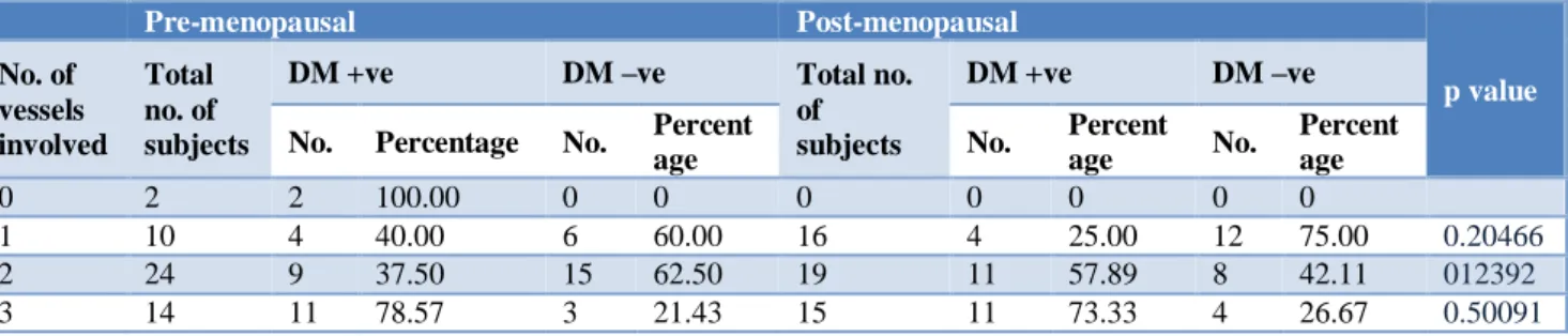
In the pre-menopausal group, 2 subjects with no vessel involvement on angiography were diabetics whereas 40.00% of subjects with single vessel involvement, 37.50% of subjects with double vessel involvement and 78.57% subjects with triple vessel involvement on angiography were found to be diabetics. In post-menopausal group, 25.00% of subjects with single vessel
involvement, 57.89% subjects with double vessel involvement and 73.33% subjects with triple vessel involvement on angiography were found to be diabetics. But the differences found were not statistically significant between the two groups. It is worthwhile to note that in patients presenting with triple vessel disease, 78.50% (11 out of 14) in the pre-menopausal group and 73.33% (11 out of 15) in the post-menopausal group were diabetics (Table 4).
Table 4: Distribution of subjects according to number of vessels involved and diabetic status.
Pre-menopausal Post-menopausal
p value
No. of vessels involved
Total no. of subjects
DM +ve DM –ve Total no.
of subjects
DM +ve DM –ve
No. Percentage No. Percent
age No.
Percent
age No.
Percent age
0 2 2 100.00 0 0 0 0 0 0 0
1 10 4 40.00 6 60.00 16 4 25.00 12 75.00 0.20466
2 24 9 37.50 15 62.50 19 11 57.89 8 42.11 012392
3 14 11 78.57 3 21.43 15 11 73.33 4 26.67 0.50091
Figure 2: Distribution of subjects according to type of vessel involved.
Further, the distribution of subjects according to vessel involved was studied. 18% of pre-menopausal women had involvement of LAD as compared to 22% of post-menopausal women but the difference was not statistically significant (p value = 0.33000). 2% of post-menopausal women had involvement of LCX whereas no subject in pre-menopausal group had involvement of LCX and the difference was statistically significant (p value = 0.04801). 8% of post-menopausal women had involvement of RCA whereas no subject in pre-menopausal group had involvement of RCA and the difference found was statistically significant. (p value = 0.04801). 2% of pre-menopausal women had involvement of LMCA where no subject in post-menopausal group had involvement of LMCA and the difference was statistically significant (p value =
0.04801). In the pre-menopausal group, 18% of subjects had involvement of LAD + LCX as compared to 16% subjects in post-menopausal group (difference was not significant). 22% subjects had involvement of LAD + RCA in pre-menopausal group as compared to 14% subjects in post-menopausal group but the difference was not statistically significant (p value = 0.15848). 8% subjects in both pre-menopausal and post-menopausal group had involvement of LCX + RCA. Data wise28% subjects in pre-menopausal group had involvement of LAD+LCX + RCA (triple vessel disease) as compared to 30% subjects in post-menopausal group. The difference was not statistically significant (p value = 0.74871) (Figure 2).
There was no significant difference between the EF (Ejection fraction) values between the two groups (p value – 0.20625). Pre-menopausal women (mean EF; 46.78 + 10.33), Post– menopausal women (mean EF; 46.25 + 9.35).
DISCUSSION
The annual incidence of cardiovascular disease varies according to menopausal status. Greater loss of physical functioning in post-menopausal women leads to greater weight gain, insulin resistance and hypertension. Weight gain is mainly attributed to central obesity which is linked to waning of estrogen production. Changes in the lipid profile during the menopausal transition are contributory risk factors to coronary artery disease in peri-menopausal women. There is an increased prevalence of other risk factors such as diabetes mellitus, hypertension and metabolic syndrome in the post-menopausal period and are well linked to increased risk 0
5 10 15 20 25 30
18
0 0 2
18 22
8 28
22
2 8
0 16
14
8 30
P
e
r
c
e
n
tage
o
f
su
b
je
c
ts
of CAD. In this study, 52% of subjects in both pre and post-menopausal group had past history of DM showing that diabetic status was not influenced by menopause. Matthews KA et al found in the SWAN study (Study of Women’s Health Across the Nation) that glucose and insulin were not influenced by menopause in this cohort study.4 No differences were found in blood glucose and
insulin levels among pre-menopausal and post-menopausal women in a study done in Netherlands by Peters HW et al and Manson JE et al in Nurses’ Health Study found that diabetes was associated with a marked increase in myocardial infarction.5,6 British Birth Cohort
Study found that at 53 years, HbA1C varied by menopausal status group.7 In the Healthy Women Study,
fasting blood glucose levels were greater during post-menopausal period.8
There was no significant difference in the past history of hypertension in both pre and postmenopausal women. Akhoshi MS et al found that menopause did not affect the systolic blood pressure.9 In a longitudinal study in
Melbourne, Australia it was found that changes in diastolic blood pressure during menopause were related to increasing age of women independent of menopause.10
In a cross-sectional study by Peters HW et al, no difference in systolic and diastolic blood pressure was found between pre-menopausal and post-menopausal women.5 However, in the study, pre-menopausal women
presented with significantly higher systolic blood pressure and pulse pressure than their post-menopausal counterparts.11 Estimating the interaction effect between
blood pressure and menopausal status in the overall cohort demonstrated that both systolic blood pressure and pulse pressure were more potent risk factors for coronary artery disease in the pre-menopausal group.
In this study 48% subjects in pre-menopausal group in our study had family history of cardiovascular diseases as compared to 30% subjects in post-menopausal group showing a higher prevalence of the family history of cardiovascular diseases in the pre-menopausal group (p=0.053). However, the difference was not statistically significant.
The post-menopausal women were physically inactive with a mean activity score of 1.32 as compared to pre-menopausal women with mean activity score of 1.59 (statistically significant). I-Min Lee et al demonstrated an inverse association between relative intensity of physical activity and risk of developing coronary heart disease.12
Amy RW et al demonstrated both physical inactivity and BMI play an important role in development of Type 2 diabetes.13 In the longitudinal study in Melbourne,
Australia, it was found that BMI increased with decreased levels of exercise and that increased physical activity in the workplace lowered triglycerides/HDL cholesterol ratios.10
Chang CJ et al in a study conducted on Chinese women found that through the menopausal transition, the BMI
and total body fat percentage were increased significantly and post-menopausal women showed higher android fat percentage and centrality index.14 Central obesity was the
major independent factor associated with worsened cardiovascular disease risk factors after menopause. Ley CJ et al found a marked increase in android fat and a decrease in gynoid fat in post-menopausal women.15
British Birth Cohort Study showed that at 53 years, body mass index and waist circumference varied by menopausal status group.7 In a study by Gower BA et al,
it was found that post-menopausal women had greater total body fat, summed central skin folds and estimated intraabdominal fat than pre-menopausal women.16 In
study done by Green KA et al, the factor that had the most far reaching adverse influence on cardiovascular disease risk in menopausal women was high BMI.17 Neze
O et al found an increase in WHR (Wait Hip Ratio) originated from an increase in Waist Circumference but BMI and body weight were maintained during menopause.18 However, in a study in Melbourne,
Australia, it was found that changes in BMI during menopause were related to women’s increasing age.10
Peters HW et al didn’t find any difference in BMI and Waist-Hip ratio between pre and post-menopausal women in their study.5 Singh PN et al and Nurses’ Health
study have shown that a high BMI increases the relative risk of CAD.19,6 Interheart study showed that there was a
linear correlation between the incidence of CAD and W: H ratio.20
The post menopausal women were heavier than the pre-menopausal women with a mean weight of 67.9±12.54 kgs and mean BMI of 28.36±4.64 as compared to mean weight of 65.54±10.73 kgs and mean BMI of 27.22±3.97 in pre-menopausal women which was however not statistically significant. Also, the difference between the W: H ratio between the pre-menopausal women and post-menopausal women was not significant (p value = 0.21512).
Pre-menopausal women had higher LDL-C (110.57±51.45 mg/dL vs. 97.10±48.73 mg/dL) and triglyceride levels (168.86±87.81 mg/dL vs. 149.80±91.27 mg/dL) and lower HDL-C levels (41.38±19.37 mg/dL vs. 47.31±29.88 mg/dL) as compared to their post-menopausal counterparts. Total cholesterol (172.0±54.28 mg/dL vs. 173.0±55.88 mg/dL) was not different between the two groups. However, the differences in the individual lipid profile parameters between the two groups did not show statistical significance.
In the longitudinal study in Melbourne Australia, it was found that the only change dependent on the final menstrual period was a significant decrease in HDL cholesterol and the rate of decrease was maximal around 9 months after menses ceased.10 Chang CJ et al found in
Cohort Study, it was found that BMI, WC, total cholesterol and LDL-C varied by the menopausal status.7
Peters HW et al found that compared to pre-menopausal women, post-menopausal women had significantly increased levels of total cholesterol, LDL-C and Apo lipoprotein-B.5 The difference was present within 3 years
after onset of menopause but no difference was found in HDL-C, triglycerides and Apo lipoprotein A1 levels. Gower BA et al also found that post-menopausal women had higher plasma concentrations of total cholesterol, LDL-C and triglycerides than pre-menopausal women.16
In a study done by Torng PL et al, total cholesterol increased after menopause, LDL-C was dependent on age and obesity and HDL-C declined irrespective of the menopausal status.21
There was no difference in the metabolic syndrome between the pre-menopausal and post-menopausal women as 16% subjects in both the groups had metabolic syndrome.
In the postmenopausal group, 2% subjects had involvement of LCX whereas 8% had involvement of RCA while no subject in the pre-menopausal group had involvement of LCX and RCA. 2% subjects in the pre-menopausal group had involvement of LMCA white no subject in the post-menopausal group had involvement of LMCA. No other significant difference between the two groups was found as regards the angiographic findings.
On analysing the data of angiography in diabetics and non-diabetics, about 75% subjects in both the pre- and post- menopausal women who presented with triple vessel disease had diabetes mellitus. This highlights the well known fact that diabetes mellitus is an important risk factor for extensive atherosclerosis (triple vessel disease). There was no difference in the 2D Echo Ejection fraction findings between the two groups. Study suggests that the incidence of diabetes and hypertension was similar in both the groups and the family history of CAD was found to be more prevalent among pre-menopausal women. The activity index was lower in the post-menopausal women.
The body composition i.e. the weight, BMI, WC, HC and W: H ratio was not different between pre and post-menopausal women. Noteworthy was the fact that both the pre- and post- menopausal women who presented with CAD had a high BMI and increased waist circumference thus highlighting that obesity is important risk factor for CAD in women.
LDL-C and triglycerides were higher in the menopausal women and HDL-C was lower in the pre-menopausal women. Total cholesterol was similar in both the groups.
CONCLUSION
Differences in the risk factor profile between the two groups in our study will be helpful in targeting the
preventive measures and the therapies in both pre-menopausal and post-pre-menopausal women. We suggest that additional risk factors i.e. serum estrogen, hs-CRP, lipoprotein (a) should be investigated to further improve our understanding of the cardiovascular risk factor profile among pre-menopausal and post-menopausal women.
ACKNOWLEDGEMENT
Authors wish to acknowledge the contribution of Dr. Khushpreet Kaur and Dr. Naresh Sood in this research work.
Funding: No funding sources Conflict of interest: None declared Ethical approval: Not required
REFERENCES
1. Mosca L, Jones WK, King KB, Ouyang P, Redberg RF, Hill MN. Awareness, perception and knowledge of heart disease risk and prevention among women in the United States. American Heart Association Women’s Heart Disease and Stroke Campaign Task Force. Arch Fam Med. 2000;9:506-15.
2. Kannel WB, Hjortland MC, McNamara PM, Gordon T. Menopause and risk of cardiovascular disease: Framingham study. Ann Intern Med. 1976; 85:447-52.
3. Matthews KA, Meilahn E, Kuller LH, Kelsey SF, Caggiula AW, Wing RR. Menopause and risk factors for coronary heart disease. New Engl J Med. 1989;321:698-9.
4. Matthews KA, Crawford SL, Chae CU. Are changes in cardiovascular disease risk factors in midlife women due to chronological ageing or to the menopausal transition? J Am Coll Cardiol. 2009; 54:2366-73.
5. Peters HW, Westendrop ICD, Hak AE, Grobbee DE, Stehouwer CDA, Hofman A, Witteman JCM. Menopausal status and risk factors for cardiovascular disease. J Intern Med. 1999; 246:521-8.
6. Manson JE, Colditz GA, Stampfer MJ, Willet WC. A prospective study of maturity-onset diabetes mellitus and risk of coronary heart disease and stroke in women. Arch Int Med 1991;151:1141-7. 7. Kuh D, Langenberg C, Hardy R, Kok H, Cooper R,
Butterworth S, et al. Cardiovascular risk at 53 yrs of age in relation to the menopause transition and use of hormone replacement therapy: a prospective British Birth Cohort Study. Int J Obstet Gynaecol. 2005;112:476-85.
8. Matthews KA, Kuller LH, Sutton-Tyrrell K. Changes in the cardiovascular risk factors during the perimenopause and post menopause and carotid artery atherosclerosis in healthy women. Stroke. 2001;32:1104-11.
menopause on serum cholesterol, body mass index, and blood pressure. Atherosclerosis. 2001 May 1;156(1):157-63.
10. Do KA, Green A, Guthrie JR, Dudley EC, Burger HG, Dennerstein L. Longitudinal Study of Risk Factors for Coronary Heart Disease Across the Menopausal Transition. Am J Epidemiol. 2000; 151:584-93.
11. Gierarch GL, Kelsey SF, Olson MB, Pepine CJ, Reis SE, Rogers WJ, et al. Hypertension, Menopause and Coronary Artery Disease Risk in the Women’s Ischemia Syndrome Evaluation (WISE) Study. J Am Coll Cardiol. 2006;47:50-8. 12. Lee IM, Sesso HD, Oguma Y, Paffenbarger Jr RS.
Relative intensity of physical activity and risk of coronary heart disease. Circulation. 2003 Mar 4;107(8):1110-6.
13. Weinstein AR, Sesso HD, Lee IM, Cook NR, Manson JE, Buring JE, et al. Relationship of physical activity vs body mass index with type 2 diabetes in women. JAMA. 2004 Sep 8;292(10):1188-94.
14. Chang CJ, Wu CH, Yao WJ, Yang YC, Wu JS, Lu FH. Relationships of age, menopause and central obesity on cardiovascular disease risk factors in Chinese women. Int J Obes. 2000; 24:1699-704. 15. Ley CJ, Lees B. Sex and menopause associated
changes in body fat distribution. Am J Clin Nutr 1992;55:990-4.
16. Gower BA, Nagy TR, Goran MI, Toth MJ, Poehlman ET. Fat distribution and plasma lipid-lipoprotein concentrations in pre and post-menopausal women. Int J Obes. 1998;22:605-11.
17. Green KA, Guthrie JR, Dudley E, Burger HD. Longitudinal study of risk factors for coronary risk factors for coronary heart disease across the menopausal transition. Menopause 2000;151:584-93.
18. Neze O, Ergin S, Senay M, Yusuf O. Body fat distribution and cardiovascular disease risk factor in pre and post-menopausal obese women with similar BMI. Endocrine J. 2002;49:503-9.
19. Singh PN, Haddad, Ella, Knutse, Synnove F, Fraser GE. The effect of menopause on the relation between weight gain and mortality among women. Menopause. 2001;8:314-20.
20. Yusuf S, Hawken S, Ounpuu S, Bautista L, Franzosi MG, Commerford P, et al. On behalf of the Interheart study investigators. Obesity and the risk of myocardial infarction in 2700 participants from 52 countries: a case control study. Lancet. 2005; 366:1640-9.
21. Torng PL, Sung FC, Chien KL, Huang SC, Chow SN. Effects of menopause on intra-indivitual changes in serum lipids, blood pressure and body weight-the Chin-Shan community cardiovascular cohort study. Atheroscler. 2002;161:409-15.