675
© 2018 by the Serbian Biological Society How to cite this article: Mitrović MB, Tatalović NR, Nikolić-Kokić AL, Ciraj-Bjelac OF, Krstić NE, Oreščanin-Dušić ZS, Krstić DŽ, Jovanović ZM, Blagojević DP, Lazarević-Macanović MV. Influence of absorbed radiation dose following computed tomography on the antioxidative status in rabbit testicles. Arch Biol Sci. 2018;70(4):675-80.
Influence of absorbed radiation dose following computed tomography on the
antioxidative status in rabbit testicles
Marko B. Mitrović1, Nikola R. Tatalović2, Aleksandra L. Nikolić-Kokić2, Olivera F. Ciraj-Bjelac3, Nikola E.
Krstić1, Zorana S. Oreščanin-Dušić2, Dragana Ž. Krstić4, Zoran M. Jovanović4, Duško P. Blagojević2,* and
Mirjana V. Lazarević-Macanović1
1Department of Radiology and Radiation Hygiene, Faculty of Veterinary Medicine, University of Belgrade, Bulevar
oslobođenja 18, 11000 Belgrade, Serbia
2Department of Physiology, Institute for Biological Research “Siniša Stanković”, University of Belgrade, Bulevar despota
Stefana 142, 11060 Belgrade, Serbia
3Radiation Protection Department, Vinča Institute of Nuclear Sciences, University of Belgrade, Mike Petrovića Alasa 12-14,
11351 Belgrade, Serbia
4Department of Physics, Faculty of Science, University of Kragujevac, Radoja Domanovića 12, 34000 Kragujevac, Serbia
*Corresponding author: [email protected]
Received: April 13, 2018; Revised: May 23, 2018; Accepted: June 12, 2018; Published online: June 15, 2018
Abstract: In recent years, computed tomography (CT) has become very common in veterinary medicine. It is well known that testicles are organs with high radiosensitivity and their function can be impaired even after exposure to low radiation doses. In this work, we calculated the absorbed radiation doses after CT was performed with different voltage/current levels and correlated it with the activity of antioxidant enzymes in rabbit testicles. Two hours after CT, the activities of catalase (CAT) and glutathione peroxidase (GSH-Px) were increased in the testicles of animals that received an absorbed dose of 29.2 mGy. The same changes, along with elevated glutathione reductase (GR) activity, were observed after 7 days in animals that received the highest absorbed dose(46.3 mGy). It would appear that absorbed doses above 27.8 mGy provoked the antioxidant reaction but the time scale of the reaction was dose-dependent. Examination of the obtained results revealed that the main denominator of CT influence was a higher current. Our results suggest that CT influences the antioxidant status in rabbit testicles. The changes in antioxidant enzyme activities were dose- and time-dependent and influenced by the applied current.
Key words: computed tomography; oxidative stress; antioxidant enzymes; testicles; rabbit
INTRODUCTION
In sexually mature males, the testicles are metaboli-cally very active, consuming significant amounts of energy for spermatogenesis. Spermatogenesis implies an extremely high rate of cells division, being capa-ble of generating approximately 1000 sperm cells per second. High metabolic activity is accompanied by intensive oxygen consumption in the mitochondria of germinal epithelial cells that may consequently result in the production of substantial amounts of free radi-cals [1]. Male germ cells are more susceptible to oxida-tive stress than somatic cells, because their membranes contain more polyunsaturated fatty acids [2]. Thus,
oxidative stress plays an important role in the etiology of sperm malformation, altered function, the sperm count profile and male infertility [3]. Testicles have a well-developed enzymatic antioxidant system [1].
to those received after CT [4]. Radiation doses from a single, routine CT examination belong to the category of low radiation doses defined as doses in the range of near zero up to about 100 mSv of low-LET radiation [5], which corresponds to an absorbed dose of 100 mGy. Therefore, it can exert only stochastic effects.
It is well known that testicles are organs with high radiosensitivity and their function can be impaired even after exposure to low radiation doses [6]. Ac-cording to literature data, spermatogonia cells are par-ticularly sensitive to radiation and can be damaged by exposure to doses lower than 0.1 Gy [7]. At the cellular level, the harmful effects of ionizing radiation are due to the production of free radicals from water radiolysis that causes direct damage to DNA molecules [8].
The main objective of this study was to calculate the absorbed radiation doses in rabbit testicles after CT performed by variation of voltage and current, and to correlate these values with the activity of anti-oxidant enzymes. Although it is well known that ex-posure to x-radiation results in oxidative stress, there are no literature data on the influence of diagnostic doses of x-radiation emitted during CT on the oxida-tive status in testicles.
MATeRIAls AND MeThODs
The study was conducted in agreement with existing ethical norms and was approved by the Permission of the Ministry of Agriculture and Environmental Pro-tection – Veterinary Directorate, Republic of Serbia, No. 323-07-03455/2015-05/5. Experiments were per-formed on mature New Zealand white rabbit males in accordance with the ARRIVE (Animal Research: Reporting of In Vivo Experiments) guidelines for re-porting experiments involving animals [9]. The rab-bits were kept under standard laboratory conditions (12 h light, 12 h dark, 21±2°C ambient temperature). All animals were housed in individual cages and
pro-vided with standard diet and tap water adlibitum.
Animals and grouping
Sixty-six experimental rabbits were divided into 11 groups consisting of 6 animals each. Three groups were not exposed to radiation and served as controls. Rabbits from the NT (not treated) group were killed
without any treatment, while animals from groups
A1 and A2 were anesthetized and served as the
anes-thetized controls. Rabbits from the remaining eight groups (I1, I2, II1, II2, III1, III2, IV1 and IV2) were anes-thetized to ensure still positioning during examination and subjected to CT using different CT protocols (two different values of voltage and current-amperage in the x-ray tube were applied). For anesthesia, a keta-mine hydrochloride (Ketamidor 10%, Richter Pharma, Austria) was used and administered intramuscularly (i.m.) (35 mg/kg body weight). Prior to anesthesia, a premedication by i.m. application of xylazine hy-drochloride (Xylased, Bioveta, Czech Republic) was performed (5 mg/kg body weight). All animals were
killed by decapitation. Rabbits from groups A1, I1, II1,
III1 and IV1 were killed after 2 h, while rabbits from
groups A2, I2, II2, III2 and IV2 were killed after 7 days. Immediately after sacrifice, testicle samples were col-lected and stored in liquid nitrogen before determina-tion of the activities of antioxidant enzymes.
CT examination protocols
CT examinations of rabbits were performed using a CT SOMATOM AR STAR (Siemens Medical Systems, Erlangen, Germany). CT examinations were per-formed using the following examination protocols:
Groups I1 and I2: tube voltage (U) 110 kV; tube
cur-rent and exposure time product (It) 63 mAs; exposure
time (t) 1 s; slice thickness 10 mm; Groups II1 and II2:
tube voltage (U) 130 kV; tube current and exposure time product (It) 63 mAs; exposure time (t) 1 s; slice
thickness 10 mm; Groups III1 and III2: tube voltage
(U) 110 kV; tube current and exposure time product (It) 105 mAs; exposure time (t) 1 s; slice thickness 10
mm; Groups IV1 and IV2: tube voltage (U) 130 kV;
tube current and exposure time product (It) 105 mAs; exposure time (t) 1 s; slice thickness 10 mm.
Dose quantities
simu-lations included properties of the CT scanner and 110 kV and 130 kV x-ray spectra generated using the soft-ware tool SEPC78 (Institute of Physics and Engineer-ing in Medicine, IPEM). A mathematical model of the rabbit was generated based on representative animal CT images. Each voxel of the testicles was assigned with relevant tissue parameters in terms of density and mass attenuation coefficient. As an outcome of the Monte Carlo simulations, the energy deposited in each voxel was registered, and was further used for
organ dose calculation. A total of 107 photon histories
were followed, using cut-off energy of 1 keV.
Determination of antioxidant enzyme activities Thawed testicles were homogenized and sonicated in 0.25 M sucrose, 1 mM ethylenediaminetetraacetic acid and 0.05 M Tris-HCl buffer (pH 7.4) before
centrifu-gation (90 min at 105000×g). The supernatant was
used for enzyme activity measurements. Enzymatic essays were based on spectrophotometric measure-ments of absorbance changes. Measuremeasure-ments of ab-sorbance were performed using a Shimadzu UV-160 spectrophotometer (Shimadzu Scientific Instruments, Shimadzu Corporation, Kyoto, Japan). Total superox-ide dismutase (SOD) activity was determined by the adrenaline method [11]. One SOD unit was defined as the amount of the enzyme necessary to decrease the rate of adrenalin autooxidation by 50% at pH 10.2. For determination of SOD2 activity, the assay was performed after sample incubation with 8 mM KCN. SOD1 activity was calculated from the differ-ence between total SOD and SOD2 activities. CAT ac-tivity was estimated by monitoring hydrogen peroxide consumption [12]. The activity of GSH-Px was deter-mined using t-butyl hydroperoxide as a substrate and estimated by calculating NADPH consumption [13]. GR activity was measured as the rate of NADPH oxi-dation concomitant with glutathione disulfide (GSSG) reduction [14]. Specific activities were expressed per mg of tissue protein. The protein concentration was measured by the method of Lowry [15].
statistical analysis
All values were expressed as the mean±SEM. Statisti-cal evaluation was Statisti-calculated by two-way ANOVA, with factors: anesthesia (A) and the time of death (T),
the absorbed dose (D) and the time of death (T) and
post hoc compared using Tukey’s HSD t-test. For all comparisons, p<0.05 was considered as significant.
ResUlTs
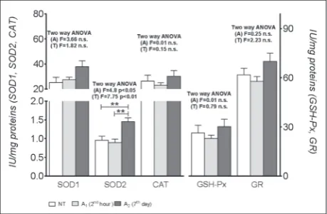
The absorbed doses in testicles were calculated by computational dosimetry and are listed in Table 1. Since the rabbits were anesthetized prior to CT, the effect of anesthesia on antioxidant enzyme activities was checked. Our results showed that anesthesia had no effect on the activities of antioxidant enzymes in testicles, except for SOD2. There was a statistically significant increase in SOD2 activity after 7 days in comparison to non-anaesthetized rabbits (Fig. 1). Table 1. Parameters of different CT examination protocols and absorbed doses in rabbit testicles.
Groups U (kV) It (mAs) t (s) D (mGy)
I1 and I2 110 63 1 17.5
II1 and II2 130 63 1 27.8
III1 and III2 110 105 1 29.2
IV1 and IV2 130 105 1 46.3
U – voltage; It – current; t – time; D – absorbed doses
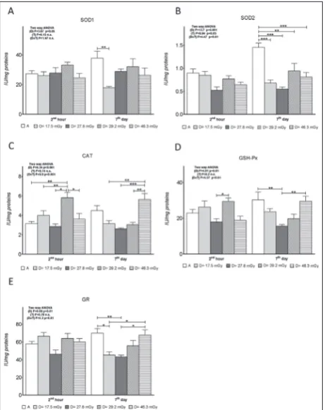
compared with the anesthetized control group. How-ever, 7 days following CT, there were no differences in SOD2 activity in rabbit testicles between groups that received different absorbed doses (Fig. 2B).
CAT activity increased after 2 h in the group that received 29.2 mGy and was at the control level after 7 days (Fig. 2C). On the other hand, after 7 days, CAT activity was elevated in the group of rabbits that re-ceived the highest amount of radiation (46.3 mGy).
GSH-Px activity was lower in the group of animals that received an absorbed dose of 27.8 mGy when compared to a dose of 29.2 mGy after 2 h (Fig. 2D). Lower GSH-Px activity in the first group (27.8 mGy) persisted after 7 days as compared to the anesthetized control group and the group that received the maxi-mal absorbed dose (46.3 mGy).
There were no significant differences in GR activity between experimental groups 2 h after CT when
com-pared to the anesthetized control group. However, after 7 days, a significant decrease was noted in animals that received absorbed doses of 17.5 and 27.8 mGy when compared to the anesthetized control animals (Fig. 2E).
DIsCUssION
Our results suggest that CT influences the antioxidant status in rabbit testicles, with the changes in antioxi-dant enzymes activity being time- and dose-depend-ent and influenced by the applied anesthesia. There are no literature data about the influence of ketamine and xylazine on the antioxidant enzyme activities in rabbit testicles. In our experiments, there were no changes in SOD1, CAT, GSH-Px and GR activities in animals that were anesthetized using ketamine and xylazine in comparison to the non-anesthetized con-trol group. However, there was a significant increase in SOD2 activity after 7 days. This implies that the applied anesthesia influenced mitochondrial ROS pro-duction, which should be considered in the interpreta-tion of data. There are reports that many anesthetics have profound effects on mitochondrial membranes at concentrations as low as those known to produce gen-eral anesthesia, and they can destabilize lipid-protein interactions [16-18]. Zaugg et al. [19] showed that the intravenous anesthetics R-ketamine exert pronounced mitochondrial effects that are reflected on ROS pro-duction. Therefore, the elevation of SOD2 in testicles observed in our experiment can be considered as the effect of anesthesia on mitochondrial ROS homeosta-sis. Thus, the effects of CT on antioxidant enzyme ac-tivities were compared to the acac-tivities of antioxidant enzymes in testicles of anesthetized control animals.
Two hours after CT we did not observe a general effect of x-radiation on antioxidant enzyme activities in comparison to the anesthetized group of animals; however, subtle increases in CAT and GSH-Px ac-tivities were observed in the group of animals that received the absorbed dose of 29.2 mGy in compari-son with the other groups. In our experiment, the same changes, along with elevated GR activity, were detected after 7 days in animals that received the
high-est absorbed dose(46.3 mGy). This means that the
absorbed doses above 27.8 mGy provoked an anti-oxidant reaction, and for these doses, the time scale of the reaction was dose-dependent.
Fig. 2. The effect of different absorbed doses on: A – SOD1 activ-ity; B – SOD2 activity; C – CAT activity; D – GSH-Px activity;
Since there was no effect on the activities of anti-oxidant enzymes in rabbits that received the absorbed dose of 27.8 mGy, it would appear that the dose threshold range was very narrow. Our previous re-sults obtained in erythrocytes indicated that the dose threshold was about 25 mGy [20]. In the present study, we also found that doses below that level did not pro-duce any significant changes in antioxidant enzyme activities, but the threshold was a little higher (27.8 mGy). These results can be interpreted as a tissue-spe-cific sensitivity. On the other hand, a radiation level just above 27.8 mGy had a significant impact on the antioxidant defense in testicles, but after a relatively short time (2 h after exposure), as compared to the higher dose that required a longer adaptive period. It has been documented that local radiation as low as 0.35 Gy induced a response in testicles, but mutagenic changes were detected at 0.5 Gy [21,22].
The elevation of CAT and GSH-Px activities sug-gests that the concentration of hydrogen peroxide was raised above a tolerable ROS level, and there was a need for its faster elimination. Both CAT and GSH-Px me-tabolize hydrogen peroxide, but with different enzyme
constants, i.e. CAT removes H2O2 faster and is
physio-logically operative at higher concentrations of hydrogen peroxide [23,24]. At the same time, GSH-Px is efficient
at lower cellular H2O2 levels, but it also metabolizes
lipid peroxides. Since in our study GSH-Px was elevated along with CAT, it seems that the ROS attack involved lipid molecules as well. This was especially important for rabbits that received the highest absorbed dose, since an additional elevation of GR activity found in these rabbits suggested that increased glutathione medi-ated antioxidant activity and its faster turnover. If the received absorbed dose was about 29.2 mGy, the re-sponse was rapid and occurred after 2 h. If the dose was about 70% higher, an elevation in antioxidant enzymes was noted after 7 days. The antioxidant response after 7 days was stronger, but it required more time to be ex-pressed. The time effect on antioxidant enzyme activity after irradiation is already known [4,25]. However, the precise dynamics and sequence of changes, particularly of individual antioxidant components, depend on the dose, time, tissue and antioxidant [26].
In our experiment, different CT protocols were performed by changing the voltage and amperage
(current) parameters. We found that CAT activity in the testicles at 2 h post CT was increased when a low voltage/high current was applied, as compared to a high voltage/low current, or a low voltage/low current.
Moreover, on the 7th day, the CT examination that was
carried out with the use of high voltage/high current resulted in increased CAT activity, when compared to CAT activities recorded in animals where CT ex-amination was performed with other combinations of applied voltage and current parameters. Addition-ally, our results showed that GSH-Px and GR activities were also increased in high-current CT conditions. It appears that the main element influencing radiation effects and subsequent changes in antioxidant activity is a high current. A higher current provides a higher density of radiation per surface unit and intensifies interactions between molecules, while voltage influ-ences x-ray penetration and energy.
CONClUsIONs
Our results show that CT produced changes in the antioxidant status of rabbit testicles, suggesting a ROS imbalance. The effects were prominent above the absorbed dose of 27.8 mGy and were expressed on different time scales. The effects of the dose slightly above the threshold dose of 29.2 mGy were noted after 2 h, but the effects of much higher doses (48.2 mGy) were noted after 7 days. Since the applied anesthetics, as an integral part of this procedure, also influenced antioxidant enzyme activities, it can be concluded that CT disturbs the ROS balance in rabbit testicles.
Funding: This work was financed by the Ministry of Educa-tion, Science and Technological Development, Republic of Ser-bia, Grant 173014, “Molecular mechanisms of redox signaling in homeostasis: adaptation and pathology”, and Grant 175061, “Antioxidant protection and potentials for differentiation and re-generation of mesenchymal stem cells from various tissues during the aging process”.
Mirjana Lazarević-Macanović, Zorana Oreščanin-Dušić wrote the second draft of the manuscript. Mirjana Lazarević-Macanović, Duško Blagojević edited the manuscript. Nikola Krstić performed the CT procedure. Zoran Jovanović created the three-dimensional mathematical model of the rabbit. Dragana Krstić performed a Monte Carlo simulation of the CT examination. Olivera Ciraj-Bjelac calculated the absorbed dose.
Conflict of interest disclosure: The authors declare that there is no conflict of interests.
ReFeReNCes
1. Aitken RJ, Roman SD. Antioxidant systems and oxidative stress in the testes. Oxid Med Cell Longevity. 2008;1(1):15-24. 2. Bas H, Kalender S. Antioxidant Status, Lipid Peroxidation
and Testis-histoarchitecture Induced by Lead Nitrate and Mercury Chloride in Male Rats. Braz Arch Biol Technol. 2016;59:e16160151.
3. Acharya UR, Mishra M, Patro J, Panda MK. Effect of vita-mins C and E on spermatogenesis in mice exposed to cad-mium. Reprod Toxicol. 2008;25:84-8.
4. Peltola V, Parvinen M, Huhtaniemi I, Kulmala J. Compari-son of Effects of 0.5 and 0.3 Gy X-Irradiation on Lipid Per-oxidation and Antioxidant Enzyme Function in Rat Testis and Liver. EJBMSR. 1993; 14(4):267-74.
5. National Research Council (U.S.) Committee to Assess Health Risks from Exposure to Low Level of Ionizing Radia-tion. Health risks from exposure to low levels of ionizing radiation : BEIR VII, Phase 2. Washington, D.C.: National Academies Press; 2006. 406 p.
6. Howell SJ, Shalet SM. Spermatogenesis after cancer treat-ment: damage and recovery. J Natl Cancer Inst Monogr. 2005;34:12-7.
7. Kovacs GT, Stern K. Reproductive aspect of cancer treat-ment: an update. Med J Aust. 1999;170:495-7.
8. Suntharalingam N. Basic Radiobiology. In: Podgorsak EB, editor. Review of Radiation Oncology Physics, A Handbook for Teachers and Students. Vienna: International Atomic Energy Agency; 2003. p. 397-412.
9. Kilkenny C, Browne WJ, Cuthill IC, Emerson M, Alt-man DG. Improving Bioscience Research Reporting: The ARRIVE Guidelines for Reporting Animal Research. PLOS Biol. 2010;8(6):e1000412.
10. Monte Carlo Team. MCNP–a General Monte Carlo N-Par-ticle Transport Code (X-5 Monte Carlo Team, Version 5). Vol. 1: Overview and Theory. Los Alamos, NM: Los Alamos National Laboratory; 2003. LA- UR-03-1987.
11. Mirsa HP, Fridovic I. The role of superoxide anion in the autoxidation of epinephrine and a simple assay for superox-ide dismutase. J Biol Chem. 1972;247(10):3170-5.
12. Beutler E. Catalase. In: Beutler E, editor. Red Cell Metabo-lism, A Manual of Biochemical Methods. New York: Grune and Stratton Inc; 1982. p. 105-6.
13. Paglia DE, Valentine WN. Studies on the quantitative and qualitative characterization of erythrocyte glutathione per-oxidase. J Lab Clin Med. 1967;70(1):158-69.
14. Glatzle D, Vuillemier JP, Weber F, Decker K. Glutathione reductase test with whole blood, a convenient procedure for the assessment of riboflavin status in humans. Experientia. 1974;30(6):665-7.
15. Lowry OH, Rosenbrough NJ, Farr A, Randall RJ. Protein measurement with the Folin phenol reagent.J Biol Chem. 1951;193:265-75.
16. Kowaltowski AJ, Seetharaman S, Paucek P, Garlid KD. Bio-energetic consequences of opening the ATP-sensitive K channel of heart mitochondria. Am J Physiol Heart Circ Physiol. 2001;280:649-57.
17. Murata M, Akao M, O’Rourke B, Marbán E. Mitochondrial ATP-sensitive potassium channels attenuate matrix Ca(2+) overload during simulated ischemia and reperfusion. Possi-ble mechanism of cardioprotection. Circ Res. 2001;89:891-8. 18. Zaugg M, Lucchinetti E, Spahn DR, Pasch T, Schaub MC.
Volatile anesthetics mimic cardiac preconditioning by prim-ing the activation of mitochondrial K(ATP) channels via multiple signaling pathways. Anesthesiology. 2002;97:4-14. 19. Zaugg M, Lucchinetti E, Spahn DR, Pasch T, Garcia C,
Schaub MC. Differential Effects of Anesthetics on Mito-chondrial K(ATP) Channel Activity and Cardiomyocyte Protection. Anesthesiology. 2002;97:15-23.
20. Mitrović BM, Ciraj-Bjelac FO, Oreščanin-Dušić SZ, Blagojević PD, Nikolić-Kokić LA, Tatalović RN, Krstić EN, Lazarević-Macanović VM. Influence of radiation dose in computed tomography on antioxidant enzyme activity in rabbit erythrocytes. Nucl Technol Radiat. 2017;32(4):342-9. 21. Sanderman TF. The effects of X-irradiation on male human
fertility. Br J Radiol. 1966;39:901-7.
22. Lahdetie J, Parvinen M. Meiotic micronuclei induced by x-rays in early spermatids of the rat. Mutat Res. 1981;81:103-15. 23. Halliwel B, Gutteridge JMC. Oxygen toxicity, oxygen radicals,
transition metals and desease. Biochem J. 1984;219:1-14. 24. Halliwel B, Clement MV, Long LH. Hydrogen peroxide in
the human body. FEBS Lett. 2000;486:10-3.
25. Korać B, Blagojević D, Saičić ZS, Buzadžić B, Spasić MB, Petrović VM. Effects of acute x-ray irradiation on antioxi-dative defence in the rats chronically exposed to cold. Arch Biol Sci. 1993;46(3-4):87-93.