261
© 2017 by the Serbian Biological Society How to cite this article: Vujičić M, Vasić B, Nikolić I, Saksida T, Stojanović I. The role of NUPR1 in lymphocyte proliferation and apoptosis. Arch Biol Sci. 2017;69(2):261-7.
The role of NUPR1 in lymphocyte proliferation and apoptosis
Milica Vujičić, Bobana Vasić, Ivana Nikolić, Tamara Saksida and Ivana Stojanović*
Department of Immunology, Institute for Biological Research “Siniša Stanković”, University of Belgrade, Bulevar despota
Stefana, 11060, Belgrade, Serbia
*Corresponding author: [email protected]
Received: July 7, 2016; Revised: October 7, 2016; Accepted: October 7, 2016; Published online: October 14, 2016
Abstract: Nuclear protein 1 (NUPR1) is a transcription cofactor that senses stressful conditions and modulates cellular response by promoting or inhibiting apoptosis. NUPR1 is usually highly expressed in tumor cells where it enables them to adapt and resist environmental stress or chemotherapeutic compounds. NUPR1 can be involved in cell proliferation. Data about the involvement of NUPR1 in the proliferation and apoptosis of lymphocytes are scarce. Therefore, in this study we focused on the role of NUPR1 in lymphocyte physiology and found that NUPR1 might be involved in the initiation of their proliferation. Lymphocytes were isolated from the cervical lymph nodes of C57BL/6 mice. NUPR1 expression subsided 24 h after the induction of proliferation by a mitogen. Also, stressful conditions after cell isolation led to increased NUPR1 mRNA and protein expression in vitro that coincided with cell apoptosis. Similarly, apoptosis induction by staurosporine, a broad-range protein kinase inhibitor, led to increased NUPR1 expression. In addition, NUPR1 inhibition by small-interfering RNA prevented the staurosporine-induced apoptosis (judging from decreased caspase activity) in the whole cell population of cervical lymph nodes. However, NUPR1 absence was irrelevant to the induction of apoptosis in CD3+ T lymphocytes, suggesting that NUPR1 is probably a mediator of apoptosis in other immune cell populations within the lymph node, such as B lymphocytes. In conclusion, our results suggest that NUPR1 is important for the initiation of lymphocyte cell division and for the apoptotic process of non-T cells during stressful conditions.
Key words: apoptosis; cervical lymph node; apoptosis; lymphocyte; nuclear protein 1; proliferation
INTRODUCTION
Nuclear protein 1 (NUPR1) is a small chromatin pro-tein that senses stress signals from the environment and enables cells to adapt and to resist apoptosis. NUPR1 is a protein of 8 kDa (hence its other name, p8) that can interfere with histone modifications and bind to gene promoters, thereby modulating gene expression. NUPR1 is important for the resistance of tumor cells to apoptosis induction since it spe-cifically leads to the transactivation of the immediate early response 3 (Ier3) gene in pancreatic cancer cells that protects cells from Fas- or tumor necrosis fac-tor (TNF)-induced apoptosis [1]. Also, NUPR1 binds thymosin and interferes with the apoptotic cascade in HeLa cells. The absence of NUPR1 renders HeLa cells susceptible to staurosporine-induced apoptosis [2]. Therefore, it is not surprising that elevated NUPR1 expression is found in breast cancer cells, pancreatic carcinoma cells and adenocarcinoma [3,4]. In breast
cancer cells, the overexpression of NUPR1 leads to the activation of the phosphatidylinositide 3-kinase (PI3K)/Protein kinase B (PKB) (Akt) signaling path-way, cyclin-dependent kinase inhibitor 1 (CDKN1A; p21Cip1) phosphorylation and relocalization from the
β-cell proliferation [8]. Also, NUPR1 negatively regu-lates the proliferation of myocytes and promotes myo-genic differentiation [9,10]. As regards immune cells, it was shown that the number of all immune cells is comparable between NUPR1-/- and NUPR1+/+, though
the apoptosis rate under normal and inflammatory conditions is increased in the absence of NUPR1, es-pecially in CD3+ cells [11].
To date, there are no data about the role of NUPR1 in the proliferation and stress-induced apoptosis of immune cells. Therefore, in this study we investigated the correlation of NUPR1 mRNA and protein expres-sion and mitogen-triggered immune cell proliferation or apoptosis induced either by the process of cell iso-lation or by staurosporine, a broad-spectrum protein kinase inhibitor.
MATERIALS AND METHODS
Animals
Female C57BL/6 mice were bred and kept in the ani-mal facility of Institute for Biological Research “Siniša Stanković”, and their use and all experimental proce-dures were approved by the Ethics Committee of the Institute (App. No 01-07/15), which are in accordance with Directive 2010/63/EU. The mice were maintained under standard conditions with a 12-h day/night rhythm and fed with standard rodent chow and water.
Isolation of lymphocytes from cervical lymph nodes
C57BL/6 mice (8-12 weeks of age) were euthanized by CO2 affixation and cervical lymph nodes were col-lected in tubes filled with phosphate-buffered saline (PBS). Lymph nodes were passed through 40-μm cell strainers and dispersed into a single cell suspension. After centrifugation at 500 g for 5 min, cervical lymph node cells (CLNC) were resuspended in RPMI-1640 medium containing 25 mM HEPES, 2 mM L-gluta-mine, 5% FCS (PAA Chemicals, Pasching, Austria), penicillin/streptomycin (Sigma-Aldrich, St. Luis, MO, USA) and 5 µM β-mercaptoethanol, counted by try-pan blue exclusion test (Sigma-Aldrich) and placed in 24-well plates at 37°C in a 5% CO2 incubator. CLNC
were cultured in the media alone or in the presence of 1 μg/mL concanavalin A (ConA), or with 1 μM staurosporine (Sigma-Aldrich).
CD3+ lymphocyte purification
CLNC (1 x 10⁷) were resuspended in 5 mL of
RPMI-1640 and 10% FCS. For separation of adherent and nonadherent cells, a column made of nylon wool was soaked first in 5 mL of medium (RPMI-1640 and 10% FCS) and the resuspended cells were overlaid. The col-umn with cells was maintained for 1 h at 37°C in a 5% CO2 incubator and nonadherent cells were eluted with warm medium (2 x 5 mL). The recovery of cells was about 2x10⁶; the purity of CD3+ cells was 92.2±8.2%
(determined by flow cytometry analysis).
NUPR1 inhibition by small interfering RNA (siRNA)
In a 24-well tissue culture plate, 1×10⁶ CLNC were
placed in 1 mL of antibiotic-free medium supplement-ed with 10% FCS. After overnight incubation, the cells were transfected according to the manufacturer’s in-structions (SantaCruz Biotechnology Inc., Santa Cruz, CA, USA). Briefly, incubation in transfection medium containing NUPR1 or control small-interfering RNA (siRNA) lasted for 6 h. The cells were then washed and placed in RPMI-1640 medium with 10% FCS and antibiotics. After overnight incubation (18 h), 1 μM staurosporine was applied for 3 h before the cells were collected and their apoptosis was measured by deter-mining caspase activity by flow cytometry. Transfected CLNC were also challenged for 24 h with ConA and proliferation was measured by the 3-(4,5-dimethyl-2-thiazolyl)-2-5-diphenyl-2H-tetrazolium bromide (MTT) assay.
Apoptosis detection
with active caspases (an indicator of apoptosis), CLNC were stained with Apostat (pan-caspase inhibitor) (1:100) (R&D, Minneapolis, MN, USA) for 30 min at 37°C in a 5% CO2 incubator, washed in PBS and analyzed on the flow cytometer.
Proliferation assay
After 24 h of treatment with ConA, control CLNC or siRNA NUPR1-transfected CLNC were treated with MTT solution (0.5 mg/ml) for 2 h at 37°C in a 5% CO2 incubator. In the presence of oxygen derived from mitochondria MTT is reduced to dark formazan crystals, serving as an indicator of cell respiration and indirectly, of cell number and proliferation [12]. The formazan crystals were then dissolved in DMSO. The absorbance was measured in an automated microplate reader (LKB 5060-006, LKB Instruments, Vienna, Austria) at 540 nm and the background at 670 nm was subtracted. The proliferation index was calculated as the ratio between proliferation of ConA-stimulated cells and control cells.
Immunoblot
CLNC (106) were disrupted in lysis buffer
contain-ing 62.5 mM Tris-HCl (pH 6.8, 25°C), 2% w/v SDS, 10% glycerol, 50 mM DTT, 0.01% w/v bromophenol blue, and were subjected to electrophoresis on a 15% SDS-polyacrylamide gel. All samples were electro-transferred to polyvinylidene difluoride membranes at 5 mA/cm2, using a semi-dry blotting system
(Fast-blot B43, Biorad, Munich, Germany). The (Fast-blots were blocked with 5% w/v bovine serum albumin (BSA) in PBST buffer (80 mM Na2HPO4; 20 mM NaH2PO4; 100 mM NaCl; 0.1% Tween-20) and probed with specific rabbit antibodies raised against β-actin (1:500), tu-bulin (1:500) (Abcam, Cambridge, UK) and NUPR1 (1:500) (SantaCruz Biotechnology) in 1% blocking buffer, followed by incubation with secondary anti-body donkey anti-rabbit HRP at 1:10000 (GE Health-care, Buckinghamshire, England) in 1% blocking buffer. Detection was performed by the substrate for horseradish peroxidase (HRP), Luminata Crescendo (Milipore Corporation, Billerica, AM, USA); photo-graphs were made using X-ray films (Kodak, USA). Protein production was calculated by Scion Image
Alpha 4.0.3.2 (Scion Corporation, Frederick, MD, USA) and was expressed relative to the production of β-actin or tubulin.
RNA isolation, reverse transcription and Real-time PCR
CLNC were lysed using RNAzol (Metabion, Mar-tinsried, Germany) and RNA was obtained; reverse transcription to cDNA and real-time PCR was per-formed as previously described [13]. The primer pairs for β-actin were 5'-GACCTGACAGACTACC-3 and 5'-GGCATAGAGGTCTTTACGG-3' (NM_007393.2); NUPR1 primer pairs were purchased as Quantitect Primer Assays from Qiagen (Germantown, MD, USA). NUPR1 mRNA expression was calculated ac-cording to the formula 2-(Ct-Cta), where C
t is the cycle
threshold of the gene of interest and Cta is the cycle threshold value of the housekeeping gene (-actin). Data were obtained from a real-time PCR apparatus and quantitatively analyzed using SDS 2.1 software (Applied Biosystems, Woolston, UK).
Statistical analysis
Statistical analysis was performed using the Student’s t test (Statistica 6.0; StatSoft,Inc., Tulsa, OK, USA). The results were presented as means±standard deviation (SD); p<0.05 were considered statistically significant.
RESULTS
The relation of NUPR1 and mitogen-triggered lymphocyte proliferation
to ConA for 24 h in optimal culture conditions, the expression of NUPR1 mRNA subsided (Fig. 1A). This phenomenon was accompanied by lower NUPR1 pro-tein expression (Fig. 1B) after 24 h cultivation with mitogen, suggesting that NUPR1 expression and proliferation are inversely correlated. However, at-tenuation of NUPR1 mRNA expression with specific siRNA resulted in impaired ConA-stimulated CLNC proliferation (Fig. 1C). This implies that the activity of NUPR1 is probably necessary, at least for the initiation of lymphocyte proliferation. Notably, the efficiency of
NUPR1 mRNA silencing was 100% (Fig. 1D).
NUPR1 expression correlates with stress-induced apoptosis
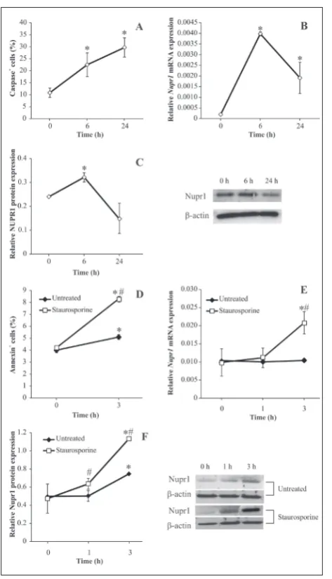
Interestingly, lymphocytes isolated from cervical lymph nodes and immediately tested for the presence of apoptotic cells have a small rate of 8% of apoptosis (Fig. 2A). This coincides with a relatively high ex-pression of NUPR1 mRNA (Fig. 2B), probably due to stressful events during the cell isolation procedure. However, when these cells are placed in a culture plate with optimal conditions for their survival, the apoptosis rate increases over time and reaches a steady state after 24 h (Fig. 2A). Simultaneously, we tested
NUPR1 mRNA expression and found that its expres-sion transiently increases up to 6 h and then subsides after 24 h (Fig. 2B). Seemingly, the NUPR1 expression precedes apoptosis in lymphocytes. Also, the presence of the NUPR1 protein (Fig. 2C) correlates well with the expression of mRNA. What is more, lymphocyte apoptosis induced by staurosporine (an antibiotic that unselectively inhibits protein kinases by blocking the ATP-binding domain; Fig. 2D) leads to an increase in NUPR1 mRNA and protein expression (Fig. 2 E, F), suggesting a positive correlation between NUPR1 action and apoptosis induction.
NUPR1 does not promote apoptosis in T lymphocytes
In order to confirm the suggested role of NUPR1 in apoptosis induction in lymphocytes, NUPR1 mRNA was blocked using siRNA. When NUPR1 was in-hibited, staurosporine was not as efficient in induc-ing apoptosis as when NUPR1 was active (Fig. 3A). However, when we isolated CD3+ cells, staurosporine
provoked apoptosis of these cells, as in the control cultures (Fig. 3B), suggesting that NUPR1 is not man-datory for apoptosis induction in T lymphocytes.
DISCUSSION
Our study has revealed that NUPR1 could have a role in the initiation of lymphocyte proliferation stimu-lated by mitogen as well as in apoptosis induction of non-T cells in the lymph nodes of C57BL/6 mice. NUPR1 expression is usually high in tumor cells and it is thought that it stimulates their proliferation and drug resistance. However, it might have an opposite role in normal, non-transformed somatic cells since its deletion enables the rapid proliferation of pancre-atic beta cells [8]. In this study, two opposing results were obtained: attenuation of NUPR1 impaired lym-phocyte proliferation, but also, NUPR1 expression was significantly lower 24 h after the administration
of mitogen. Seemingly, NUPR1 might be mandatory for the initiation of proliferation, but it is not needed for subsequent steps of cell division and therefore its expression subsides. The observed lower NUPR1 ex-pression after ConA stimulation cannot be an artifact since the non-treated mixed lymphocyte population in the culture increase their NUPR1 expression com-pared to the baseline level detected immediately after cell isolation from the lymph nodes. This
upregula-Fig. 2. The role of NUPR1 in immune cell apoptosis. Cervical lymph node cells were either tested immediately after isolation (0 h) or kept in RPMI-1640 and 5% FBS medium in the incubator for 6 h or 24 h. A – Apoptosis detected by the determination of caspase activity (flow cytometry). B – NUPR1 mRNA expression determined by real-time PCR. C – NUPR1 protein expression estimated by immunoblotting. A representative blot is displayed adjacent to the graph. D – Cervical lymph node cells treated with staurosporine (1 μM); apoptosis was determined by annexin-FITC staining immediately after isolation (0 h) or 3 h after the treat-ment. E – NUPR1 mRNA expression and F – NUPR1 protein expression 1 h and 3 h after staurosporine administration. The results of one representative experiment out of three with similar results are displayed. * denotes p<0.05 between values detected in cells at time points 6 h and 24 h vs. 0 h (A, B, C), or 3 h vs, 0 h (D, E, F); # denotes p<0.05 between values detected in staurosporine-treated vs. unstaurosporine-treated cells.
tion of NUPR1 is probably related to the initiation of cell apoptosis due to cell manipulations during the isolation procedure. It is remarkable that CLNC kept in the optimal conditions for their survival, exhibit around 30% of apoptosis after 24 h in culture. In addi-tion, apoptosis induction by staurosporine also stimu-lates NUPR1 expression. This is, however, in conflict with data obtained from the cervical cancer cell line (HeLa cells), where NUPR1 binds the anti-apoptotic protein prothymosin α and blocks staurosporine-induced apoptosis [2]. The observed discrepancy in the relation between induced apoptosis and NUPR1 expression could be in the nature of the investigated cells, meaning that immortalized cells have a ent physiology and thus NUPR1 might have a differ-ent function. The final proof that NUPR1 is actually related to CLNC apoptosis induction was obtained from the experiments where NUPR1 expression was inhibited by specific siRNA that interfered with trans-lation of the NUPR1 protein. The observed reduction in cell apoptosis in the absence of NUPR1 clearly sug-gests that NUPR1 is mandatory for the initiation of apoptosis by staurosporine. However, CD3+ cells were
not protected from apoptosis induction when NUPR1 was absent. Although our results do not imply an anti-apoptotic role of NUPR1, Weis et al. [11] suggested that NUPR1 functions as a blocker of CD3+
lympho-cyte apoptosis since in both physiological conditions and after induction of inflammation, NUPR1 deletion coincides with increased CD3+ apoptosis. Due to
ex-perimental restrictions we were not able to identify the possible immune cell population in the lymph node that requires NUPR1 expression for initiation of the apoptotic process, but it can be speculated that these might be B lymphocytes that represent the sec-ond predominant population in cervical lymph nodes (around 30%). This percentage actually coincided with the usual number of B cells in cervical lymph nodes in healthy animals (our unpublished results). Indeed, it was shown that B cells can succumb to staurospo-rine-induced apoptosis in vitro [14]. Staurosporine is a broad-spectrum protein kinase inhibitor and it initiates apoptosis in both normal and transformed cells [15,16]. Although we cannot ignore the fact that NUPR1 is important for the execution of apoptosis in other immune cell populations present in lymph nodes, such as dendritic cells (1.5-2.5%), macrophages (3-4%) and NK cells (1-2.5%), these cells are relatively
low in number, and their participation in the obtained amount of apoptotic cells is probably negligible.
Finally, it can be concluded that NUPR1 is impor-tant for initiation of the proliferation of T cells and the initiation and propagation of apoptosis in immune cells other than T lymphocytes.
Acknowledgments: This work was supported by the Ministry of
Education, Science and Technological Development, Republic of Serbia, Project No. OI 173013.
Conflict of interest disclosure: We declare no conflict of interest.
REFERENCES
1. Hamidi T, Algul H, Cano CE, Sandi MJ, Molejon MI, Rie-mann M, Calvo EL, Lomberk G, Dagorn JC, Weih F, Urru-tia R, Schmid RM, Iovanna JL. Nuclear protein 1 promotes pancreatic cancer development and protects cells from stress by inhibiting apoptosis. J Clin Invest. 2012;122(6):2092-103. 2. Malicet C, Giroux V, Vasseur S, Dagorn JC, Neira JL, Iovanna
JL. Regulation of apoptosis by the p8/prothymosin alpha complex. Proc Natl Acad Sci U S A. 2006;103(8):2671-6. 3. Mohammad HP, Seachrist DD, Quirk CC, Nilson JH.
Reex-pression of p8 contributes to tumorigenic properties of pituitary cells and appears in a subset of prolactinomas in transgenic mice that hypersecrete luteinizing hormone. Mol Endocrinol. 2004;18(10):2583-93.
4. Su SB, Motoo Y, Iovanna JL, Xie MJ, Mouri H, Ohtsubo K, Matsubara F, Sawabu N. Expression of p8 in human pancre-atic cancer. Clin Cancer Res. 2001;7(2):309-13.
5. Pommier RM, Gout J, Vincent DF, Cano CE, Kaniewski B, Martel S, Rodriguez J, Fourel G, Valcourt U, Marie JC, Iovanna JL, Bartholin L. The human NUPR1/P8 gene is transcriptionally activated by transforming growth fac-tor beta via the SMAD signalling pathway. Biochem J. 2012;445(2):285-93.
6. Carracedo A, Lorente M, Egia A, Blazquez C, Garcia S, Gir-oux V, Malicet C, Villuendas R, Gironella M, González-Feria L, Piris MA, Iovanna JL, Guzmán M, Velasco G. The stress-regulated protein p8 mediates cannabinoid-induced apopto-sis of tumor cells. Cancer Cell. 2006;9(4):301-12.
7. Salazar M, Carracedo A, Salanueva IJ, Hernandez-Tiedra S, Lorente M, Egia A, Vázquez P, Blázquez C, Torres S, García S, Nowak J, Fimia GM, Piacentini M, Cecconi F, Pandolfi PP, González-Feria L, Iovanna JL, Guzmán M, Boya P, Velasco G. Cannabinoid action induces autophagy-mediated cell death through stimulation of ER stress in human glioma cells. J Clin Invest. 2009;119(5):1359-72.
9. Sambasivan R, Cheedipudi S, Pasupuleti N, Saleh A, Pav-lath GK, Dhawan J. The small chromatin-binding protein p8 coordinates the association of anti-proliferative and pro-myogenic proteins at the myogenin promoter. J Cell Sci. 2009;122(Pt 19):3481-91.
10. Sambasivan R, Pavlath GK, Dhawan J. A gene-trap strategy identifies quiescence-induced genes in synchronized myo-blasts. J Biosci. 2008;33(1):27-44.
11. Weis S, Schlaich TC, Dehghani F, Carvalho T, Sommerer I, Fricke S, Kahlenberg F, Mössner J, Hoffmeister A. p8 defi-ciency causes siderosis in spleens and lymphocyte apoptosis in acute pancreatitis. Pancreas. 2014;43(8):1277-85. 12. Mosmann T. Rapid colorimetric assay for cellular growth
and survival: application to proliferation and cytotoxicity assays. J Immunol Methods. 1983;65(1-2):55-63.
13. Stojanovic I, Saksida T, Nikolic I, Nicoletti F, Stosic-Grujicic S. Macrophage migration inhibitory factor deficiency
pro-tects pancreatic islets from cytokine-induced apoptosis in vitro. Clin Exp Immunol. 2012;169(2):156-63.
14. Souvannavong V, Lemaire C, Chaby R. Lipopolysaccharide protects primary B lymphocytes from apoptosis by prevent-ing mitochondrial dysfunction and Bax translocation to mitochondria. Infect Immun. 2004;72(6):3260-6.
15. Belmokhtar CA, Hillion J, Segal-Bendirdjian E. Stauro-sporine induces apoptosis through both caspase-depen-dent and caspase-indepencaspase-depen-dent mechanisms. Oncogene. 2001;20(26):3354-62.