A TRANSCRIPTOMIC COMPARISON OF PHYSIOLOGICAL RESPONSES TO IRON AND LIGHT IN SOUTHERN OCEAN DIATOMS
Carly Moreno
A thesis submitted to the faculty at the University of North Carolina at Chapel Hill in partial fulfillment of the requirements for the degree of Master of Science in the Marine Sciences
Department in the College of Arts and Sciences.
Chapel Hill 2015
Approved by:
Adrian Marchetti
Nicolas Cassar
ii
iii
ABSTRACT
Carly Moreno: A transcriptomic comparison of physiological responses to iron and light in Southern Ocean diatoms
(Under the direction of Adrian Marchetti)
Iron and light are two important abiotic factors that influence diatom growth and
distribution in the Southern Ocean (SO). Through a combination of physiological and
transcriptomic approaches, I have explored the molecular underpinnings of nine SO diatoms that
allow for adaptation and/or acclimation to low iron and light conditions. SO diatoms used in this
study ranged across five orders of magnitude in size and displayed various degrees of resistance
to iron and light limitation. Specifically, we investigated the presence or absence of 22 key
genes involved in iron acquisition and homeostasis, photosynthesis, and nitrogen assimilation.
SO diatoms have a variety of unique resource utilization strategies coupled with gene repertoires
that allow them to take advantage of ecological niches or play important roles in phytoplankton
blooms. Certain diatom genes, such as B12-independent methionine synthase (MetE) and
flavodoxin, were found to exhibit biogeographical patterns in distribution that favor
iv
To my family – for their love and support
v
ACKNOWLEDGMENTS
This work would not have been possible without the many individuals who guided and
supported me along the way. First, I thank those who supplied me with seawater from the
Western Antarctic Peninsula on which I performed diatom isolations: Rachel Eveleth, Yajuan
Lin, and Naomi Shelton. A very special thanks to Sarah Davies (UNC) for guidance and
instruction for polar diatom transcriptome analysis. I thank Jihyuk Kim and Elaine Monbureau
(UNC) for help in creating Matlab figures and gene distribution maps. I thank Matt Kanke,
Natalie Cohen, and Weida Gong for assistance with transcriptome and python scripting. I am
grateful to Spencer Nelson and Jamal Benjamin (UNC) for culturing of diatom isolates used in
this study. Last, but not least, I would like to thank my beautiful family – my grandmother, my
sisters, and my two fathers – for their amazing support these past three years. With their
unwavering love, I will always have the courage and motivation to get through difficult times.
This work was partially funded by the NSF Antarctic Organisms and Ecosystems
Program and the Gordon and Betty Moore Foundation. Partial funding was also made possible
by the Gates Millennium Scholarship Program for which I remain continuously grateful for their
vi
TABLE OF CONTENTS
LIST OF TABLES……….vii
LIST OF FIGURES………..viii
LIST OF ABBREVIATIONS……….ix
THESIS OBJECTIVES………....1
CHAPTER 1: THE EFFECTS OF IRON AND LIGHT ON SOUTHERN OCEAN DIATOM PHYSIOLOGY Introduction……….……….4
Oceanography of the Southern Ocean: primary production, iron and light, and the rapidly changing Western Antarctic Peninsula………...4
Effects of iron and light on polar diatom physiology and metabolism………9
Materials and Methods………...12
Results………15
Discussion………..25
CHAPTER 2: INVESTIGATING THE TRANSCRIPTOMES OF SEVEN DIATOMS IN RELATION TO IRON AND LIGHT STATUS Introduction………33
Materials and Methods………..……..………..………..…...38
Results and Discussion………...42
APPENDIX 1: REFERENCE GENES USED IN BLASTP TO MMETSP……….….63
vii
APPENDIX 3: PetF SUPPLEMENTARY……….………...…66
APPENDIX 4: FLDA SUPPLEMENTARY……….………….…69
APPENDIX 5: MetE SUPPLEMENTARY……….……….…….72
viii
LIST OF TABLES
Table 1.1. Polar diatom isolates information…….………...16
Table. 2.1. Summary of protein substitutions………35
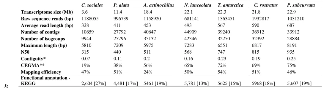
Table 2.2. Statistics of sequencing, assembly, and quality metrics……….……..43
ix
LIST OF FIGURES
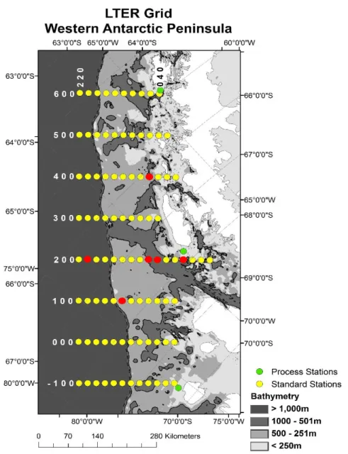
Figure 1.1. Map of PalmerLTER sampling grid along the WAP…….…….………..8
Figure 1.2. 18S rDNA phylogenetic tree of MMETSP diatoms and SO diatoms...…….…...…..17
Figure 1.3 A-D. Iron effect. ………….……….20
Figure 1.4 A-D. Light effect. ………….………...21
Figure 1.5 A-D. Combined iron and light effect……….………...22
Figure 1.6 A-I. Comparison of growth rates of all diatoms in all treatments………....23
Figure 1.7. A-C. µmax, Fe effect, and light effect as a function of biovolume ……….…………24
Figure 2.1. Contiguity of N. lanceolata transcriptome……….………...……….….………46
Figure 2.2. N. lanceolata transcriptome completeness………...……….….….………47
x
LIST OF ABBRREVIATIONS
AOX mitochondrial alternative oxidase
ASSY argininosuccinate synthase
Chl a chlorophyll a
Cu copper
CREG cellular repressor of E1A-stimulated genes
CYTC6 cytochrome c6
Fe iron
FLDA flavodoxin
FRE ferric reductase
FTN ferritin
FTR iron (III) permease
HNLC high-nutrient, low chlorophyll
ISIP iron-starvation induced protein
LTER Long-Term Ecological Research project
MCU multi-copper oxidase
MetE B12-independent methionine synthase MetH B12-dependent methionine synthase
MMETSP Marine Microbial Eukaryote Transcriptome Sequencing Project
Mn/Fe SOD manganese/Fe superoxide dismutase
NCP net community productivity
NiR nitrate reductase
xi
NRAMP natural resistance associated macrophage protein
PCYN plastocyanin
PET photosynthetic electron transport
PetF ferredoxin
POC particulate organic carbon
RHO rhodopsin
ROS reactive oxygen species
SO Southern Ocean
1
THESIS OBJECTIVES
Southern Ocean ecosystems are being altered due to anthropogenic climate change,
potentially resulting in shifts in the composition, diversity, and growth of primary producers,
including diatoms. Diatoms are a key group of phytoplankton in the SO and serve as the base of
most marine food webs in Antarctic polar waters (Saba et al. 2014). Since the Palmer long term
ecological research (LTER) project was established 20 years ago along the western Antarctic
Peninsula (WAP), researchers have recorded dramatic changes in the ecology and oceanography
or the region, especially at lower latitudes north of Palmer station (Saba et al. 2014). The WAP is
a particularly important area to study because in the past 50 years it has experienced an increase
in the mean annual air temperature of 7°C and a decline in sea ice extent and duration (Ducklow
et al. 2013), phytoplankton biomass (Montes-Hugo et al. 2009), krill (Saba et al. 2014), and
Adelie penguin populations (Ducklow et al 2007).
Within the Southern Ocean, the two main environmental variables that have been
identified as influencing phytoplankton growth are iron and light availability. Iron is supplied
naturally to the SO in by aeolian deposition, melting of sea ice, resuspension of sediments, and
deep winter mixing; however, rapid biological uptake and particle scavenging create a
high-nutrient, low chlorophyll (HNLC) condition in which primary productivity is limited by iron
availability (Cassar et al. 2007; Edwards and Sedgewick 2001; Death et al. 2014). The
mixed-layer depth, which influences mean light levels, is an important driver of phytoplankton growth
and the occurrence of blooms. Deep winter mixed layers (>100m) can result in phytoplankters,
like diatoms, experiencing considerable periods of light limitation (Sallée et al. 2010). As
warming climate changes the dynamics of light and iron availability in the SO, there could be
2
warming and freshening of the surface layer would enhance vertical stratification, resulting in
elevated mean irradiances; however, increased stratification might lead to more seasonal nutrient
limitation. The interactive effects of these environmental factors are still missing from models of
primary productivity.
Although there have been recent advances in our understanding of how iron and light
influence the physiology of SO diatoms, such as the possible lack of an interactive effect
between iron and light (Strzepek et al., 2012), few studies have investigated the molecular
underpinnings of the distinct physiological responses of polar diatoms to iron and light
limitation. The main objective of this study is to identify the genetic and cellular mechanisms
that may mediate a specific adaptation or acclimation to low iron and light in SO diatoms using
transcriptomics. Transcriptomics is the study of an organism’s mRNA transcripts, or expressed
genes, under specific circumstances using high-throughput sequencing. The benefits of analyzing
transcriptomes as opposed to genomes is they relate directly to the physiological status of the cell
and they are much easier to sequence than genomes. We also utilized publically available
transcriptomes from the Marine Microbial Eukaryote Transcriptome Project (Keeling et al.
2013), in conjunction with transcriptomes of SO diatoms I recently isolated, to understand the
molecular bases of physiological adaptations to low iron and light conditions.
From Western Antarctic waters, I have isolated three species of polar pennate diatoms
(Navicula lanceolata, Fragilariopsis cylindrus and Pseudo-nitzschia subcurvata) and six centric
diatoms (Thalassiosira antarctica, Actinochilus actinocyclus, Proboscia alata., Chaetoceros
debilis, Chaetoceros rostratus, and Eucampia antarctica) and performed a detailed examination
of their growth under varying ironand light conditions, along with transcriptomic analysis. We
3
previously been identified as being affected by iron and or light. Ultimately, by investigating
gene expression repertoires in conjunction with the effects of varying iron and light conditions
on growth, we can gain a better understanding of how differences in cellular mechanisms can
have far-reaching effects on diatom distribution and bloom occurrence, as well as processes that
4
CHAPTER 1: THE PHYSIOLOGICAL EFFECTS OF IRON AND LIGHT LIMITATION ON SOUTHERN OCEAN DIATOMS
Introduction
Diatoms are key organisms in determining net community production, and the overall
health of the Antarctic ecosystem (Ducklow, et al., 2007). Understanding the physiological
adaptation to changing light and iron regimes of polar diatoms along the western Antarctic
Peninsula is crucial to defining the role they have in sustaining Antarctic food web structure and
to what extent they can potentially affect biogeochemical cycles in the Southern Ocean. This
chapter will focus on the physiological adaptations of diatoms to limiting iron and light
conditions in terms of biophysical properties such as growth rate, photosynthetic efficiency and
biovolume, while providing essential background information.
Oceanography of the Southern Ocean: primary production, iron and light, and the rapidly changing Western Antarctic Peninsula
The Southern Ocean encircling Antarctica is an important driver of ocean circulation and
climate, biogeochemical cycling, and polar ecosystem structure and productivity (Sabine et al.,
2004, Gruber et al., 2009, Smith and Comiso, 2007). Although the Southern Ocean (SO) is a
relatively small ocean basin, it serves as the main intersection between the Atlantic, Pacific, and
Indian Oceans as well as between surface and deep waters. The principle current, the Antarctic
Circumpolar Currrent (ACC), is comprised of complex oceanographic zones and fronts with
5
Antarctic continent are particularly important as sites of upwelled waters rich in nutrients and
CO2 (Arrigo et al., 2008). When SO waters subsequently subduct to form subantarctic
intermediate and mode waters, they can ultimately fuel productivity in low latitudes when they
return to the surface (Sarmiento et al., 2004).
Primary production in the SO is highly variable spatially and temporally. Intense
phytoplankton blooms can occur when environmental conditions are favorable (Arrigo et al.,
2008). Production “hot spots’ include polynyas (regions of open water surrounded by sea ice)
(Tremblay and Smith, 2007), the seasonal ice zone (Smith and Nelson, 1986), and narrow
continental shelves (Sweeney, 2003). The phytoplankton communities in these regions, mainly
comprised of diatoms and prymnesiophytes such as Phaeocystis, are responsible for the large
quantity of dissolved inorganic carbon (DIC) fixed into organic matter by photosynthesis (Arrigo
et al., 2010). This organic matter can be recycled in surface waters, remineralized at depth, or
transported to deep ocean sediments via the biological pump, the efficiency of which determines
how much CO2 the ocean can draw down (Ducklow et al., 2001). Phytoplankton, especially
diatoms, also serve as the base of the polar marine food web, and support a wide variety of
Antarctic fauna like krill, fish, penguins, and whales (Saba et al., 2014). However, despite
plentiful nitrate and phosphate in the surface waters throughout the Southern Ocean, primary
productivity is patchy at best (Moore and Abbott, 2000).
In the Southern Ocean, abundant nutrients remain unutilized in the euphotic zone (Boyd,
2002; Falkowski 1998). It is a high-nutrient low-chlorophyll (HNLC) region in which
phytoplankton growth and biomass are limited by the availability of the micronutrient iron and
low light (Boyd, 2002). The high nutrient concentrations correspond to lower than expected
6
sources of iron (Sokolov and Rintoul, 2007). Iron is naturally added to the SO by aeolian
deposition, sea ice melt water, upwelling, and wind driven mixing over shallower coastal regions
(Cassar et al., 2007; Edwards and Sedgewick 2001; Death et al., 2014). Iron limitation is
supported by findings from artificial iron fertilization experiments, where large amounts of iron
are added to surface waters, leading to massive phytoplankton blooms (Boyd et al., 2000). Thus,
if more iron were available to phytoplankton, it is predicted that primary production would
increase and blooms would be more common (de Baar et al., 2005).
All organisms have a requirement for iron because it is an essential component of
enzymes required for photosynthesis, respiration, reduction of oxidized nitrogen and sulfur
compounds, and nitrogen fixation (Raven, 1999). Iron limitation in diatoms results in decreased
growth rates, chlorophyll-a contents, and photosynthetic efficiency (Timmermans et al., 2010;
Arrigo et al., 2010; Strzepek et al., 2012, Alderkamp et al., 2012). Iron limitation can also cause
diatoms to change their silicon-to-nitrogen consumption ratios. During periods of low iron input,
diatoms take up more silicon relative to nitrogen from seawater to incorporate in their frustules,
leaving surface waters depleted in silicic acid (Takeda, 1998). One proposed mechanism for
increased silica precipitation on frustules of iron-limited diatoms is the upregulation of spermine
synthase, a key enzyme in the polyamine synthesis pathway, which is associated with
precipitating silica on the cell wall (Nunn et al., 2013). This regulation, along with decreasing
cell size, could have evolved to allow faster sinking to the deep ocean possibly resulting in
reduced bacterial colonization and degradation (Marchetti & Cassar, 2009; Nunn et al., 2013). In
this way diatoms essentially control the silicon cycle and indirectly the global carbon cycle
7
Light is another important factor in regulating productivity in the surface mixed layer.
Because phytoplankton drift freely in the oceans, they are sensitive to dynamic physical ocean
processes such as stratification, changes in mixed layer depth, and sea ice extent and duration
(Deutsch, et al., 2009). In the SO, light availability varies daily and seasonally. Because the
mixed layer can also be very deep, phytoplankton may spend considerable periods of time below
the euphotic zone. This means diatoms must be able to exploit highly variable light environments
in order to survive, or lie dormant when light is absent for long periods of time. It has been
proposed that light is the major limiting factor during the austral spring and autumn (Smith and
Gordon, 1997), whereas Fe limits primary production during the summer (van Oijen, et al.,
2004). However, co-limitation of iron and light is also possible at different times and regions
(Tremblay and Smith, 2007; Boyd et al., 2002; Sedwick et al., 2007).
The WAP has been studied intensively for the past two decades by the Palmer
Long-Term Ecological Research Project (LTER; Fig. 1). This is an area of rapidly changing climate,
declining sea ice coverage, and deglaciation (Schofield et al., 2010). It is also experiencing the
fastest increase in mean atmospheric temperature in the world, with an increase of 7°C since
1951 (Meredith, 2005). Because biological systems in the WAP are sensitive to ice seasonality,
the recent warming and decrease in sea ice has resulted in significant decreases in phytoplankton
8
2009), as well as decreases in primary production, krill abundance, and the Adelie penguin
populations that are found north of Palmer Station (Saba et al., 2014).
9
Effects of iron and light on polar diatom physiology and metabolism
It is well known that iron and light are the primary environmental bottom-up controls that
influence primary productivity and diatom abundances in the Southern Ocean (Boyd, 2002;
Strzepek et al., 2012). Recent investigations have revealed multiple physiological mechanisms
for increasing iron acquisition, efficiently allocating intracellular iron, and decreasing iron
requirements in polar diatoms in order to adapt or acclimate to low iron environments. These
include reducing cellular to-carbon ratios (Strzepek et al., 2012), substituting
iron-containing redox catalysts with proteins that can use other metal cofactors (Peers and Price,
2006), activating a high-affinity iron uptake system (Raven, 1990), performing luxury uptake of
iron (Marchetti et al., 2009), and increasing surface area-to-volume (SA:V) ratios (Sunda &
Huntsman, 1995). Increasing SA:V ratios are useful under nutrient deficiency because it
decreases the diffusive boundary layer and increases iron uptake rates.
Intracellular iron requirements, or quotas, are measured as Fe:C ratios and are a measure
of the iron demands of the cell. Fe requirements of a diatom can be inferred by either measuring
the concentration of Fe in the medium that supports growth and calculating a half-saturation
constant in relation to growth rates (Kµ), or by measuring their intracellular Fe content. Diatoms
isolated from the SO have particularly low iron contents and higher iron use efficiencies, or the
amount of C fixed per mole of Fe per day (Marchetti and Maldonado, 2016). Strzepek et al.
(2011) found that intracellular iron concentrations normalized to either carbon or cell volume
were extremely low for SO diatoms. For example, Proboscia inermis had the lowest cellular
Fe:C ratio recorded (0.4 µmol:mol with a relative growth rate of 0.6 d-1 in relation to the high iron treatment), while the model temperate diatom, T. oceanica, had an Fe:C of 0.58 µmol:mol
10
iron requirements that were at least twofold lower than other examined oceanic species.
Although the diatoms used in their study were large (>20 um in some cases), and thus, had lower
cell SA:V ratios, they appeared to compensate for this by greatly reducing their iron
requirements.
Raven et al. (1999) theoretically calculated that most of the iron required by a cell is
allocated to catalytic proteins used in photosynthetic electron transport, specifically the
photosynthetic units (PSUs). Iron is used in the major photosynthetic complexes including:
photosystem I (PS1), photosystem II (PSII), and the cytochrome b6f complex. The iron
requirements of the cell can be reduced by changing the ratio of each complex in response to
growth, irradiance, and iron availability (Raven et al., 1999; Sunda and Huntsman 1997). For
example, because PSII requires less iron than PSI and cytochrome b6f, oceanic diatoms can
become more enriched in PSII relative to the other complexes (Strzepek and Harrison, 2004;
Green et al., 1991).
Because the photosynthetic architecture of diatoms requires 50-80% of the available
intracellular iron, photoacclimation and iron interactions are intimately linked (Raven 2013).
Laboratory studies have documented distinct physiological responses to low irradiances when
cells are grown in iron-limiting conditions such as increasing the number or size of
photosynthetic subunits, altering light harvesting elements, and decreasing Chl a contents.
(Timmermans et al., 2010; Arrigo et al., 2010; Strzepek et al., 2012, Alderkamp et al., 2012).
Although some of these strategies have higher iron requirements, polar diatoms appear to have
the ability to maintain low cellular iron demands when grown under low irradiances. Strzepek et
al. (2012) hypothesized that polar diatoms may increase the size rather than the number of PSU
11
cytochrome b6f complex and they perform photosynthetic electron transport (PET). By
increasing the size of the PSU’s, they could lower their iron requirements and grow at a higher
relative growth rate. This strategy would contradict previous research regarding the antagonistic
relationship between light and iron, in which photoacclimation strategies to low light cause
cellular iron requirements to increase (Raven et al. 1990).
The interaction of iron and light in the SO is particularly important for diatoms because
they can experience environmental conditions both low in iron availability, and with variable
light levels due to deep mixed layers. For example, F. cylindrus, a common bloom former in the
Southern Ocean, has been shown to thrive in shallow mixed layers where light levels can be
high, and under sea ice where light levels just below the ice can be as low as 0.1% of surface
irradiance. This is because at high light levels, F. cylindrus can maintain low concentrations of
photosynthetic pigments and high concentrations of photoprotective pigments, while in low light
regimes, this species can absorb green light between 500-575nm, which is found underneath sea
ice (Arrigo et al., 2010). This could be due to F. cylindrus containing the chromophore retinal, a
pigment used in rhodopsin (Marchetti et al., 2015). Rhodopsin (RHO) is a light-driven proton
pump that could potentially act as an alternative to photosynthesis in low iron conditions. While
the chromophore retinal requires a small amount of iron, the rhodopsin protein uses a much
smaller amount than the photosynthetic apparatus. This could indicate that diatoms may lower
their iron requirements through activating the rhodopsin gene. Pseudo-nitzschia granii has been
shown to increase transcripts of RHO under low iron conditions (Marchetti et al. 2015).
However, it is still unclear if the rhodopsin in diatoms is producing ATP as an alternative to
12
Understanding the physiological adaptation to changing light and iron availability of
polar diatoms along the WAP is crucial to defining their role in the Antarctic food web and to
what extent they can potentially affect biogeochemical cycles in the SO. The goal of this chapter
was to understand how iron and light limitation affects the growth rate, photosynthetic
efficiency, and biovolume of polar diatoms. We sought to determine if polar diatoms grown
under iron and light limitation were indeed co-limited by iron and light, or if they exhibited other
types of resource limitation. Ultimately, this research will expand our existing knowledge of how
iron and light interactions affect polar and temperate species (Strzepek et al. 2012; Sunda &
Huntsman et al. 1997)
Materials and methods
Diatom isolation and identification - Eight species of diatoms were isolated from the
Western Antarctic Peninsula along the PalmerLTER sampling grid in March 2014 (see Fig. 1.1
for locations). Isolations were performed using an Olympus CKX41 inverted microscope by
single cell isolation with micropipette (Anderson 2005). The identification of diatom species was
performed by morphological characterization and 18S rRNA gene sequencing. DNA was
extracted with the DNeasy Plant Mini Kit according to the manufacturer’s protocols (Qiagen).
Amplification of the nuclear 18S rDNA region was achieved with standard PCR protocols using
eukaryotic-specific, universal 18S forward and reverse primers. Primers were obtained from
Medlin et al. (1982) and are as follows: 18AF 5’-AACCTGGTTGATCCTGCCAGT-3’ and
18BR 5’- TGATCCTTCTGCAGGTTCACCTAC -3’. The length of the region amplified is
approximately 1600 base pairs (bp). Pseudo-nitzschia species are difficult to identify by 18S
13
subcurvata was provided through sequencing of the 18S-ITS1-5.8S regions. Amplification of
this region was performed with 18SF-euk and 5.8SR_euk primers according to Hubbard et al.
(2008), which are as follows: 18SF-euk 5’-CTTATCATTTAGAGGAAGGTGAAGTCG-3’ and
5.8SR-euk 5’-CTGCGTTCTTCATCGTTGTGG-3’. PCR products were purified using either
QIAquick PCR Purification Kit (Qiagen) or ExoSAP-IT (Affymetrix) and sequenced by Sanger
DNA sequencing (Genewiz). Sequences were edited using Geneious Pro software
(http://www.geneious.com, Kearse et al., 2012) and BLASTn sequence homology searches were
performed against the NCBI nucleotide non-redundant (nr) database to determine species with a
cutoff identity of 98%. In the case of P. subcurvata, a specific polymorphic fragment of the ITS1
region was further investigated by aligning PnAll primers (Hubbard et al., 2008) to the
18S-ITS1-5.8S region, resulting in a fragment of 171 bp. Hubbard et al. (2008) found the length of
this fragment to be unique to a given Pseudo-nitzschia species despite very similar sequences.
My analysis of this fragment resulted in the highest sequence homology and length similarity, an
ITS1 fragment of 171 bp, to P. subcurvata (Strain: 1-F, Accession number: DQ329205).
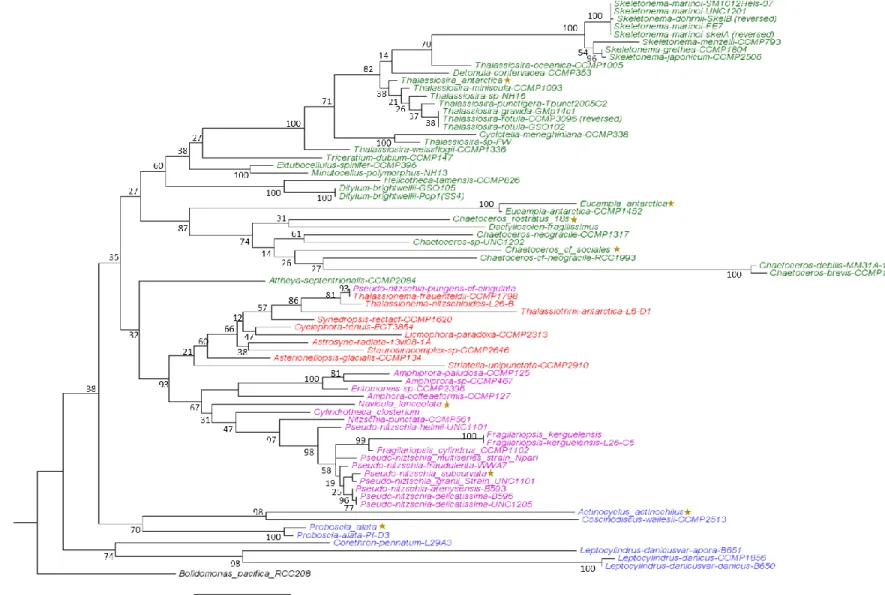
Diatom phylogenetic analysis was performed with Geneious Pro and included 71
additional diatom 18S rDNA sequences from publically available genomes and transcriptomes,
including all those in the Marine MicroEukaryote Transcriptome Sequencing Project (MMETSP)
database. Diatom sequences were trimmed to the same length and aligned with MUSCLE (Edgar
2004). A phylogenetic tree was created in Mega with the Maximum-likelihood method of tree
reconstruction, the Jukes-Cantor genetic distance model (Jukes and Cantor 1969), and 100
bootstrap replicates.
Growth conditions, physiological characteristics and biovolumes - Isolates were
14
photons m-2 s-1 (standard light) and with media containing two iron concentrations. Cultures were grown in synthetic seawater medium, AQUIL, enriched with filter sterilized vitamin and trace
metal (buffered with 100 μmol L-1 EDTA) solutions as well as 300 μmol L-1 nitrate, 200 μmol L -1 silicate and 20 μmol L-1 phosphate. Premixed Fe-EDTA (1:1) was added separately for total Fe
concentrations of either 1370 nmol L-1 (pFe 19) or 3.1 nmol L-1 (pFe 21.7) to achieve high iron and low iron media, respectively. All media preparation and subsampling were performed under
a positive-pressure, trace metal clean laminar flow hood. Cultures were grown in acid-washed 28
mL polycarbonate centrifuge tubes (Nalgene) and maintained in exponential phase by dilution.
Specific growth rates were calculated from the linear regression of the natural log of in vivo
chlorophyll a fluorescence using a Turner 10-AU fluorometer (Brand et al. 1981).
Photophysiological parameters were measured with a Fluorescence Induction Relaxation
System (FIRe) (Satatlantic). Samples were dark acclimated for at least 10 minutes and
measurements were taken of each culture for photosynthetic efficiency (Fv:Fm), and functional
absorption cross-section of PSII, (σPSII). FIRe parameters were set to measure single turnover
flash of PSII reaction centers (single closure event) with a sample delay of 100, and a total of 50
samples (Gorbunov and Falkowski, 2004).
Cell dimensions and biovolume measurements - To estimate biovolumes (V) of each
diatom species, frustules were viewed using an Olympus BX61 Upright Wide Field Microscope
with the differential interference contrast (DIC) imaging mode and a 60X/1.42 Oil PlanApo N
objective lens. Valve apical length (AL), transapical width (TW), and pervalvar height (PH)
dimensions were estimated with Scion Image software (Informer Technologies, Inc.). Diatom
15
modeling the cells after geometric shapes. Pennate diatoms were modeled after elliptic prisms
and centric diatoms were modeled after cylinders. The equations used are:
Cylinder: 𝑉 =𝜋 4∙ h ∙ d
2
Prism on elliptic base: 𝑉 =𝜋
4 ∙ 𝐴𝐿 ∙ 𝑇𝑊 ∙ 𝑃𝐻
For all species except T. antarctica, at least four cells were imaged and measured. T. antarctica
had a sample size n = 1.
Data analysis - Statistical analyses of growth rates and photophysiological data were
performed with SigmaPlot 12.5 (SysStat Software Inc.). To test for significant differences
between treatments, Two-Way Analysis of Variance (ANOVA) was performed with a
significance level set to p<0.05. ANOVA also tests for normality using Shapiro-Wilks and Equal
Variance tests. Because ANOVA does not test all interactions, an unpaired t-test was performed
between –FeLL and +FeSL for µ, Fv:Fm, or σPSII. All tests passed the Shapiro-Wilks Normality
tests unless otherwise stated, in which case p-values are representative of the Mann-Whitney
Rank Sum test. Post-hoc Tukey tests were also performed in order to determine which treatments
differed significantly (p < 0.05). Plots were created using Matlab software.
Results
Diatom isolates – Isolates collected for this study include two raphid pennate diatoms:
Navicula lanceolata and Pseudo-nitzschia subcurvata; four radial centric diatoms: Actinocyclus
actinochilus, Eucampia antarctica, Proboscia alata, and Thalassiosira antarctica; and two
bi-multipolar centrics: Chaetoceros cf sociales and Chaetoceros rostratus. Our diatom collection
does not include an araphid pennate. The diatoms were isolated from the following stations:
16
mid-shelf regions (Table 1). Despite originating from a relatively small geographic region off the
Western Antarctic Peninsula, these diatoms represent diverse lineages. Full-length 18S rDNA
sequences were successfully obtained for eight of the isolates which were aligned with 76 18S
rDNA sequences obtained from the MMETSP diatom database (Fig. 1.2). Of the diatoms
sequenced as part of the MMETSP, four of our polar isolate transcriptomes are unique, as A.
actinochilus, N. lanceolata, C. rostratus, and C. sociales have not been previously sequenced.
Transcriptome sequencing of E. antarctica was unsuccessful within our first round of
transcriptome sequencing; however, the MMETSP database also contains a sequenced strain of
this species.
Table 1.1. Isolates with strain designation, sampling location along the PalmerLTER, and accession numbers of best BLAST hit in NCBI Genbank.
Polar diatoms Strain ID Date Isolated
PalmerLTER
Station Latitude Longitude GenBank best hit Isolator
Thalassiosira antarctica UNC1401 Mar-14 200.180 -66.5795 -72.7391 DQ514874.1 C. Moreno
Eucampia antarctica UNC1402 Mar-14 200.020 -67.6418 -70.2688 X85389.1 C. Moreno
Actinocyclus actinochilus UNC1403 Mar-14 400.040 -66.2540 -67.3366 AY485506.1 C. Moreno
Navicula lanceolata UNC1404 Mar-14 100.040 -68.1121 -72.3461 KC771158.1 C. Moreno
Proboscia alata UNC1405 Mar-14 200.040 -67.5111 -70.5890 AY485525.1 C. Moreno
Pseudo-nitzschia subcurvata UNC1406 Mar-14 200.040 -67.5111 -70.5890 AY485490.1 C. Moreno
Chaetoceros cf sociales UNC1407 Mar-14 200.-020 -67.2956 -69.6654 HM581778.1 C. Moreno
Chaetoceros rostratus UNC1408 Mar-14 200.-020 -67.2956 -69.6654 X85391.1 C. Moreno
Figure 1.2. Phylogenetic tree generated through Maximum-likelihood analysis of 18S rDNA sequences from MMETSP diatoms and new isolates from the WAP denoted with a yellow star. Centric diatoms are highlighted in green (bi-polar) and blue (radial) and pennate diatoms are
highlighted in purple (raphid) and red (araphid). Node labels show percent consensus support for tree arrangement, and branch lengths represent divergence between two nodes in the tree (substitutions per site of sequence alignment). The outgroup of the rooted tree is Bolidomonas pacifica RCC208, a eukaryotic, picoplanktonic heterokont.
18
Growth parameters – Growth rates are expressed as specific growth rate (µ; d-1) or as relative growth rate in relation to the maximum specific growth rate (µmax) determined in
iron-replete and standard light conditions (µ:µmax). Other physiological parameters such as Fv:Fm and
σPSII are expressed in specific terms. Nutrient limitation of iron and light, or an additive
interaction of both these resources, was diagnosed in an isolate if the mean relative growth rate
and Fv:Fm were significantly reduced (p < 0.05).
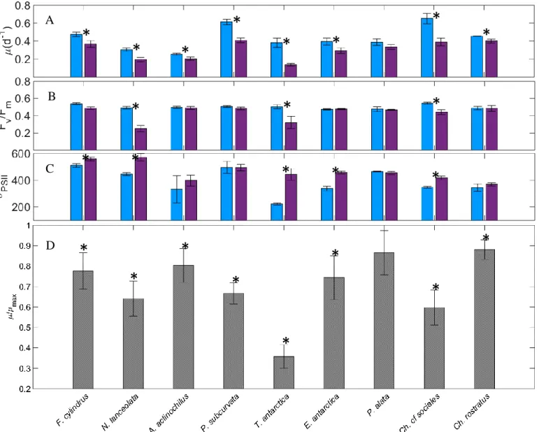
Among the isolates grown in iron-replete, standard light conditions (+FeSL), µ max ranged
from 0.25 d-1 (A. actinochilus) to 0.72 d-1 (P. subcurvata; Fig. 1.3A); F
v:Fm values were between
0.473 (E. antarctica) and 0.545 (C. sociales; Fig. 1.3B); and σPSII ranged from 222 for T. antarctica to 511 in F. cylindrus (Fig. 1.3C).
The relative growth rates of all isolates in low iron cultures (-FeSL) were reduced by 12 –
64%. Except for P. alata, all diatoms experienced some degree of iron limitation, as their µ:µmax
were significantly reduced (ANOVA, p<0.05) (Fig. 1.3D). Fv:Fm is known to decrease in
iron-limited cells; however, in our study this measure was less responsive to iron stress than was
growth rate. However, for N. lanceolata, T. antarctica, and C. cf sociales, Fv:Fm was reduced 19
– 50% (ANOVA, p<0.05), indicating these diatoms were experiencing moderate to severe
nutrient stress (Fig. 1.3B). In addition, σPSII was observed to significantly increase (ANOVA,
p<0.05) under low iron conditions, which is also characteristic of Fe limitation in diatoms (Fig.
1.3C). While a decrease in Fv:Fm was not observed in F. cylindrus and E. antarctica, an increase
in σPSII was observed.
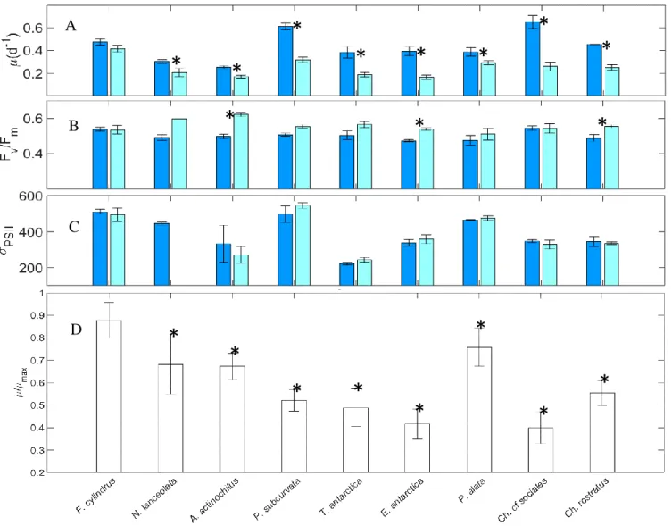
Low irradiance (+FeLL) physiologically stressed all diatoms, except F. cylindrus,
resulting in reductions in growth rates (Fig. 1.4A). For the remaining species, µ:µmax was
19
actinochilus, P. subcurvata, C. rostratus, and E. antarctica, Fv:Fm significantly increased from 7
– 25% (p < 0.05; Fig. 1.4B). σPSII did not significantly change from +FeSL treatments (Fig.
1.4C).
When cells were grown under combined low iron and low light growth conditions
(-FeLL), all isolates experienced significant decreases in µ:µmax of at least 35% (p < 0.05; Fig.
1.5A) relative to +FeSL. However, decreases in Fv:Fm were only evident in F. cylindrus
(Mann-Whitney, p = 0.01), P. alata, and N. lanceolata (Fig. 1.5B). An increase in σPSII of ~30%
(p<0.05) was observed for F. cylindrus, P. alata (Mann-Whitney; p = 0.024), and C. sociales
(Fig. 1.5C). Steady-state measurements of Fv:Fm and σPSII in T. antarctica and N. lanceolata
under -FeLL conditions were not obtainable due to cessation of growth in these isolates.
We also determined interactive effects of low iron and light in our isolates (Fig. 1.6 A-I).
Diatoms exhibiting no interactive effects include C. cf sociales, P. subcurvata, A. actinochilus,
C. rostratus, N. lanceolata, and T. antarctica. Growth rates in these –FeLL cultures were not
significantly different from either –FeSL or +FeLL treatments. Diatoms that only appeared to be
limited by light were E. antarctica and C. rostratus. C. rostratus also showed a small degree of
iron limitation, but only in light saturating conditions. P. alata and F. cylindrus both exhibited
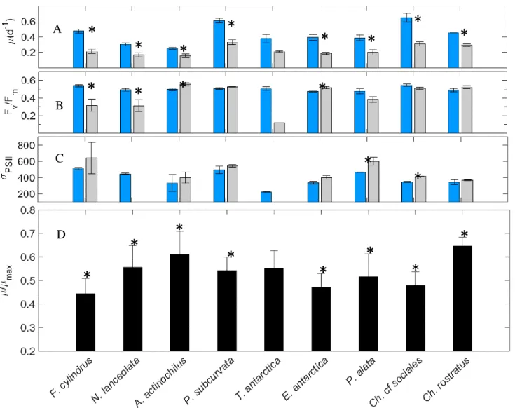
Figure 1.3. A-D. Iron effect. Comparison of specific growth rate (A), Fv:Fm (B), and σPSII (C) in +FeSL and –FeSL conditions. Relative values of growth rates for each isolate are shown in D. Error bars represent the standard error of the mean (n > 3 for all +FeSL and –FeSL treatments). Isolates labeled with an asterisk indicate the –FeSL treatment is significantly different from the +FeSL treatment (Two-Way ANOVA, p < 0.05).
D
*
*
*
*
*
*
*
*
*
*
*
*
*
*
*
*
*
*
*
*
*
*
*
*
A
B
C
D
Figure 1.4. A-D. Light effect. Comparison of specific growth rate (A), Fv:Fm (B), and σPSII (C) in +FeSL and +FeLL conditions. Relative values of growth rates for each isolate are shown in D. Error bars represent the standard error of the mean (n > 3 for +FeSL; n > 3 +FeLL treatments, except for N. lanceolata Fv:Fm and σPSII n = 1). Treatments labeled with an asterisk are significantly different from the +FeSL treatment (Two-Way ANOVA, p < 0.05).
*
*
*
*
*
*
*
*
*
*
*
*
*
*
*
*
*
*
*
A
B
C
D
Figure 1.5. A-D. Combined Fe and light effect. Comparison of specific growth rate (A), Fv:Fm (B), and σPSII (C) in +FeSL and –FeLL conditions. Relative values of growth rate for each isolate are shown in D. Error bars represent the standard error of the mean (n > 3 for +FeSL; n > 3 -FeLL treatments, except for T. antarctica Fv:Fm and σPSII n = 1; N. lanceolata Fv:Fm and σPSII n = 2). Treatments labeled with an asterisk are significantly different from the +FeSL treatment (Student’s t-test, p<0.05).
*
*
*
*
*
*
*
*
*
*
*
*
*
*
*
*
*
*
*
*
*
*
A
B
C
D
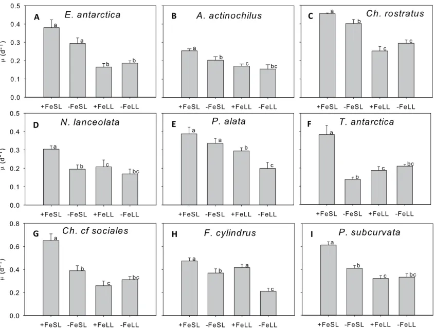
Figure 1.6. A-I. Comparison of growth rates of each isolate in each treatment. Letters denote significant differences in growth rates (ANOVA, p < 0.05.; Student’s t-test or Mann-Whitney, p < 0.05). Error bars represent one standard error of the mean.
A B C
D E F
G H I
24
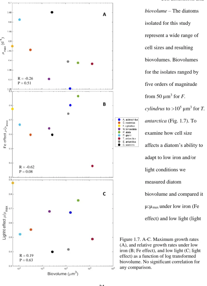
Cell dimensions and
biovolume – The diatoms
isolated for this study
represent a wide range of
cell sizes and resulting
biovolumes. Biovolumes
for the isolates ranged by
five orders of magnitude
from 50 µm3 for F.
cylindrus to >105 µm3 for T. antarctica (Fig. 1.7). To
examine how cell size
affects a diatom’s ability to
adapt to low iron and/or
light conditions we
measured diatom
biovolume and compared it
µ:µmax under low iron (Fe
effect) and low light (light
Figure 1.7. A-C. Maximum growth rates (A), and relative growth rates under low iron (B; Fe effect), and low light (C; light effect) as a function of log transformed biovolume. No significant correlation for any comparison.
R = -0.62 P = 0.08 R = -0.26 P = 0.51
A
R = 0.19 P = 0.63
B
25
effect) conditions. In replete conditions, µmax was not significantly correlated with biovolume (r
= -0.26, p = 0.51). Similarly, under low iron and low light conditions there was not a significant
correlation between µ:µmax and biovolume (iron; r = -0.06, p = 0.08 and light; r = 0.19, p = 0.63).
Discussion
This is the first study in which the physiology of nine Southern Ocean diatoms have been
directly compared. The number and diversity of diatoms, including both centrics and pennates,
isolated from this region is valuable as a way to understand how diatoms are able to survive
under low iron and light conditions. Diatoms are a diverse lineage among the stramenopiles and
those that reside in HNLC waters employ unique adaptations and acclimations to variable
environmental conditions, such as low Fe:C ratios, and high relative growth rates under low light
and iron availability (Strzepek et al. 2012) .
Maximum specific growth rates determined for F. cylindrus,E. antarctica, P. alata, T.
antarctica, and A. actinochilus are comparable to previously published growth rates of the same
or closely related species (Strzepek et al. 2012; Agusti & Duarte 2000;Timmermans et al. 2004).
Strzepek et al. (2012) used DFB, a stronger siderophore than EDTA, resulting in slightly lower
growth rates in the two diatoms used in their study (E. antarctica, P. inermis). To my
knowledge, growth rates under varying iron and light conditions of three of our diatoms have not
previously been published, including C. rostratus, C. sociales, and N. lanceolata.
As a way to measure degree of iron limitation, the photophysiological measurements
Fv:Fm and σPSII were also collected. In iron-limited cells, Fv:Fm is expected to decline as the
light-harvesting antenna systems transfer electrons to PSII reaction centers less efficiently. This
26
(Green et al 1991; Petrou et al. 2014; Boyd et al. 2000; Trimborn et al. 2014). Conversely, σPSII
characteristically increases under iron-limited conditions as a result of an increase in the ratio of
PSII antenna complexes relative to reaction center complexes (Green et al. 1991). The increase
in σPSII has also been proposed to be due to an increase in the size of the absorption cross section
area (Strzepek et al. 2012).
In our low iron treatment, this pattern was evident in only C. cf sociales, T. antartica, and
N. lanceolata, indicating these diatoms were experiencing moderate to severe iron limitation. P.
alata did not exhibit any of the characteristics associated with iron limitation. While a decrease
in µ:µmax and Fv:Fm was not observed in F. cylindrus and E. antarctica, an increase in σPSII was
seen, perhaps implying these species were just experiencing the onset of iron-limited growth
when grown in the low iron medium used in this study.
Light limitation resulted in reduced µ:µmax in all diatoms except for F. cylindrus, which
did not exhibit significant reduction in µ, Fv:Fm, or σPSII, indicating this species was not
experiencing light stress under the low light condition used in this study. For most other diatoms
growing under low light, neither Fv:Fm nor σPSII changed relative to the replete treatment, except
for A. actinochilus, E. antarctica, and C. rostartus, in which Fv:Fm actually increased. These
trends in Fv:Fm and σPSII might suggest Southern Ocean diatoms have different mechanisms to
maintain a healthy photosynthetic apparatus along with high photosynthetic efficiency, despite a
reduced growth. Interestingly, diatoms that overwinter in the SO and experience months of
darkness appear to retain a functional photosynthetic electron transport chain (Peters and Thomas
1996). This would help them rapidly acclimate to more favorable light conditions when the
mixed layer depth shoals in spring. These observations suggest Fv:Fm and σPSII are not good
27
In many of these treatments, µ, Fv:Fm, and σPSII were appreciably decoupled. In terms of
iron limitation, this may indicate several of our diatoms were only moderately iron-limited under
our culture conditions. In order to produce growth and physiological parameters with
characteristics of iron limitation, a further reduction in iron concentrations and/or a stronger iron
chelator, such as desferrioxamine B mesylate (DFB), would need to be added to the culture
medium. Under light-limiting conditions, Fv:Fm did not decrease, indicating SO diatoms may not
have been experiencing much stress and maintained efficient electron transfer between light
harvesting antennae and reaction centers. It may also indicate that SO diatoms have unknown
novel mechanisms for maintaining high levels of photosynthetic efficiency. In terms of light
limitation in SO diatoms, Fv:Fm may not be a good indicator of stress. µ was the most responsive
physiological parameter for iron and/or light stress in our study. Overall, SO diatoms have larger
σPSII and lower maximum Fv:Fm than temperate diatoms, which allows them to take advantage of
low iron and light conditions (Strzepek et al. 2012 & Sugget et al. 2009)
Three environmental stress responses were observed: no interaction between iron and
light, an additive interaction between iron and light, and a response in which light was the main
limiting resource (Fig. 1.6). A stress response with no interaction is defined as a response in
which resource limitation of two potential limiting nutrients is no greater than one of the two
limiting nutrients by itself. In this example, reducing both iron and light produces the same
reduction in µ as though either iron or light were limiting. C. cf sociales, P. subcurvata, A.
actinochilus, C. rostratus, N. lanceolata, and T. antarctica growth can be classified as having no
interaction between iron and light as –FeLL cultures were not significantly different from either
–FeSL or +FeLL treatments (Fig. 1.6). This would indicate a synergistic relationship between
28
found that in temperate diatoms, low-light, iron-limited cells had reduced growth rates compared
to high-light, iron-limited cells due to increased iron demands in order to efficiently use the
photosynthetic electron transport chain under low light. It is interesting the diatoms that exhibit
this growth response include among the smallest and largest diatoms used in this study. Small
diatoms may be able to cope with low iron and low light without a decrease in growth because of
increased SA:V ratios, decreased diffusive boundary layers, and increased iron uptake rates.
Similar findings were observed in C. brevis, a small diatom grown in natural SO water (without
EDTA) which thrived under low iron and low light conditions (Timmermans et al. 2001; Oijen et
al. 2004). In contrast, large diatoms are able to exhibit this growth pattern between iron and light
because they minimize their Fe:C ratios (Strzepek et al 2012). SO diatoms also have the ability
to reduce iron strongly bound to organic complexes such as EDTA, DFB, and other
siderophores, possibly making the pool of bioavailable iron larger in this region (Strzepek et al.
2011).
E. antarctica demonstrated only light limitation in this study, with no significant iron
effect on growth. It is possible that E. antarctica was not iron-limited in our study as we used
EDTA and not a stronger siderophore such as DFB. Previous researchers, who added DFB to
induce iron limitation in this species (Strzepek et al. 2011), also observed that E. antarctica had
high relative growth rates and near-constant Fe:C ratios even in low irradiances. In the case of C.
rostratus, the patterns in growth are slightly more complicated, but are mainly driven by
irradiance as well. Under light saturating conditions, low iron availability resulted in slightly
reduced growth rate (12%; p < 0.001), however, a decrease in light resulted in a much larger
29
less pronounced in low light cultures than light saturated cultures, indicating C. rostratus is more
sensitive to change in light than iron.
P. alata and F. cylindrus demonstrated an additive interaction between iron and light. In
both of these isolates, growth rates were most reduced in the combined low iron and low light
treatment compared to either variable alone. These two diatoms are also at the extremes of the
size distribution among our diatoms. P. alata and F. cylindrus are the second to largest and the
smallest cells, respectively, yet have similar functional characteristics in relation to iron and light
limitation. The lack of synergistic effects observed for most of the isolates support the proposal
by Strzepek et al. (2012), in which iron and light limitation in SO diatoms result in a distinctive
photoacclimatory response that increases the number but not the size of PSU, which minimizes
iron demands.
Phytoplankton cell size is known to correlate well with resource utilization and growth
(Epply et al 1969). Nutrient uptake rates decrease due to diffusion limitation with increasing cell
size (Marchetti and Maldonado, 2016), and growth typically decreases with increasing size
(Banse 1976). Thus, small cells should dominate HNLC regions; however, iron kinetics are not
the only factor responsible for community composition, allowing many large diatoms to exist in
the SO for several biophysical reasons. First, they have large vacuoles with high nutrient storage
capabilities, which could be useful under fluctuating nutrient regimes (Raven 1987). Second,
their large size can serve as protection from zooplankton grazing and allows them to control their
depth in the euphotic zone by varying their ballast (Armstrong et al. 2002). At the biochemical
level, SO diatoms also have adaptive strategies to acquire iron and efficiently use light so they
can survive in the SO. When cells are iron limited they can substitute iron-containing redox
high-30
affinity iron uptake system (Raven, 1990), perform luxury uptake of iron (Marchetti et al., 2009),
and reduce Fe:C ratios (Strzepek et al., 2012). However, the physiological trade-off of reducing
iron quotas in response to iron limitation is that open ocean diatoms compromise their ability to
quickly acclimate to rapidly changing irradiance (Strzepek and Harrison, 2004).
Diatoms in this study displayed a weak, insignificant negative correlation between
decreasing growth rate and increasing biovolume. This poor correlation between maximum
growth rates and biovolume was also found among other SO diatoms (Strzepek et al. 2011).
Maximum growth rate appears to be species specific and in the SO is likely determined by
temperature and nutrient resource (nutrient) utilization strategies of each diatom. In our survey, it
appears there are several types of diatoms exhibiting different resource utilization strategies,
including diatoms that grow quickly and utilize nutrient pulses like C. cf sociales and P.
subcurvata (highumax but low u:umax in –FeLL); diatoms that grow slowly but can also take
advantage of nutrient pulses perhaps by having large storage capacities such as E. antarctica and
P. alata (large vacuoles); and diatoms that have low growth rates but can survive well under
severe resource limitation, such as N. lanceolata and C. rostratus (high u:umax in –FeLL).
Interestingly, the ability of SO diatoms to cope with low iron and low light also does not
scale with biovolume. This could be a result of not being iron limited in our medium, but it also
suggests that large, SO diatoms have efficient mechanisms and gene repertoires that can maintain
higher µ:µmax under these stressful conditions. On the other hand, smaller cells, such as F. cylindrus, P. subcurvata, and C. rostratus are able to maintain relatively high µ:µmax, perhaps
due to their increased SA:V ratios.
The oceanography of the sites from which these diatoms were isolated could partially
31
stations (200.-020 and 100.040) and likely represent coastal and/or sea ice dependent diatoms
adapted to higher iron concentrations and shallower mixed layers associated with upwelling
along the shelf. T. antarctica was isolated from an off-shelf region (200.180). Thalassiosira
species have been shown to be associated with coastal areas and sea ice blooms (Lin et al 2015;
Garibotti et al. 2005). P. alata and E. antarctica were isolated from a more southerly station
associated with the sea ice edge bloom (200.040) where iron and light can interact
synergistically. These large diatoms would benefit from their storage-adapted lifestyle in this
region as the sea ice edge can be a source of nutrients and iron pulses and is associated with a
shallower mixed layer.
Because of the importance of light in the SO, further research should investigate the
effects of high light or diel light cycles on these diatoms. SO diatoms overwinter in near
darkness or within sea ice where light levels routinely reach 0.1% of surface irradiance for
months at a time. Further research with SO diatoms should investigate the elemental ratios
(C:N:Si) including iron quotas and iron-use-efficiencies (amount of C per Fe used per day) to
understand how these diatoms change their cellular composition in times of iron stress and how
these physiological adaptations could relate to biogeochemical cycles. In particular, during the
PalmerLTER 2014 field season, the same year in which these diatoms were isolated, it was
observed that four diatom genera, including Probosica, Thalassiosira, Pseudo-nitzschia, and
Stellarima, contributed to 88% of the net community productivity (NCP) variance (Lin et al.
submitted). Thus, understanding cellular and molecular mechanisms of iron and light limitation
within specific diatoms could help in our understanding of how these particularly important
32
Phenotypic variability in growth rates and photophysiology as a function of iron and/or
light status observed among the diatoms examined in this study could likely be explained by
differences in gene repertoires in relation to iron-requiring processes such as photosynthesis and
nitrogen assimilation. In the next chapter, I will investigate if SO diatoms show a correlation
between presence (or inferred absence) of particular genes and the physiological parameters
discussed in this chapter. Elucidating the genotypic variability among SO diatoms could help in
understanding phytoplankton community composition and succession, nutrient cycling, and
carbon and silica sequestration in HNLC regions, which is especially important in light of
33
CHAPTER 2: INVESTIGATING THE TRANSCRIPTOMES OF SEVEN DIATOMS IN RELATION TO IRON AND LIGHT STATUS
Introduction
The first chapter addressed phenotypic acclimation to iron and light limitation. Here, I
will investigate genotypic adaptations that have possibly resulted from selective pressure from
low iron and light conditions. Despite numerous physiological investigations (Strzepek et al.,
2012; Arrigo et al., 2010; Alderkamp et al., 2012) on the effects of iron and light limitation on
Southern Ocean diatoms, there are no studies that use modern molecular techniques such as
next-generation sequencing in conjunction with physiological studies to elucidate how SO diatoms
cope with light and iron stress (Park et al. 2010; Strauss, 2012).
Diatoms are a diverse lineage comprised of two major clades. Centrics are older and
divided further into radial or bi-multipolar varieties; pennates diverged later and are comprised
of araphid and raphid pennates (Kooistra et al. 2007). The first two diatoms to have their
genomes sequenced, the centric T. pseudonana and the pennate P. tricornutum, have been shown
to be more divergent than fish and humans despite their relatively recent evolution (Bowler et al.
2010). In ancient oceans diatoms became successful 190 Mya, when the atmosphere contained
almost eight times higher CO2 concentrations and higher levels of dissolved inorganic iron
(Armbrust 2009). Because iron has a high capacity as an electron donor and acceptor, diatoms
34
other biochemical reactions. Unfortunately, in modern oceanic environments iron is rapidly
oxidized and precipitated. The selective pressure in low iron environments has resulted in
diatoms evolving strategies including luxury storage, protein substitution, photoacclimation, and
reduced cell size. In this study we investigated 22 genes (isogroups) from nine different
metabolic groups including photosynthetic iron-dependent and independent proteins, iron
storage, high-affinity iron-uptake, transport proteins, vitamin synthesis, superoxide dismutase
and alternative oxidase, nitrogen assimilation, and proteins involved in the urea cycle.
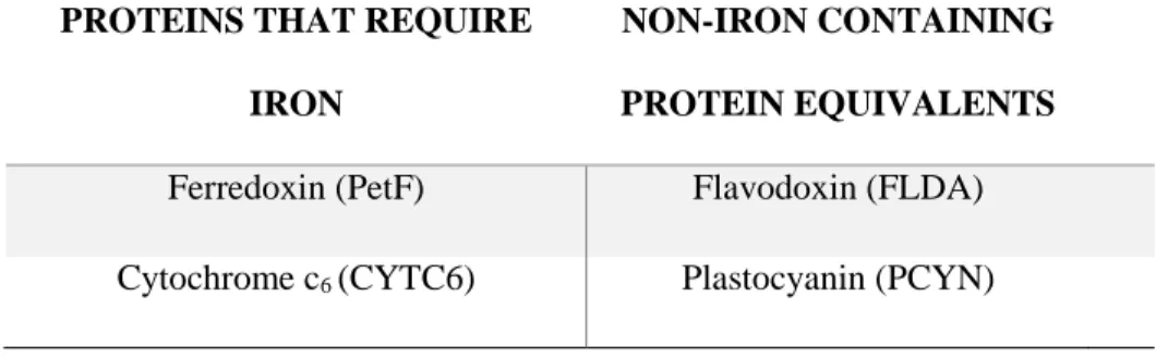
We investigated two protein substitution pairs that are used in photosynthesis and that
can be exchanged for each other in low iron conditions. The first protein substitution pair we
investigated was cytochrome c6 (CYTC6) and plastocyanin (PCYN). All complete diatom
genome sequences have a gene encoding the iron-containing electron transporter CYTC6 (Raven
et al., 2013), but only examined diatoms that come from iron-limited regions, such as T.
oceanica and F. cylindrus, appear to have genes that encode for PCYN, an alternative,
copper-containing electron transporter (Peers and Price, 2006). In addition, several diatoms, including P.
inermis, transcribe more than one copy of PCYN (Groussman et al 2015). T. oceanica
constitutively expresses plastocyanin even under iron-replete conditions, suggesting that
plastocyanin expression is a permanent adaptation to open ocean, low iron environments (Peers
and Price, 2006).
The second major protein substitution that can occur under low iron conditions is the
substitution of ferredoxin (PetF) for flavodoxin (FLDA). PetF is a non-heme iron-sulfur protein
that serves as the terminal electron acceptor and reduces NADP+ to NADPH which, together
with ATP, provides energy to drive the light-independent Calvin Cycle. PetF has a high redox
35
environments, diatoms have been found to replace PetF with FLDA, a protein that contains
flavin-mononucleotide as a prosthetic group, as the redox cofactor instead of iron (La Roche et
al. 1996). The ratio of PetF to FLDA has been used as a molecular indicator of iron stress (La
Roche et al. 1996); however, multiple copies of FLDA (I and II) exist in certain diatom species
and may not all be regulated by iron availability (Whitney et al. 2011, Groussman et al. 2015).
Table 2.1. Summary of protein substitutions in the photosynthesis electron transport chain
Two other proteins involved in photosynthesis were investigated, one that requires iron
and one that does not. The first protein, plastid terminal oxidase (PTOX), is used in an
alternative electron photosynthetic pathway from PSII to PTOX. PTOX requires two iron atoms,
but this short pathway provides an electron shunt after PSII allowing photosynthesis to bypass
the PSI complex, which requires 12 Fe atoms (three 4Fe-4S centers; Behrenfeld & Milligan
2013). In some diatoms, rhodopsins (RHO) can also be used as an iron/light management
strategy. Diatom RHO is a putative light-driven proton pump that can be used for ATP synthesis
and requires a small amount of iron for retinal synthesis. Recently, Marchetti et al. (2015) found
that RHO in a temperate diatom, P. granii, was highly expressed at both the gene and protein
level under iron-limited growth conditions. This suggests that RHO might be used when
photosynthesis is compromised at low iron concentrations.
PROTEINS THAT REQUIRE
IRON
NON-IRON CONTAINING
PROTEIN EQUIVALENTS
36
Physiological and molecular studies of T. oceanica and P. tricornutum have shown that
diatoms employ a high affinity iron uptake system under low iron conditions. A ferric reductase
(FRE) first reduces Fe(III) to Fe(II), which is then re-oxidized by a multi-copper oxidase (MCU)
which is paired with or in close proximity to an iron permease (FTR) that transports Fe(III) into
the cell (Marchetti & Maldonado, 2016). Interestingly, SO diatoms can acquire iron complexed
to strong organic ligands; however, the mechanisms are unknown (Strzepek et al. 2011).
There are a number of other controlled iron uptake systems that diatoms can use. An
iron-starvation induced protein 2a (ISIP2a) has been shown to be widely expressed among
phytoplankton (Morrissey et al. 2015). It is highly expressed under low iron conditions and
concentrates Fe(III) at the cell surface without the use of MCU (Morrissey et al. 2015). In T.
oceanica and P. tricornutum, other low-iron responsive genes include ISIP1, ISIP3, natural
resistance-associated macrophage proteins (NRAMP), and cellular repressor of E1A-genes
(CREG; Allen et al. 2008). Transcripts for these genes have been identified as putative iron
receptors because they are up-regulated under iron limitation and targeted to the secretory
pathway (Lommer et al. 2012; Allen et al. 2008).
Once iron has reached the interior of the cell, it can be used immediately or stored for
later use. Luxury uptake is the ability to acquire and safely store iron in excess of what the cell
needs to grow, and often employs storage proteins called ferritin. Almost all raphid pennate
diatoms for which whole-genome sequences or transcriptomes are available, such as those in the
MMETSP, contain a ferritin gene, whereas ferritin genes in centric diatoms appear to be more
randomly distributed (Groussman et al. 2015).
Iron is used in many other aspects of diatom metabolism besides photosynthesis. For
37
(NiR), require iron cofactors. In a proteomic study of T. pseudonana, iron-limited cells were
found to significantly up-regulate their nitrogen recycling pathways, resulting in conservation of
iron normally allocated to NR, NiR, and other reduced nitrogen assimilation steps (Nunn et al.,
2013; Lommer et al., 2012). In addition, Nunn et al. (2013) suggest further adaptations under
iron limitation, including recycling of N in the urea cycle to create polyamines for nitrogen
storage and silica precipitation. As a representative of the urea cycle, we chose argninosuccinate
synthase (ASSY). It is one of the first enzymes in the urea cycle and was found to be highly
expressed in an iron-enriched metatranscriptome study in an HNLC region (Marchetti et al.
2012).
In addition to iron, diatoms require vitamin B12, which has been shown to be limiting in
certain regions of the SO (Bertrand and Saito 2007). To our current knowledge B12 is only
synthesized by bacteria. Diatom requirements for this nutrient depend on the type of methionine
synthase they possess. Some diatoms only contain MetH, which requires B12, while other
diatoms also contain the B12-independent methionine synthase MetE. Studies have shown that
iron and B12 can act synergistically to enhance growth in iron-limited waters (Koch et al. 2011).
In the SO, the ability to retain MetE would be advantageous as these areas can be co-limited by
both nutrients. For example, F. cylindrus, a major bloom former in the SO, contains MetE
allowing it to flourish in conditions that might be limiting to other phytoplankton that are
auxotrophic for B12 such as following a large iron-induced bloom (Ellis et al. 2015)
Diatoms minimize reactive oxygen species (ROS) resulting from metabolic byproducts
by using superoxide dismutases (SOD), which convert superoxide to a molecular oxygen and
hydrogen peroxide (Peers & Price 2004). There are four isoforms of SODs, containing either
38
times of iron limitation (Wolfe-Simon et al. 2006). Many algae have both Fe-SOD and Mn-SOD;
by expressing Mn-SOD, they can decrease their iron demands, especially when iron availability
is low. Mitochondrial alternative oxidases (AOX) are similar to SODs in their structure and
activity, and help to reduce ROS in the mitochondria (Maxwell et al 1999). They have a lower
requirement for iron than other terminal oxidases (Bowler et al. 2010). Electrons from
photosynthesis can also be transported via the malate shunt to the mitochondria, where AOX
again can help with reducing ROS (Yoshida et al 2007).
These are just a few of the many genes affected by iron (and possibly light) limitation.
Some have been shown to have a direct role in iron homeostasis, photosynthesis, and nitrogen
assimilation, while others have putative functions in other pathways that are affected by iron and
light. The focus here was to investigate the presence or absence of their transcripts within the
transcriptomes of the polar diatom isolates described in Chapter 1, the genome of F. cylindrus,
and other diatom transcriptomes made available by the MMETSP (Keeling, et al., 2014). Over
46 different diatoms were sequenced as part of this program, which has facilitated a better
understanding of the various strategies diatoms employ to cope with iron and light limitation, as
well as their ecology and influence on marine biogeochemistry.
Materials and Methods
RNA extractions, transcriptome library preparation and sequencing - Cultures grown for
high throughput sequencing of mRNA were grown in acid-washed 2L polycarbonate bottles in
iron-replete (pFe 19) conditions and standard light (90 μmol photons m-2 s-1). After reaching late
exponential/early stationary phase, cultures were harvested onto polycarbonate filters (3.0 m