Cover Page
The handle
http://hdl.handle.net/1887/19756
holds various files of this Leiden University
dissertation.
Author
: Naber, Hildegonda Petronella Henriëtte
Title
: TGF-beta and BMP in breast cancer cell invasion
TGF
‐β
and
BMP
in
breast
cancer
TGF
‐β
and
BMP
in
breast
cancer
cell
invasion
Proefschri
ter verkrijging van
de graad van Doctor aan de Universiteit Leiden,
op gezag van de Rector Magnificus prof. mr. P.F. van der Heijden, volgens besluit van het College voor Promo es
te verdedigen op woensdag 5 september 2012
klokke 16.15 uur
door
Hildegonda Petronella Henrië e Naber
geboren te ‘s‐Gravenhage
Promo ecommissie
Promotor: Prof. Dr. P. ten Dijke
Co‐promotores: Dr. E. Pardali
Dr. T. van Laar
Overige leden: Prof. Dr. B van de Water
Prof. Dr. P. Devilee
Prof. Dr. R.C. Hoeben
Prof. Dr. A. Sonnenberg (Nederlands Kanker Ins tuut, Amsterdam)
Dr. G. van der Pluijm
ISBN 978‐94‐6182‐135‐5
Cover: Rosa Gloire de Dijon
The studies described in this thesis were performed at the department of Molecular and Cellular Biology, Leiden University Medical Center.
Contents
Chapter 1 General Introduc on 7
Chapter 2 Role of TGF‐ in tumor stroma 59
Current Cancer Drug Targets 2008 8:466‐72
Chapter 3 Spheroid assay to measure TGF‐β‐induced invasion 79
Journal of Visualized Experiments 2011 (57). pii: 3337.
Chapter 4 The TGF‐/Smad pathway induces breast cancer cell invasion through theup‐ regula on of matrix metalloproteinase 2 and 9 in a spheroid invasion model system 93
Breast Cancer Research and Treatment 2011 128:657‐666
Chapter 5 BMP‐7 inhibits TGF‐‐induced invasion of breast cancer cells through inhibi on of integrin 3 expression 123
Cellular Oncology 2012 35:29‐38
Chapter 6 Key regulators of EMT are essen al for TGF‐‐induced invasion by promo ng single cell invasion 149
In prepara on
Chapter 7 Iden fica on of kinases involved in BMP signalling 165
In prepara on
Chapter 8 Summary and discussion 179
Chapter
1
1.
TGF
‐
superfamily
The Transforming Growth Factor (TGF)‐ superfamily comprises a large collec on of struc‐
turally related cytokines which regulate a mul tude of biological processes. These include
TGF‐s, Bone Morphogene c Proteins (BMPs), Growth Differenta on Factors (GDFs), Ac‐
vins, Inhibins, Nodal and An ‐Müllerian Hormone (AMH) [1]. Its prototypical member,
TGF‐, was discovered as a factor capable of transforming the cellular morphology and
promo ng growth of normal rat kidney cells in so agar [2]. Three isoforms of TGF‐ exist
in humans, namely TGF‐1, TGF‐2, TGF‐3, and they share 60‐80 % amino acid homology
in their mature forms [3]. Knockout mice of either of these isoforms showed severe de‐
fects and died in utero or shortly a er birth [4‐9] (Table 1). These phenotypes emphasize
the importance of TGF‐ during development. In adult life, dysregula on of TGF‐ signal‐
ing has been reported in vascular disorders, fibrosis and cancer.
BMPs were ini ally discovered for their capacity to induce bone forma on [10‐12]. Subse‐
quent studies showed that they have an important role in pa erning during embryonic
development [13]. Dele on of specific BMP isoforms in mice can lead to defects in the
vascular system, cardiac system, nervous system or the skeleton [14‐22] (Table 1), thus
highligh ng the diversity in func on of the different BMPs. However, some BMP knockout
mice have only a mild phenotype [23,24] (table 1), sugges ng redundancy between BMPs.
Indeed, BMP5/7 double knockout mice die during early development, whereas the single
mutants are not embryonic lethal [25].
1.1
Smad
signaling
TGF‐ family members usually act as homodimers, although some may act heterodimers,
such as the BMP‐2/BMP‐7 dimer [1]. These dimers mediate their effects through binding
to type I and type II transmembrane receptors, which have a strong Ser/Thr kinase ac vity
and a weaker Tyr‐kinases ac vity [26]. Ligand binding can be regulated by co‐receptors,
the type III receptors. Upon ligand‐induced complex forma on, the type II receptor phos‐
phorylates the type I receptor. The type I receptor phosphorylates receptor regulated
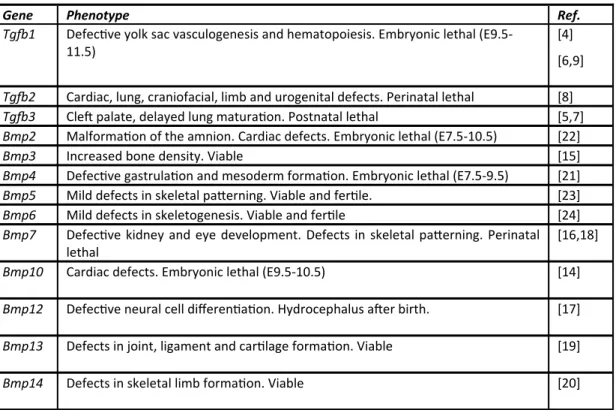
Table 1 Knockout studies of TGF‐s and BMPs in mouse models. Tg 1 phenotype depends on gene c background
Gene Phenotype Ref.
Tg 1 Defec ve yolk sac vasculogenesis and hematopoiesis. Embryonic lethal (E9.5‐
11.5)
[4]
[6,9]
Tg 2 Cardiac, lung, craniofacial, limb and urogenital defects. Perinatal lethal [8]
Tg 3 Cle palate, delayed lung matura on. Postnatal lethal [5,7]
Bmp2 Malforma on of the amnion. Cardiac defects. Embryonic lethal (E7.5‐10.5) [22]
Bmp3 Increased bone density. Viable [15]
Bmp4 Defec ve gastrula on and mesoderm forma on. Embryonic lethal (E7.5‐9.5) [21]
Bmp5 Mild defects in skeletal pa erning. Viable and fer le. [23]
Bmp6 Mild defects in skeletogenesis. Viable and fer le [24]
Bmp7 Defec ve kidney and eye development. Defects in skeletal pa erning. Perinatal
lethal
[16,18]
Bmp10 Cardiac defects. Embryonic lethal (E9.5‐10.5) [14]
Bmp12 Defec ve neural cell differen a on. Hydrocephalus a er birth. [17]
Bmp13 Defects in joint, ligament and car lage forma on. Viable [19]
Bmp14 Defects in skeletal limb forma on. Viable [20]
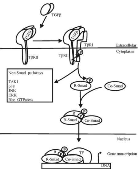
Smads (R‐Smads) at their C‐terminal Ser‐residues [1,27‐32].
Smads consist of two Mad homology (MH) domains, with a linker region in between. In R
‐Smads, these two MH domains (MH1 and MH2) bind to each other, thus inhibi ng each
other. Phosphoryla on of the R‐Smad by the type I receptor releases the MH1 and MH2
domain of the R‐Smads from each other. This allows the MH2 domain of the R‐Smad to
associate with the common‐Smad (Co‐Smad), Smad4. In addi on, the nuclear localiza on
signal of the R‐Smad is unmasked, enabling the complex to translocate into the nucleus.
In the nucleus the Smad complex can affect transcrip on together with other transcrip‐
on factors [1,26] (Figure 1).
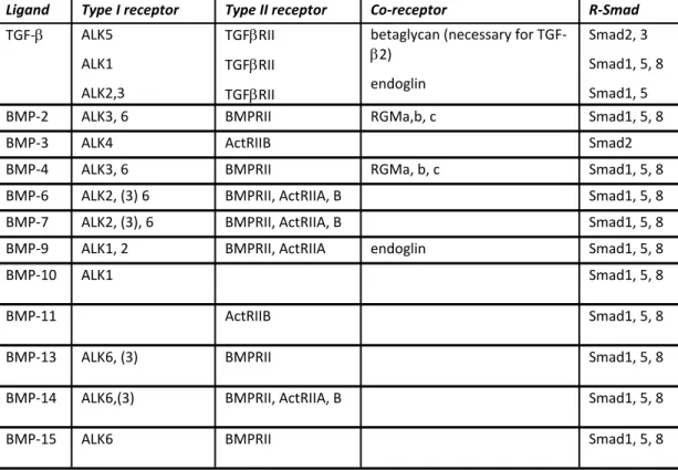
TGF‐ binds ini ally to the TGF‐ type II receptor (TGF‐RII) on the cell surface. Because of its low affinity, TGF‐2 also requires binding to the TGF‐RIII ‐glycan before binding to the type II receptor [32]. Binding of TGF‐ to the type II receptors induces a conforma‐
ALK5 [33]. ALK5 phosphorylates the R‐Smads 2 and 3 (Table 2).
Upon transloca on into the nucleus, Smad3 can bind directly to the DNA at GTCT/AGAC
sequences through its MH1 domain. Smad2 is not able to bind directly to DNA, because it
has addi onal amino acids in its MH1 domain [1]. A plethora of transcrip on factors has
been shown to interact with Smad2/3, such as CREB binding protein (CBP)/p300, SP1 [39]
and c‐Jun [40]. Some transcrip on factors only interact only with Smad3, such as FOXO
family members [41] and ATF3 [42].
Forma on of the BMP‐receptor complexes occurs through simultaneous recruitment of
the type I and type II receptor [33]. BMPs use Ac vin receptor‐like kinase (ALK) 1,2, 3 or 6
as type I receptor and BMP type II receptor (BMPRII) and Ac vin type II receptor (ActRII)
A or B as type II receptor. Not all BMPs can bind to all these receptors. For example, BMP‐
9 can bind to ALK1 and ALK2 [34,35], whereas BMP‐7 can bind to ALK2, 3 and 6, but not
to ALK1 [36]. An excep on is BMP‐3, as it signals via ALK4 and ActRII to Smad2, to Figure 1 Basic signaling scheme. Upon binding of TGF‐/BMP to the receptors, the type II receptor
phosphorylates the type I receptor, which then phosphorylates the R‐Smad. This induces a conforma‐
onal change in these Smads, enabling them to interact with the Co‐Smad and these complexes trans‐
counteract bone differen a on [37,38]. Ligand‐receptor binding of BMP‐2 and 4 is en‐
hanced by GPI‐anchored proteins of the repulsive guidance molecule (RGM) family, such
as RGMb/DRAGON and RGMc/hemojuvelin [30]. Ac va on of ALK1, 2, 3 or 6 by type II
receptors results in phosphoryla on of the R‐Smads 1,5 and 8 (Table 2). Upon transloca‐
on to the nucleus, these BMP‐R‐Smads can bind directly to the DNA at sites containing
BMP Response Elements and GC‐rich sequences [26]. In addi on, BMP‐R‐Smad complexes
also interact with transcrip on factors, such as Runx2 and Schnurri2 [30].
In certain cell types, TGF‐ also induces ac va on of R‐Smads 1 and 5 through the type I
receptor ALK1. ALK5 was required for this ac va on, whereas ALK1 inhibited ALK5‐
Smad2/3 signaling [43]. The co‐receptor endoglin (CD105) poten ates TGF‐‐ALK1 signal‐ ing over ALK5 signaling [44]. Recent reports also suggested that TGF‐ could signal through
the type‐I receptor ALK2 and 3 to ac vate Smad1 and 5 in epithelial cells (Table 2). This
process also requires ALK5 and resulted in the forma on of so‐called "mixed" R‐Smad complexes, consis ng of BMP‐R‐Smads and TGF‐‐R‐Smads [45].
Ligand Type I receptor Type II receptor Co‐receptor R‐Smad
TGF‐ ALK5
ALK1
ALK2,3
TGFRII
TGFRII
TGFRII
betaglycan (necessary for TGF‐
2)
endoglin
Smad2, 3
Smad1, 5, 8
Smad1, 5
BMP‐2 ALK3, 6 BMPRII RGMa,b, c Smad1, 5, 8
BMP‐3 ALK4 ActRIIB Smad2
BMP‐4 ALK3, 6 BMPRII RGMa, b, c Smad1, 5, 8
BMP‐6 ALK2, (3) 6 BMPRII, ActRIIA, B Smad1, 5, 8
BMP‐7 ALK2, (3), 6 BMPRII, ActRIIA, B Smad1, 5, 8
BMP‐9 ALK1, 2 BMPRII, ActRIIA endoglin Smad1, 5, 8
BMP‐10 ALK1 Smad1, 5, 8
BMP‐11 ActRIIB Smad1, 5, 8
BMP‐13 ALK6, (3) BMPRII Smad1, 5, 8
BMP‐14 ALK6,(3) BMPRII, ActRIIA, B Smad1, 5, 8
BMP‐15 ALK6 BMPRII Smad1, 5, 8
Table 2 Receptor binding of the ligands. Receptors in brackets indicate that only weak binding
1.2
Non
‐
Smad
signaling
Besides Smad ac va on, TGF‐ and BMPs may induce non‐Smad signaling, such as MAPK and Rho GTPase signaling. For example, ac va on of p42/44 MAP kinases by TGF‐ oc‐
cured through direct phosphoryla on of ShcA by ALK5. Phosphorylated ShcA recruits
Grb2 and Sos. This complex ac vates RAS, which ac vates the Raf‐MEK‐ERK cascade [46]. Interes ngly, recruitment of Shc and Grb2 to the TGF‐ type II receptor was enhanced by
phosphoryla on of Thr284 by Src at the type II receptor, but this resulted p38 ac va on
[47].
TGF‐ and BMPs also induce ac va on of TGF‐ ac vated kinase‐1 (TAK‐1). TAK‐1 is able to ac vate p38 and JNK pathways in response to TGF‐ and BMPs. Ac va on of TAK‐1 by TGF‐ occured by the ubiqui n ligase Tumor Necrosis Factor Associated Factor (TRAF)‐ 6 in the TGF‐ receptor complex, possibly through poly‐ubiqui na on of TAK‐1 [48]. TAK‐
1 is also ac vated in response to BMPs, although it is not able to bind BMP receptors by
itself. This requires interac on with the X‐linked inhibitor of apoptosis (XIAP), which acts
as a scaffold [49].
Conflic ng data exist about the role of RhoA in TGF‐ signaling. Ini al studies showed that RhoA is ac vated by TGF‐[50,51]. However, in the ght junc ons of polarized epi‐
thelial cells, TGF‐RII phosphorylated Par6, leading to recruitment of the E3 ligase
Smurf1 and subsequent ubiqui na on and degrada on of RhoA [52,52]. Perhaps the lo‐
caliza on in the cell determines if the response is either inhibi on or s mula on of RhoA
ac vity.
1.3
Regula on
of
TGF
‐
and
BMP
signaling
Because of their important role in development and ssue homeostasis, ac vity of TGF‐
and BMPs are ghtly regulated. This regula on occurs at mul ple stages of signaling,
ranging from regulated secre on to degrada on in de nucleus. A large number of these
regulatory proteins are induced by TGF‐ and BMPs themselves, thus func oning in feed‐
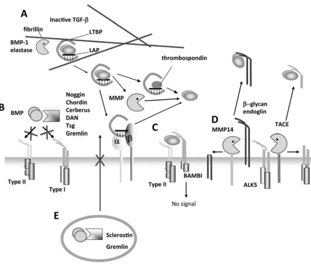
1.3.1 Extracellular control of TGF‐/BMP signaling
TGF‐ and BMPs are synthesized as large precursor proteins with a long N‐terminal pro‐
pep de and a shorter C‐terminal mature pep de [53]. Disulfide bridges link the two
pep des at conserved cysteine knots, with the excep on of BMP‐15 [26]. Cleavage of
the prodomain occurs in the Golgi apparatus by furin proteases. The propep de remains
non‐covalently a ached to the mature pep de. The TGF‐ propep de is referred to as
latency associated pep de (LAP) as it prevents binding of the protein to its receptor. In
most cells, the LAP is covalently bound to latent TGF‐ binding proteins (LTBPs). This
protein links the complex to the extracellar matrix (ECM), especially to the ECM compo‐
nent fibrillin [53]. Release of the complex from the ECM occurs through proteoly c
cleavage by elastase or the protease BMP‐1 [1,26,53,54]. TGF‐ is subsequently released
from the LAP by degrada on of LAP by matrix metalloproteinases (MMPs) or scavenging
of LAP by other proteins such as thrombospondin or integrins v6, v5, v8, [53,55,56] (figure 2A).
The prodomain of all BMPs, except BMP‐11, does not interfere with their ac vity, but
may regulate mature protein stability and processing [26]. Regula on of BMPs may al‐
ready occur at the level of secre on. For example, the BMP antagonist Gremlin binds
intracellularly to BMP‐4, thereby preven ng secre on [57]. Also Scleros n inhibits BMP‐
7 secre on by binding to both the prodomain and mature domain of BMP‐7 [58] (Figure
2E).
Bio‐availability of BMPs is generally regulated extracellularly by natural antagonists,
such as Noggin, twisted gastrula on (Tsg), the Chordin protein family and the DAN pro‐
tein family [59]. These proteins bind to BMPs, thus preven ng binding to the receptor
(Figure 2B). One antagonist usually targets a subset of BMPs. For example, Noggin binds
with high affinity to BMP‐2, 4 and 7, but not to other BMPs [60]. Some of the BMP‐
antagonist complexes are able to form ternary complexes with other inhibitory proteins.
For example, the binding of Chordin to BMP4 is enhanced by Tsg [59].
TGF‐ and BMPs can also be bound by the decoy receptor BMP and Ac vin membrane‐
kinase domain and consequently sequesters ligands from the ac ve receptors [61] (Figure
2C). Another mechanism of modula ng ligand ac vity extracellularly is shedding of the
extracellular domain of the receptor. This results in receptors that are unable to bind lig‐
ands, whereas the soluble extracellular domain may prevent the ligands from binding to
funcional receptors by scavenging them. For example, the TGF‐ co‐receptors endoglin
and betaglycan can be cleaved by matrix metalloproteinase (MMP) 14 [62,63]. In addi on, cleavage of the extracellular domain of ALK5 by TNF conver ng enzyme (TACE) resulted
in decreased Smad3 ac va on [64] (figure 2D).
1.3.2 Regula on at the receptor level
Signaling can be enhanced by accessory proteins which facilitate the recruitment of R‐
Smads to the receptors. Recruitment of R‐Smads 2 and 3 to ALK5 occurs through Smad
anchor for receptor ac va on (SARA) [26,32]. The transmembrane protein Transmem‐
brane prostate androgen‐induced RNA(TMEPAI) competed with SARA for interac ng with
R‐Smads, thus sequestering Smads from the ac va ng receptor complex [76]. For BMP
signaling, the R‐Smads 1, 5 and 8 can be recruited to the type I receptors by endofin [77]
or CD44 [78], also resul ng in enhanced Smad signaling.
A common mechanism of regula ng receptor signaling is internaliza on and degrada on
of the receptors. Also the TGF‐ receptors can be internalized and degraded, which occurs
through caveolin‐Smad7 posi ve vesicles. However, internaliza on through clathrin coat‐
ed pits promotes Smad signaling, which are enriched in SARA [79]. Similarly, the
BMPRII can be targeted for degrada on through caveolae, whereas endocytosis of BMP
receptors via clathrin coated pits promotes the transcrip onal ac vity of BMP‐Smads [80].
Recently Smad2 and 3 were shown to be dephosphorylated by MTMR4 in endosomes [81].
If these phosphatase regulate signaling in clathrin coated vesicles or aid the termina on of
signaling in caveolin posi ve vesicles remains to be determined.
Another pos ransla onal modifica on which modulates protein func on is covalent link‐
age of the small ubiqui n‐like protein SUMO. For example, ac vated ALK5 can be sumoy‐
lated by the ligase Ubc9, which results in enhanced recruitment of Smad3 [82]. If other
Figure 2 Extracellular regula on of TGF‐superfamily signaling. A. Inac ve TGF‐ is bound to latency
associated pep de (LAP) which is covalently linked to latent TGF‐ binding protein (LTBP). This complex
is targeted to the extacellular matrix compenent fibrillin, from which it can be released by BMP‐1 and
elastase. Further processing of the complex is achieved through MMPs or by scavenging the LAP‐LTBP to
integrins or thrombospondin. B. Natural antagonists are secreted by cells and bind to BMPs thus pre‐
ven ng receptor liga on. C. The decoy receptor BAMBI binds ligands and forms a receptor complex, but
can not transduce signals. D. ALK5 or the co‐receptors endoglin and ‐glycan can be cleaved by TACE or
MMP14, respec vely. Shedding of the extracellular domain scavenges ligands from receptors. E. Scle‐
ros n and Gremlin prevent the secre on of BMPs
ALK5 is also known to interact with regulatory subunits of protein phosphatase 2 (PP2)
[83,83]. More recently, two different regulatory subunits of PP2, B and B, were shown to have different effects on TGF‐ signalling, with B promo ng and B inhibi ng signal‐
ling [84]. However, it remains to be established if PP2 dephosphorylates the receptor.
ALK3 can be dephosphorylated by the phosphatase Dullard, which also promotes inter‐
1.3.3 I‐Smads
Inhibitory (I)‐Smads, Smad6 and Smad7, are well‐known nega ve regulators of TGF‐
superfamily signaling. I‐Smads have a similar MH2 domains as the R‐Smads, enabling
them to interact with the ac vated type I receptors and thus compe ng with R‐Smads
for binding [1]. Smad7 binds both to TGF‐ and BMP receptors, whereas Smad6 has a
preference for BMP‐receptors [26]. In addi on, I‐Smads are involved in the degrada on of TGF‐RI. They interact with the E3 ligases Smurf 1 and 2, resul ng in the degrada on
of the I‐Smads and the bound type I receptor [65,66]. Smad7 is also able to recruit the E3
ligases WWP1/Tiul and Nedd4‐2/Nedd4L which also resulted in degrada on of the type I
receptor [67‐69]. Furthermore, Smad7 may recruit PP1c, a regulatory subunit of PP1,
thus promo ng dephosphoryla on of ALK5 [70]. A similar mechanism involving Smad7
and PP1c has been implicated in the regula on of ALK1 [71].
Although the I‐Smads predominantly act on the type I receptor, they also inhibit R‐Smad
and Smad4 func on. Smad6 competed with Smad4 for binding to Smad1, thus inhibi ng
propaga on of the BMP signal [72]. Smad7 acted as an adaptor protein for ubiqui na on
of Smad2 and Smad4 by Nedd4‐2 and WWP1, thus promo ng its degrada on [68,69,73].
Furthermore, Smad6 might be a transcrip onal repressor of BMP target genes [74].
Smad7 competed with Smad2/Smad4 complexes in the nucleus for binding sites in the
promoters of targets genes [75] (Figure 3).
1.3.4 Smad linker phosphoryla on
Besides phosphoryla on at their C‐terminal Ser residues by the type I receptor, R‐Smads
may also be phosphorylated at different sites. These phosphoryla ons modulate the sta‐
bility and nuclear localiza on of the Smads. For example, Smad2 phosphoryla on by cal‐
modulin kinase II at Ser240, prevents its nuclear localiza on [86]. Protein kinase C phos‐
phorylated Smad3 at Ser37 and Ser70, which interferes with Smad3 DNA binding [87].
Smad1 can be phosphorylated in the linker region between the two MH domains by MAP
kinases in response to mitogens [88]. This primes Smad1 for linker phosphoryla on by
ubiqui na on and degrada on [89]. GSK3 also phosphorylates Smad3 at Thr66, which
promotes its ubiqui na on and subsequent degrada on [90]. Thus, the phosphoryla on
of Smads in the linker region integrates signals from different other signaling pathways,
such as the Wnt an mitogen pathways, into TGF‐ and BMP signaling pathways.
However, linker phosphoryla on of Smad1 or Smad2/3 is also induced by BMPs or TGF‐ themselves. Casein kinase (CK) 1 phosphorylated Smad3 in a ligand dependent manner
and promotes its ubiqui na on and degrada on [91]. This might be mediated by the
ubiqui n ligase ROC1, which was the first ubiqui n ligase iden fied for Smad3 [92]. The
kinases cyclin dependent kinase (CDK) 8 and 9 phosphorylates both BMP and TGF‐ R‐
Smads in the linker region [93]. This creates a docking site for Smurf1 in Smad1, whereas
the ubiqui n ligase Nedd4‐2 binds to linker phosphorylated Smad2 and 3, resul ng in
their degrada on [93,94]. This process could be enhanced by further linker phosphoryla‐
on by GSK3‐ [95]. However, the CDK8/9 phosphoryla on sites also served as docking
site for posi ve regulators, such as the transcrip onal co‐ac vator Yes Associated Pro‐
tein (YAP) for Smad1 [93].
This mechanism might also explain why MAPK‐mediated linker phosphoryla on of
Smad2/3 seems to have opposing effects. Ini al reports suggested that Smad2/3 linker
phosphoryla on prevents nuclear localiza on [96,97]. Others, however, reported that
Smad2 is stabilized by MAPK phosphoryla on, thus promo ng transcrip onal ac vity
[98]. The amount of YAP versus Smurf1 and Nedd4/2 present in the cell may determine
the outcome of linker phosphoryla on.
1.3.5 Ski and SnoN
The co‐repressors Ski and SnoN regulate TGF‐ and BMP signaling in the nucleus. They
bind simultaneously to Smad4 and the R‐Smad, resul ng in dissocia on or inac va on of
the complex [99,100]. Both Ski and SnoN are able to bind to Smad2/3/4, whereas Ski is
also able to bind Smad1 and 5 [101]. Thus, SnoN acts as an inhibitor for TGF‐ signaling, whereas Ski also inhibit both TGF‐ and BMP signaling.
regulators. The ubiqui n ligase Arkadia binds to Smad7 and reduces its levels [102]. More
recently, it was discovered that Arkadia also targets Ski and SnoN for degrada on [103‐
105]. This occurs upon ligand s mula on and required phosphorylated Smad2 or Smad3
[103,104]. This mechanism provides a posi ve feedback loop in TGF‐ signaling.
1.3.6 Smad nucleocytoplasmic shu ling
Although the described signaling model implies that Smads only are present in the nucle‐
us upon ligand s mula on, Smad2, 3 and 4 are con nuously shu ling between cytoplasm
and nucleus. S mula on with TGF‐ increased the nuclear pool of Smads [106] by reduc‐
ing the rate by which Smads were exported from the nucleus [107]. Mathema cal model‐
ing of the kine cs implied that Smad complexes are imported faster than exported and
Smads are dephosphorylated in the nucleus [108]. Indeed, Smad1,2 and 3 were
dephosphorylated by the nuclear phosphatase protein phosphatase, Mg2+/Mn2+ depend‐
ent (PPM)1A [109,110], The faster import of Smad2/3/4 complexes may be mediated by
the transcrip onal regulator Tafazzin (TAZ), which enhanced Smad nuclear import [111].
Furthermore, pos ransla onal modifica ons could alter Smad localiza on. For example,
mono‐ubiqui na on at Lys519 of Smad4 by ectodermin (Ecto) inhibits Smad2/3 complex
forma on and promotes Smad4 nuclear export [112], a process which was reversed by
the deubiqui na ng enzyme FAM/ USP9x [113]. However, Smad4 is also mono‐
ubiqui nated at Lys507, resul ng in enhanced forma on of Smad3/4 complexes [114].
Smads are also modified by sumoyla on. Smad4 can be sumoylated by protein inhibitor
of ac vated STAT (PIAS)y and ubiqui n conjuga ng enzyme (Ubc) 9, which results in in‐
creased stability and transcrip onal ac vity [115,116]. However, sumoyla on of Smad3
by PIASy resulted in decreased transcrip onal ac vity [117], by promo ng its nuclear ex‐
port [118]. On the other hand, the SUMO ligase PIAS3 recruited the co‐ac vator CBP/
p300, hereby promo ng signaling [119]. Thus, the effect of sumoyla on on TGF‐ signal‐
ing depends on which Smad and ligase are involved.
Recently, poly‐ADP‐ribose polymerase(PARP) 1 was iden fied as a mediator of Smad‐ADP
ribosyla on. This modifica on occurred in Smad2/3/4 complexes at the promoters,
resul ng in dissocia on of the complex and decreased transcrip onal responses [120].
Whether a similar mechanism operates in BMP signaling remains to be determined.
Figure 3 Intracellular regula on of TGF‐and BMP signaling. SARA enhances signaling by presen ng
Smad2‐3 to the type I receptor. TMEPAI competes with SARA for Smad2/3 and inhibits TGF‐signaling.
BMP‐Smads (1/5/8) can be presented by endofin or CD44. Smad6 competes with Smad1/5/8 for the
type I receptor. Smad7 competes with all R‐Smads for the type I receptor and recruits ubiqui n ligases,
such as Smurf1, 2, WWP and Nedd4‐2 to target the receptors, Smad2 and Smad4 for degrada on.
Smad7 also recruits the phosphatase PP2 to the type I receptor. Sumoyla on of the TGF‐ type I recep‐
tor (ALK5) and Smad4 by Ubc9 enhances signalling. Phosphoryla on of Smads at the linker sites may
result in ubiqui na on and degrada on, but may also result in enhanced signalling, for example
through associa on with the transcrip on factor YAP. Nuclear entrance of Smads is promoted by TAZ. In
the nucleus, R‐Smads can be desphosphorylated by PPM1A, thus termina ng signalling. Also Ski, SnoN,
Smad6 and Smad7 may inhibit Smad‐dependent transcrip on, which on their turn are targeted for deg‐
rada on by the ubiqui n ligase Arkadia. Sumoyla on of Smad3 by PIASy promotes nuclear export. Mono
‐ubiqui na on of Smad4 at L519 by Ecto also promotes nuclear export, which can be reversed by the de
‐ubiqui na ng enzyme FAM. Mono‐ubiqui na on of Smad4 at another residue, L507 promotes nuclear
1.4
Interplay
between
TGF
‐
and
BMP
signaling
The TGF‐ and BMP signaling pathways share several common mediators, such as Smad4,
which may result in antagonism between these two pathways. The Smad4 pool may thus
be a limi ng factor if BMP and TGF‐ signaling pathways are ac vated simultaneously,
thus resul ng in reduced ac va on in one of the pathways. Indeed, compe on for
Smad4 has been observed during Xenopus development [121]. Furthermore, TGF‐ and
BMPs have opposing effects on the transcrip on of genes, such as Id2 and Id3 [122]. How‐
ever, studies on digit chondrogenesis in chicken embryos demonstrated posi ve effects of
the TGF‐ pathway on the BMP pathway by repression of Smad6 and BAMBI [123]. The
effect of TGF‐ on BMP signaling depended on ming and dura on of the co‐s mula on
[124], thus emphasizing the importance of the context of the signals.
The interplay between TGF‐ and BMP‐7 in fibrosis has been extensively studied. TGF‐
promotes fibrosis, which can be reversed by BMP‐7 [125‐128]. This ac on of BMP‐7 oc‐
currs through inhibi on of TGF‐‐induced transdifferena on of epithelial cells to mesen‐
chymal cells [128]. A similar phenomenon was reported in prostate cancer [129]. Further
studies have demonstrated that BMP‐7 antagonizes Smad3 signaling by inducing SnoN
expression [130].
2.
Breast
cancer
Breast cancer is one of the leading causes of death in women. Most morbidity and mor‐
tality does not arise from the primary tumor, but from distant metastasis, especially
bone metastasis which causes severe pain and increased risk of pathologic fractures
[131]. Gene expression profiling primarily dis nguish between estrogen receptor (ER)
posi ve and estrogen nega ve breast tumors, with the la er having worse prognosis [132
‐134]. ER nega ve breast cancer cells can be further classified into Erbb2/Her2/Neu posi‐
ve, basal cell like and normal breast‐like subtypes, with the basal cell like breast cancer
having the worst prognosis and the normal breast‐like the best prognosis
subtype A, B and C, with luminal C having the worst and luminal A having the best progno‐
sis [132,133,135,136]. Furthermore, the clinical outcome can be predicted based on a 70
gene signature in the primary tumor [134]. This led to the hypothesis that metasta c traits
are already acquired during early tumorigenesis [137].
2.1
3
‐
dimensional
culture
models
Most of the data on cancer cell biology are based on studies of cell monolayers grown on
flat and hard plas c. However, this does not recapitulate the 3 dimensional (3D) organiza‐
on and mul cellular complexity of ssues and tumors in vivo [138‐140]. Several 3D mod‐
els have been developed over the past decades. Cells can be either completely embedded
within in a matrix or placed on top of a matrix or a polymeric scaffold [138,139]. This can
be achieved using single cell suspensions or mul cellular spheroids. Spheroids are clus‐
tered masses of cells, which are formed under non‐adhesive condi ons [139].
These models have been proven to recapitulate ssues in 3D. For example, non‐
transformed breast epithelial cells organized themselves into acinar structures when
grown in a 3D basement membrane matrix (Matrigel), thus resembling the architecture of
a normal breast duct [141]. Transformed cells failed to organize themselves in such a man‐
ner [142]. The composi on of the matrix provides important cues for this organiza on,
since normal breast epithelial cells could not organize themselves in a collagen type I ma‐
trix [143]. Increased expression of this collagen has been associated with metastasis [144]
and dense matrixes containing high levels of collagen I promoted tumorigenesis, invasion
and metastasis [145]. Increased matrix density resulted in increased signaling through the
focal adhesion kinase (FAK) and ERK, thus promo ng cell growth [146]. Further crosslink‐
ing of collagen by lysyl oxidase also promoted tumor progression through enhanced sig‐
naling by adhesion molecules [147].
Furthermore, mul cellular spheroids showed the same resistance to cytotoxic drugs as
their parental cell line in vivo, whereas cells in monolayers failed to do so [148]. In subse‐
quent studies, cells in 3D cultures were more resistant to cytotoxic drugs compared to
resistant to apoptosis, because adhesion to the matrix provides survival signals [149]. Fur‐
thermore, expression of drug transporters was higher in 3D cultures [154]. On the other
hand, breast cancer cells showed increased sensi vity to the Her2 an body Trastuzamab
in spheroids, because Her2 homodimeriza on is preferred over Her2/Her3 heterodimeri‐
za on compared to monolayers [155]. Furthermore, breast cancer cells in 3D were more
sensi ve to MEK inhibitors than in monolayers [153]. These studies demonstrated the
usefulness of 3D models in cancer biology.
2.2
TGF
‐
in
breast
cancer
TGF‐ has a dual role in carcinogenesis. In the early stages it has a growth inhibitory and pro‐apopto c effect, whereas at the later stages of cancer, TGF‐ promotes invasion and
metastasis [27,29]. This was elegantly demonstrated using the MCF10A system of cell
lines [156]. The MCF10A cell lines originate from MCF10A1 (M‐I), a spontaneously immor‐
talized breast cell line [157]. This cell line was transformed with oncogenic RAS, resul ng
in MCF10AT (M‐II), which forms premalignant lesions in mice [158]. From the carcinomas,
which arose a er xenogra ing in nude mice, MCF10CA1h (M‐III) and MCF10CA1a were
isolated (M‐IV). M‐III was isolated from a low grade carcinoma, while M‐IV was isolated
from a high grade carcinoma, which metastatasized to the lung [159]. Loss of TGF‐ sig‐
naling caused oncogenic transforma on in the premalignant M‐II and a more aggressive
phenotype in low‐grade M‐IIII. In M‐IV however, inhibi on of TGF‐ signaling resulted in
decreased metastasis [156]. In line with its tumor suppressor func on, loss of TGF‐RII
expression in benign hyperplasic lesions correlated with progression to invasive breast
carcinoma [160]. Furthermore, inhibi on of TGF‐RII expression correlated with tumor
grade [161]. In line with its s mulatory role in cancer progression, TGF‐β is frequently
overexpressed in breast cancer and its expression correlates with poor prognosis and me‐
tastasis [162‐165]. Moreover, studies in mouse models demonstrated that inhibi on of TGF‐‐signaling in breast cancer cells reduces metastasis [166‐170]. Also gene signatures represen ng presence or absence of TGF‐ s mula on have been used to iden fy corre‐
la ons between TGF‐ signaling and clinical outcome of breast cancer. In ER nega ve
However, in ER posi ve breast cancer a TGF‐ signaling deficient gene signature correlat‐
ed with poor prognosis, especially in luminal A breast carcinomas [172]. (reviewed in
[28]). These findings suggest that the role of TGF‐ is context‐dependent.
2.3
BMPs
in
breast
cancer
The role of BMPs in breast cancer is not so well characterised. Blockade of BMP signaling
by a dominant nega ve BMPRII s mulates cell growth [173]. Expression analysis has
shown that BMP‐4, 6 and BMP‐7 mRNA are expressed in breast cancer, while BMP‐2
mRNA expression is decreased compared to normal ssue [174‐176]. BMP‐7 expression is
reported to be associated with bone metastasis [177]. However, BMP‐7 expression has
also been inversely correlated with bone metastasis and BMP‐7 reduced metastasis in a
mouse model [178]. A study on the effects of BMP‐7 in different breast cancer cell lines
demonstrated differen al effects on prolifera on and migra on, thus indica ng that the Figure 4 The MCF10A system of cell lines. MCF10A1 (M‐I) is a spontaneously immortalized normal epi‐
thelial breast cell line. This cell line was transformed with oncogenic RAS to give rise to MCF10AT (M‐II).
M‐II was xenogra ed several mes in nude mice, which ul mately gave rise to tumors. From these tu‐
mors, MCF10CA1h (M‐III) was isolated, a low grade carcinoma, as well as MCF10CA1a (M‐IV), a high
effect of BMP‐7 might be dependent on the cellular context [179]. In prostate cancer the
response to BMP‐7 depended on androgen dependency of the prostate cancer cells [180].
Similarly, estrogen dependency might affect the outcome of BMP signaling in breast can‐
cer. This is supported by the no on that expression of ALK6 correlates with high tumor
grade and poor prognosis of ER posi ve breast cancer only [181]. Interes ngly, Smad4 has
been proposed to be a co‐repressor for ER [182].
BMP‐2 has been shown to inhibit breast cancer cell prolifera on through induc on of p21
[183‐185]. Another study demonstrated BMP‐2 promoted the expression of the tumor
suppressor PTEN [186]. On the other hand, BMP2 induced tumor angiogenesis, invasion
and hormone independent growth [187,188]. The effects of BMP‐2 on breast cancer cells
might be biphasic, since different genes are expressed in response to prolonged exposure,
compared to short exposure to BMP‐2 [189].
The effect of BMPs on breast cancer cell growth may depend on other growth factors pre‐
sent. For example, BMP‐4 enhanced Fibroblast Growth Factor (FGF), Epidermal Growth
Factor (EGF) or Hepatocyte Growth Factor (HGF) induced cell growth at subop mal con‐
centra ons, whereas BMP‐4 alone had no effect [190]. In addi on, BMP‐6 and BMP‐7 in‐
hibited estrogen receptor‐induced prolifera on [191].
Other BMPs implicated in breast cancer include BMP‐10 and BMP‐15, whose expression
correlated with good prognosis [192,193]. BMP‐10 nega vely affected cell growth and
migra on in MDA‐MB231 cells [193]. How BMP‐15 inhibits tumor progression remains to
be established.
2.4
Breast
cancer
metastasis
The process of metastasis can be divided in different steps. First, a cancer cell must shed
many of its epithelial characteris cs, invade into the surrounding ssue to enter the circu‐
la on, subsequently survive in the circula on, then extravasate and colonize a metasta c
2.4.1 Losing epithelial characteris cs: epithelial to mesenchymal transi on (EMT)
Epithelial to mesenchymal transi on (EMT) is a process in which epithelial cells acquire a
more mo le, mesenchymal phenotype. This process occurs normally during embryon‐
icdevelopment. However, it also occurs in pathologic condi ons, such as fibrosis and can‐
cer [195]. Cancer cells with a more mesenchymal phenotype were observed at the inva‐
sive edge of the tumor [196]. This led to the hypothesis that the process of EMT is in‐
volved in the invasion and escape of the cancer cells from the tumors. The observa on
that cancer cells which have undergone EMT resembled the cancer stem cells, a few cells
which can establish a whole tumor [197], suggests the importance of EMT in the patholo‐
gy of cancer.
In normal breast ssue, epithelial cells form a ght layer of cells. The cells are polarized
and are connected to each other through adherens junc ons and desmosomes. Epithelial
are also ghtly associated with the underlying basement membrane. During EMT these
epithelial characteris cs are lost, marked by the loss of the adherens junc ons proteins E‐
cadherin and Zona Occludens (ZO)‐1. At the same me mesenchymal markers are induced
such as ‐smooth muscle ac n (‐SMA), vimen n and N‐cadherin [195,198‐200].
The downregula on E‐cadherin is the most studied event in EMT. Several transcrip on
factors are able to bind to so‐called E‐boxes in the E‐cadherin promoter, such as Snail
[201,202], Slug [203], ZEB1/EF1 [204] and ZEB2/SIP1 [205]. Snail and Slug are zinc finger
proteins of the Snail family that recognize E2 box type elements C/A (CAGGTG). Zinc finger
E‐box binding homeobox (ZEB)1 and ZEB2 are zinc finger proteins that bind to bipar te E‐
boxes (CACCT and CACCTG) [198]. Furthermore, the basic Helix Loop Helix (bHLH) factor
Twist is also able to induce EMT, although it does not directly regulate E‐cadherin [200].
The expression of these transcrip onal regulators has been associated with poor progno‐
sis and metastasis (reviewed in [198,199]).
These transcrip on factors also downregulate components of the adherens junc ons [206
‐208] and desmosomes [209,210]. Recent studies have iden fied target genes that are
involved in maintaining the polarity of epithelial cells, such as Crumbs3 and Lethal giant
single non‐coding RNAs which bind to complementary sequence in mRNAs and prevent
their transla on. They induced either a miRNA which targets epithelial genes [214] or re‐
pressed a miRNA which targets posi ve regulators of EMT, such as the transcrip on factor
themselves [215‐217].
TGF‐ is a potent inducer of EMT, by inducing these transcrip on factors. This is mediated by the TGF‐‐ALK5‐Smad pathway [166,218,219]. In addi on signaling through the polari‐
ty protein Par6 in adherens junc ons, also plays a role in TGF‐‐induced EMT [52]. Fur‐
thermore, ac va on of MAPK signaling in specialized domains of the cell membrane, lipid
ra s, has also been implicated in TGF‐‐induced EMT [220].
HMGA2 is a Smad dependent target gene and acts as a master regulator of EMT. It is in‐
volved in the upregula on of Snail, Slug and Twist by TGF‐ [221]. TGF‐‐induced upregu‐
la on of ZEB1 and ZEB2 is regulated by Ets transcrip on factors in a Smad dependent
manner [222]. TGF‐ not only induces expression of Slug, Snail, ZEB1, ZEB2 and TWIST, but TGF‐‐ac vated Smads may also cooperate with these transcrip on factors. For exam‐
ple, Snail interacted with the Smad3/4 complex to repress the transcrip on of E‐cadherin
[223]. Also the induc on of Snail by HMGA2 involved interac on between Smads and
HGMA2 [224].
TGF‐ also cooperates with other pathways to induce EMT [195]. For, example TGF‐ in‐ duced components of the Notch signaling pathway, which are essen al for TGF‐‐induced EMT [225]. In addi on, RAS enhanced TGF‐‐induced EMT [226]. Further analysis revealed
that the downstream Raf‐Erk pathway is involved in this process, whereas the PI3K path‐
way merely protects from TGF‐ induced growth arrest [227]. Coopera on of TGF‐ and
the Raf‐ERK pathway resulted in synergis c ac va on of Snail [228].
During development, BMPs are involved in EMT during neural crest cell induc on
[195,200]. The role of BMPs in oncogenic EMT has been less well studied. BMP‐2 induced
an EMT phenotype in colorectal cancer cells in the absence of PI3K signaling [229]. BMP‐4
induced EMT in ovarian cancer cells, possibly by ac va on of RhoA [230]. BMP‐2, 4 and to
a lesser extent BMP‐7 also induced EMT in pancrea c cancer cells, which was enhanced
also induced EMT [230,231].
On the other hand, BMP‐7 antagonized TGF‐‐induced EMT in kidney cells, normal mam‐
mary epithelial cells and prostate cancer cells [123,122]. Rather, BMP‐7 induces the re‐
verse process, mesenchymal to epithelial transi on (MET) [232]. In the developing kidney,
BMP‐7 induced MET by inducing p38‐ATF‐2 and integrin linked kinase (ILK) ac vity
[233,234]. This effect of BMP‐7 was biphasic, since low doses s mulated p38 ac vity,
whereas high doses of BMP‐7 inhibited p38 ac vity [233].
BMP‐6 has been shown to induce E‐cadherin expression by repressing ZEB1 in breast can‐
cer cells [235]. This repression might be the consequence of repression of AP‐1 and ex‐
pression of miR192 [236]. The repression of both ZEB1 and AP1 reduced levels of mir21
[237], which has been implicated in TGF‐‐induced EMT [238]. Taken together, BMP‐2 and
4 seem to promote EMT, whereas BMP‐6 and 7 seem to inhibit EMT.
2.4.2 Invasion
Having lost their epithelial phenotype, cancer cells are able to invade the surrounding s‐
sue, allowing them to the reach the vasculature. Several factors are known to induce inva‐
sion of breast cancer cells [187,229,241,242], whereas BMP‐7 and BMP‐10 inhibited this
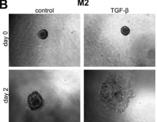
process [Chapter 4, [193]. Cancer cells can either invade as a single cell or collec vely as a
cohesive mass [243]. Intravital imaging studies revealed that both modes of mo lity occur in experimental breast tumors, while TGF‐ s mulated the single cell mo lity [244]. These
single cells moved along collagen fibers towards blood vessels [245,246]. The process of
invasion requires interac on with the extracellular matrix (ECM). Adhesion to the ECM
provides the trac on necessary for movement, which is mediated by integrins [247]. In‐
tegrins are heterodimeric cell surface receptors that recognize specific sequences in the
ECM, such as the RGD sequence. At least 24 integrins heterodimers exist by the combina‐
on of one of the 18 ‐subunits with one of the 8 ‐subunits [248‐250]. Upon adhering to
the ECM, integrins cluster in the plane of the membrane, which induces downstream sig‐
naling. Most integrins ac vate FAK and Src family kinases [247‐249]. A subset of integrins, namely 51, 11, v3 and 46, recruits the adaptor protein SHC in a Src‐dependent
signals, in concert with signals from growth factors, regulate migra on [247‐249] in an
invasion‐promo ng manner.
In breast cancer, expression of integrin 64 correlated with increased tumor grade and
decreased survival [248], although this integrin mediates firm adhesion to the basement
membrane in hemidesmosomes in normal breast ssue [251]. However, phosphoryla on
of integrin 4 by EGF‐receptor‐ac vated Src kinase Fyn or protein kinase C results in de‐
struc on of hemidesmosomes and more invasive behavior of cancer cells [252,253]. This
process might be enhanced by TGF‐, which induced clustering of this integrin with the
EGF receptor Her2 by ac va ng Src kinase [254]. Furthermore, expression of integrin 64 in the 4 nega ve breast cancer cell line MD‐MB465 promoted its invasive behavior [255].
Integrin 64 interacts also with the receptor for HGF, Met, and is required for invasive growth in response to this growth factor [256].
Expression of integrin v3 correlated with bone metastasis in breast cancer [257,258].
Overexpression of this integrin enhanced migra on [259] and bone metastasis of cancer
cells [260,261]. Invasion is further promoted by associa on and coopera on of integrin
v3 with growth factor receptors, such as the EGFR or TGF‐RII, which enhanced growth
factor signaling [247,250,262]. Integrin v3 is a TGF‐ target gene [263] and promoted TGF‐ signaling through phosphoryla on of TGF‐RII by Src [47,264], thus func oning in a
feed forward loop. Integrin v3 is also able to ac vate latent TGF‐ in vitro, but it re‐ mains unresolved if this also occurs in vivo [56]. Furthermore, integrin v3 enhanced in‐
vasion through associa on with MMP2 and MMP9, thus promo ng local ECM degrada‐
on at the cell surface [265,266].
MMP2 and MMP9 belong to the protein family of matrix metalloproteinases, which
cleave the components of the ECM [267,268]. MMPs are secreted as inac ve zymogens,
which are ac vated upon cleavage. Ac vity of the MMPs is controlled by endogenous in‐
hibitors, such as thromospondin [267]. Both MMP1 and MMP2 expression has been cor‐
related with breast cancer metastasis, whereas MMP9 seems to have a dual role in cancer
[268].
MMPs do not only promote tumor progression through degrada on of the ECM, but also
by processing signaling and adhesion molecules [267,268]. For example, cell‐surface‐
bound MMP9 ac vates TGF‐, thus promo ng invasion through TGF‐ signaling [269].
Since TGF‐ is able to induce the expression of MMP9 [270‐272], TGF‐ promotes ac va‐ on of other latent TGF‐present. Furthermore, TGF‐ induced sialoprotein (BSP), which
is able to accelerate ac va on of MMP2, MMP3 and MMP9 [273,274]. BSP is also able to associate with integrin v3 and MMP2 and this complex enhanced tumor invasion [275]. Thus, TGF‐ may promote invasion through induc on of several key players of invasion.
2.4.3 Intravasa on
Having reached a vessel, a cancer cell can enter the blood or lympha c system. Using
intravital imaging, it has been observed that cancer cells enter the blood through amoe‐
boid movement through the vessel wall [245]. This process is s mulated by perivascular
macrophages by secre on of EGF and Colony S mula on Factor (CSF) 1 [276]. Proteo‐
ly c ac vity, provided both by MMPs and urokinase plasminogen ac vator (uPA) is im‐
portant for the intravasa on process [277].
4.4 Survival in the circula on
Once in the blood stream, cancer cells o en aggregate with platelets and leukocytes,
which protects them from shear forces. This process is mediated through integrins and
selec ns [249]. For example, integrin v3 enhances binding of breast cancer cells to platelets [278]. Furthermore, platelets protect tumor cells from natural killer cells [279].
Protec on from lysis by the complement system may be achieved by expression of BSP
[274].
2.2.4.5 Extravasa on
Extravasa on of cancer cells appears in a similar fashion as leukocyte extravasa on, alt‐
hough different molecules are involved. First cells adhere loosely to the endothelium in a
process called rolling, then the cells firmly adhere to the endothelium and transmigrate
[280]. Leukocytes may aid extravasa on by ac ng as a bridge between tumor cells and
endothelial cells through integrin v3 [282]. Tissues with fenestrated capillaries, such as
the bone marrow and the liver, are more readily traversed by tumor cells than ssues
with capillaries fully lined with endothelial cells, such as the lung [283].
Expression of genes involved in vascular remodeling, namely epiregulin, cyclo‐oxygenase
2, MMP1 and MMP2 enabled extavasa on of breast tumor cells into the lung [284]. Induc‐ on of angiopoie n‐like 4 by TGF‐ in the primary tumor enhanced vessel permeability in
the lung, thus promo ng extravasa on in the lung [171]. Similar to intravasa on, macro‐
phages enhanced breast cancer extravasa on in lung metastasis [285].
Extravasa on into the brain is hampered by the blood‐brain barrier [283]. Therefore,
breast cancer cells that are able to metastasize to the brain not only express the same
vascular remodeling genes as cells which metastasize to the lung, but also ST6GALNAC5.
This sialyltransferase catalyzes the addi on of sialic acid to cell surface molecules, which
are important for adhesion to brain endothelial cells and their passage through the blood
brain barrier [286].
2.4.6 Coloniza on
Most breast cancer that infiltrate into distant organs die due to restric ons in the host
microenvironment [283]. However, other organs provide an environment in which the
disseminated breast cancer cells may survive. Already in 1889, Paget discovered that
breast cancer cells are osteotropic, leading to the "seed and soil hypothesis"; the cancer
cell ("the seed") can only grow in a suitable environment ("the soil") [287]. In the bone
environment, breast cancer cells usually induce destruc ve osteoly c lesions. Less com‐
mon are osteoblas c lesions, in which bone is synthesized [131]. Noggin seems to play an
important role in determining which type of lesions occurs [288]. Other "soils" include the
lungs and brain. The focus of this sec on will be on bone metastasis.
Once extravasated, breast cancer cells do not immediately colonize the bone [289]. In‐
stead, the single cells or cells micrometastasic clusters reside in the bone marrow, either
being dormant or prolifera ng at a slow rate [283]. Survival of breast cancer cells may be
mediated by Src, which can be induced by several growth factors present in the bone mar‐
The presence of bone marrow micrometastasis correlated with metastasis [291], indi‐
ca ng that the cells in the micrometastasis eventually acquire the ability to colonize or
manipulate the environment to enable coloniza on. TGF‐ is one of the growth factors
highly present in the bone environment and ac ve TGF‐ signaling has been detected in breast cancer metastasis [292,293]. However, abroga on of TGF‐ signaling in macrome‐ tastasis had no effect, indica ng the importance of TGF‐ signaling in early stages [292].
In early stages of tumor growth, angiogenesis is crucial for a metastasis to grow. TGF‐
induced expression of VEGF, a cri cal mediator of angiogenesis, in a Smad3 dependent
manner. Indeed, breast cancer cells with Smad3 knockdown grew slower due to reduced
angiogenesis [169]. The parathyroid hormone‐related protein (PTHrP) is involved in bone
metastasis forma on by ac va ng the bone‐resorbing cells, osteoclasts [131]. Ectopic Figure 5 Role of TGF‐ and BMPs in the metasta c cascade. In normal epithelium TGF‐ inhibits cell
growth. Upon malignant transforma on, TGF‐, as well as BMP‐2 and BMP‐4 promote epithelial to
mesenchymal transforma on (EMT) resul ng in more mo le mesenchymal cells. BMP‐6 and BMP‐7
counteract this process. Invasion of these cells is promoted by TGF‐, BMP‐2 and BMP‐4, whereas BMP
‐7 and BMP‐10 inhibit it. Cancer cells then intravasate, survive in the circula on and extravasate,
where they establish micrometastasis. TGF‐ promotes further coloniza on, which allows the cancer
expression of PTHrP reverted the reduc on in osteoly c lesions observed in metastasis of
cells expressing a dominant nega ve TGF‐RII, thus indica ng an important role for this protein in TGF‐‐induced bone metastasis forma on [170]. Furthermore, TGF‐ is able to
induce a set of genes which have been iden fied as drivers of breast cancer metastasis,
namely IL‐11, CTGF, CXCR4 and MMP1 [294]. These genes were found to be expressed in
breast cancer cells which preferen ally metastasize to the bone and overexpression of these genes coopera vely promoted bone metastasis [295]. TGF‐ also induced expres‐
sion of cyclo‐oxygenase 2 (COX2), which enhanced breast cancer bone metastasis [296].
All of the above men oned studies used MDA‐MB231 cells, which form osteoly c lesions.
In osteoblas c breast cancer lesions, endothelin‐1 is an important mediator of bone for‐
ma on, which can also be induced by TGF‐ [294,297]. Since BMPs are also potent induc‐
ers of bone forma on, one would a expect a role for them in osteoblas c metastasis. In‐
deed, in prostate cancer BMP‐6 promotes osteoblas c metastasis [298]. However, expres‐
sion of BMP‐2 in MDA‐MB231 cells s ll gives rise to osteoly c lesions [299]. Thus, differ‐
ent BMPs might have different roles in bone metastasis.
3.
Outline
of
the
thesis
A large number of studies has been performed to understand the biology of TGF‐ and
BMPs. These have highlighted the dual role of TGF‐ in cancer cells; in early stages it ham‐
pers prolifera on and promotes apoptosis, whereas in later stages it promotes invasion
and metastasis. However, TGF‐ also acts on the environment of the tumor, the tumor
stroma. This will be discussed in Chapter 2.
To inves gate TGF‐‐induced invasion of breast cancer cells experimentally, we estab‐
lished a 3D spheroid invasion model, which is introduced in Chapter 3. Using this model,
we characterized the role of Smads and MMPs in breast cancer cell invasion, which is de‐
scribed in Chapter 4.
BMP‐7 has been shown to inhibit bone metastasis of breast cancer, possibly by antagoniz‐
ing TGF‐‐induced EMT. In Chapter 5, we exploited our spheroid system to study the
crosstalk between TGF‐ and BMP‐7 in invasion. We also characterized the genes which
play a crucial role in this crosstalk.
EMT has been proposed to be a crucial step before invasion. In Chapter 6, we analyzed
the role of the key mediators of EMT in invasion of breast cancer cells.
Compared to TGF‐‐signaling, BMP‐signaling has been less well studied. Therefore, we
sought to iden fy kinases involved in BMP‐signaling. The results are described in Chapter
7.
Finally, the conclusions of the results described in this thesis are summarized and dis‐
cussed in Chapter 8.
4.
References
1. ten Dijke,P. and Heldin,C.‐H. (2006) The Smad family. In ten Dijke,P. and Heldin,C.‐H. (eds.) Smad Signal Transduc on. Springer, pp 1‐13.
2. Roberts,A.B., Anzano,M.A., Lamb,L.C., Smith,J.M., and Sporn,M.B. (1981) New class of transforming growth factors poten ated by epidermal growth factor: isola on from non ‐neoplas c ssues. Proc.Natl.Acad.Sci.U.S.A, 78, 5339‐5343.
3. Yu,L., Border,W.A., Huang,Y., and Noble,N.A. (2003) TGF‐ isoforms in renal fibrogene‐ sis. Kidney Int., 64, 844‐856.
4. Dickson,M.C., Mar n,J.S., Cousins,F.M., Kulkarni,A.B., Karlsson,S., and Akhurst,R.J. (1995) Defec ve haematopoiesis and vasculogenesis in transforming growth factor‐1 knock out mice. Development, 121, 1845‐1854.
5. Kaar nen,V., Voncken,J.W., Shuler,C., Warburton,D., Bu,D., Heisterkamp,N., and Groffen,J. (1995) Abnormal lung development and cle palate in mice lacking TGF‐3 indicates defects of epithelial‐mesenchymal interac on. Nat.Genet., 11, 415‐421. 6. Kulkarni,A.B., Huh,C.G., Becker,D., Geiser,A., Lyght,M., Flanders,K.C., Roberts,A.B.,
Sporn,M.B., Ward,J.M., and Karlsson,S. (1993) Transforming growth factor‐1 null muta‐ on in mice causes excessive inflammatory response and early death.
Proc.Natl.Acad.Sci.U.S.A, 90, 770‐774.
7. Proetzel,G., Pawlowski,S.A., Wiles,M.V., Yin,M., Boivin,G.P., Howles,P.N., Ding,J., Fergu‐ son,M.W., and Doetschman,T. (1995) Transforming growth factor‐3 is required for sec‐ ondary palate fusion. Nat.Genet., 11, 409‐414.
8. Sanford,L.P., Ormsby,I., Gi enberger‐de Groot,A.C., Sariola,H., Friedman,R., Boivin,G.P., Cardell,E.L., and Doetschman,T. (1997) TGF2 knockout mice have mul ple develop mental defects that are non‐overlapping with other TGF‐ knockout phenotypes. Devel
opment, 124, 2659‐2670.
9. Shull,M.M., Ormsby,I., Kier,A.B., Pawlowski,S., Diebold,R.J., Yin,M., Allen,R., Sidman,C., Proetzel,G., Calvin,D., and . (1992) Targeted disrup on of the mouse transforming growth factor‐1 gene results in mul focal inflammatory disease. Nature, 359, 693‐699. 10. Celeste,A.J., Iannazzi,J.A., Taylor,R.C., Hewick,R.M., Rosen,V., Wang,E.A., and Woz‐ ney,J.M. (1990) Iden fica on of transforming growth factor‐ family members present in bone‐induc ve protein purified from bovine bone. Proc.Natl.Acad.Sci.U.S.A, 87, 9843‐ 9847.