Design of protein-protein interactions via
β
-strand pairing
Peter Benjamin Stranges
A dissertation submitted to the faculty of the University of North Carolina at Chapel Hill in partial fulfillment of the requirements for the degree of Doctor of Philosophy in the Department of Biochemistry and Biophysics.
Chapel Hill 2012
Approved by:
Brian Kuhlman Ph.D.
Nikolay Dokholyan Ph.D.
Jane Richardson
John Sondek Ph.D.
c 2012
Abstract
PETER BENJAMIN STRANGES: Design of protein-protein interactions via β-strand pairing
(Under the direction of Brian Kuhlman Ph.D.)
The design of new protein-protein interfaces is a test of our understanding of protein
interaction biophysics and can provide new tools to understand cell biology. Methods to
accurately design high-affinity interactions have not been established, making it necessary to
devise new strategies to facilitate the design process. Solvent exposed main chain atoms on
β-strands are prone to interact with other exposed strands and could serve as the basis for the design of a novel interaction. This dissertation describes the application ofβ-strand pairing to design homodimeric and heterodimeric complexes. It also addresses the successes and failures
in computational interface design to determine how design methods need to be improved.
One of the most common ways that proteins interact is the formation of symmetric
ho-modimer. A way to test the hypothesis thatβ-strand mediated interactions can be accurately designed is to redesign a monomeric protein to form a symmetric homodimer viaβ-strand pair-ing. A computational method in Rosetta was used to find monomeric proteins with exposed
β-strands then redesign them to form a symmetric homodimer by pairing exposedβ-strands to form an intermolecular β-sheet. A crystal structure of one designed complex closely matches the computational model (RMSD = 1.0 ˚A). This work demonstrates thatβ-strand pairing can be used to computationally design new interactions with high accuracy
After successful design of a homodimer, β-strand pairing can be extended design to het-erodimers. A computational protocol is described that identifies proteins with exposed strand
capable of pairing with an exposed strand on a target protein. The interface of the identified
protein is then redesigned to allow it to bind to the target protein. Experimental testing
of proteins designed to bind RalA and PCSK9 show that no interaction is made. Directed
evolution of the scaffold proteins could allow them to bind to their target.
Most computational protein interface designs from our laboratory and others fail to form
when tested experimentally. Successful and failed protein interface designs were examined to
see if they provided answers about what works in interface design. Successful designs were, in
general, more hydrophobic than failed designs and had few designed hydrogen bonds buried
at the interface. Rosetta designed hydrogen bonds were found not to match hydrogen bond
distributions observed in high resolution crystal structures. The hydrogen bonding portion
of the energy function needs to be improved to allow for design of polar interfaces similar to
To the Flying Spaghetti Monster.
Acknowledgements
First and foremost I need to thank my advisor Brian Kuhlman giving me a challenging
project and sticking with it in the face of two years of disappointing experimental results.
When I felt like I was hitting a wall with my research Brian provided an endless source of
ideas and insight. I am also grateful that he got me into cycling.
Every member of the Kuhlman lab has contributed to my project in some way, though
some deserve need additional mention here. Andrew Leaver-Fay, Steven Lewis and Doug
Renfrew were instrumental in introducing me to computational protein design and making me
confortable with programming. Matt O’Meara contributed the intellectual and computational
basis for inspecting how Rosetta designs hydrogen bonds at protein-protein interfaces. Tim
Jacobs is a much appreciated collaborator and source of ideas in the design of novel binders to
PCSK9. Bryan Der has been an enormas help in developing approached to protein interface
design as well as a constant motivating force.
The members of Rosetta Commons have contributed the basis for most of the
computa-tional methods described in this thesis. The progress I made was only possible because of
the thousands of lines of code written by others. I especially need to thank Ingemar Andr´e
for developing and answering questions about symmetric modeling and Sarel Fleishman for
developing the RosettaScripts protocol.
Several faculty members at Bowdoin College piqued my interest in science and laboratory
research. I especially need to thank my advisors Mike Palopoli and Amy Johnson for getting me
involved in research labs and telling me I should do science instead of going to law school. My
development as a competent researcher was ensured by the expert tutelage of Sasha Chervonsky
It seems clich´e to thank one’s family here, but it needs to be done for several reasons. My
mom and dad made sure they were the first people I saw when I woke up after bumping my
head on the asphalt. My brother then took time to come to Chapel Hill and stayed with me
until I had my senses back. In addition my mom and brother have been constant source of
support, humor, and a reminder that it is possible to leave Chapel Hill sometimes.
There are several folks that need to be thanked for their contributions to my life outside
of the laboratory. First is my initial roommate, Andrew Parsons, for demonstrating the effort
required to be a successful graduate student, the joys of cycling up a 17% grade every morning,
and introducing me to the little spot of heaven that is Carrburritos. My second roommate,
Colin Deicth, has been a great friend and fellow political junkie since high school. I have been
fortunate to find soccer and volleyball teammates that are as much fun to play with as they
are to hang out with off the field/court. Finally, my classmates Jon Edwards, Aaron Hobbes,
and Patrick Lackey have been some of the best friends I could hope for.
Table of Contents
Abstract . . . iii
List of Tables . . . xii
List of Figures . . . xiii
List of Abbreviations . . . xv
1 Introduction . . . 1
1.1 Protein-protein interaction design . . . 1
1.2 Computational protein interface design . . . 3
1.2.1 Interface design using Rosetta . . . 5
1.3 Complications in computational protein interface design . . . 6
2 Computational design of a symmetric homodimer usingβ-strand assembly 22
2.1 Introduction . . . 22
2.2 Materials and Methods . . . 24
2.2.1 Search Method for Homodimer Scaffolds . . . 24
2.2.2 Homodimer Design and Selection . . . 26
2.2.3 Evaluation of Designs . . . 26
2.2.4 DNA Construct and Protein Production . . . 27
2.2.5 Multiangle Light Scattering . . . 27
2.2.6 Analytical Ultracentrifugation Sedimentation Equilibrium . . . 27
2.2.7 Fluorescence Polarization Assay . . . 28
2.2.8 Homodimerization Fluorescence Polarization Fitting Procedure . . . 28
2.2.9 Crystallization and Structure Refinement . . . 29
2.3 Results . . . 30
2.3.1 Scaffold Search Protocol . . . 30
2.3.2 Design Protocol . . . 32
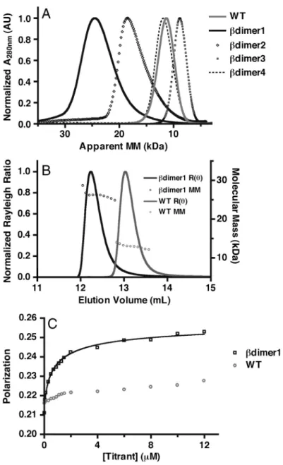
2.3.3 Determining Oligomeric Status . . . 33
2.3.4 Homodimer Binding Affinity . . . 35
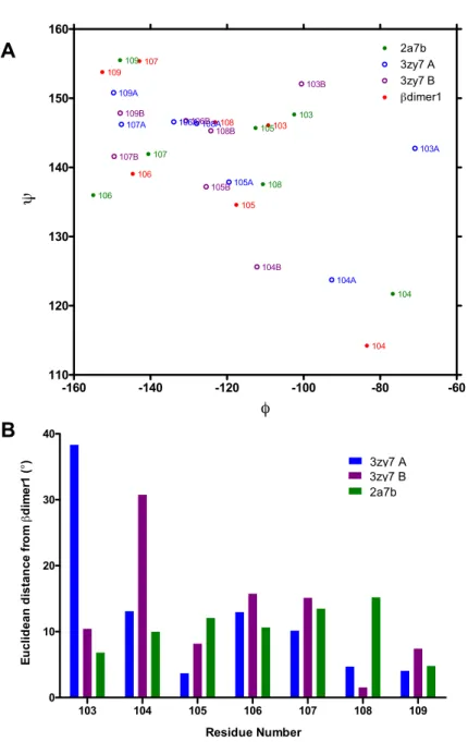
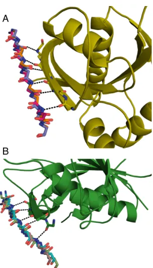
2.3.5 Crystal Structure of theβdimer1 . . . 37
2.4 Discussion . . . 39
2.5 Supporting Information . . . 41
3 Computational design of aβ-strand mediated heterodimer . . . 52
3.1 Introduction . . . 52
3.2 Materials and Methods . . . 55
3.2.1 Idealized binding strand construction . . . 55
3.2.2 Scaffold search procedure . . . 56
3.2.3 Interface design of selected scaffolds . . . 57
3.2.4 Protein expression and purification . . . 58
3.2.5 Yeast expression of designs . . . 59
3.2.6 ITC binding measurements . . . 59
3.3 Results . . . 60
3.3.1 Idealized interaction strand model . . . 60
3.3.2 Scaffold search protocol . . . 60
3.3.3 Selection of interface designs . . . 60
3.3.4 Affinity measurements of designed binders to RalA . . . 63
3.3.5 Yeast display of proteins to bind PCSK9 . . . 63
3.4 Discussion . . . 67
3.4.1 Future direction: evolving binders to PCSK9 . . . 67
3.4.2 Computational library generation . . . 68
3.5 Supporting Information . . . 70
4 A comparison of successful and failed computational protein interface
de-signs . . . 77
4.1 Introduction . . . 77
4.2 Methods . . . 79
4.2.1 Definition of an interface residue . . . 79
4.2.2 Input structure energy minimization . . . 81
4.2.3 Interface analysis protocol . . . 81
4.2.4 Polar burial definition . . . 82
4.2.5 Determining hydrogen bond geometry . . . 82
4.3 Results . . . 83
4.3.1 Set of designed interfaces . . . 83
4.3.2 Definition of a successful design . . . 84
4.3.3 Designed interfaces are small . . . 86
4.3.4 Successful designs have few polar interactions . . . 88
4.3.5 Rosetta designs suboptimal hydrogen bonds . . . 91
4.3.6 Other observations . . . 92
4.4 Discussion . . . 96
4.5 Supporting Information . . . 99
4.5.1 Figures . . . 99
4.5.2 Protein structures and models used . . . 103
4.5.3 Analysis protocol . . . 109
4.5.4 Hydrogen bond features . . . 110
5 Conclusion . . . 116
5.1 Additional applications ofβ-strand interface design . . . 116
5.2 Difficulties in β-strand interface design . . . 117
5.3 Improvements in interface design methodology . . . 120
5.4 Future of computational interface design . . . 121
A Fluorescence polarization titrations and fitting protocol . . . 129
A.1 Titration calculations . . . 129
A.2 Homodimer fitting protocol for Prism . . . 131
List of Tables
2.1 Computational evaluation of designed homodimer models . . . 33
2.2 Molecular mass of designed homodimers in solution . . . 35
S2.1 Data collection and refinement statistics . . . 46
3.1 Computational evaluation of designed heterodimer models . . . 63
4.1 Successful and failed interface designs . . . 86
S4.1 Native heterodimers and homodimers . . . 103
List of Figures
1.1 Model of β-strand interaction . . . 8
1.2 Natural β-strand mediated protein interactions . . . 9
2.1 Search and design protocol for a symmetric homodimer . . . 31
2.2 Computational designs used in experiments . . . 34
2.3 Homodimer molecular mass and affinity . . . 36
2.4 Computational model to crystal structure comparison . . . 38
S2.1 AUC determination of molecular mass . . . 41
S2.2 AUC determination of affinity . . . 42
S2.3 Comparison of interactingβ-strands . . . 43
S2.4 Unmodeled salt bridge in crystal structure . . . 43
S2.5 Clashes prevent register shift . . . 44
S2.6 Backbone torsion angles of interacting strands . . . 45
3.1 β-strand mediated interactions to RalA . . . 54
3.2 β-strand mediated interactions to PCSK9 . . . 55
3.3 Idealized paired strand conformation . . . 61
3.4 Heterodimer scaffold search method . . . 62
3.5 Designed binders to RalA . . . 64
3.6 Designed binders to PCSK9 . . . 65
3.7 ITC measurement of designed binders to RalA . . . 66
4.1 Interface residue vector definition . . . 80
4.2 Computational interface design targets . . . 85
4.3 Size of designed and natural interfaces . . . 87
4.4 Interface energy density designed and natural interfaces . . . 89
4.5 Polar content of designed and natural interfaces . . . 92
4.6 Buried polars and buried hydrogen bonds at interfaces . . . 93
4.7 Buried hydrogen bonds at designed interfaces . . . 94
4.8 Interface hydrogen bond geometry . . . 95
S4.1 Distance from water to protein atoms . . . 100
S4.2 Hydrogen bond Lambert azimuthal equal-area projection . . . 101
S4.3 Interface backbone hydrogen bond geometry . . . 102
S4.4 Change in solvation and hydrogen bond energy . . . 102
5.1 Problems with identified exposed β-strands . . . 119
List of Abbreviations
AUC Analytical ultra centrifugation
DSSP Database of secondary structure of proteins
FACS Fluorescence activated cell sorting
FP Fluorescence polarization
∆Gbind Calculated change in energy upon protein-protein binding
ITC Isothermal titration calorimetry
MALS Multi-angle light scattering
PDB Protein Data Bank
REU Rosetta energy units
RMSD Root mean square deviation
SASA Solvent accessible surface area
∆SASA change in SASA upon protein-protein binding
SEC Size exclusion chromatography
Chapter 1
Introduction
The energetics that govern how a protein folds from an extended chain into a final
con-formation are understood in principle (1), but reliable techniques to accurately predict final
structure based on sequence information alone have proved elusive (2). Additionally, a clear
picture of how, once folded, a protein interacts with other proteins has not been elucidated.
A complete understanding of how these processes work cannot be determined until the forces
stabilizing the folded and interacting states are explained. De novoprotein and protein-protein interaction design provides one way of comprehending these forces. The de novo design of a protein means rationally choosing a sequence that will fold into a target final structure (3).
Failed and successful designs can provide valuable information about the forces involved in
stabilizing a three-dimensional structure. De novo protein interface design extends this idea to design a sequence capable of forming an interaction between two or more protein chains.
A designed interface that forms experimentally can provide valuable information about the
physical chemistry of protein-protein association (4).
1.1
Protein-protein interaction design
Protein-protein interaction networks monitor the internal and external state of a cell and
act as signaling circuits to transmit information and change the phenotype of the cell. Genome
sequencing and proteomic databases provide a nearly complete list of the available modules for
high throughput techniques have allowed some of these interaction networks to be mapped
(6), but a complete understanding of cell signaling remains out of reach. Like protein design,
the design of new signaling circuits provides a test of our understanding of a basic biological
process. An understanding of which interactions work, which do not, and why they do not can
provide valuable information about natural protein-protein interactions (5). Novel methods
to alter natural cellular interactions provide a wealth of information about the regulation and
response of the signaling circuit (7).
The design of new protein-protein interactions has provided valuable tools to undersand the
way nature constructs interactions and modify cell signaling systems. Many natural proteins
exist as large complexes of small subunits that form symmetric interfaces in order to construct
the larger, functional, protein (8). Grueninger et al. used a simple mutagenesis approach to construct ordered, symmetric oligomers out of monomeric proteins (9). This demonstrates a
mechanism that nature may use to build multimeric proteins. Protein interactions that can
be turned on and off with light have be used to control cell motility (10; 11) and membrane
recruitment of certain proteins (12). These tools provide the ability to selectively alter
pro-tein function in different areas of the cell. Modular domain recombination of propro-tein-propro-tein
interaction domains can create new cellular responses by providing alternative protein
interac-tion networks (reviewed in (13) and (14)). This method recombines protein domains to form
non-native interactions with a scaffold that serves to orient and control the flow of
informa-tion. This method has been used to tune and improve metabolic flux (15), activate guanine
exchange factors with a non-native input (16) and engineer feedback loops into MAP kinase
signaling (17). These studies demonstrate that engineered protein-protein interactions can
control output and tease apart natural protein pathways.
One of the most common ways to obtain a new protein-protein interaction is though
directed evolution (18). Phage display (19), yeast display (20) and ribosomal display (21)
have provided a wide variety of affinity reagents and protein functions used in cell biology. For
example the fusion of a PDZ domain to an FNIII domain followed by directed evolution of
the FNIII domain allowed the construct to recognize alternate PDZ binding peptides (22). A
multiple laboratory effort engineered high affinity binders to 20 SH2 domains using a variety of
directed evolution techniques (23). It now appears that it is possible to use directed evolution
to obtain an affinity reagent to any target that can be purified. A remaining challenge is
obtaining binders that recognize a conformational state, post translational modification, or
are specific for only one member of a protein family.
Biological engineering has made it evident that more tools are needed to control
cellu-lar machinery. Some of the primary needs are orthogonal protein interaction pathways with
variable affinity, specificity (5), and computer aided design strategies to design new logic
circuits(24). Metabolic pathway design requires protein interactions that can be predictably
regulated and have tunable affinity for certain upstream and downstream effectors (25).
Com-putational methods of modifying and designing new protein interactions can provide the tools
to meet the needs of biological engineering (26) .
1.2
Computational protein interface design
Computational protein design attempts to search the sequence and conformation of a
polypeptide chain to minimize the energy of the structure. All computational design protocols
have two components, a search function to explore sequence and conformational space and
an energy function that evaluates the fitness of a particular search model (27). Successful
computational design of to monomeric proteins quickly suggested that these same models
could be applied to engineer protein-protein interactions (28).
The idea that the molecular structure of proteins is responsible for their interaction has
been recognized since the time that the first few crystal structures of protein complexes were
determined (29; 30). However, the ability to accurately model how two proteins will form
a complex has proved elusive (31). De novo computational interface design is a test of our understanding of the forces governing protein-protein interactions. A typical computational
design run involves modeling the rigid body orientation of two or more protein chains in a
conformation that will allow them to interact. Next, the computational protocol designs amino
acids near the modeled interaction to stabilize the complex state. The design goal determines
on a natural protein to enable it to bind to another natural protein. In this case the natural
protein that is left unchanged is called the target protein and the redesigned protein is called
the scaffold.
Computational interface design has helped create new interactions and functions that
na-ture has not sampled. Recent progress in computational interaction design is reviewed in
(26) and (32). Computational design provides several advantages over directed evolution and
modular domain recombination. Most importantly, computational design can be used to
tar-get a specific interaction site for design and constrain the design to satisfy a particular goal.
For example Reina et al. used a computational protocol to predictably engineer a Class I PDZ domain to recognize Class I and Class II target peptides (33). Similarly, Shifman and
colleagues were able to design mutations to alter specificity of calmodulin for different target
peptides (34; 35). Another group was able to find an array of substrates for a protein
chap-erone by investigating the sequences allowed to bind to a target cite on the chapchap-erone (36).
An antibody’s affinity for its target can be improved beyond in vivo affinity maturation by computationally optimizing interface electrostatics (37). Computational design also allows for
favoring one type of interaction (positive design) while simultaneously disfavoring an alternate
interaction (negative design). This idea has been used to design heterodimers that do not
interact with off target proteins (38; 39; 40).
Computational protein-protein interaction design has the potential to create proteins
ca-pable of predictably changing cell signaling. One exciting advance is the development of a
computational framework to design a multi-function protein hubs capable of interacting with
a variety of partners (41). To achieve this goal computational design will need to be able to
repeatably engineer novel protein-protein interactions. The majority of successful
computa-tional designs addressed in the reviews above involve the redesign of an existing interaction for
increased affinity or altered specificity. Most success computational novel interaction design
in-volves idealized systems such asα-helical pairing (42; 38; 40) or hot spot grafting (43; 44). New approaches to interface design are needed to create a toolkit capable to predictably modifying
cellular function.
1.2.1 Interface design using Rosetta
The Rosetta suite of macromolecular modeling software (45) was used for all protein design
steps in this work. The energy function is comprised of terms for all aspects thought to be
important to protein structure (46).
Eprotein =Wlj atrElj atr+Wlj repElj rep+WHbondEHbond+WsolvationEsolvation+WaaEaa+
WpairEpair+WramaErama+WrotamerErotamer −Wref erenceEref erence
These terms include physically based potentials to capture van der Waals interactions
(Wlj atrElj atr andWlj repElj rep) and solvation energy (WsolvationEsolvation). The other,
knowl-edge based terms, are parameterized based on high resolution structures in the Protein Data
Bank (PDB). Hydrogen bond energy (WHbondEHbond) is calculated based on the distance and
angles between the acceptor and donor atoms (47). The other terms account for
electro-static pairing (WpairEpair), torsional preferences (WramaEramaand WrotamerErotamer) and the
contextual preferences of amino acids ( WaaEaa and Wref erenceEref erence). Rosetta’s search
function is Metropolis Monte Carlo (48) with simulated annealing (49), which allows the
se-quence and conformation of a protein to be quickly sampled during a design simulation but
does not guarantee finding a global minimum.
Rosetta is well suited to design new protein-protein interactions (50). Rosetta has the
ability to perform rigid body protein docking (51), backbone conformation sampling (52), and
sequence design (53). The recent development of an XML scripting language allows many
design and sampling modules to be easily incorporated into a single simulation. (54). Rosetta
can increase the affinity of a protein interaction (55), alter the specificity of existing interactions
(56; 57; 58; 59) and stabilize an interaction to serve as a biosensor (60). More recently, Rosetta
has proven to be capable of designing novel protein interactions (61; 62; 63). These successes
are addressed in Chapter 4. The next step is to move beyond from proof of concept designs to
1.3
Complications in computational protein interface design
The design of a protein-protein interactions from scratch has proven to be a very difficult
problem (reviewed in (26; 32)). Few designed interfaces have been experimentally verified
to form a complex that matches the computational model. Computational interface design
combines two challenging modeling goals, rigid body orientation between protein chains and
the design of sequence that will allow those chains to interact (28). Most computational designs
of a de novo interface have no measurable affinity (64). The ones that do interact often have low affinity (KD > 100 µM) (4; 65) or do not form the expected complex (66). Directed
evolution can help overcome low affinities by sampling additional sequences that were not in
the designed model (61). Computational redesign of existing protein-protein interactions for
enhanced affinity has also been plagued by similar problems. Many of the mutations predicted
to increase affinity actually weaken the interaction or the redesigned proteins (67; 68).
A protein interface presents several modeling challenges that are not relevant for design
of a monomeric protein. Transient protein interactions are more polar than protein cores (69)
and require the desolvation of polar atoms to be offset by an interface spanning hydrogen bond
(70). A transient interaction requires the proteins involved to be stable in both the bound
and unbound state. Mutations that favor an interaction could cause one of the monomers to
become unstable and aggregate. Monomeric proteins only need to be designed in the context of
the final folded structure (53; 71). Buried water molecules can bridge hydrogen bonds between
molecules and stabilize the complex (72). So far, attempts to model water mediated hydrogen
bonds have not yielded encouraging results (73). Electrostatic complementation is another
driving force behind protein-protein interaction (70; 74). Optimizing long range electrostatic
steering can enhance the affinity of an interaction by increasing the on rate (kon) (75; 76).
Though useful, intensive electrostatics calculations are impractical during design because of
speed and the need for a pairwise additive potential (28). A survey of successful and failed
protein interface designs revealed that electrostatic complementation was not designed in most
computational models (64). Recent advances in simplifying electrostatic models could allow
their incorporation into design simulations (77).
Many groups have employed specialized energy functions to redesign protein-protein
inter-actions for altered specificity or increased affinity. The requirement for specialized interface
potentials should not be necessary, although different residues are preferred in interfaces versus
proteins as a whole (69) the same packing density and hydrogen bond geometry is observed
in monomers and across protein interfaces (78). The results of these studies have provided
no consistent insight into energy function improvements. Different studies found success by
increasing the weight of interchain interactions (35), down weighting the solvation terms of
the energy function (68; 79), specifically favoring the electrostatic component of the energy
function (37), and training the energy function on mutational data for a specific interface (67).
The need for modified energy functions for protein interfaces is more likely to be indicative of
a flaw in design methodology and sampling than in the energy function.
The research highlighted here suggests that one hurdle to computational interface design
is the correct modeling of hydrogen bonds and electrostatics. It is imperative to devise new
strategies that can overcome this and other persistent complications faced in computational
protein-protein interaction design. The successes and failures of interface design can also be
examined to glean any information that indicates why many designs fail and only few succeed.
1.4
Using
β
-strands for protein-protein interface design
“Stealing” the sequence information or local structure from natural protein-protein
inter-faces is a prudent way to design a new interactions or inhibit existing ones. (80). It allows
us to borrow information from the billions of years life has spent engineering interactions and
reapply it for our needs. Nature uses a variety of different motifs, folds, and sequences to form
the basis of a protein-protein interface (81). One common interface architectural motif is
pair-ing solvent accessible β-strands to form an intermolecularβ-sheet (Figure 1.1). These paired β-strands form hydrogen bonds between the main chain atoms in a antiparallel orientation (82). Remaut and Waksman (83) surveyed heterodimeric protein interactions that result in
Ras family form a complex with their ubiquitin-like effectors via an intermolecular β-sheet (84) (Figure 1.2C). Antiparallel cross-chain β-strand pairing also represents 8.8% of contacts observed in homodimeric proteins (Figure 1.2D) (82). Paired β-strands in homodimers are often longer than those involved in forming hetero-complexes (85).
Target
Protein
!
Scaffold
Protein
!
Figure 1.1: Model of heterodimer β-strand interaction. A target protein (blue) with an exposed β-strand forming a complex with another scaffold protein (red) with an exposed strand. Black dashed lines represent main chain hydrogen bonds formed across the interface.
Interface design based onβ-strand pairing presents a solution to two problems in compu-tational protein interface design: assuring orientation specificity and satisfying hydrogen bond
potential. β-strand interactions are geometrically constrained by hydrogen bonding between the main chain atoms and the twist and sheer observed inβ-sheets has been well documented (90; 91; 92). This specificity for a particular hydrogen bonds arrangement should prevent the
formation of a complex that does not match the design model (66). Complementary strand
pairing across an interface helps satisfy some of the hydrogen bond potential without the need
for sequence design. Design can then proceed from well established side-chain preferences for
β-sheet formation and stability (93; 94; 95; 96).
The intrinsic potential of exposed β-strands to self-assemble makes them an attractive starting point for interface design. β-strands are often calledsticky because of their tendency
A!
B!
C!
D!
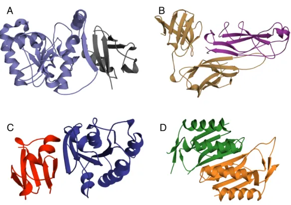
Figure 1.2: Naturalβ-strand mediated protein interactions. A) Crystal structure of thymine DNA glycosylase (light blue) conjugated to SUMO-1 (grey) (PDB ID: 1WYW) demonstrating β-strand addition(86). B) Crystal structure of the chaperone protein PapD (beige) in complex with PapK (purple) (PDB ID: 1PDK) demonstrating fold completion (87). C) Canonical β -strand mediated GTPase-effector interaction demonstrated by the complex of RAP1A (blue) with c-Raf1(red) (PDB ID: 1GUA) (88) D) Symmetric homodimer molybdopterin synthase (PDB ID: 1NVJ) with anti parellel β-strands at the interface between the two chains (green and orange) (89).
to interact with other strands. This is evident inβ-strand pairing in crystal contacts (97; 98; 99; 53) and the propensity ofβ-sheet proteins to uncontrollably assemble into amyloid-like fibrils (100; 101). Exposed strands are so prone to interact with each other that nature uses negative
design elements such as strand kinks, charged residues, or occlusion with a loop to prevent
nonspecific β-strand interactions (102) Solvent accessible strands that interact with another protein tend to be longer and have fewer negative design elements than their counterparts
strand could cause the designed protein to uncontrollably self assemble. While this is a distinct
possibility, explicit negative design steps were not required to redesign a natural β-sandwich protein (71), which suggests negative design elements exist in the backbone of exposed strands.
Several studies have shown the efficacy of usingβ-strands to alter protein function. Cyclic peptide inhibitors of protein-protein interactions and protease activity are thought to bind to
their targets via formation of a intermolecular β-sheet (105; 106). Directed evolution experi-ments have generated β-strand pairing between an antibody and its antigen (107) as well as phage display targets with an exposed strand (108). Amyloid fibrillization can be inhibited by
a peptide designed to form a terminating β-strand on a growing fibril (109) and an evolved β-strand presenting protein (110).
The following chapters seek to provide a solution to some of the difficulties in
protein-protein interaction design. First, I introduce β-strand pairing at protein-protein interfaces as a way to ensure orientation and affinity at a de novo interface. I demonstrate that pairing solvent exposedβ-strands, followed by sequence design at the interface, can lead to the accurate design of a symmetric homodimer in Chapter 2, beginning on page 22. Chapter 3 (page 52),
extends this idea to the design of a binder to a natural protein using β-strand pairing at the targeted interface. I describe a general method to find proteins capable of forming an
intermolecular β-sheet. Finally, Chapter 4 (page 77) examines successful and failed attempts to computationally design novel interfaces and finds that most successful designs have few
designed polar interactions at the interface.
References
1. Dill, K. A. (1990) Dominant forces in protein folding. Biochemistry29, 7133–7155
2. Moult, J., Fidelis, K., Kryshtafovych, A., and Tramontano, A. (2011) Critical assess-ment of methods of protein structure prediction (CASP)–round IX. Proteins 79 Suppl 1, 1–5
3. Betz, S. F., Raleigh, D. P., and DeGrado, W. F. (1993) De novo protein design: from molten globules to native-like states. Current Opinion in Structural Biology3, 601– 610
4. Huang, P.-S., Love, J. J., and Mayo, S. L. (2007) A de novo designed protein protein interface. Protein science : a publication of the Protein Society 16, 2770–4
5. Lim, W. a. (2010) Designing customized cell signalling circuits. Nature reviews. Molecular cell biology 11, 393–403
6. Breitkreutz, A., Choi, H., Sharom, J. R., Boucher, L., Neduva, V., Larsen, B., Lin, Z.-Y., Breitkreutz, B.-J., Stark, C., Liu, G., Ahn, J., Dewar-Darch, D., Reguly, T., Tang, X., Almeida, R., Qin, Z. S., Pawson, T., Gingras, A.-C., Nesvizhskii, A. I., and Tyers, M. (2010) A global protein kinase and phosphatase interaction network in yeast. Science (New York, N.Y.)328, 1043–6
7. Bennett, M. R., Pang, W. L., Ostroff, N. A., Baumgartner, B. L., Nayak, S., Tsimring, L. S., and Hasty, J. (2008) Metabolic gene regulation in a dynamically changing environment. Nature 454, 1119–22
8. Janin, J. (2008) Biochemistry. Dicey assemblies. Science (New York, N.Y.)319, 165–6
9. Grueninger, D., Treiber, N., Ziegler, M. O. P., Koetter, J. W. A., Schulze, M.-S., and Schulz, G. E. (2008) Designed protein-protein association. Science (New York, N.Y.)
319, 206–9
11. Hahn, K. M. and Kuhlman, B. (2010) Hold me tightly LOV. Nature methods 7, 595, 597
12. Levskaya, A., Weiner, O. D., Lim, W. A., and Voigt, C. A. (2009) Spatiotemporal control of cell signalling using a light-switchable protein interaction. Nature 461, 997–1001
13. Koide, S. (2009) Generation of new protein functions by nonhomologous combinations and rearrangements of domains and modules. Current opinion in biotechnology 20, 398–404
14. Good, M. C., Zalatan, J. G., and Lim, W. A. (2011) Scaffold proteins: hubs for con-trolling the flow of cellular information. Science (New York, N.Y.) 332, 680–6
15. Dueber, J. E., Wu, G. C., Malmirchegini, G. R., Moon, T. S., Petzold, C. J., Ullal, A. V., Prather, K. L. J., and Keasling, J. D. (2009) Synthetic protein scaffolds provide modular control over metabolic flux. Nature biotechnology 27, 753–9
16. Yeh, B. J., Rutigliano, R. J., Deb, A., Bar-Sagi, D., and Lim, W. A. (2007) Rewiring cellular morphology pathways with synthetic guanine nucleotide exchange factors. Nature447, 596–600
17. Bashor, C. J., Helman, N. C., Yan, S., and Lim, W. A. (2008) Using engineered scaffold interactions to reshape MAP kinase pathway signaling dynamics. Science (New York, N.Y.)319, 1539–43
18. J¨ackel, C., Kast, P., and Hilvert, D. (2008) Protein design by directed evolution. Annual review of biophysics37, 153–73
19. Sidhu, S. S. and Koide, S. (2007) Phage display for engineering and analyzing protein interaction interfaces. Current opinion in structural biology 17, 481–7
20. Gai, S. A. and Wittrup, K. D. (2007) Yeast surface display for protein engineering and characterization. Current opinion in structural biology17, 467–73
21. Zahnd, C., Amstutz, P., and Pl¨uckthun, A. (2007) Ribosome display: selecting and evolving proteins in vitro that specifically bind to a target. Nature methods 4, 269–79
22. Ferrer, M., Maiolo, J., Kratz, P., Jackowski, J. L., Murphy, D. J., Delagrave, S., and Inglese, J. (2005) Directed evolution of PDZ variants to generate high-affinity detection reagents. Protein engineering, design & selection : PEDS 18, 165–73
23. Colwill, K. and Gr¨aslund, S. (2011) A roadmap to generate renewable protein binders to the human proteome. Nature Methods 8, 551–8
24. Clancy, K. and Voigt, C. A. (2010) Programming cells: towards an automated ’Genetic Compiler’. Current opinion in biotechnology21, 572–81
25. Holtz, W. J. and Keasling, J. D. (2010) Engineering static and dynamic control of synthetic pathways. Cell140, 19–23
26. Mandell, D. J. and Kortemme, T. (2009) Computer-aided design of functional protein interactions. Nature chemical biology 5, 797–807
27. Street, A. G. and Mayo, S. L. (1999) Computational protein design. Structure 7, R105–R109
28. Kortemme, T. and Baker, D. (2004) Computational design of protein-protein interac-tions. Current opinion in chemical biology 8, 91–7
29. Chothia, C. and Janin, J. (1975) Principles Of Protein-Protein Recognition. Nature
256, 705–708
30. Jones, S. and Thornton, J. M. (1996) Principles of protein-protein interactions. Proceedings of the National Academy of Sciences of the United States of America
93, 13–20
31. Lensink, M. F. and Wodak, S. J. (2010) Docking and scoring protein interactions: CAPRI 2009. Proteins78, 3073–84
32. Karanicolas, J. and Kuhlman, B. (2009) Computational design of affinity and specificity at protein-protein interfaces. Current opinion in structural biology19, 458–63
34. Shifman, J. M. and Mayo, S. L. (2002) Modulating calmodulin binding specificity through computational protein design. Journal Of Molecular Biology 323, 417–423
35. Yosef, E., Politi, R., Choi, M. H., and Shifman, J. M. (2009) Computational Design of Calmodulin Mutants with up to 900-Fold Increase in Binding Specificity. Journal Of Molecular Biology385, 1470
36. Kota, P., Summers, D. W., Ren, H.-Y., Cyr, D. M., and Dokholyan, N. V. (2009) Iden-tification of a consensus motif in substrates bound by a Type I Hsp40. Proceedings of the National Academy of Sciences of the United States of America106, 11073–8
37. Lippow, S. M., Wittrup, K. D., and Tidor, B. (2007) Computational design of antibody-affinity improvement beyond in vivo maturation. Nature biotechnology25, 1171–6
38. Havranek, J. J. and Harbury, P. B. (2003) Automated design of specificity in molecular recognition. Nature structural biology10, 45–52
39. Bolon, D. N., Grant, R. A., Baker, T. A., and Sauer, R. T. (2005) Specificity versus stability in computational protein design. Proceedings of the National Academy of Sciences of the United States of America 102, 12724–9
40. Grigoryan, G., Reinke, A. W., and Keating, A. E. (2009) Design of protein-interaction specificity gives selective bZIP-binding peptides. Nature 458, 859–864
41. Humphris, E. L. and Kortemme, T. (2007) Design of multi-specificity in protein inter-faces. PLoS computational biology3, e164
42. Harbury, P. B., Plecs, J. J., Tidor, B., Alber, T., and Kim, P. S. (1998) High-resolution protein design with backbone freedom. Science (New York, N.Y.) 282, 1462–7
43. Liu, S., Liu, S. Y., Zhu, X. L., Liang, H. H., Cao, A. N., Chang, Z. J., and Lai, L. H. (2007) Nonnatural protein-protein interaction-pair design by key residues grafting. Proceedings of The National Academy of Sciences 104, 5330–5335
44. Azoitei, M. L., Ban, Y.-E. A., Julien, J.-P., Bryson, S., Schroeter, A., Kalyuzhniy, O., Porter, J. R., Adachi, Y., Baker, D., Pai, E. F., and Schief, W. R. (2012) Computational design of high-affinity epitope scaffolds by backbone grafting of a linear epitope. Journal of molecular biology415, 175–92
45. Leaver-Fay, A., Tyka, M., Lewis, S. M., Lange, O. F., Thompson, J., Jacak, R., Kauf-man, K., Renfrew, P. D., Smith, C. A., Sheffler, W., Davis, I. W., Cooper, S., Treuille, A., Mandell, D. J., Richter, F., Ban, Y.-E. A., Fleishman, S. J., Corn, J. E., Kim, D. E., Lyskov, S., Berrondo, M., Mentzer, S., Popovi´c, Z., Havranek, J. J., Karanicolas, J., Das, R., Meiler, J., Kortemme, T., Gray, J. J., Kuhlman, B., Baker, D., and Bradley, P. (2011) ROSETTA3: an object-oriented software suite for the simulation and design of macromolecules. Methods in enzymology 487, 545–74
46. Rohl, C. A., Strauss, C. E. M., Misura, K. M. S., and Baker, D. (2004) Protein structure prediction using Rosetta. Methods in enzymology383, 66–93
47. Kortemme, T., Morozov, A. V., and Baker, D. (2003) An orientation-dependent hydro-gen bonding potential improves prediction of specificity and structure for proteins and protein-protein complexes. Journal of molecular biology326, 1239–59
48. Metropolis, N., Rosenbluth, A. W., Rosenbluth, M. N., Teller, A. H., and Teller, E. (1953) Equation Of State Calculations By Fast Computing Machines. Journal Of Chemical Physics21, 1087–1092
49. Kirkpatrick, S., Gelatt, C. D., and Vecchi, M. P. (1983) Optimization by simulated annealing. Science (New York, N.Y.)220, 671–80
50. Kaufmann, K. W., Lemmon, G. H., Deluca, S. L., Sheehan, J. H., and Meiler, J. (2010) Practically useful: what the Rosetta protein modeling suite can do for you. Biochemistry49, 2987–98
51. Wang, C., Bradley, P., and Baker, D. (2007) Protein-protein docking with backbone flexibility. Journal of molecular biology 373, 503–19
52. Smith, C. A. and Kortemme, T. (2008) Backrub-like backbone simulation recapit-ulates natural protein conformational variability and improves mutant side-chain prediction. Journal Of Molecular Biology380, 742–756
53. Kuhlman, B., Dantas, G., Ireton, G. C., Varani, G., Stoddard, B. L., and Baker, D. (2003) Design of a novel globular protein fold with atomic-level accuracy. Science (New York, N.Y.) 302, 1364–8
55. Sammond, D. W., Eletr, Z. M., Purbeck, C., Kimple, R. J., Siderovski, D. P., and Kuhlman, B. (2007) Structure-based protocol for identifying mutations that enhance protein-protein binding affinities. Journal of molecular biology371, 1392–404
56. Kortemme, T., Joachimiak, L. A., Bullock, A. N., Schuler, A. D., Stoddard, B. L., and Baker, D. (2004) Computational redesign of protein-protein interaction specificity. Nature Structural & Molecular Biology 11, 371–379
57. Joachimiak, L. A., Kortemme, T., Stoddard, B. L., and Baker, D. (2006) Computa-tional design of a new hydrogen bond network and at least a 300-fold specificity switch at a protein-protein interface. Journal of molecular biology361, 195–208
58. Sammond, D. W., Eletr, Z. M., Purbeck, C., and Kuhlman, B. (2010) Computational design of second-site suppressor mutations at protein-protein interfaces. Proteins78, 1055–65
59. Kapp, G. T., Liu, S., Stein, A., Wong, D. T., Rem´enyi, A., Yeh, B. J., Fraser, J. S., Taunton, J., Lim, W. A., and Kortemme, T. (2012) Control of protein signaling using a computationally designed GTPase/GEF orthogonal pair. Proceedings of the National Academy of Sciences of the United States of America
60. Jha, R. K., Wu, Y. I., Zawistowski, J. S., MacNevin, C., Hahn, K. M., and Kuhlman, B. (2011) Redesign of the PAK1 autoinhibitory domain for enhanced stability and affinity in biosensor applications. Journal of molecular biology413, 513–22
61. Fleishman, S. J., Whitehead, T. A., Ekiert, D. C., Dreyfus, C., Corn, J. E., Strauch, E.-M. M., Wilson, I. A., and Baker, D. (2011) Computational design of proteins targeting the conserved stem region of influenza hemagglutinin. Science 332, 816– 821
62. Sammond, D. W., Bosch, D. E., Butterfoss, G. L., Purbeck, C., Machius, M., Siderovski, D. P., and Kuhlman, B. (2011) Computational design of the sequence and structure of a protein-binding peptide. Journal of the American Chemical Society
133, 4190–2
63. Der, B. S., Machius, M., Miley, M. J., Mills, J. L., Szyperski, T., and Kuhlman, B. (2012) Metal-Mediated Affinity and Orientation Specificity in a Computationally Designed Protein Homodimer. Journal of the American Chemical Society134, 375– 385
64. Fleishman, S. J., Whitehead, T. A., Strauch, E. M., Corn, J. E., Qin, S., Zhou, H. X., Mitchell, J. C., Demerdash, O. N., Takeda-Shitaka, M., Terashi, G., Moal, I. H., Li, X., Bates, P. A., Zacharias, M., Park, H., Ko, J. S., Lee, H., Seok, C., Bourquard, T., Bernauer, J., Poupon, A., Aze, J., Soner, S., Ovali, S. K., Ozbek, P., Tal, N. B., Haliloglu, T., Hwang, H., Vreven, T., Pierce, B. G., Weng, Z., Perez-Cano, L., Pons, C., Fernandez-Recio, J., Jiang, F., Yang, F., Gong, X., Cao, L., Xu, X., Liu, B., Wang, P., Li, C., Wang, C., Robert, C. H., Guharoy, M., Liu, S., Huang, Y., Li, L., Guo, D., Chen, Y., Xiao, Y., London, N., Itzhaki, Z., Schueler-Furman, O., Inbar, Y., Potapov, V., Cohen, M., Schreiber, G., Tsuchiya, Y., Kanamori, E., Standley, D. M., Nakamura, H., Kinoshita, K., Driggers, C. M., Hall, R. G., Morgan, J. L., Hsu, V. L., Zhan, J., Yang, Y., Zhou, Y., Kastritis, P. L., Bonvin, A. M., Zhang, W., Camacho, C. J., Kilambi, K. P., Sircar, A., Gray, J. J., Ohue, M., Uchikoga, N., Matsuzaki, Y., Ishida, T., Akiyama, Y., Khashan, R., Bush, S., Fouches, D., Tropsha, A., Esquivel-Rodriguez, J., Kihara, D., Stranges, P. B., Jacak, R., Kuhlman, B., Huang, S. Y., Zou, X., Wodak, S. J., Janin, J., and Baker, D. (2011) Community-wide assessment of protein-interface modeling suggests improvements to design methodology. J Mol Biol414, 289–302
65. Jha, R. K., Leaver-Fay, A., Yin, S., Wu, Y., Butterfoss, G. L., Szyperski, T., Dokholyan, N. V., and Kuhlman, B. (2010) Computational design of a PAK1 binding protein. Journal Of Molecular Biology 400, 257–270
66. Karanicolas, J., Corn, J., Chen, I., Joachimiak, L., Dym, O., Peck, S., Albeck, S., Unger, T., Hu, W., Liu, G., Delbecq, S., T.´aMontelione, G., P.´aSpiegel, C., Liu, D., and Baker, D. (2011) A de novo protein binding pair by computational design and directed evolution. Molecular Cell 42, 250–260
67. Potapov, V., Reichmann, D., Abramovich, R., Filchtinski, D., Zohar, N., Ben Halevy, D., Edelman, M., Sobolev, V., and Schreiber, G. (2008) Computational Redesign of a Protein-Protein Interface for High Affinity and Binding Specificity Using Modular Architecture and Naturally Occurring Template Fragments. Journal Of Molecular Biology384, 109–119
68. Filchtinski, D., Sharabi, O., R¨uppel, A., Vetter, I. R., Herrmann, C., and Shifman, J. M. (2010) What makes Ras an efficient molecular switch: a computational, bio-physical, and structural study of Ras-GDP interactions with mutants of Raf. Journal of molecular biology399, 422–35
69. Ofran, Y. and Rost, B. (2003) Analysing six types of protein-protein interfaces. Journal of molecular biology325, 377–87
71. Hu, X., Wang, H., Ke, H., and Kuhlman, B. (2008) Computer-based redesign of a beta sandwich protein suggests that extensive negative design is not required for de novo beta sheet design. Structure (London, England : 1993)16, 1799–805
72. Reichmann, D., Phillip, Y., Carmi, A., and Schreiber, G. (2008) On the contribution of water-mediated interactions to protein-complex stability. Biochemistry47, 1051–60
73. Jiang, L., Kuhlman, B., Kortemme, T., and Baker, D. (2005) A ”solvated rotamer” approach to modeling water-mediated hydrogen bonds at protein-protein interfaces. Proteins 58, 893–904
74. Schreiber, G. (2002) Kinetic studies of proteinprotein interactions. Current Opinion in Structural Biology 12, 41–47
75. Selzer, T., Albeck, S., and Schreiber, G. (2000) Rational design of faster associating and tighter binding protein complexes. Nature structural biology7, 537–41
76. Kiel, C., Selzer, T., Shaul, Y., Schreiber, G., and Herrmann, C. (2004) Electrostatically optimized Ras-binding Ral guanine dissociation stimulator mutants increase the rate of association by stabilizing the encounter complex. Proceedings of the National Academy of Sciences of the United States of America 101, 9223–8
77. Lippow, S. M. and Tidor, B. (2007) Progress in computational protein design. Current opinion in biotechnology 18, 305–11
78. Cohen, M., Reichmann, D., Neuvirth, H., and Schreiber, G. (2008) Similar chemistry, but different bond preferences in inter versus intra-protein interactions. Proteins72, 741–53
79. Sharabi, O., Yanover, C., Dekel, A., and Shifman, J. M. (2011) Optimizing energy functions for protein-protein interface design. Journal of computational chemistry
32, 23–32
80. London, N., Raveh, B., Movshovitz-Attias, D., and Schueler-Furman, O. (2010) Can self-inhibitory peptides be derived from the interfaces of globular protein-protein interactions? Proteins78, 3140–9
81. Lo Conte, L., Chothia, C., and Janin, J. (1999) The atomic structure of protein-protein recognition sites. Journal of molecular biology 285, 2177–98
82. Guharoy, M. and Chakrabarti, P. (2007) Secondary structure based analysis and clas-sification of biological interfaces: identification of binding motifs in protein-protein interactions. Bioinformatics (Oxford, England)23, 1909–18
83. Remaut, H. and Waksman, G. (2006) Protein-protein interaction through beta-strand addition. Trends In Biochemical Sciences 31, 436–444
84. Kiel, C., Foglierini, M., Kuemmerer, N., Beltrao, P., and Serrano, L. (2007) A genome-wide Ras-effector interaction network. Journal of molecular biology370, 1020–32
85. Hoskins, J., Lovell, S., and Blundell, T. L. (2006) An algorithm for predicting protein-protein interaction sites: Abnormally exposed amino acid residues and secondary structure elements. Protein science : a publication of the Protein Society 15, 1017– 29
86. Baba, D., Maita, N., Jee, J.-G., Uchimura, Y., Saitoh, H., Sugasawa, K., Hanaoka, F., Tochio, H., Hiroaki, H., and Shirakawa, M. (2005) Crystal structure of thymine DNA glycosylase conjugated to SUMO-1. Nature435, 979–82
87. Sauer, F. G., F¨utterer, K., Pinkner, J. S., Dodson, K. W., Hultgren, S. J., and Waks-man, G. (1999) Structural basis of chaperone function and pilus biogenesis. Science (New York, N.Y.) 285, 1058–61
88. Nassar, N., Horn, G., Herrmann, C., Block, C., Janknecht, R., and Wittinghofer, A. (1996) Ras/Rap effector specificity determined by charge reversal. Nature structural biology 3, 723–9
89. Rudolph, M. J., Wuebbens, M. M., Turque, O., Rajagopalan, K. V., and Schindelin, H. (2003) Structural studies of molybdopterin synthase provide insights into its catalytic mechanism. The Journal of biological chemistry 278, 14514–22
90. Chothia, C. and Janin, J. (1981) Relative Orientation Of Close-Packed Beta-Pleated Sheets In Proteins. Proceedings Of The National Academy Of Sciences Of The United States Of America-Biological Sciences78, 4146–4150
91. Nesloney, C. L. and Kelly, J. W. (1996) Progress towards understandingβ-sheet struc-ture. Bioorganic & medicinal chemistry 4, 739–66
93. Minor, D. L. and Kim, P. S. (1994) Measurement Of The Beta-Sheet-Forming Propen-sities Of Amino-Acids. Nature 367, 660–663
94. Minor, D. L. and Kim, P. S. (1994) Context Is A Major Determinant Of Beta-Sheet Propensity. Nature371, 264–267
95. Smith, C. K., Withka, J. M., and Regan, L. (1994) A thermodynamic scale for the beta-sheet forming tendencies of the amino acids. Biochemistry33, 5510–7
96. Smith, C. K. and Regan, L. (1995) Guidelines for protein design: the energetics of beta sheet side chain interactions. Science (New York, N.Y.)270, 980–2
97. Tereshko, V., Uysal, S., Koide, A., Margalef, K., Koide, S., and Kossiakoff, A. A. (2008) Toward chaperone-assisted crystallography: protein engineering enhancement of crystal packing and X-ray phasing capabilities of a camelid single-domain antibody (VHH) scaffold. Protein Sci 17, 1175–1187
98. Koide, S. (2009) Engineering of recombinant crystallization chaperones. Curr Opin Struct Biol19, 449–457
99. Nauli, S., Kuhlman, B., Le Trong, I., Stenkamp, R. E., Teller, D., and Baker, D. (2002) Crystal structures and increased stabilization of the protein G variants with switched folding pathways NuG1 and NuG2. Protein Sci 11, 2924–2931
100. Wang, W. and Hecht, M. H. (2002) Rationally designed mutations convert de novo amyloid-like fibrils into monomeric beta-sheet proteins. Proceedings of the National Academy of Sciences of the United States of America 99, 2760–5
101. Yeates, T. O. and Padilla, J. E. (2002) Designing supramolecular protein assemblies. Curr Opin Struct Biol12, 464–470
102. Richardson, J. S. and Richardson, D. C. (2002) Natural beta-sheet proteins use negative design to avoid edge-to-edge aggregation. Proceedings of the National Academy of Sciences of the United States of America 99, 2754–9
103. Makabe, K., Yan, S., Tereshko, V., Gawlak, G., and Koide, S. (2007)β-strand flipping and slipping triggered by turn replacement reveal the opportunistic nature of beta-strand pairing. Journal of the American Chemical Society 129, 14661–14669
104. Makabe, K. and Koide, S. (2008) The Promiscuity of Beta-Strand Pairing Allows for Rational Design of Beta-Sheet Face Inversion. Journal of the American Chemical Society 130, 14370–14371
105. Loughlin, W. A., Tyndall, J. D. A., Glenn, M. P., and Fairlie, D. P. (2004) β-strand mimetics. Chemical Reviews 104, 6085–6118
106. Khakshoor, O. and Nowick, J. S. (2008) Artificial β-sheets: chemical models of β -sheets. Current Opinion In Chemical Biology12, 722–729
107. Ni, Y. G., Di Marco, S., Condra, J. H., Peterson, L. B., Wang, W., Wang, F., Pandit, S., Hammond, H. A., Rosa, R., Cummings, R. T., Wood, D. D., Liu, X., Bottomley, M. J., Shen, X., Cubbon, R. M., Wang, S.-p., Johns, D. G., Volpari, C., Hamuro, L., Chin, J., Huang, L., Zhao, J. Z., Vitelli, S., Haytko, P., Wisniewski, D., Mitnaul, L. J., Sparrow, C. P., Hubbard, B., Carf´ı, A., and Sitlani, A. (2011) A PCSK9-binding antibody that structurally mimics the EGF(A) domain of LDL-receptor reduces LDL cholesterol in vivo. Journal of lipid research52, 78–86
108. Gilbreth, R. N., Truong, K., Madu, I., Koide, A., Wojcik, J. B., Li, N.-S., Piccir-illi, J. A., Chen, Y., and Koide, S. (2011) Isoform-specific monobody inhibitors of small ubiquitin-related modifiers engineered using structure-guided library design. Proceedings of the National Academy of Sciences108, 7751–7756
109. Sievers, S. A., Karanicolas, J., Chang, H. W., Zhao, A., Jiang, L., Zirafi, O., Stevens, J. T., Mnch, J., Baker, D., and Eisenberg, D. (2011) Structure-based design of non-natural amino-acid inhibitors of amyloid fibril formation. Nature 475, 96–101
Chapter 2
Computational design of a symmetric
homodimer using
β
-strand assembly
The work in this chapter has been published in Proceedings of the National Academy of Sciences (2011) 108: pp 20562-7. (1). Mike Miley helped up screening conditions for crystallography and screened crystals for diffraction. Mischa Machius collected the X-ray
diffraction data and helped determine the structure. Ashutosh Tripathy designed the AUC
experiments to measure affinity and size of the protein constructs.
2.1
Introduction
Protein-protein interactions and assemblies are essential for a wide array of cellular
pro-cesses. The ability to rationally design unique protein interactions could provide scaffolds
for functional reactions and new reagents for perturbing and monitoring cellular processes.
Computational approaches for interface design have advanced rapidly in recent years and have
allowed interactions to be engineered for increased affinity or altered specificity (2; 3). One
long-standing goal is the creation of unique interactions. Thus far, most computational designs
of new interactions have involved either the pairing ofα-helices (4; 5; 6; 7) or binding of anα -helix to an open groove on a target (8; 9; 10; 11). Other methodologies have focused on grafting
side-chain interactions from a known interaction onto another scaffold (8; 12; 13). There have
been two examples of structurally confirmed unique computational interface designs (7; 8),
of constructing an interface are necessary to mimic the ways nature forms protein-protein
interactions (14).
There are many examples of naturally occurring protein heterodimers, homodimers, and
larger complexes whereβ-strands from each chain associate to form an intermolecularβ-sheet (15);β-strand pairing has also been observed in evolved antibody-antigen interactions (16) and monobody-target interfaces selected from phage display libraries (17). It has been proposed
thatβ-strand pairing is so favorable that naturally occurring proteins often use negative design to avoid edge-to-edge association. In one study, 75 monomeric β-sheet proteins were visually examined to see if they contained structural features that would be predicted to disfavor β -sheet formation across their edge strands (18). In almost every case, one or more negative
design elements were present including prolines, strategically placed charges, very short edge
strands, loop coverage, and irregular edge strands. The propensity of exposed β-strands to pair is reinforced by observations of intermolecular β-sheet formation at crystal contacts of crystallization chaperones (19; 20) and designed proteins (21; 22) with exposed strands. In
addition to providing affinity,β-strand interactions are geometrically constrained (23), which could provide a stable building block for designing interactions with a predetermined binding
orientation. The intrinsic preference of β-strands to interact suggests that they may serve as a good anchor point for de novo interface design.
Formation of symmetric homodimers is one of the most common ways that proteins interact
(24). Symmetric oligomerization provides increased stability, strict control over the number of
protein units in the assembly, and low-energy structures (25). A survey of secondary structure
at interfaces found that strand pairing represents 8.8% of contacts in homodimers (15). Paired
β-strands at a homodimer interface are typically antiparallel (15) and longer than noninterface forming exposed strands (26). Protecting elements, typical of exposed strands in monomeric
proteins, are less prevalent at β-strand mediated protein interfaces (18; 26).
There have been few successful rational designs of β-strand mediated protein interac-tions. Peptides that form β-strand mimetics are therapeutically used to inhibit proteases or protein-protein interactions(27; 28). One approach targeted amyloid fibrils by computationally
took the sequence of the β-strand of a known β-strand mediated homodimer and embedded it in a cyclic peptide (30). A crystal structure of the peptide showed it formed an antiparallel
β-strand paired dimer as predicted (31). However, there have been no structurally verified computational designs of a unique protein-protein interaction between two domains where the
interface contains interactions between β-strands.
Here, we redesign a monomeric protein to form a symmetric homodimer via an
intermolec-ular β-sheet. To design a β-strand mediated homodimer, we first identified structures in the protein database with exposed β-strands that could self-associate by β-strand pairing. We then used symmetric docking and sequence optimization (32) to create favorable interactions
surrounding the interacting strands. Four designs were experimentally characterized and one
was found to adopt the structure of the computational model.
2.2
Materials and Methods
2.2.1 Search Method for Homodimer Scaffolds
To find possible starting structures for homodimer design, we computationally scanned
through a set of 5,500 high-resolution crystal structures. All computational steps were
per-formed in the Rosetta3 suite of macromolecular protein modeling software (33). We defined a
β-strand as surface-exposed if it met three criteria: (i) five sequential residues had β-strand secondary structure as judged by the Database of Secondary Structure of Proteins algorithm
(34); (ii) there were no backbone-backbone hydrogen bonds formed by every other residue in the strand; and (iii) every other residue had fewer than 16 neighboring residues, or had 16-30 neighbors and a SASA per atom greater than 2.0 ˚A2. Residues are defined as neighbors if their
Cβ to Cβ distance is < 10 ˚A. An example command line used to find the exposed strands follows:
./exposed_strand_finder.<exe> -database <rosetta_database> -l <list_of_inputs>
-ignore_unrecognized_res true -packing::pack_missing_sidechains -out::nooutput > exposed_list
We then created a potential homodimer. The axis of an exposed β-strand was defined as a vector from the Cα atom on the first residue of the strand to the Cα atom on the final residue of the strand. Another vector is defined at the center residue of the strand from
the carbonyl carbon to carbonyl oxygen. A final vector is drawn perpendicular to the two
vectors described through the Cα atom of the residue at center of the strand. The antiparallel homodimer is constructed by copying the protein and rotating the copy 180◦ about this axis.
The copied chain is then translated away from the original by 6.0 ˚A to create a starting point
for evaluation. The copied chain was then translated along the axis of the exposed strand
in steps of 7 ˚A to identify alternate conformations that have no clashing backbone atoms.
As a final filter, we check to make sure there are no backbone-backbone clashes between the
two chains. The identification of exposed β-strands generated from the above protocol was used as input for the next step. The command line used to make potential homodimers with
interacting strands of five residues was
./homodimer_maker.<exe>
-database <rosetta_database> -s <pdb_file>
-run::chain <chain_char> -sheet_start <start_residue_#> -sheet_stop <last_residue_#> -window_size 5
To narrow down the list of alignments, we ran short symmetric design simulations followed
by side-chain and backbone minimization. These alignments were then filtered for designs that
possessed an interface area of at least 850 ˚A2, two or fewer polar atoms not forming hydrogen
bonds at the interface, and a calculated ∆Gbind of less than -15.0 Rosetta energy units. The
2.2.2 Homodimer Design and Selection
The computational homodimer interface design strategy is similar to the Dock Design
Minimize Interface protocol used previously for heterodimer design (10). Each step employed
Rosettas symmetry protocols, which can perform symmetric protein-protein docking,
symmet-ric design, and side-chain/backbone minimization (32; 35). The homodimer model generated
above was used to generate the symmetry definition and starting structure for interface
de-sign. First, the protein is symmetrically docked against itself to sample rigid-body degrees of
freedom. After the docking step, all residues within 8 ˚A of the other chain were symmetrically
designed. Finally, the backbone and side chains of all interface residues were minimized. An
example command line for this protocol is:
./homodimer_design.<exe>
-database <rosetta_database> -s <pdb_file>
-symmetry:symmetry_definition <symmdef> -nstruct 5000
-pack_min_runs 4
-make_ala_interface false -find_bb_hbond_E true -no_his_his_pairE true -disallow_res CGP -use_input_sc -ex1 -ex2
-docking:docking_local_refine true -docking:sc_min true
-docking:dock_ppk false
-symmetry:perturb_rigid_body_dofs 3 5 -out:file:fullatom
2.2.3 Evaluation of Designs
We selected which computational designs to express based on several metrics. As a first
criterion, we selected designs that were in the top 10% in backbone-backbone hydrogen-bond
energy across the interface, total Rosetta energy, and calculated ∆Gbind. We then calculated
additional metrics including interface energy density (∆Gbind/∆SASA ), RosettaHoles score
(36), and number of buried-unsatisfied at the interface. To pick out the final designs to test
experimentally, we visually inspected the designs that scored better than native interfaces in
all of these metrics.
2.2.4 DNA Construct and Protein Production
DNA sequences for the wild type and all four designs were synthesized by GenScript
USA and cloned into the pQE-80L vector as 6-His-maltose-binding protein (MBP) fusions as
described previously (10). All proteins were expressed in BL21(DE3) pLysS cells induced with
0.3 mM IPTG overnight at 18 ◦C. The proteins were purified by immobilized-nickel affinity
chromatography and then cleaved from 6-His-MBP with tobacco etch virus protease. The
cleaved proteins were again subjected to immobilized-nickel affinity chromatography to trap
the 6-His-MBP. Flow-through from the nickel column was then further purified with
size-exclusion chromatography (Superdex 75) in buffer A (20 mM MES, pH 6.0, and 150 mM
NaCl). Protein concentration was quantified based on absorbance and a predicted extinction
coefficient (ExPASy; ProtParam) of 8;480 M−1 cm−1 for wild type and βdimer4, 13;980 M−1 cm−1 forβdimer1, 12;490 M1 cm1 forβdimer2, and 9;970 M−1 cm−1 forβdimer3.
2.2.5 Multiangle Light Scattering
Samples of βdimer1, βdimer3, and the wild-type protein were concentrated to approxi-mately 300µM (4 mg/mL) in buffer A and injected onto a WTC-030S5 size-exclusion column (Wyatt Technologies) connected to a multiangle light scattering instrument (DAWN HELEOS
II; Wyatt Technologies) and a refractometer (OPTILAB rEX; Wyatt Technologies). Molecular
mass of particles in a single elution peak was calculated based light scattering data using the
ASTRA software package (Wyatt Technologies).
2.2.6 Analytical Ultracentrifugation Sedimentation Equilibrium
Sedimentation equilibrium experiments were performed using a Beckman XL-I analytical
ultracentrifuge using six-sector cells and an An-50 Ti rotor. Samples of wild-type protein,
βdimer1, and βdimerdimer3, at concentrations of 20, 40, and 60 µM in buffer A, were spun at 46,400 g until equilibrium was reached. Absorbance measurements at 280 nm were taken
every 2 h. The absorbance offset was found by meniscus depletion after spinning the samples
at 163,300 g for 6 h. The sedimentation equilibrium data were analyzed with XL-I data
The homodimer dissociation constant was measured in a similar fashion to the method
outlined above. βdimer1 was placed at in the sample cells at concentrations of 0.8, 1.5, and 2.0µM in 20 mM KH2PO4, pH 7.0 and 150 mM NaCl. The absorbance was measured at 215 nm to obtain readings sufficiently above background to reliably fit the data. The data were
analyzed using a monomer-dimer equilibrium model.
2.2.7 Fluorescence Polarization Assay
A variant ofβdimer1 with the mutation S62C was produced for labeling with thiol reactive Bodipy (507/545)-iodoacetamide (Molecular Probes). The labeling procedure was performed
as previously described (10). Buffer A supplemented with 5 mM β-mercaptoethanol was used as the binding buffer for the titrations. Bodipy-labeled βdimer1, at a concentration of 2 nM, was placed in a 1-cm path length cuvette and titrated with unlabeled protein. The
change in fluorescence polarization was measured using a Jobin Yvon Horiba Spex FluoroLog-3
instrument (Jobin Yvon, Inc.). Titration amounts were calculated as described in Appendix A
on page 129.The data were analyzed according to a homodimerization model (described below)
and fit with Prism (GraphPad Software).
2.2.8 Homodimerization Fluorescence Polarization Fitting Procedure
We derived a homodimerization model to be used when fitting the fluorescence polarization
data. This model accounts for the interaction of a protein A with itself in its labeled (A∗) and unlabeled states (A). The model was derived as follows where P is the total amount of protein in a given state:
A+AAA
A∗+AAA+A∗A∗+A∗A+AA∗
Writing this in terms ofKd and total protein concentration:
Kd=
[A]2 [AA]
[Ptotal] = [A∗total] + [Atotal]
[Pmonomer] = [A∗monomer] + [Amonomer]
[Pdimer] = [A∗A∗] + [A∗A] + [AA∗] + [AA]
[Pdimer] =
[Ptotal]−[Pmonomer]
2
Solving for the total concentration of monomeric protein gives:
[Pmonomer] =
−Kd+ q
Kd2+ 8[Ptotal]Kd
4
Any change in signal seen would come from association of a labeled and unlabeled protein. We
assume that the interactions between labeled and labeled is negligible because labeled protein
is present in low concentrations.
[A∗A] = [AA∗] = [Pdimer]
[A∗total] + [Atotal]
[Ptotal]
This model is then written to fit the change in polarization:
P olobs = (P olmax−P olmin)
[A∗A] + [AA∗] [Atotal]
−P olmin
The code for this fit is given in Appendix A on page 129.
2.2.9 Crystallization and Structure Refinement


![Dichloridobis[3 methyl 4 phenyl 5 (2 pyridyl) 4H 1,2,4 triazole κ2N1,N5]copper(II) 3 33 hydrate](data:image/gif;base64,R0lGODlhAQABAIAAAP///wAAACH5BAEAAAAALAAAAAABAAEAAAICRAEAOw==)