Heart Repair Using Nanogel-Encapsulated
Human Cardiac Stem Cells in Mice and Pigs
with Myocardial Infarction
Junnan Tang,
†,‡,§Xiaolin Cui,
∥Thomas G. Caranasos,
⊥M. Taylor Hensley,
‡,§Adam C. Vandergri
ff
,
‡,§Yusak Hartanto,
∥Deliang Shen,
†Hu Zhang,*
,∥Jinying Zhang,*
,†and Ke Cheng*
,‡,§,#†
Department of Cardiology, The First A
ffi
liated Hospital of Zhengzhou University, Zhengzhou, Henan 450052, China
‡
Department of Molecular Biomedical Sciences and Comparative Medicine Institute, North Carolina State University, Raleigh, North
Carolina 27607, United States
§
Department of Biomedical Engineering, University of North Carolina at Chapel Hill & North Carolina State University, Raleigh,
North Carolina 27607, United States
∥
School of Chemical Engineering, The University of Adelaide, Adelaide, SA 5005, Australia
⊥
Division of Cardiothoracic Surgery, University of North Carolina at Chapel Hill, Chapel Hill, North Carolina 27599, United States
#
Pharmacoengineering and Molecular Pharmaceutics, Eshelman School of Pharmacy, University of North Carolina at Chapel Hill,
Chapel Hill, North Carolina 27599, United States
*
S Supporting InformationABSTRACT:
Stem cell transplantation is currently
imple-mented clinically but is limited by low retention and
engraftment of transplanted cells and the adverse e
ff
ects of
in
fl
ammation and immunoreaction when allogeneic or
xenogeneic cells are used. Here, we demonstrate the safety
and e
ffi
cacy of encapsulating human cardiac stem cells
(hCSCs) in thermosensitive poly(
N
-isopropylacrylamine-co
-acrylic acid) or P(NIPAM-AA) nanogel in mouse and pig
models of myocardial infarction (MI). Unlike xenogeneic
hCSCs injected in saline, injection of nanogel-encapsulated
hCSCs does not elicit systemic in
fl
ammation or local T cell
in
fi
ltrations in immunocompetent mice. In mice and pigs with acute MI, injection of encapsulated hCSCs preserves cardiac
function and reduces scar sizes, whereas injection of hCSCs in saline has an adverse e
ff
ect on heart healing. In conclusion,
thermosensitive nanogels can be used as a stem cell carrier: the porous and convoluted inner structure allows nutrient,
oxygen, and secretion di
ff
usion but can prevent the stem cells from being attacked by immune cells.
KEYWORDS:
cardiac stem cells, nanogel, myocardial infarction, biomaterials, mouse model, pig model
A
s a promising approach to tissue repair, multiple types
of stem cells have entered the stage of clinical
testing.
1−4However, their e
ffi
cacy is limited by low
retention and engraftment of transplanted cells, together with
the potential risk of in
fl
ammation and immunoreaction when
allogeneic or xenogeneic cells are used.
5−7Heart diseases
including myocardial infarction (MI) and heart failure remain
the leading cause of death worldwide.
8Even with the most
advanced pharmacological and medical device treatment
methods, mortality and morbidity of heart disease stay high.
9Cardiac tissue engineering and stem cell transplantation
approaches aim at
de novo
cardiac regeneration after
injury.
10−12Clinical outcomes of cardiac stem cell (CSC)
therapy are hampered by low cell retention rate and side e
ff
ects
associated with immune rejection if allogeneic cells are used.
5,13Injectable hydrogels have been used to treat MI, and the
studies have been demonstrated to improve cardiac function
via
LaPlace
’
s Law (increased wall thickness and reduced wall
stress).
14Various natural polymers such as
fi
brin,
15colla-gen,
16,17Matrigel,
18chitosan,
19,20keratin,
21and hyaluronic
acid
22,23have been investigated as injectable hydrogels to treat
MI. They have excellent biocompatibility and can promote cell
migration, proliferation, and/or di
ff
erentiation, leading to
ultimate heart regeneration/repair.
24However, the drawbacks
of natural polymers hampering their clinical applications are
their batch-to-batch variation and expensive cost.
25Synthetic
Received: February 13, 2017
Accepted: September 1, 2017
Published: September 20, 2017
Article
www.acsnano.org Cite This: ACS Nano 2017, 11, 9738-9749
Figure 1. Synthesis of P(NIPAM-AA) nanogel and characterization of nanogel-encapsulated CSCs. (A) Schematic showing the synthesis of P(NIPAM-AA) nanogel by emulsion polymerization. (B) FTIR spectra of P(NIPAM-AA) thermoresponsive nanogel. (C) Temperature-dependent hydrodynamic diameter,dh, for 1 mg/mL of P(NIPAM-AA) nanogel in PBS. (D) Temperature-dependent shrinkage ratiodh(T)/dh
(25°C) in PBS. (E) Comparison of 30 mg/mL of P(NIPAM-AA) nanogel in PBS at sol state (25°C) and gel state (37°C). (F) Temperature-dependent dynamic rheological moduli of 30 mg/mL of P(NIPAM-AA) nanogel. Black closed circle corresponds to the elastic (or storage) modulus (G′), and the red circle corresponds to the viscous (or lose) modulus (G″). (G) Color-depth projection confocal image showing the morphology of CSCs encapsulated in the nanogel. Scale bar, 20μm. (H) SEM image showing CSCs in the P(NIPAM-AA) nanogel. Scale bar, 20μm. (I) Representativefluorescent image showing the morphology of CSCs cultured in nanogel. Scale bar, 10μm. (J) Proliferation of CSCs cultured in P(NIPAM-AA) nanogel (red line) or on tissue culture plate (TCP) (blue line). (K−M) Release of insulin-like growth factor (IGF)-1, vascular endothelial growth factor (VEGF), and stromal cell-derived factor (SDF)-1 from hCSCs encapsulated in nanogel (red bar) or on TCP (blue bar) at various time points determined by ELISA;*indicatesP< 0.05 when compared to the other group.
polymers hold the potential to replace natural polymers as
injectable hydrogels to treat MI.
26For example, the copolymer
of poly(
N
-isopropylacrylamine-
co
-acrylic acid) or
P(NIPAM-AA) with hydroxyethyl methacrylate/poly(trimethylene
carbo-Figure 2. Impact of nanogel-encapsulated xenogeneic cardiac stem cells on cell retention and systemic inflammation in mice. (A) Schematic image indicating the general animal study design. (B)Ex vivofluorescent imaging of mouse hearts at day 7 after injection of hCSCs in PBS or hCSCs in nanogel. (C) Quantitative PCR analysis of human cell retention in the mouse hearts (n= 3 animals per group). (D) Circulating levels of pro-inflammatory factors were remarkably elevated in mice treated with hCSCs in PBS compared to those treated with hCSCs encapsulated in polymer. (E−G) Fluorescent images revealing the presence of CD3+T cells, CD8+T cells, and CD68+macrophage cells
(green) in hearts injected with hCSCs (red) in PBS or nanogel at day 7 (n= 3 animals per group). Scale bar, 100μm;*indicatesP< 0.05.
nate) (HEMAPTMC) has been used to treat chronic infarcted
myocardium in animal models.
27One appealing regenerative medicine strategy for MI is
encapsulating stem cells such as CSCs inside the hydrogels and
deliver the cell-laden hydrogels into the damage tissues.
28−30Here, we propose the use of P(NIPAM-AA) nanogel, a
synthetic injectable carrier to encapsulate human CSCs in
mouse and pig models of MI. The nanogel serves as a
sca
ff
olding material to enhance cell retention and as a barrier to
prevent T cells from entering and attacking the encapsulated
CSCs. The treatment ultimately resulted in augmented cardiac
function and stimulation of endogenous regeneration.
RESULTS
Synthesis and Characterization of P(NIPAM-AA)
Nanogel.
As an injectable hydrogel material, it is a solution
at room temperature but changes into a soft gel at 37
°
C.
P(NIPAM-AA) nanogel was synthesized by emulsion
polymer-ization (
Figure 1
A). Fourier transform infrared (FTIR)
spectroscopy analysis was employed to identify functional
groups of the synthesized nanogel (
Figure 1
B). Peaks at the
bands around 1640 and 1550 cm
−1as well as the peak of 1450
cm
−1represent the chemical bonds of NIPAM.
31,32The band
around 1710 cm
−1is assigned to the C
O bond in AA, which
is also con
fi
rmed by the titration (
Supporting Information
Figure S1A
), indicating the successful copolymerization with
NIPAM. Dynamic light scattering was applied to determine the
hydrodynamic diameter (
d
h) of the nanogel dispersions at
di
ff
erent temperatures in phosphate-bu
ff
ered saline (PBS) to
demonstrate that synthesized P(NIPAM-AA) is
thermores-ponsive (
Figure 1
C). The
d
hvalue dramatically decreases
around a temperature of 30
−
35
°
C, which corresponds to the
volume phase transition temperature (VPTT) of this nanogel.
To further illustrate the nanogel phase transition behaviors with
temperature, the shrinkage ratio (
d
h(
T
)/
d
h(20
°
C) was plotted
against temperature (
Figure 1
D). For a temperature beyond 30
°
C, the shrinkage ratio decreases, signifying that the size of the
nanogel shrinks. The nanogel sol
−
gel phase change was also
observed (
Figure 1
E). At 37
°
C, the balance between
hydrophobic attractions and electrostatic repulsions o
ff
ered
by the carboxyl groups of the nanogel results in a gel state for
the hydrogel.
31The rheological study was further conducted to
characterize the mechanical properties of the nanogel shown in
Figure 1
F. At a lower temperature, the nanogel dispersions are
at the sol state, where the loss modulus (
G
″
) is greater than the
elastic modulus (
G
′
). The point at which the value of
G
′
is
Figure 3. Injection of nanogel-encapsulated human cardiac stem cells reduces myocardial apoptosis and promotes angiomyogenesis. (A) General design of animal study to explore the possible treatment of nanogel-encapsulated hCSCs in a mouse model of MI. (B) Fluorescent images of TUNEL+ apoptotic cells (red) in nanogel alone or hCSCs in nanogel-treated hearts at 3 weeks. (C) Quantitative analysis of TUNEL+apoptotic cells (n= 3 animals per group). Scale bar, 100μm. (D) Representative images revealing Ki67-positive cardiomyocyte nuclei (green) in nanogel or hCSCs in nanogel-treated hearts at 3 weeks. (E) Quantitative analysis of Ki67-positive nuclei both in scar zone and border zone (n= 3 hearts per group). Scale bar, 100μm. (F) Representative images showing vWF-positive endothelial cells (green) in nanogel or hCSCs in nanogel-treated hearts at 3 weeks. Scale bar, 200μm. (G) High-magnification image showing vessel formation (green) surrounding the injected nanogel-encapsulated CSCs (red). Scale bar, 50μm. (H) Numbers of vWF-positive endothelial cells were quantified both in scar zone and border zone (n= 3 hearts per group);*indicatesP< 0.05.
greater than
G
″
is considered as gelation temperature
T
gel.
From
Figure 1
F, the
T
gelof the nanogels is around 32
−
33
°
C,
which is close to their VPTT. Scanning election microscopy
(SEM) was utilized to analyze the structure of the resultant
hydrogel network (
Supporting Information Figure S1B
), which
reveals the minuscule pore size of the network.
P(NIPAM-AA) Nanogel-Encapsulated Human Cardiac
Stem Cells.
Confocal microscopy (
Figure 1
G) and SEM
(
Figure 1
H) revealed the morphology of human cardiac stem
cells (hCSCs) in the P(NIPAM-AA) nanogel. Live/dead
staining (
Figure 1
I; red = dead, green = live) showed 3D
CSC clusters in the nanogel, distinct from the cell morphology
on tissue culture plates (TCP, as control) (
Supporting
Information Figure S2A,B
). Nevertheless, cell viability was
not compromised by the nanogel (
Supporting Information
Figure S2C
). Nanogel encapsulation did not a
ff
ect the
proliferation of CSCs (
Figure 1
J) and the release of various
regenerative factors (
Figure 1
K
−
M), including insulin-like
growth factor (IGF)-1, vascular endothelial growth factor
(VEGF), and stromal cell-derived factor (SDF)-1 from CSCs.
12To test the biocompatibility of the nanogel with
cardiomyo-cytes, neonatal rat cardiomyocytes (NRCMs) were cultured in
the nanogel or on TCP for 7 days. Although NCRMs exhibited
a unique morphology in the nanogel (
Supporting Information
Figure S3A
−
C
) compared to their counterparts cultured on
TCP, their viability was not compromised (
Supporting
Information Figure S3D
). In addition, NRCMs exhibited
similar cell viability when cultured in conditioned media
collected from CSCs cultured on TCP or in nanogel
(
Supporting Information Figure S4A
−
C
). The nanogel had
no negative e
ff
ects on NRCM contractility, which is an
important cellular function of NRCMs (
Supporting
Informa-tion Figure S4D
). These compound data sets indicated that the
P(NIPAM-AA) nanogel was nontoxic to cardiac stem cells and
cardiomyocytes.
Injection of Nanogel-Encapsulated CSCs Does Not
Elicit Rejection in Normal Mice and in Pigs.
To evaluate
the systemic in
fl
ammation and local T cell in
fi
ltration to
P(NIPAM-AA) nanogel-encapsulated hCSCs,
immunocompe-tent CD1 mice were intramyocardially injected with hCSCs in
PBS or hCSCs encapsulated in the nanogel. Mice were
sacri
fi
ced 7 days after injection. Hearts and blood were
collected for cell engraftment and systemic/local immune
response analysis (
Figure 2
A).
Ex vivo
fl
uorescent imaging
revealed that nanogel encapsulation signi
fi
cantly boosted cell
retention (
Figure 2
B) in the heart. To have an accurate analysis
of cell retention, quantitative PCR on human SRY gene was
performed (
Figure 2
C). The results con
fi
rmed boosted cell
retention from nanogel encapsulation. Mouse in
fl
ammatory
protein array showed that the plasma levels of
pro-in
fl
ammatory factors were remarkably elevated in mice treated
with hCSCs in PBS compared to those treated with hCSCs
Figure 4. Injection of nanogel-encapsulated human cardiac stem cells augments cardiac function in a mouse model of MI. (A) Representative Masson’s trichrome-stained myocardial sections 3 weeks after treatment. (B−D) Quantitative analyses of viable myocardium (B), scar size (C), and infarct thickness (D) from the Masson’s trichrome images (n= 5 animals per group). (E,F) LVEFs determined by echocardiography at baseline (4 h post-MI) (E) and 3 weeks afterward (F) (n= 6 animals per group). (G) Treatment effects calculated as the change of LVEFs from baseline to end point;*indicatesP< 0.05 when compared to“MI”group;#indicatesP< 0.05 when compared to“MI + hCSCs”group; & indicatesP< 0.05 when compared to“MI + nanogel”group.
encapsulated in nanogel (
Figure 2
D). This is consistent with
previous studies indicating that xenogeneic CSC
trans-plantation could induce systemic in
fl
ammatory response.
13Histology revealed greater numbers of DiI-labeled CSCs
detected in heart (
Figure 2
E
−
G), con
fi
rming the
ex vivo
imaging data. Micrographs of hematoxylin and eosin (H&E)
staining also indicated no structure damage and T cell
in
fi
ltration on spleen and heart sections obtained from mice
injected with P(NIPAM-AA) nanogel at 21 days (
Supporting
Information Figure S5A,B
). These data sets also con
fi
rmed that
nanogel-encapsulated hCSC treatment did not elicit local T cell
in
fi
ltration or exacerbate cardiac in
fl
ammation as only negligible
amounts of CD3
+T 175 cells, CD8
+T cells, or CD68
+macrophage cells (green) were 176 identi
fi
ed in the heart
(
Figure 2
E
−
G). Severe rejection was observed evidently in
mouse hearts intramyocardially injected with human CSCs in
PBS contrastively (
Figure 2
E
−
G). Additionally, injection of
nanogel-encapsulated hCSCs in pig heart did not induce
structural or functional damage of the kidney and the liver
(
Supporting Information Figure S5C
−
H
).
Nanogel-Encapsulated CSC Therapy Reduces
Apop-tosis but Promotes Angiomyogenesis.
Mouse model of
MI was created by ligation of the left anterior descending artery
(LAD) (
Figure 3
A). Immunocompetent normal CD1 mice
were used. Immediately after MI induction, animals were
randomized into the following four groups: (1) MI + hCSCs in
nanogel, intramyocardially injected with 1
×
10
5hCSCs in 50
μ
L of P(NIPAM-AA) nanogel; (2) MI + hCSCs in PBS,
intramyocardially injected with 1
×
10
5hCSCs in 50
μ
L of
PBS; (3) MI + nanogel alone, intramyocardially injected with
50
μ
L of P(NIPAM-AA) nanogel; (4) MI control MI surgery
without any treatment. Echocardiography was performed 4 h
post-MI as the baseline and 3 weeks afterward as the end point.
Less apoptotic nuclei were detected by TUNEL staining in
hearts treated with nanogel-encapsulated hCSCs (red nuclei,
Figure 3
B,C). In addition, cycling cardiomyocytes (Ki67
+/
alpha-SA
+cells; green nuclei,
Figure 3
D,E) were more evident
in the hearts treated with nanogel-encapsulated hCSCs.
Furthermore, treatment with nanogel-encapsulated hCSCs
augmented capillary densities in the post-MI heart (
Figure
3
F
−
H). Also, patent blood vessels could be detected
Figure 5. Nanogel encapsulation boosted cell retention in pig hearts. (A) Study design of the pig experiment. (B) Schematic images showing intramyocardial injection of nanogel-encapsulated CSCs in a pig heart. (C) Fluorescent micrograph and (D) quantitative analysis showing the presence of CD3+T cells (red) in MI alone (white bar) or nanogel-encapsulated CSC (red bar)-treated hearts at 24 h (n= 3 animals per group). Scale bar, 100μm. (E) Macroscopic images revealing infarct area on multiple slices of an infarcted pig heart. (F) Representativeex vivofluorescent images and quantitative analysis offluorescent intensities of pig hearts 24 h after injection of hCSCs in PBS (blue bar) or hCSCs in nanogel (red bar);*indicatesP< 0.05.
surrounding the injected nanogel-encapsulated hCSCs (
Figure
3
G).
Nanogel-Encapsulated CSC Therapy Ameliorates
Ventricular Dysfunction and Fibrosis in Mice with
Acute MI.
Masson
’
s trichrome staining was performed 3
weeks after treatment (
Figure 4
A); the results showed that
nanogel-alone treated heart (orange bars,
Figure 4
B
−
D)
exhibited heart protection as compared to the MI (control)
group (white bars,
Figure 4
B
−
D) to some extent. hCSCs
injected in PBS did not confer any therapeutic bene
fi
ts (blue
bars,
Figure 4
B
−
D). Injection of hCSCs encapsulated in
nanogel generated the largest therapeutic bene
fi
t in the MI
heart (red bars,
Figure 4
B
−
D). Left ventricular ejection
fractions (LVEFs) were detected at baseline (4 h post-infarct)
Figure 6. Nanogel-encapsulated CSC therapy reduces scar and preserves cardiac function in pigs with acute MI. (A) Featured Masson’s trichrome-stained myocardial sections 4 weeks after treatment in the infarct area and quantitative analysis of scar transmurality. (B) LVEFs determined by echocardiography at baseline (before infarct), post-MI (48 h post-infarct), and 4 weeks afterward. (C) Treatment effects calculated as the change of LVEFs from post-MI to end point. (D) Representative images indicating alpha-SA+cardiomyocyte (green) in
hearts treated with hCSCs in PBS or hCSCs in nanogel (n= 3 hearts per group) at 4 weeks. Quantitative analysis of alpha-SA+cardiomyocyte.
Scale bar, 100μm. (E) Representative images exhibiting alpha-SMA+vasculatures (green) in hearts treated with hCSCs in PBS or hCSCs in
nanogel (n= 3 hearts per group) at 4 weeks. The numbers of alpha-SMA+vasculatures were quantified. Scale bar, 200μm;*indicates
P<
0.05.
and 3 weeks post-MI. LVEFs from the four treatment groups
were similar at baseline (
Figure 4
E). Three weeks later, the
LVEFs in MI alone or hCSC-treated animals deteriorated
continuously (white and blue bars,
Figure 4
F), whereas the
nanogel-treated animals exhibited some degree of LVEF
preservation (orange bar,
Figure 4
F). Injection of hCSCs in
nanogel led to the highest LVEFs at 3 weeks (red bar,
Figure
4
F). When we calculated the treatment e
ff
ects (
i.e.
, change of
LVEFs from baseline), it was clear that both MI alone and MI +
hCSCs had negative treatment e
ff
ects; nanogel alone preserved
cardiac functions, and hCSCs in nanogel robustly boosted
cardiac functions (
Figure 4
G).
Nanogel-Encapsulated CSC Therapy in Pigs with
Acute MI.
We then evaluated the therapeutic e
ff
ects of
nanogel-encapsulated hCSCs in a pig model of acute MI
induced by LAD ligation (
Figure 5
A). Twenty minutes after
ligation, mini-pigs were intramyocardially injected with PBS,
hCSCs in PBS, or nanogel-encapsulated hCSCs. (
Figure 5
B).
Injection of nanogel-encapsulated hCSCs did not increase the
numbers of CD3-positive T cells in the post-MI heart (
Figure
5
C,D). Macroscopic images of pig heart indicated the infarct
area on each heart slice (
Figure 5
E).
Ex vivo
fl
uorescent
imaging revealed that nanogel encapsulation signi
fi
cantly
boosted acute cell retention (
Figure 5
F) 24 h after injection
in the pig heart. Four weeks after treatment, Masson
’
s
trichrome staining revealed that nanogel-encapsulated CSC
therapy reduced scar transmurality (
Figure 6
A). As an indicator
of cardiac function, LVEFs were measured at baseline (before
infarct), post-infarct (48 h post-infarct), and end point (4
weeks post-MI). LVEFs were similar at baseline for all groups
and 4 h post-MI (
Figure 6
B). LVEF deterioration was evident
in hearts treated with hCSCs but not in those treated with
nanogel-encapsulated hCSCs over the 4 week time course
(
Figure 6
C). Four weeks after treatment, cycling
cardiomyo-cytes (alpha-SA
+cells; green nuclei,
Figure 6
D) were more
evident in the hearts treated with nanogel-encapsulated hCSCs.
Furthermore, treatment with nanogel-encapsulated hCSCs
increased the numbers of alpha-SMA
+vasculatures in the
post-MI heart (
Figure 6
E).
DISCUSSION
Ischemic heart disease, especially MI, is the major reason for
morbidity and mortality worldwide.
8Ischemia can cause
irreversible loss of cardiomyocytes, followed by in
fl
ammation,
fi
brosis, and cardiac dysfunction.
33Despite the development of
new medications and devices, heart failure can occur in a large
number of MI patients. The therapeutic e
ff
ects of stem cells in
heart repair have been investigated in the last two decades. It
has been clear that short-term cell retention rate and long-term
cell engraftment rate were consistently poor in the heart
regardless of the delivery routes and cell types. The poor
vascularization of the injected area and the in
fl
ammation and
immune reaction associated with allogeneic cell transplantation
are the major hurdles for cell retention after delivery.
34We
hypothesize that cell encapsulation technologies may overcome
these hurdles.
6,35,36Here, we synthesized thermosensitive poly(NIPAM-AA)
nanogel with enhanced
−
COOH, which could provide a
hydrophilic environment for cells proliferation and engraftment
(
Figure 1
). It has been demonstrated that this material is able
to promote stem cell proliferation and clustering,
37,38which
leads to enhanced cell function and survival rate.
25The porous
structure of the nanogel can maximize mass transport of
nutrients, oxygen, and secretion of regenerative factors from the
encapsulated cells (
Figure 1
and
Supporting Information Figure
S1
).
CSCs have been tested in laboratory animal model studies
10and in recent clinical trials
1,39for the treatment of MI. Like
other cell types, CSCs also su
ff
er from low retention after
injection into the heart. In the present study, we investigated
the potential of thermosensitive P(NIPAM-AA)
nanogel-encapsulated human CSCs for the treatment of MI in both
small (mouse) and large (pig) models.
Mounting lines of evidence have suggested that paracrine
mechanisms play vital roles in CSC-mediated cardiac repair.
CSCs secrete VEGF, IGF-1, and SDF-1, which can contribute
to the neovascularization, inhibition of apoptosis, and
recruit-ment of endogenous stem cells into the injured area.
40Nanogel
encapsulation did not a
ff
ect the viability and proliferation of
CSCs and cardiomyocytes, suggesting its excellent
biocompat-ibility (
Figure 1
and
Supporting Information Figures S2
−
S4
).
In addition, the release of various regenerative factors
(including VEGF, IGF-1, and SDF-1) by CSCs was not
a
ff
ected by nanogel encapsulation (
Figure 1
).
One potential risk of allogeneic stem cell (including CSCs)
transplantation is the possibility of triggering immune rejection
and in
fl
ammation. Previous studies have demonstrated the
bene
fi
t of using hydrogels to encapsulate and deliver stem cells
to treat MI.
30,32,41−43However, in those approaches, either
syngeneic models were used to avoid immune responses or the
host immune system was suppressed to tolerate allogeneic or
xenogeneic stem cells. Particularly, the test of human cells was
normally done in immunode
fi
cient animals. Here, we showed
that injection of P(NIPAM-AA) nanogel-encapsulated hCSCs
in immunocompetent mice did not trigger signi
fi
cant systemic
in
fl
ammation or local in
fi
ltration of T cells and macrophages
(
Figure 2
and
Supporting Information Figure S5A
). In line with
the absence of immune reaction, a larger amount of
nanogel-encapsulated hCSCs was observed in the injected heart (
Figure
2
B,C,E
−
G), suggesting that nanogel encapsulation could
enhance cell retention. In addition, injection of
nanogel-encapsulated hCSCs in pig heart is nontoxic to the kidney and
the liver (
Supporting Information Figure S5C
−
H
). These
compound data sets suggested that the nanogel sca
ff
olding
material provided a barrier to prevent the entrance of T cells by
the small pore size and capillary force generated by the porous
structure.
The mechanisms underlying the therapeutic bene
fi
ts of
nanogel-encapsulated CSC therapy are likely to be complicated.
Our
fi
ndings indicated that the P(NIPAM-AA)
nanogel-encapsulated hCSCs promoted post-MI cardiac repair by the
inhibition of apoptosis and promotion of angiomyogenesis
(
Figure 3
). Collectively, these favorable actions lead to reduced
fi
brosis and improved cardiac function (
Figure 4
). Fast
degrading natural polymers do not support long-term support
to the heart.
34,35In contrast, synthetic polymers cannot be
quickly removed by enzyme activities.
study to exaggerate the immune reaction. In real scenarios, we
expect the nanogel will provide a shield for allogeneic stem cells
or induced pluripotent cells, which are likely to trigger immune
reaction in the host tissue. In addition, the polymer carrier can
drastically improve cell retention rate.
Our study has several limitations. First, we applied
permanent vessel ligation in both mouse and pig models.
Certainly, this is not what happens in real clinical situations
where patients normally get coronary reperfusion. Second,
minimally invasive delivery of biomaterials to the heart has been
a challenge. In the current setting, open chest surgery is needed
to expose the heart for direct muscle injection of the
biomaterial/stem cell construct. Nevertheless, advanced
equip-ment and technology have been developed to perform
percutaneous endomyocardial injection (
e.g.
, NOGA-Myostar
injection).
CONCLUSION
Our
fi
ndings indicated that synthetic porous nanogel can act as
a favorable cell carrier for allogeneic/xenogeneic cell therapies.
In particular, P(NIPAM-AA) nanogel blocks immune cells from
entering while permitting the release of regenerative factors to
promote regeneration. Taken together, nanogel-encapsulated
hCSC therapy represents a safe and e
ff
ective method for heart
repair.
MATERIALS AND METHODS
Synthesis of Poly(P)(NIPAM-AA) Nanogel. N
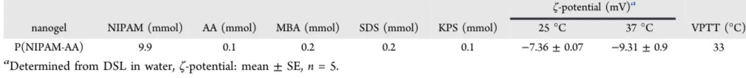
-Isopropylacryla-mide (NIPAM,99%+),N,N-methylenebis(acrylamide) (MBA, 98%+), potassium persulfate (KPS, 99%+), and sodium dodecyl sulfate (SDS, 98.5%+) were bought from Sigma-Aldrich. Acrylic acid (AA, 99.5%) was purchased from Acros Organics Co. (New Jersey, USA). NIPAM was purified prior to synthesis through recrystallization inn-hexane and dried in vacuum at room temperature. Free radical emulsion polymerization was carried out to synthesize P(NIPAM-AA) nanogel. Based on the recipe inTable 1, 9.9 mmol (1.1203 g) of NIPAM, 0.1 mmol (6.86μL) of AA, 0.2 mmol (31 mg) of MBA, and 0.2 mmol of SDS (57.9 mg) were dissolved in 97 mL of water. Then we poured the liquid into a 250 mL three-neckedflask attached with a condenser and a mechanical stirrer. Before being moved to a 70°C oil bath, the system was degassed for 30 min. Three milliliters of KPS aqueous solution (0.1 mmol, 27 mg) was injected into the system to start the polymerization. The polymer synthesis was carried out for 5 h with continuous stirring under the protection of a nitrogen atmosphere at 70°C. Once the polymerization wasfinished, the solution was purified by membrane dialysis (cutoffMwof 12−14 kDa) against Milli-Q water
for a week with daily water change. After purification, nanogels were concentrated by heating to 70°C. Two hundred microliters of the concentrated nanogel dispersion was dried at 70°C for 48 h. The concentration was calculated.
Dynamic Light Scattering Measurement. A Zetasizer
(Mal-vern, Nano-ZS) was utilized to measure the hydrodynamic diameter (dh) andζ-potential of P(NIPAM-AA) nanogels (1.0 mg/mL in PBS
buffer) at different temperatures. An autocorrelator was used to collect dynamic light scattering data.
Rheological Characterization.A universal stress rheometer SR5
(Rheometric Scientific) with a 40 mm cone plate geometry was used to perform dynamic oscillation experiments for 30 mg/mL nanogel dispersions. The gap was setup at 0.0483 mm, and the temperature
was controlled by a Peltier system connected with a water bath. The elastic (storage) modulus G′ and viscous (loss) modulus G″were examined at different temperatures from 20 to 40°C. The stress was
fixed at 0.1 Pa and the frequency at 0.1 Hz. The experiment was carried out in the linear viscoelastic region.
Hydrogel Morphologies. Nanogel dispersions (30 mg/mL) in
physiological saline buffer (pH is approximately 7.2) were put into a 37°C water bath to form physical gel. Once the gel was formed, the sample was quenched by liquid nitrogen and then under vacuum using a Christ Alpha 2-4 LD free dryer. A Philips XL 30 FEG scanning electron microscopy was used to observe the hydrogel morphology after being coated with platinum at an acceleration voltage of 20 kV. Derivation and Culture of Human Cardiac Stem Cells. Human CSCs were derived as previously descried using the cardiosphere method.44 In brief, heart tissues were cut into tiny pieces and washed with PBS and digestion of collagenase (Sigma, St. Louis, MO). Tissue fragments were cultured as“cardiac explants”on a plate coated with 0.5 mg/mLfibronectin (BD Biosciences, San Jose, CA) in IMDM supplemented with FBS, 0.5% gentamicin (Gibco, Life Technologies, California, USA), 0.1 mM 2-mercaptoethanol (In-vitrogen), and 1%L-glutamine (Invitrogen). After about 7−14 days, we collected cardiac explanted-derived cells with 0.25% trypsin (Gibco) and then seeded them in ultralow attachment flasks (Corning) for cardiospheres. After several days, cardiosphere-derived cardiac stem cells were formed by seeding harvested cardiospheres onfi bronectin-coated plates and being incubated in 5% CO2at 37°C.
P(NIMAP-AA) Nanogel Encapsulation of Human CSCs. Human CSCs were collected in culture media and mixed with 10× PBS and 50 mg/mL P(NIPAM-AA) nanogel liquid in a ratio of 1:1:3 and then warmed in a 37 °C incubator for gelation to occur. The culture was maintained in IMDM (Invitrogen) containing 20% FBS. Human CSC morphology, viability, and proliferation in the nanogel were compared to that of hCSCs cultured on normal TCP. For cell viability, 1×10⁵hCSCs were cultured in 125μL of P(NIPAM-AA) nanogel or on TCP on a 96-well plate for 7 days and then detected with the live/dead viability/cytotoxicity kit (Invitrogen). Cell morphology (e.g., cell body elongation) was calculated according to the image analysis results from ImageJ software. For cell proliferation, 1×10⁵hCSCs were seeded in 125μL of P(NIPAM-AA) nanogel or
on TCP on a 96-well plate, and we used counting kit-8 (Dojindo Molecular Technologies, Rockville, MD) to quantify cellular proliferation at days 1, 3, and 5. Absorbance rate was read by a microplate reader (Tecan Sunrise, Switzerland). Confocal images were captured by a ZEISS LSM 880 confocal microscope (Carl Zeiss, Oberkochen, Germany).
Scanning Electron Microscopy for P(NIPAM-AA)
Nanogel-Encapsulated Human CSCs.The morphology of P(NIPAM-AA)
nanogel-encapsulated hCSCs was studied by SEM (Philips XL30 scanning microscope, The Netherlands). The specimen was scanned and photographed under the microscope at an acceleration voltage of 15 kV.
In Vitro Cytokine Release of P(NIPAM-AA)
Nanogel-Encapsulated Human CSCs. Three hundred microliters of 50
mg/mL of P(NIPAM-AA) nanogel (with 1×10⁵hCSCs) was placed
into one well of a 24-well plate and incubated with 1 mL of FBS-free media. To study the continuous release of growth factors, we collected the conditioned media at day 3, 5, and 9 and added back fresh media into the well to be conditioned for the next time point. The expressions of IGF-1, VEGF, and SDF-1 in the conditioned media were determined by ELISA kits (R&D Systems, Minneapolis, MN; B-Bridge International, Cupertino, CA).
Table 1. Protocol for the Synthesis of P(NIPAM-AA) Nanogel
ζ-potential (mV)a
nanogel NIPAM (mmol) AA (mmol) MBA (mmol) SDS (mmol) KPS (mmol) 25°C 37°C VPTT (°C)
P(NIPAM-AA) 9.9 0.1 0.2 0.2 0.1 −7.36±0.07 −9.31±0.9 33
aDetermined from DSL in water,ζ-potential: mean±SE,n= 5.
Biocompatibility of P(NIPAM-AA) Nanogel with
Cardiomyo-cytes.To examine the biocompatibility of P(NIPAM-AA) nanogel,
1.5×10⁵neonatal rat cardiomyocytes (NRCMs) were collected in 25
μL of IMDM containing 10% FBS and mixed with 25μL of 10×PBS and 50 mg/mL of P(NIPAM-AA) nanogel solution and then cultured on a 96-well plate in a 37°C incubator for 3 days. NRCM morphology and viability in the polymer nanogel were characterized and compared to the NRCMs cultured on TCPs. The live/dead viability/cytotoxicity kit (Invitrogen) was applied to reflect cell viability and morphology. Beating NRCMs were observed using time-lapse imaging.
Secretion of CSCs Cultured in P(NIPAM-AA) Nanogel.Three
hundred microliters of P(NIPAM-AA) nanogel (with 1×10⁵hCSCs)
was placed into each well of a 24-well plate and incubated with 1 mL of FBS-free media. As the control, 1×10⁵hCSCs were seeded onto conventional TCPs. Conditioned media were collected at day 3 and plated onto NRCMs for 3 days. A live/dead viability/cytotoxicity kit (Invitrogen) was used for the determination of cell viability and morphology.
Immunogenicity of Polymer-Encapsulated hCSCs in
Immu-nocompetent Mice. Male CD1 mice received one of the two
treatments randomly: (1) “hCSCs in PBS” group, intramyocardial injection of 1×10⁵human CSCs in 50μL of PBS; (2)“hCSCs in nanogel”group, intramyocardial injection of 1 × 10⁵ human CSCs encapsulated in 50 μL of P(NIPAM-AA) nanogel. To enable
fluorescent imaging and histological detection, hCSCs were labeled with red fluorophore DiI. Seven days after injection, mice were sacrificed to harvest the heart and blood. IVIS XenogenIn VivoImager (Caliper Lifesciences, Waltham, MA) was used forex vivofluorescent imaging. Afterward, the heart was frozen in OCT compound and sectioned at 10μm thickness for histology analysis. Vein blood was harvested in a EDTA tube and centrifuged for 20 min at 2000 rpm to get plasma and stored in−80°C. Mouse inflammation antibody array C1 (Raybio, Norcross, GA) was used for the evaluations of inflammatory proteins in the plasma.
Mouse Model of Acute Myocardial Infarction.All animal work
was approved by the Institutional Animal Care and Use Committee at North Carolina State University. Mouse MI model was generated as previously described.45,46Generally, male CD1 mice were anesthetized with isoflurane mixed with oxygen inhalation. The heart was exposed by a minimally invasive left thoracotomy, and LAD was ligated permanently for induction of acute MI. After LAD ligation, the heart was to receive one of the following four treatments randomly: (1) MI + hCSCs in nanogel, intramyocardially injected with 1×10⁵hCSCs in
50 μL of P(NIPAM-AA) nanogel; (2) MI + hCSCs in PBS, intramyocardially injected with 1 × 10⁵ hCSCs in 50 μL of PBS; (3) MI + nanogel, intramyocardially injected with 50μL of P(NIPAM-AA) nanogel; (4) MI alone, MI surgery without any injection. The hCSCs or nanogels were prelabeled with Texas Red-X succinimidyl ester (1 mg/mL, Invitrogen) for detection.
Cell Engraftment Assay by Quantitative PCR. Animals were
sacrificed, and their hearts were excised to obtain an actual measurement of the number of cells engrafted. Real-time PCR experiments using the human-specific repetitive Alu sequences were conducted. The whole heart was weighed and homogenized. Genomic DNA was isolated from aliquots of the homogenate with the DNAeasy minikit (Qiagen). The TaqMan assay (Applied Biosystems) was used to quantify the number of transplanted cells with the human Alu sequence as the template.
Hematoxylin and Eosin Staining.To evaluate possible immune
responses to the injected nanogel, major organs from the injected mice and pigs were harvested. H&E staining was performed on tissue sections. Slides werefixed in hematoxylin (Sigma-Aldrich, MO, USA) for 5 min at room temperature and then rinsed in running water for 2 min. Afterward, the slides were decolorized in acid alcohol for 2 s and rinsed again in sodium bicarbonate for 5 dips, and the container was rinsed out with dehydrant after 95% iso for 30 s. Slides were thenfixed in eosin solution (Sigma-Aldrich, MO, USA) for 2 min and then washed in 100% dehydrant (Richard-Allan Scientific, MI, USA) and subsequent xylene solution (VWR, PA, USA) three times. The slides
were digitally photographed and analyzed by independent pathologists blinded to treatment allocations.
Heart Morphometry.After the echocardiography detection at 3
weeks, mice were euthanized and hearts were harvested and frozen in OCT compound. Specimens were sectioned at 10μm thickness with 100μm intervals. Masson’s trichrome staining was performed with a HT15 trichrome staining (Masson) kit (Sigma-Aldrich). Stained slides were placed in PathScan Enabler IV slide scanner (Advanced Imaging Concepts, Princeton, NJ) for image collection. NIH ImageJ software was used for the measurement of morphometric parameters in each section.47 Values from three sections per heart (5 hearts from each group) were determined and averaged.
Cardiac Function Evaluation.Under inhaled isoflurane−oxygen
mixture anesthesia, the transthoracic echocardiography procedure was performed by a cardiologist and detected by a Philips CX30 ultrasound system coupled with a L15 high-frequency probe. Hearts were imaged 2D in long-axis views at the level of the greatest LV diameter. LVEFs were determined by measurement from views taken from the infarcted area.
Immunohistochemistry Staining.Heart cryosections werefixed
with 4% paraformaldehyde, permeabilized, and blocked with protein block solution (DAKO, Carpinteria, CA) with 0.1% saponin (Sigma) and then incubated with the primary antibodies overnight at 4 °C. Primary antibodies were listed as follows: rabbit anti-CD3 (ab16669, Abcam, Cambridge, United Kingdom), mouse anti-CD8 alpha (mca48r, abd Serotec, Raleigh, NC), mouse anti-CD68 (ab955, Abcam), mouse anti-alpha sarcomeric actin (a7811, Sigma), rabbit anti-Ki67 (ab15580, Abcam), rabbit anti-vWF (ab6994, Abcam), and a smooth muscle actin antibody (A5228, Sigma). FITC- or Texas-Red secondary antibodies obtained from Abcam Company were incubated and conjoined with related primary antibodies. For evaluation of cell apoptosis, heart cryosections were incubated with TUNEL solution (Roche Diagnostics GmbH, Mannheim, Germany) and counter-stained with DAPI (Life Technology, NY, USA). Images were taken by an Olympus epi-fluorescence microscopy system as previously described.48,49
Pig Studies.Acute MI was induced in female mini-pigs (8−10 kg)
by permanent ligation of LAD. Twenty minutes later, 10 million nanogel-encapsulated hCSCs were injected into the peri-infarct area in 10 sites (1 million for each site). Control animals received injection of hCSCs suspended in PBS. After the procedures, the animals recovered. Successful induction of MI was verified by ST elevation on an ECG. At three time points (baseline, 48 h post-MI, and 4 weeks after treatment), LVEFs were determined by echocardiography using a SIUI Apogee 1200v veterinary ultrasound system. Blood was collected at day 0 and day 28 for ALT, AST, urea, and creatinine analysis (DiaSys Diagnostic Systems). From the cryosections, Masson’s trichrome staining was performed, and images were taken from the infarct area. Scar transmurality was analyzed.
Statistical Analysis.All results are expressed as mean±standard
deviation. Comparison between two groups was performed by a two-tailed Student’sttest. One-way ANOVA test was used for comparison among three or more groups with Bonferroni post-hoc correction. Differences were considered statistically significant whenPvalues were <0.05.
ASSOCIATED CONTENT
*
S Supporting InformationThe Supporting Information is available free of charge on the
ACS Publications website
at DOI:
10.1021/acsnano.7b01008
.
Additional biocompatibility of P(NIPAM-AA) nanogel,
immune response to injected P(NIPAM-AA) nanogel in
mice and pigs (
)
AUTHOR INFORMATION
Corresponding Authors
*
E-mail:
[email protected]
.
*
E-mail:
[email protected]
.
ORCID
Xiaolin Cui:
0000-0001-5118-0169Adam C. Vandergri
ff
:
0000-0001-7614-4834 Author ContributionsJ.T., J.Z., H.Z., and K.C. conceived the study; J.T., X.C.,
M.T.H., A.C.V., Y.H., D.S., and T.G.C. performed the
experiments and collected data; J.T., X.C., H.Z., and K.C.
wrote the paper; J.Z., H.Z., and K.C. provided
fi
nancial support.
J.T., X.C., and T.G.C. contributed equally to this work.
Notes
The authors declare no competing
fi
nancial interest.
ACKNOWLEDGMENTS
This work is supported by funding University of
Adelaide-NCSU Starter Grant, U.S. National Institutes of Health
(HL123920 and HL137093), NC State University Chancellor
’
s
Faculty Excellence Program, NC State Chancellor
’
s Innovation
Fund, University of North Carolina General Assembly Research
Opportunities Initiative grant, National Natural Science
Foundation of China (81370216, 81570274), and China
Scholarship Council (J.T.). H.Z. acknowledges the
fi
nancial
support from The MAWA, and X.C. thanks the divisional
scholarship from The University of Adelaide.
REFERENCES
(1) Bolli, R.; Chugh, A. R.; D’Amario, D.; Loughran, J. H.; Stoddard, M. F.; Ikram, S.; Beache, G. M.; Wagner, S. G.; Leri, A.; Hosoda, T.; et al. Cardiac Stem Cells in Patients with Ischaemic Cardiomyopathy (SCIPIO): Initial Results of a Randomised Phase 1 Trial.Lancet2011, 378, 1847−57.
(2) Sayed, N.; Liu, C.; Wu, J. C. Translation of Human-Induced Pluripotent Stem Cells: From Clinical Trial in a Dish to Precision Medicine.J. Am. Coll. Cardiol.2016,67, 2161−76.
(3) Hunsberger, J. G.; Rao, M.; Kurtzberg, J.; Bulte, J. W.; Atala, A.; LaFerla, F. M.; Greely, H. T.; Sawa, A.; Gandy, S.; Schneider, L. S.; et al. Accelerating Stem Cell Trials for Alzheimer’s Disease.Lancet Neurol.2016,15, 219−230.
(4) Bartunek, J.; Davison, B.; Sherman, W.; Povsic, T.; Henry, T. D.; Gersh, B.; Metra, M.; Filippatos, G.; Hajjar, R.; Behfar, A.; et al. Congestive Heart Failure Cardiopoietic Regenerative Therapy (CHART-1) Trial Design.Eur. J. Heart Failure2016,18, 160−8.
(5) van Berlo, J. H.; Molkentin, J. D. An Emerging Consensus on Cardiac Regeneration.Nat. Med.2014,20, 1386−93.
(6) Dolgin, E. Encapsulate This.Nat. Med.2014,20, 9−11. (7) Cheng, K.; Shen, D.; Hensley, M. T.; Middleton, R.; Sun, B.; Liu, W.; De Couto, G.; Marban, E. Magnetic Antibody-Linked Nano-́ matchmakers for Therapeutic Cell Targeting.Nat. Commun.2014,5, 4880.
(8) Mozaffarian, D.; Benjamin, E. J.; Go, A. S.; Arnett, D. K.; Blaha, M. J.; Cushman, M.; de Ferranti, S.; Després, J. P.; Fullerton, H. J.; Howard, V. J.; et al. Heart Disease and Stroke Statistics–2015 Update: a Report from the American Heart Association.Circulation2015,131, e29−e322.
(9) Braunwald, E. The War against Heart Failure: the Lancet Lecture. Lancet2015,385, 812−824.
(10) Cheng, K.; Malliaras, K.; Smith, R. R.; Shen, D.; Sun, B.; Blusztajn, A.; Xie, Y.; Ibrahim, A.; Aminzadeh, M. A.; Liu, W.; et al. Human Cardiosphere-Derived Cells from Advanced Heart Failure Patients Exhibit Augmented Functional Potency in Myocardial Repair. JACC Heart Fail.2014,2, 49−61.
(11) Li, T. S.; Cheng, K.; Lee, S. T.; Matsushita, S.; Davis, D.; Malliaras, K.; Zhang, Y.; Matsushita, N.; Smith, R. R.; Marban, E.́ Cardiospheres Recapitulate a Niche-Like Microenvironment Rich in Stemness and Cell-matrix Interactions, Rationalizing their Enhanced
Functional Potency for Myocardial Repair.Stem Cells2010,28, 2088− 98.
(12) Malliaras, K.; Marbán, E. Cardiac Regeneration Validated.Nat. Biotechnol.2015,33, 587.
(13) Malliaras, K.; Li, T. S.; Luthringer, D.; Terrovitis, J.; Cheng, K.; Chakravarty, T.; Galang, G.; Zhang, Y.; Schoenhoff, F.; Van Eyk, J.; et al. Safety and Efficacy of Allogeneic Cell Therapy in Infarcted Rats Transplanted with Mismatched Cardiosphere-Derived Cells. Circu-lation2012,125, 100−12.
(14) Nguyen, M. M.; Gianneschi, N. C.; Christman, K. L. Developing Injectable Nanomaterials to Repair the Heart.Curr. Opin. Biotechnol. 2015,34, 225−31.
(15) Christman, K. L.; Fok, H. H.; Sievers, R. E.; Fang, Q.; Lee, R. J. Fibrin Glue Alone and Skeletal Myoblasts in a Fibrin Scaffold Preserve Cardiac Function after Myocardial Infarction. Tissue Eng.2004,10, 403−9.
(16) Dai, W.; Wold, L. E.; Dow, J. S.; Kloner, R. A. Thickening of the Infarcted Wall by Collagen Injection Improves Left Ventricular Function in Rats: a Novel Approach to Preserve Cardiac Function after Myocardial Infarction.J. Am. Coll. Cardiol.2005,46, 714−9.
(17) Huang, N. F.; Yu, J.; Sievers, R.; Li, S.; Lee, R. J. Injectable Biopolymers Enhance Angiogenesis after Myocardial Infarction.Tissue Eng.2005,11, 1860−1866.
(18) Ou, L.; Li, W.; Zhang, Y.; Wang, W.; Liu, J.; Sorg, H.; Furlani, D.; Gäbel, R.; Mark, P.; Klopsch, C.; et al. Intracardiac Injection of Matrigel Induces Stem Cell Recruitment and Improves Cardiac Functions in a Rat Myocardial Infarction Model. J. Cell Mol. Med. 2011,15, 1310−1318.
(19) Lu, W. N.; Lü, S. H.; Wang, H. B.; Li, D. X.; Duan, C. M.; Liu, Z. Q.; Hao, T.; He, W. J.; Xu, B.; Fu, Q.; et al. Functional Improvement of Infarcted Heart by Co-injection of Embryonic Stem Cells with Temperature-Responsive Chitosan Hydrogel. Tissue Eng., Part A2009,15, 1437−1447.
(20) Wang, H.; Zhang, X.; Li, Y.; Ma, Y.; Zhang, Y.; Liu, Z.; Zhou, J.; Lin, Q.; Wang, Y.; Duan, C.; et al. Improved Myocardial Performance in Infarcted Rat Heart by Co-injection of Basic Fibroblast Growth Factor with Temperature-Responsive Chitosan Hydrogel. J. Heart Lung Transplant.2010,29, 881−887.
(21) Shen, D.; Wang, X.; Zhang, L.; Zhao, X.; Li, J.; Cheng, K.; Zhang, J. The Amelioration of Cardiac Dysfunction after Myocardial Infarction by the Injection of Keratin Biomaterials Derived from Human Hair.Biomaterials2011,32, 9290−9299.
(22) Ifkovits, J. L.; Tous, E.; Minakawa, M.; Morita, M.; Robb, J. D.; Koomalsingh, K. J.; Gorman, J. H., 3rd; Gorman, R. C.; Burdick, J. A. Injectable Hydrogel Properties Influence Infarct Expansion and Extent of Postinfarction Left Ventricular Remodeling in an Ovine Model. Proc. Natl. Acad. Sci. U. S. A.2010,107, 11507−12.
(23) Yoon, S. J.; Fang, Y. H.; Lim, C. H.; Kim, B. S.; Son, H. S.; Park, Y.; Sun, K. Regeneration of Ischemic Heart Using Hyaluronic Acid-Based Injectable Hydrogel.J. Biomed. Mater. Res., Part B2009,91B, 163−171.
(24) McGarvey, J. R.; Pettaway, S.; Shuman, J. A.; Novack, C. P.; Zellars, K. N.; Freels, P. D.; Echols, R. L., Jr; Burdick, J. A.; Gorman, J. H., 3rd; Gorman, R. C.; et al. Targeted Injection of a Biocomposite Material Alters Macrophage and Fibroblast Phenotype and Function Following Myocardial Infarction: Relation to Left Ventricular Remodeling.J. Pharmacol. Exp. Ther.2014,350, 701−9.
(25) Lin, R. Z.; Chang, H. Y. Recent Advances in Three-dimensional Multicellular Spheroid Culture for Biomedical Research.Biotechnol. J. 2008,3, 1172−84.
(26) Ungerleider, J. L.; Christman, K. L. Concise Review: Injectable Biomaterials for The Treatment of Myocardial Infarction and Peripheral Artery Disease: Translational Challenges and Progress. Stem Cells Transl. Med.2014,3, 1090−1099.
(27) Fujimoto, K. L.; Ma, Z.; Nelson, D. M.; Hashizume, R.; Guan, J.; Tobita, K.; Wagner, W. R. Synthesis, Characterization and Therapeutic Efficacy of a Biodegradable, Thermoresponsive Hydrogel Designed for Application in Chronic Infarcted Myocardium.Biomaterials2009,30, 4357−4368.
(28) Ruvinov, E.; Cohen, S. Alginate Biomaterial for the Treatment of Myocardial Infarction: Progress, Translational Strategies, and Clinical Outlook: from Ocean Algae to Patient Bedside. Adv. Drug Delivery Rev.2016,96, 54−76.
(29) Levit, R. D.; Landazuri, N.; Phelps, E. A.; Brown, M. E.; García,́ A. J.; Davis, M. E.; Joseph, G.; Long, R.; Safley, S. A.; Suever, J. D.; et al. Cellular Encapsulation Enhances Cardiac Repair.J. Am. Heart Assoc.2013,2, e000367.
(30) Cheng, K.; Blusztajn, A.; Shen, D.; Li, T. S.; Sun, B.; Galang, G.; Zarembinski, T. I.; Prestwich, G. D.; Marban, E.; Smith, R. R.; et al.́ Functional Performance of Human Cardiosphere-Derived Cells Delivered in anin situ Polymerizable Hyaluronan-Gelatin Hydrogel. Biomaterials2012,33, 5317−5324.
(31) Cavus, S.; Gurdag, G. Noncompetitive Removal of Heavy Metal Ions from Aqueous Solutions by Poly [2-(acrylamido)-2-methyl-1-propanesulfonic acid-co-itaconic acid] Hydrogel.Ind. Eng. Chem. Res. 2009,48, 2652−2658.
(32) Lin, X.; Tang, D.; Yu, Z.; Feng, Q. Stimuli-Responsive Electrospun Nanofibers from Poly (N-isopropylacrylamide)-co-poly (acrylic acid) Copolymer and Polyurethane.J. Mater. Chem. B2014,2, 651−658.
(33) Heusch, G.; Gersh, B. J. The Pathophysiology of Acute Myocardial Infarction and Strategies of Protection Beyond Reperfu-sion: a Continual Challenge.Eur. Heart J.2017,38, 774−784.
(34) Sanganalmath, S. K.; Bolli, R. Cell Therapy for Heart Failure: a Comprehensive Overview of Experimental and Clinical Studies, Current Challenges, and Future Directions. Circ. Res. 2013, 113, 810−34.
(35) Vegas, A. J.; Veiseh, O.; Gürtler, M.; Millman, J. R.; Pagliuca, F. W.; Bader, A. R.; Doloff, J. C.; Li, J.; Chen, M.; Olejnik, K.; et al. Long-Term Glycemic Control Using Polymer-Encapsulated Human Stem Cell-Derived Beta Cells in Immune-competent Mice.Nat. Med.2016, 22, 306−311.
(36) Blocki, A.; Beyer, S.; Dewavrin, J. Y.; Goralczyk, A.; Wang, Y.; Peh, P.; Ng, M.; Moonshi, S. S.; Vuddagiri, S.; Raghunath, M.; et al. Microcapsules Engineered to Support Mesenchymal Stem Cell (MSC) Survival and Proliferation Enable Long-term Retention of MSCs in Infarcted Myocardium.Biomaterials2015,53, 12−24.
(37) Shen, Z.; Bi, J.; Shi, B.; Nguyen, D.; Xian, C.; Zhang, H.; Dai, S. Exploring Thermal Reversible Hydrogels for Stem Cell Expansion in Three-dimensions.Soft Matter2012,8, 7250−7257.
(38) Cui, X.; Dini, S.; Dai, S.; Bi, J.; Binder, B.; Green, J.; Zhang, H. A Mechanistic Study on Tumour Spheroid Formation in Thermosensi-tive Hydrogels: Experiments and Mathematical Modelling.RSC Adv. 2016,6, 73282−73291.
(39) Makkar, R. R.; Smith, R. R.; Cheng, K.; Malliaras, K.; Thomson, L. E.; Berman, D.; Czer, L. S.; Marban, L.; Mendizabal, A.; Johnston, P.́ V.; et al. Intracoronary Cardiosphere-Derived Cells for Heart Regeneration after Myocardial Infarction (CADUCEUS): a Prospec-tive, Randomised Phase 1 Trial.Lancet2012,379, 895−904.
(40) Li, T. S.; Cheng, K.; Malliaras, K.; Smith, R. R.; Zhang, Y.; Sun, B.; Matsushita, N.; Blusztajn, A.; Terrovitis, J.; Kusuoka, H.; et al. Direct Comparison of Different Stem Cell Types and Subpopulations Reveals Superior Paracrine Potency and Myocardial Repair Efficacy with Cardiosphere-Derived Cells.J. Am. Coll. Cardiol.2012,59, 942− 53.
(41) Wang, T.; Jiang, X. J.; Tang, Q. Z.; Li, X. Y.; Lin, T.; Wu, D. Q.; Zhang, X. Z.; Okello, E. Bone Marrow Stem Cells Implantation with
α-cyclodextrin/MPEG−PCL−MPEG Hydrogel Improves Cardiac Function after Myocardial Infarction.Acta Biomater.2009,5, 2939− 2944.
(42) Lu, W. N.; Lü, S. H.; Wang, H. B.; Li, D. X.; Duan, C. M.; Liu, Z. Q.; Hao, T.; He, W. J.; Xu, B.; Fu, Q.; et al. Functional Improvement of Infarcted Heart by Co-Injection of Embryonic Stem Cells with Temperature-responsive Chitosan Hydrogel. Tissue Eng., Part A2009,15, 1437−47.
(43) Wang, H.; Liu, Z.; Li, D.; Guo, X.; Kasper, F. K.; Duan, C.; Zhou, J.; Mikos, A. G.; Wang, C. Injectable Biodegradable Hydrogels for Embryonic Stem Cell Transplantation: Improved Cardiac
Remodelling and Function of Myocardial Infarction. J. Cell Mol. Med.2012,16, 1310−20.
(44) Cheng, K.; Ibrahim, A.; Hensley, M. T.; Shen, D.; Sun, B.; Middleton, R.; Liu, W.; Smith, R. R.; Marbán, E. Relative Roles of CD90 and C-kit to the Regenerative Efficacy of Cardiosphere-Derived Cells in Humans and in a Mouse Model of Myocardial Infarction.J. Am. Heart Assoc.2014,3, e001260.
(45) Andrade, J. N.; Tang, J.; Hensley, M. T.; Vandergriff, A.; Cores, J.; Henry, E.; Allen, T. A.; Caranasos, T. G.; Wang, Z.; Zhang, T.; et al. Rapid and Efficient Production of Coronary Artery Ligation and Myocardial Infarction in Mice Using Surgical Clips.PLoS One2015, 10, e0143221.
(46) Gao, E.; Lei, Y. H.; Shang, X.; Huang, Z. M.; Zuo, L.; Boucher, M.; Fan, Q.; Chuprun, J. K.; Ma, X. L.; Koch, W. J. A Novel and Efficient Model of Coronary Artery Ligation and Myocardial Infarction in the Mouse.Circ. Res.2010,107, 1445−1453.
(47) Cheng, K.; Li, T. S.; Malliaras, K.; Davis, D. R.; Zhang, Y.; Marbán, E. Magnetic Targeting Enhances Engraftment and Functional Benefit of Iron-Labeled Cardiosphere-Derived Cells in Myocardial Infarction.Circ. Res.2010,106, 1570−81.
(48) Tang, J.; Shen, D.; Caranasos, T. G.; Wang, Z.; Vandergriff, A. C.; Allen, T. A.; Hensley, M. T.; Dinh, P.-U.; Cores, J.; Li, T.-S.; et al. Therapeutic Microparticles Functionalized with Biomimetic Cardiac Stem Cell Membranes and Secretome.Nat. Commun.2017,8, 13724. (49) Luo, L.; Tang, J.; Nishi, K.; Yan, C.; Dinh, P.-U.; Cores, J.; Kudo, T.; Zhang, J.; Li, T.-S.; Cheng, K. Fabrication of Synthetic Mesenchymal Stem Cells for the Treatment of Acute Myocardial Infarction in Mice.Circ. Res.2017,120, 1768−1775.