ESTIMATING 3D DEFORMABLE MOTION FROM A SERIES OF FAST 2D MRI IMAGES
Jason M. Brown
A thesis submitted to the faculty of the University of North Carolina at Chapel Hill in partial fulfillment of the requirements for the degree of Master of Science in the Department of Biomedical Engineering.
Chapel Hill 2016
ABSTRACT
JASON M. BROWN: Estimating 3D Deformable Motion from a series of Fast 2D MRI Images (Under the direction of David Lalush)
In this application, we estimated patient-specific 3D deformable motion in the abdomen from
a series of fast 2D images. CLARET (Correction via Limited-Angle Residues in External Beam
Therapy) is an image registration method that has been used to estimate 3D deformable motion from
2D X-ray images. This work generalizes CLARET and extends it to use with MRI images of the
abdomen. Using CLARET to predict the 3D motion of a subject from a set of 2D projection images
has the potential to be used in fast MRI imaging of dynamic processes. The method begins with
acquisition of a 4D respiratory-gated image set using a gradient-echo sequence. From the 4D set,
a patient-specific motion model was derived, as well as a regression relationship between the 3D
anatomy and 2D slice images taken with a specific geometry. The second dataset was a series of
fast 2D gradient-echo images of the same subject, which are used via the regression relationship to
estimate the 3D body poses at each time point. Before testing on the acquired 2D dataset, CLARET
was tested on a simulated dataset which confirmed the method accurately predicted random warps
of the dataset. In a free breathing experiment, the CLARET procedure gave motion estimates that
reduced alignment error mean and variance in the 2D frames. We conclude that CLARET can be
applied in an MRI setting and produces fast instantaneous motion estimates with less registration
ACKNOWLEDGEMENTS
I am grateful to my advisor Dr. David Lalush for the guidance, direction, and the willingness to
review and share knowledge provided to me over my time here at UNC. I would also like to thank Dr.
Hongyu An for her valuable input and knowledge in respect to my research. To my lab-mates Meher
Juttukonda and Bryant Mersereau I’ve enjoyed working beside you and I appreciate the opportunity
to learn with you.
Thank you to my parents Jim and Cheryl Brown, and my brother Connor, for their unwaivering
love and support through everything. I couldn’t wish for a better family.
Thank you to Caitlin Penry, your love and friendship have been one of the best things I’ve found
at UNC. You inspire me to discover all of the amazing things the world has to offer, and I can’t thank
you enough for the great adventures I’ve had with you.
To all my friends, especially Kyle, Mike, Sam, and Katie, thank you for the laughs and the good
times. You’ve been rocks in my life for so long and I can’t imagine finishing this without you all. I’ll
see you for beers later.
Lastly, a huge thank you to UNC Ski and Snowboard team and everyone I’ve met through it. I
fell in love with you on a chair lift, and your passion and enthusiasm made my time here at UNC
TABLE OF CONTENTS
LIST OF TABLES . . . ix
LIST OF FIGURES . . . x
LIST OF ABBREVIATIONS . . . xii
1 Introduction . . . 1
1.1 The Problem of Respiratory Motion Artifact . . . 1
1.2 Quantitative PET and Respiratory Motion Artifact . . . 2
1.3 Summary . . . 4
2 Background . . . 6
2.1 Motion Correction Versus Gating . . . 6
2.2 Incorporating Motion Fields into PET Reconstruction . . . 7
2.3 Standalone PET Motion Correction Methods . . . 9
2.4 PET/CT Motion Correction Methods . . . 10
2.5 PET/MR Motion Correction Methods . . . 11
2.6 MR Based Motion Models . . . 15
2.7 Surrogate Data Acquisition . . . 15
2.8 Training Data Acquisition and Motion Extraction . . . 18
2.9 Motion Models . . . 19
2.9.1 Single Linear Correlation Models . . . 20
2.9.2 Respiratory Variability and Complex Models . . . 20
2.9.3 Image Based Models . . . 22
2.10.1 Registration Methods . . . 23
2.11 Summarizing Ideal characteristics of an MR Based Motion Model . . . 24
3 To Generalize CLARET to work with MRI and test on generated images . . . 26
3.1 Methods . . . 26
3.1.1 Generalization of CLARET . . . 26
3.1.1.1 The set of full 3D images . . . 26
3.1.1.2 The Reduced Image . . . 27
3.1.1.3 The Problem . . . 27
3.1.1.4 Shape Space . . . 28
3.1.1.5 Machine Learning . . . 28
3.1.1.6 Estimation of motion parameters . . . 29
3.1.2 Application of CLARET . . . 30
3.1.2.1 Image Acquisition . . . 30
3.1.2.2 Image Registration . . . 30
3.1.2.3 Implementation . . . 30
3.1.2.4 Simulation Experiment . . . 31
3.1.2.5 Free Breathing Experiment . . . 31
3.1.3 Evaluation of effectiveness . . . 32
3.2 Results . . . 32
3.2.1 Simulation Experiment . . . 32
3.2.2 Free Breathing Experiment . . . 33
3.3 Discussion . . . 38
3.4 Conclusions . . . 38
4 Summary and Conclusion . . . 39
4.1 Summary . . . 39
4.2 CLARET as an MR motion model . . . 40
4.4 Future Directions . . . 43
4.4.1 Generalized CLARET . . . 43
4.4.2 Apply CLARET in a single-scan method to estimate deformable
body pose in a self-gated radial-sampled images . . . 44
LIST OF TABLES
LIST OF FIGURES
1.1 (A) Apparent activity concentration for a stationary point source, showing the expected Gaussian distribution. (B) The apparent activity concentration is stretched when the point source is oscillating, resulting in overestimating the object volume and underestimating its SUV. This image and partial
caption is from Nehmeh et al. 2008[6] . . . 3
1.2 One-dimensional superiorinferior profiles through motionless and motion-blurred tumor. This image and partial caption is from Liu et al. 2009[5]
. . . 3
2.1 Above: single respiratory phase without motion, below: all respiratory phases artificially superimposed by image blending. The extent of respi-ratory motion is visible in the left heart ventricle (black in color). The level of noise in the single phase image is much higher due to the lack of statistics. All images were reconstructed with an OSEM algorithm. This
image and caption is from Dawood et al. 2008[13] . . . 7
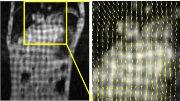
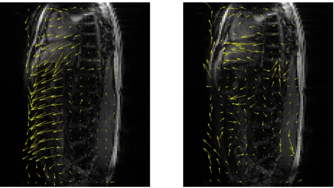
2.2 Tagged MR images with estimated motion fields (yellow box). This image
and caption is from Chun et al. 2012[32] . . . 13
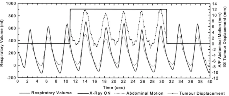
2.3 An example of a spirometer, abdominal displacement sensor, and fluo-roscopy measurement (Patient 7). Spirometry and abdominal displacement sensor measurements were acquired continuously, whereas tumor motion measurements from fluoroscopy were made for 20 s (as indicated by X-ray ON signal) at 1-min intervals. Note that the right hand scale represents both A/P abdominal motion and I/S tumor motion (mm). Error bars have been omitted for clarity. This image and caption is from Hoisak et al.
2004[42] . . . 17
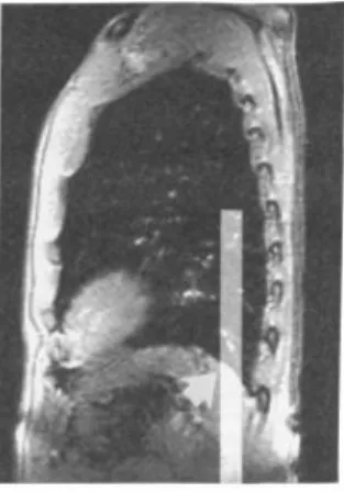
2.4 Visualization of the positioning of an MR Navigator Echo. The arrow points to the edge of the diaphragm that is tracked and used for gating, or
as a respiratory surrogate. This image is from Wang et al. 1996[47] . . . 18
2.5 Illustration of the various types of motion trajectories when modeling. The respiratory surrogate signal (a) can be modeled simply by a linear prediction (b). However, a model (c) that takes into account inhalation and exhalation separately may be more accurate. Although modeling the inhalation and exhalation separately may have discontinuity problems as shown at EI 2 in (d). This image and caption are from McClelland et al.
2013 [56] . . . 21
3.1 a slice from the simulated, warped 3D image (Left), the 2D projection derived from it as the input to CLARET (Center), and the corresponding
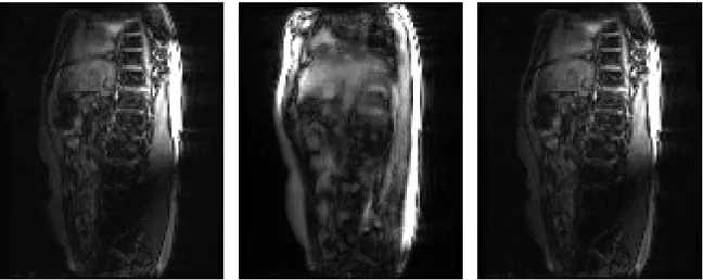
3.2 Left is a central slice of the full 3D reference image. The central image is a complex projection of the reference image into 2D. The image on the
right is an acquired 2D sagittal slab projection image. . . 33
3.3 The left image is a central slice of the reference image with the first basis deformation map superimposed, and the image on the right is the second basis deformation map. The areas of highest magnitude are where the
motion is the greatest and the highest signal was acquired. . . 34
3.4 Actual acquired 2D projection image (left) the estimated image warped from the reference 3D image and projected to 2D (center) and the estimated
image warped incorrectly with magnitude-only images (right) . . . 34
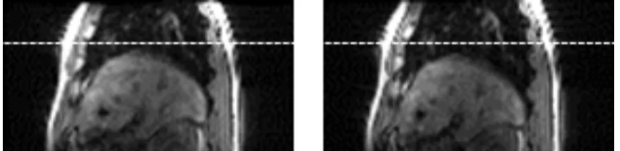
3.5 Image section showing location of the apex of the liver at (left)
end-expiration and (right) end-inspiration. . . 35
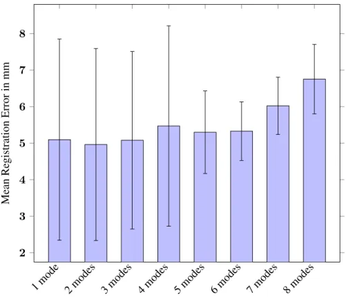
3.6 Mean registration error for different numbers of modes in the CLARET
model. Error bars indicate one standard deviation. . . 36
3.7 Graph of variation as a function of the number of eigenmodes. . . 37
3.8 Comparison of Mean Registration Error over 100 test images in the Time-Averaged Image and the CLARET estimate with 6 modes of variation. Error bars in the graph indicate standard deviation, and the differences in
LIST OF ABBREVIATIONS
MRI Magnetic Resonance Imaging
PCA Principal Component Analysis
PET Positron Emission Tomography
PET/CT Positron Emission Tomography / Computerized Tomography
PET/MR Positron Emission Tomography / Magnetic Resonance
CHAPTER 1 Introduction
Positron Emission Tomography (PET) is a molecular imaging technique that gives functional
information. In PET, a radiotracer is injected into the body and disperses throughout the body based
on the tracer’s kinetics. The tracer will localize in part of the body based on the interaction between
the tracer and the body. Since the tracer is tagged with a radionuclide, the radiotracer will decay
based on the half life of the particle. When the radiotracer decays it emits a positron particle that
then quickly interacts with a nearby electron and an annihilation event occurs which sends two
gamma energy photons in opposite directions. In PET, these gamma energy photons are detected at
simultaneous times over many annihilation events, and eventually an image is formed through the
processing of these events. Due to the length of time required for acquisition in PET (5+ minutes)
and patient breathing, there is a problem of respiratory artifact in PET images. When acquiring
a PET image in areas of significant respiratory motion such as the upper abdomen, there will be
significant blurring caused by the cyclical motion of respiration. This motion is generally in the
head-foot direction and can be up to 2 cm, but there is also additional motion in the anterior-posterior,
and left-right direction of smaller amounts[1].
1.1 The Problem of Respiratory Motion Artifact
Any time an image or scan of a patient is taken, the subject of the image needs to stay perfectly
still. If there is any movement during a scan (or even a normal photograph), the resulting image is
going to be blurry. In normal photographs, X-ray, or even CT (with a breath hold) there is not much
of a problem because the image is taken so quickly that there is not a huge opportunity to ruin the
image by moving. However, a normal PET scan is 5 or more minutes long. Even if the patient can sit
still for that amount of time, which is not always the case (source), there is no way a person could
to motion has been an accepted drawback when acquiring an image with the PET modality. However,
the introduction of PET/CT and PET/MR as well as more advanced motion modeling allows for
many methods that have attempted to correct, prevent, or reverse this kind of blurring that is caused
by respiratory motion.
1.2 Quantitative PET and Respiratory Motion Artifact
While blurring in any image is undesirable, there are some real reasons to be concerned about
respiratory motion artifact beyond the simple fact that it makes images blurry.
A potentially understated benefit to PET imaging is the quantitative nature of the modality.
There are many quantitative values that can be extracted and used from PET data [2], and simplified
measures such as standardized uptake value give a semi-quantitative measurement for PET studies
[3]. Most of these semi-quantitative measures are affected by respiratory motion artifacts in a way
that is detrimental to clinical evaluation.
For example, one semi-quantitative measure that can have clinical significance is the SUVmax value. This value is calculated as the maximum value in a region of interest, and SUVmax measure-ments have been shown to be a predictor of malignant potential where higher SUVmaxvalues are correlated with increased malignancy. SUVmaxalso has a positive correlation with tumor prolifer-ation activity as measured by the Ki-67 proliferprolifer-ation index [4]. This SUVmaxcan be thrown off due to respiratory motion, and as a result the clinical findings derived from the measure could be
incorrect. In some simulations, tumor volumes were overestimated by 130% and the SUVmaxwas underestimated by 28% [5].
Another measure, the mean lesion volume, which can be overestimated by respiratory artifact
in PET[5] is used to define targets for radiotherapy [7]. If the mean lesion is overestimated due to
respiratory motion, that overestimation could lead to irradiation of a larger part of the body than
necessary in radiotherapy. This over-irradiation could potentially be harmful in patients. Figure 1.2
gives a illustration of the blurring and localization effects of motion on a cross-section of a tumor.
One last example, total lesion glycolysis can be used as an indicator of the effectiveness of
chemotherapy [8], [9] and is proportionally based on SUV measurement changes before and after
Figure 1.1: (A) Apparent activity concentration for a stationary point source, showing the expected Gaussian distribution. (B) The apparent activity concentration is stretched when the point source is oscillating, resulting in overestimating the object volume and underestimating its SUV. This image and partial caption is from Nehmeh et al. 2008[6]
Figure 1.2: One-dimensional superiorinferior profiles through motionless and motion-blurred tumor. This image and partial caption is from Liu et al. 2009[5]
potentially yield very different results depending on the patient’s changing respiratory pattern. Thus,
a change in respiration may potentially influence the treatment plan of chemotherapy.
All of these semi-quantitative measurements are important diagnostic information that can be
used in the clinic. This means that the accuracy and preservation of quantitative measurements is
important. However, beyond these semi-quantitative measurements that are affected by the respiratory
these transmission maps for attenuation correction is important in the generation of any PET image.
Since there is motion during PET scans and not during the breath hold of CT, this transmission map
can affected by respiratory motion artifact [10].
Respiratory motion artifact is a problem that introduces both qualitative and quantitative errors
in PET images. The following sections present an overview of current research relating to the motion
correction and the many strengths and weaknesses of various methods used to perform PET motion
correction.
1.3 Summary
The problem of respiratory motion artifacts is a well studied phenomenon. For this reason, there
have been many attempts to correct for some of the qualitative and quantitative errors that have been
described above. In the next chapter, some of the current and past methods of motion correction
in PET will be described. These methods of motion correction are not limited to standalone PET
systems, the overwhelming majority of PET motion correction is in multimodality systems either
PET/CT or PET/MR.
Following the background chapter will be a chapter describing a patient-specific motion model
that can be constructed from MR images to later be used in a motion correction application. If
the motion that occurs during acquisition is known, or can be estimated, the reconstruction of the
PET image can use this information to reverse the blurring effect and artifacts caused by respiratory
motion. The challenge of reversing respiratory motion artifacts is determining the motion that
occurred during acquisition of the PET data. An image registration method called CLARET has been
used to estimate 3D deformable motion from 2D X-ray images[11]. CLARET can be extended for
use with MRI. The proposed research seeks to examine how well CLARET translates to an MRI
setting. Additionally we seek to test this methodin vivo, as well as validate the method so that
it could potentially be used in a PET motion correction application. If this method can provide a
satisfactory motion model to perform PET motion correction, we would be able to correct some
of the problems previously described such as standard uptake values and overestimating of lesion
The last chapter will be additional discussion about the proposed research. The original
appli-cation was intended for PET motion correction, thus this paper will largely be framed around this
specific application. However, the proposed research is not limited solely to this specific application,
and the generalization of this technique will allow it to be applied in many areas outside of PET
motion correction. The application of the generalized method, the limitations, and future directions
CHAPTER 2 Background
PET motion artifacts are almost unavoidable due to the long acquisition time inherent in
abdomi-nal PET imaging. Since the method was introduced, there have been many attempts to correct for
this issue. The most basic types of corrections are those that use the PET data itself, or an outside
device to attempt to quantify and correct the motion artifacts. Other methods, due to the popularity
of combination PET/CT systems, attempt to use CT to correct for respiratory motion artifacts. Lastly,
a newer type of dual modality scanner called PET/MR can use simultaneous scanning to do PET
motion correction. All of these methods, beyond using the PET data itself, attempt to create some
sort of model of the motion observed during PET scanning. Thus, it is important to understand both
the different modalities that can be used for PET motion correction as well as an understanding of
motion models and how they can be used to create an accurate representation of respiratory motion
in vivo. While there are many different sources of motion artifacts in imaging, this manuscript will
focus mainly on respiratory motion correction methods and models.
2.1 Motion Correction Versus Gating
Before delving into the background of PET motion correction and various motion estimation
methods, it is important to define what is meant by PET motion correction. A common solution to
the problem of motion artifact in images is respiratory gating. While gating will generally reduce the
amount of these artifacts due to decreased motion in each frame, there is the inherent tradeoff with
signal-to-noise ratio. The generation of PET signal is from the detection of coincident events, and
generally the larger the amount of events detected the better the signal-to-noise ratio will be. However,
when PET images are gated, the number of detected events is reduced by a factor proportional to the
number of gated frames. Consequently, the signal-to-noise ratio is also reduced due to this relative
image, the gated PET scan acquisition time must be significantly increased [12]. In other words, for
the same scan time, the finer the temporal resolution of gating becomes the more the SNR suffers
for each gated frame. Figure 2.1 illustrates the noise that occurs when using gating compared to a
combined image.
The alternative to motion gating is motion correction. In a motion corrected PET image the end
goal is to generate a single-frame image by warping or reconstructing the data to compensate for
motion. In motion corrected PET the goal is not necessarily to visualize the motion, but to recover
some of the quantitative or qualitative measures that were impaired by respiratory motion.
Figure 2.1: Above: single respiratory phase without motion, below: all respiratory phases artificially superimposed by image blending. The extent of respiratory motion is visible in the left heart ventricle (black in color). The level of noise in the single phase image is much higher due to the lack of statistics. All images were reconstructed with an OSEM algorithm. This image and caption is from Dawood et al. 2008[13]
Compared to other modalities, PET appears blurry and noisy, and reducing the SNR further
would be detrimental to a modality that already struggles for SNR. This sensitivity versus specificity
tradeoff is the reason that the focus of this manuscript will be on the single frame motion corrected
data versus the multi-frame gated data.
2.2 Incorporating Motion Fields into PET Reconstruction
Many of the methods for motion correction rely on finding a set of motion fields that can be used
to characterize motion from one point of time to another. So, before discussing how these motion
general, the motion fields are presented in a discrete number of frames, and there are many reliable
ways to incorporate those motion frames into the PET reconstruction.
The first type of motion compensated reconstruction methods uses an unconstrained or
con-strained deconvolution of the PET images to compensate for motion artifacts. In this application, the
motion data is used to generate the shape of the deconvolution and then applied to the PET dataset[14].
The problem with using deconvolution methods is that any noise in the image is amplified by the
deconvolution [14]. Since PET images are inherently noisy, deconvolution methods are not typically
used in vivo.
Although gated frames could be corrected by simply warping gated images onto a single
reference frame, a correction applied during the reconstruction process is preferred as there is a large
improvement in contrast and signal to noise ratio when the correction is applied in the reconstruction
[15]. To do this, non-rigid motion can be used in the list-mode based reconstruction algorithms
by incorporating the motion fields directly into the reconstruction system matrix. This has been
shown to give contrast improvements of 20 to 30 percent compared to performing corrections to
individual gated PET images [16]. Many of these reconstructions still use an interpolation scheme to
incorporate the motion information which reduces some of the improved statistics from applying
the motion during reconstruction [16]. However, list-mode PET reconstruction algorithms can also
incorporate motion information into a likelihood function to further reduce noise and eliminate the
need for interpolation [17].
Another method for implementing motion correction in the reconstruction scheme is to apply
the motion information to the system matrix of the reconstruction algorithm. For example, one
study created a time varying projection vector and image vector based on the motion information to
create a time varying ML-EM algorithm [18]. However, due to the time dependent update function
in the algorithm, the time to converge and create a single dataset was roughly 3 times as long as a
reconstruction that was not time varying (86 min versus 30 minute) [18].
The most popular methods of motion compensated PET reconstruction modify the sinogram bin
data and reconstruct using the modified dataset. One way to do this is to convert the sinogram data to
a 3D vector representation and then warp the 3D vector by the estimated motion using quaternion
mathematics, and then convert the resulting vector back to sinogram space[19]. Methods that modify
conventional reconstructions can be applied. However, sometimes the corrected lines of response
could fail to fill the entire 3D sinogram space resulting in artifacts[20]. For this reason a scaling
and/or rejection of lines of response outside the field of view may be needed [20].
Once these methods for incorporating the resulting motion fields into the reconstruction process
are determined, the focus becomes finding the best method for creating a motion model that would
produce the most accurate motion information. The following sections will largely focus on how
each modality will acquire motion fields and the advantages/disadvantages to each of the methods
will be listed assuming the PET reconstruction of those methods will be largely the same.
2.3 Standalone PET Motion Correction Methods
One method of a standalone PET system that attempts to solve the problem of respiratory
motion artifacts is respiratory-correlated dynamic PET [21]. In this application, a rigid block with
a radioactive point source is placed on the patient’s abdomen and then reconstructed. Following
the reconstruction, the PET sinogram data was delineated based on the position of the rigid block.
This method is very similar to gating because the result is not a combination of all of the PET data.
The only advantage to this method is that it does not require outside respiratory tracking equipment.
Thus, there is still loss of image statistics which is comparable to respiratory gated PET [21].
As previously stated, respiratory gated PET (and those similar to respiratory gated PET) divide
PET data into smaller bins, suffering from a proportional loss in the corresponding image statistics.
Instead, to correct for motion artifact, any PET data that is gated should be transformed and then
added back together during the reconstruction to recover the loss of statistics. One way this has
been done is to take respiratory gated PET data and to use optical flow methods to calculate the
motion between various image frames [13]. Once the motion information is known, the methods for
implementing a reconstruction including this motion can be performed (as mentioned above). While
this is a good start for doing motion correction, the inherently noisy PET images make it hard to get
a good motion estimation from the PET images alone.
A combination PET/CT or PET/MR scanner, which both have the advantage of better, clearer
compared to standalone PET. Thus, the following two sections will explore a few of the many
methods for motion correction that have been implemented in PET/CT and PET/MR.
2.4 PET/CT Motion Correction Methods
PET/CT is an popular dual-modality system that is been routinely used in clinical practice.
Combined PET/CT systems have been shown to have a significant advantage versus one or the other
systems alone [22]. In most standalone CT systems, the scan is done with a breath hold to eliminate
respiratory motion artifact. However, due to the long scan times, this is impossible for full PET/CT
scans, and generally only the CT part of the PET/CT scan is done with a breath hold.
One of the simplest ways to correct for motion artifacts is to do a series of breath-holds one after
another and only acquire PET data when the patient is holding his or her breath. In these breath
hold PET/CTs the patient is coached into holding their breath at a similar anatomical positions and
both the CT and PET are only acquired when the patient is in the same position. Thus, motion is
expected to be minimized in the dataset and an improved image compared to the standard PET/CT
with an increase in the median SUV by over 30% in breath hold lung tumor studies[6]. While
it may seem overly simple, breath hold PET/CT has been shown to increase detection and have
more precise localization compared to standard PET/CT [23]. Although not technically a motion
correction method, it is worth mentioning because of the simplicity and the improvement of PET
image statistics. However, much of the information that could be obtained during the rest of the
patient’s breathing has to be thrown out. Thus, this method suffers from the same problem as gating
where there either needs to be a much longer scan or the scan will suffer from reduced SNR.
Another method for correcting PET motion artifacts is to acquire a 4D PET/CT. This is a
challenge because PET/CT systems cannot be run simultaneously (unlike PET/MR). However, it can
be achieved with some help from outside hardware. By using a position monitoring system such as
the real-time position management (RPM) system (Varian Medical Systems, Palo Alto, CA), each bin
of a 4D PET acquisition can be linked to a bin from 4D CT [24]. Since the 4D datasets are aligned, a
deformable registration allows all of the 4D PET images to be reconstructed with motion information
gathered from the 4D CT data [24]. This is a brief example of a simple motion model, more of which
with free-breathing compared to many of the CT based methods that use some sort of breath hold.
However, there are still drawbacks to this method of motion correction. First, there is a significant
increase in dose compared to the standard PET/CT protocol because of the multiple acquisitions
required in 4D CT. Next, this method relies highly on the alignment of the position management
system, and there is potential for misalignment. Last, because the acquisition is sequential, there is
potential for change in motion from the CT to the PET causing inaccurate matching of the datasets.
Something as simple as a gas bubble could change the organ motion pattern and throw off the model.
This problem of sequential acquisition is a major drawback to PET/CT when compared PET/MR.
Another one of the major drawbacks to CT motion correction methods is the low contrast in soft
tissue regions. Because of the uniformity of the intensity in these regions, such as the liver, these
methods tend to have relatively low accuracy compared to MR based motion correction methods in
the same region. The soft tissue of the upper abdomen is where choosing an MR based method is
most valuable because of the benefit of enhanced soft tissue contrast in these areas compared to CT
methods. Moreover, the more information that is acquired by CT, the larger the dose is generally
going to be. The problem of low soft tissue contrast and high dose could potentially be solved by
switching to a PET/MR system.
2.5 PET/MR Motion Correction Methods
A hybrid positron emission tomography/magnetic resonance imaging (PET/MR) scanner allows
for anatomical and functional information to be acquired simultaneously [25]–[29]. Unlike PET/CT,
the MR scanner can simultaneously acquire MRI data during the full duration of the PET scan
and as an added benefit, there is no added ionizing radiation. This is advantageous to PET/MR
because motion information can constantly be observed during the entirety of the PET scan. It is this
simultaneity that is the most advantageous for PET motion correction methods. This is important
because previously, in PET/CT, the setup required the CT data to be acquired and then the PET
data would be subsequently acquired. Since the CT data is usually acquired with a breath hold, and
only at the beginning of the scan, there was very little that could be done for motion correction in
PET/CT scans. With PET/MR the scanning can be done simultaneously, up to the whole length
information that can then be used to go back and correct the PET images. Unfortunately, the speed
of acquisition is still a problem compared to the rate of detection events in PET. A typical 3D MRI
scan at 1 millimeter resolution would take significantly longer than a single respiratory cycle. For
example, a 3D-MPRAGE sequence with 1 mm isotropic resolution can take upwards of 7 minutes
[30]. However, the ability to apply different pulse sequences and acquisition methods allows for a
great opportunity to create innovate approaches to solving the problem of motion artifact in PET
images.
One of the largest attractions of MR based motion correction is the ability to do prospective
corrections. In prospective applications, motion information is derived and used to scale the magnetic
field gradients of the MR system during the acquisition in real-time. This has been shown to produce
improved image quality in vivo, and sub-millimeter registration errors in phantom experiments
[31]. However, due to the physical requirements of the MR scanner, the correction to the magnetic
field gradients can only be done in a rigid manner such as an affine transformation. While rigid
motion due to voluntary patient motion or shifting is important to take into consideration, respiratory
motion in general is going to be non-rigid in nature. It has been shown that compared to rigid or
affine motion, a correction that is elastic in nature does a better job of correcting respiratory PET
motion[16]. However, this is not to say that the application will be retrospective in manner. In fact,
the opposite is true when incorporating motion fields into the PET reconstruction. The argument
could be made that incorporating any motion information into the original reconstruction could be
seen as a prospective motion correction. The main difference between prospective and retrospective
motion correction in this case is the observation of motion information after it occurs versus the
prediction of motion information before it happens. Since prospective (predictive) motion correction
methods are limited to rigid transformations, the remainder of this manuscript will focus solely on
the retrospective motion correction methods and models.
There have been a few attempts to use the combination of MRI with PET to correct for motion
artifacts. Many of these approaches have some sort of drawback that makes it hard to use in clinical
practice. One of the most popular ways to do motion correction when PET/MRI was first introduced
was to use tagged-MRI [32], [33]. Before the introduction of PET/MRI, The most popular application
of tagged MRI was for myocardial motion information [34]. Tagged MRI uses a pulse sequence that
pattern is called the tagging pattern, and when the patient moves, the grid is deformed and the
displacement between each frame can be ascertained. This results in a motion field based on the
difference between two grids. Figure 2.2 shows an example of this grid pattern and the resulting
motion fields. This method was a good start to motion estimation during simultaneous PET/MR but
Figure 2.2: Tagged MR images with estimated motion fields (yellow box). This image and caption is from Chun et al. 2012[32]
it had a few problems. First, the acquisition of a gridded tagged MR image precluded the ability for
the MR to be used at a later time for anatomical information or hybrid imaging with the corrected
PET method. This is inherent in tagged MRI where part of the image must be nulled to get the
resulting motion information. So, while the method may produce accurate motion during the scan,
an additional scan may need to be used to generate a static MR image that has not been nulled in
the grid pattern. Additionally, the identification of motion information is based on a grid which has
been nulled compared to the foreground, therefore any areas which have little signal make it hard to
identify motion. However, this is a problem with many MR based motion correction methods, as it is
very hard to calculate motion in areas of little signal. Lastly, this method requires binning the MR
data based on some outside surrogate and allows the tagged-MR image to be formed. This binning
has two main drawbacks, one is that the respirations are divided into periodic subsets that may not
accurately represent the correct motion information at an arbitrary time point [32]. The second is that
belt or MR navigator [33]. The need for extra hardware can be a potential source of failure which
will be described in the surrogate data acquisition section later in this chapter. Many MR methods
attempt to avoid additional hardware for this reason, and the ease of implementing a fully image
based protocol. MR navigators will be explored later in the MR motion models, but they suffer from
one of the main drawbacks of tagged-MRI where nulling of large regions can actually cause artifacts
in subsequent scans.
Another method for MR based motion correction implements a registration to find a 4D set of
images that can be used for motion field generation. These motion fields are then used to reconstruct
binned PET data into a single motion corrected PET image [35]. This method uses a navigator as a
localizer to determine where the diaphragm is when certain MR slices are being acquired. Based on
the position of the diaphragm as indicated in the navigator, the 2D slices could be reordered and a set
of multiple 3D volumes could be generated. By registering the resulting 3D gated volumes a set of
motion fields were generated. In this case, all of the list-mode PET data was put into 4 bins and then
the motion fields from the 3D gated volumes were used to then reconstruct the PET data. One of
the advantages of this method is that it does not require any outside additional hardware, making
this protocol much more clinically feasible. However, this model still suffers from some of the main
problems of a gated dataset wherein the data is binned to a very small number of discrete points.
Thus if a patient has a non-regular breathing pattern, this method may suffer from reduced accuracy.
This type of MR based motion estimation that uses a registration is the basis for many different
implementations of a similar concept referred to as MR based motion models. These motion models
tend to take a bit more processing power; however they are able to more accurately model things
such as breathing variation, and data outside of a few discrete bins. Motion models are more general
in that they have a wide range of applications outside of just PET motion correction. The following
section will focus mainly on MR based motion models, but there is a wide range of modalities that
could be examined depending on the application. In this case, MR makes the most sense because
of the ability to image without increasing dose, accuracy in soft tissue regions, and potential for
2.6 MR Based Motion Models
A motion model has two main parts: the model training data and the surrogate data. Training
data requires imaging the subject in a variety of body poses to derive a shape space that represents the
range of potential motion states. The surrogate data is high-temporal-resolution image or signal data
used as input to the model to estimate the instantaneous body pose. The model relates the surrogate
data to the 3D motion state from the training images. This correspondence varies from model to
model depending on the data that is acquired. The accuracy in the relationship between the two
datasets will determine how well the model actually performs. The next few sections will outline a
few of the various ways to collect the data and how they can be used to create a motion model.
2.7 Surrogate Data Acquisition
There are many methods that can be used and/or combined with PET/MR that can generate a
signal that can be used as a surrogate for respiratory motion. The main goal of these measurements
are to be highly correlated with the true motion of the subject, and ideally should be simple to
measure. Some measurements that are the easiest to observe are those that include external hardware.
While not necessarily a MR based approach, these external devices can be used in conjunction with
an MR based model to provide a reasonably accurate representation of a subject’s respiratory motion.
One way of generating a signal that closely tracks the respiratory motion is to simply track the
abdominal displacement of a subject, and use that measurement as a surrogate for respiratory motion.
This can be done in a few different ways. First is using an elastic belt or respiratory bellows to
provide a continuous signal that tracks the expansion of the subject’s chest or abdomen. These types
of external attachments are beneficial for MR based Motion models because they provide motion
information without interfering with scanning methods. Bellows data has been shown to be highly
correlated with the position of the diaphragm [36]. However, while bellows may work as a surrogate
in ideal situations, there are many reasons to question the accuracy of respiratory bellows. First, the
position of the bellows or belt is very important in creating a repeatable and robust measurement of
the respiratory motion, and any deviation from optimal location may negatively impact the correlation
to diaphragmatic motion [36]. Next, the bellows is prone to a baseline respiratory drift which can
pads may cause additional inaccuracies in the bellows acquisition [37]. For these reasons, it may not
be ideal to use a respiratory bellows in an MR motion model by itself as a surrogate. Another type
of external equipment that can be used is a position management system that will track some sort
of point or object on the subject’s exterior. These external tracking methods have been commonly
used in radiation therapy treatments to create a target radiation dose[38]. Studies using 4D CT
have confirmed that external tracking systems correlate well with tumor motion [39], [40] However,
the location that the external tracking system is placed plays a significant roll in the correlation of
external motion to internal motion, and a poorly positioned tracking system could have substantial
negative impacts on the observed motion[41]. Also, it has been shown that external abdominal
measurements similar to these may not correlate as well with internal organs and tumors compared
to spirometry[42].
Spirometery is a common method of measuring pulmonary function through a measurement of
airflow, and has been used in the clinic and research for hundreds of years [43]. So it is no surprise
that spirometry has been attempted to be used as a representation of respiratory motion. In spirometry,
an electronic pneumotachometer measures the volume of air inhaled and exhaled by a subject, this
signal can be linearly transformed into a representation of the motion of a subject. It has been found
that this signal correlates highly with respiratory motion as measured by tumor motion and breathing
dynamics[42], [44]. Figure 2.3 gives a visualization of the types of surrogate signals that can be
measured with external tracking compared to spirometery. While studies have seen higher correlation
of spirometry compared to a method such as abdominal displacement, spirometry suffers from a
problems related to the indirect measurement of the respiratory motion. A simple predictive model is
used to translate the information obtained (volume of air) to the motion of organs or tumors, and if
something changes during the scan, the predictive model will no longer be an accurate representation
of the motion [42]. These inconsistencies can occur for a variety of reasons such as changes in the
subject’s breathing pattern, variations of internal anatomy, equipment setup, and signal drift [42],
[45]. Because of these problems it is unlikely that this could be accurately used as a surrogate without
the use of another signal to correct for any sort of drift or phase changes [42], [45]
Because of the excellent soft tissue contrast, and no added radiation dose, MRI is an excellent
candidate for image based methods of acquiring respiratory surrogates. This entails generating a
Figure 2.3: An example of a spirometer, abdominal displacement sensor, and fluoroscopy mea-surement (Patient 7). Spirometry and abdominal displacement sensor meamea-surements were acquired continuously, whereas tumor motion measurements from fluoroscopy were made for 20 s (as indi-cated by X-ray ON signal) at 1-min intervals. Note that the right hand scale represents both A/P abdominal motion and I/S tumor motion (mm). Error bars have been omitted for clarity. This image and caption is from Hoisak et al. 2004[42]
MRI can be used to observe the expansion and contraction of the diaphragm to generate a quick
measure of the position in the respiratory cycle. An example of this surrogate uses body area based
on the number of pixels in a central section of an MRI to estimate the breathing cycle[46]. By
plotting the number of pixels in the central section versus acquisition time, a relative measurement
of the respiratory signal can be resolved. This method allows for a simple image based respiratory
surrogate without additional MR pulse sequences or external devices such as those listed above and
performs similarly[46]. However, one of the advantages of MRI is the ability to create specialized
pulse sequences based on the type of information that needs to be acquired.
One of the most common pulse sequences that were designed for use in respiratory motion is
the MR navigator. In MR Navigator acquisitions, typically a 1 dimensional navigator echo is placed
on the diaphragm. Figure 2.4 gives a visualization of the location of placement of the navigator.
High contrast between the diaphragm and the lungs allows for easy quantification of the respiratory
position. Since this acquisition is usually only in 1 dimension and can be acquired extremely quickly,
a series of navigator acquisitions in rapid succession can be used as a respiratory surrogate. However,
one of the drawbacks to using MR navigators is the activation of the echo will interfere with any
subsequent MR imaging. So, if the region of interest is the diaphragm, those regions near the position
Figure 2.4: Visualization of the positioning of an MR Navigator Echo. The arrow points to the edge of the diaphragm that is tracked and used for gating, or as a respiratory surrogate. This image is from Wang et al. 1996[47]
Another option for MR based surrogates, instead of activating an entire column near the
di-aphragm, radial acquisitions can track the motion of the k-space center. It has been shown that there
is a high correlation between the center of k-space and the respiratory motion [48].This is the basis
for some self-gated MR sequences, and will be discussed in the next section.
2.8 Training Data Acquisition and Motion Extraction
The training data of an MR motion model is the data that is used to form the model which the
surrogate data will be applied to. The goal of this acquisition is different than that of the surrogate
data. In the surrogate data, the goal was to accurately represent the relatively high temporal resolution
of the respiratory cycle. In the training data, generally the goal is to acquire images that will do a
good job of extracting motion over a large region of interest.
One method to generate a set of training data is to have the subject do a breath-hold at various
positions. There have been studies that show large displacements can be observed and used to
generate a motion model by using a number of different breath hold positions[49], [50]. However, it
has also been shown that breath hold data does not represent the same type of motion that occurs
during free breathing[51].
Another way to get a dataset is to acquire respiratory gated images that will then be used to
extract motion information from. This data is generally going to be a 4D dataset that can be registered
techniques and equipment can be used in the acquisition of training data. For example, external
equipment such as spirometery, bellows, or external tracking can be used to generate 4D datasets in
MRI. The method of acquisition is to create a gated 3D dataset based on triggering of the external
equipment and then to reconstruct the images based on where they are located in the respiratory
cycle. This is a simple way to create a 4D dataset that will generally sample the various motion states
of a subject. However, when using these external methods, the same concerns as described above
should be taken into account such as baseline drift, interscan variability, and equipment placement.
As mentioned in the previous section, MR navigators can also be used to track motion. This can
be used as a triggering method to acquire 4D MRI training data. By interleaving MR Navigators and
a 3D sequence, the acquisition of each 3D image can be restricted to a certain range based on the
position of the diaphragm.
A more recent development in the acquisition of 4D MR data is the use of radial MRI to create
a ”self-gated” dataset. Unlike a standard Cartesian acquisition, a radial acquisition samples the
center of k-space each time there is a data readout. In this case, the center k-space can be thought
of as a DC term, and the variation is caused by the movement of organs in and out of the field of
view of a specific coil. In other words, if there was no movement, the DC term would be constant.
However, because the breathing motion of a subject causes parts of the body to leave the field of
view, a respiratory signal can be derived. Thus, all of the radial readouts can be binned based on their
relative position determined from the varying k-space center.
2.9 Motion Models
Once the method of acquiring the training data and the surrogate data has been decided, there
must be a way for the two datasets to interact with each other. This can be as simple as a linear
fitting between the surrogate signal and the motion information generated from the training images.
However, relationship between surrogate and training data can become extremely complex depending
2.9.1 Single Linear Correlation Models
One example of an early implementation of such modeling used a correlation between an external
and internal marker to predict where an internal marker would be at a future timepoint[52]. In this
instance a curve of the position of an internal marker was fit to a training set of 5 images and then the
surrogate signal was located on that curve using linear interpolation[52]. This allowed for a basic
linear predictor of motion based on a training dataset and a surrogate, and many models have used a
similar method of linear fitting to implement motion models. However, some studies have shown that
the correspondence between external motion and internal motion can be confounded by a number of
factors. First, the positioning of devices, breathing pattern, and time of measurement all will change
the correlation between a surrogate signal and the predicted internal motion[42], [53], [54]. Also
these relationships tend to change from person to person[53]. Moreover, breathing pattern of subjects
tends to change, and only a small number of people have a consistent relationship between internal
(tumor) motion and external surrogate signals[42]. This complexity of changing respiratory patterns
and hysteresis are not evident in the most basic linear predictions of single surrogate motion models.
2.9.2 Respiratory Variability and Complex Models
The complex nature of breathing, and the problem of creating a consistent correlation between
internal and external motion, indicate that a simple direct linear interpolation model may not work
as well as some other proposed methods. The complexity of breathing is due to a hysteresis in the
respiratory cycle, and can be explained by a few different factors. First the actual body position may
have a different location at the same surrogate value of expiration and inhalation is called intra-cycle
variability. Another area of variability is where the actual body position compared to the surrogate
signal changes from one breathing cycle to another. This is called inter-cycle variability, and the
combination of these two variations have been shown to lead to errors that can range from 1 to 5 mm
in the lung [55].
A number of different studies have attempted to create models that are more representative
of respiratory motion. A few ways this has been attempted is to model a more complex motion
trajectory, and to increase the amount of motion surrogates used in modeling. Figure 2.5 gives a
Figure 2.5: Illustration of the various types of motion trajectories when modeling. The respiratory surrogate signal (a) can be modeled simply by a linear prediction (b). However, a model (c) that takes into account inhalation and exhalation separately may be more accurate. Although modeling the inhalation and exhalation separately may have discontinuity problems as shown at EI 2 in (d). This image and caption are from McClelland et al. 2013 [56]
model Figure 2.5 (c) is an example of a model that has separate inhalation and exhalation pathways.
But since the begins and ends at the same place for each cycle, there can be no inter-cycle variability
modeled. Figure 2.5 (d) however shows an example of a model that is able to track inter-cycle
variability. The problem with this type of modeling is the discontinuity that is introduced when one
cycle ends and the next begins. It is this tradeoff between continuity and accuracy that makes it
difficult to create more complex models of motion.
To estimate motion that is more representative of breathing hysteresis, some studies have used a
mentioned before, this still does not necessarily take into account the change from one breathing
cycle to the next that has been observed in many subjects and thus is prone to errors[42].
Since the single external surrogate signals are prone to errors for a variety of reasons[42], [53],
[54], one of the proposed methods for improving motion models is to examine the effect of using
multiple surrogate signals to achieve a more accurate model fit. An example of this could be to
acquire multiple navigators positioned at different locations in conjunction with a respiratory bellows.
It has been shown that using multiple navigators in tandem could be used to refine a model and take
into account respiratory hysteresis[61].
2.9.3 Image Based Models
To avoid the problem of poor or inconsistent correlation between internal and external motion,
many models make use of image data to form the motion model. This eliminates the problems and
inconsistencies introduced when using external equipment to acquire a surrogate signal. This is
especially true in MR because there is no radiation dose, and many navigators and/or 2D images can
be acquired at a relatively fast speed.
The application of these models is typically slightly different than the methods described
previously. In many of the image based models, the 3D training images are warped to create a
representation of the surrogate signal. When the surrogate is later acquired it is compared to the
representation that has been generated from the training images, and a resulting motion estimation
can be determined based on this relationship.
One example of this type of relationship is using a projection of a 3D image (typically CT)
to create a digitally reconstructed x-ray which is then compared to a surrogate x-ray image. This
has been largely investigated in image-guided radiation therapy where there is a need for accurate
localization of tumors. However, in this application MR is seldom used during radiotherapy[62].
While there is a large body of work examining an estimation of digitally reconstructed x-rays,
there is some lack of research in the area of fully MR based motion models. In other words models
that use an MR surrogate signal as well as an MR based training dataset. Similar methods to the
image guided radiation therapy could be used to create a patient specific motion model. In the
following section is an overview of a radiation therapy approach that could be generalized and
2.10 CLARET
In the past, CLARET (Correction via Limited-Angle Residues in External Beam Therapy) has
been used to relate a set of 2D images to a corresponding set of 3D images[11]. It has been used for
estimation of 3D body pose from 2D digitally-reconstructed radiographs. We seek to use this method
of registration as it would be advantageous to estimate 3D deformable motion from a series of fast
2D images in some MRI applications such as respiratory motion tracking for PET motion correction.
By acquiring a large slab 2D image as our surrogate signal, and using CLARET as a
correspon-dence to our motion data, we create a motion model that could quickly give accurate full 3D image
estimates at very high temporal resolution. Also, while motion modeling based on gating creates
a spatially- and temporally-discrete motion model, CLARET produces a model that is continuous
in all dimensions. This enables the CLARET model to interpolate between and even extrapolate
beyond the motion states defined by discrete gated frames.
Since CLARET was originally designed to be implemented solely in an external beam therapy
application, we must first attempt to generalize CLARET so that it can be used in MRI. The next
chapter will first describe the generalization of CLARET and then the application in an MRI setting
the method for validation of CLARET in this setting will be also explained. The results will
show images generated by motion estimation, as well as acquired images. Justification for various
parameters chosen during the methods, and validation of the technique in vivo will also be presented
and discussed.
2.10.1 Registration Methods
As mentioned earlier, rigid motion due to voluntary patient motion or shifting is important to
take into consideration, respiratory motion in general is going to be non-rigid in nature. It has been
shown that compared to rigid or affine motion, a correction that is elastic in nature does a better job
of correcting respiratory PET motion[16].
Since most respiratory motion does not conform to a rigid transformation, the registration that
is used in the creation of a motion model should be nonrigid. There are many different types of
deformable registration that can be used when generating motion estimates. Two of the most common
Demons registration, first proposed by J.P. Thirion[63] is based on an analogy with
thermody-namic concepts. In this application, the image matching is thought to be a diffusion process. The
boundary of the reference image is thought of as a semi-permeable membrane which drives the
deformation of a moving image. This is an iterative technique that creates a displacement vector
for each voxel based intensity differences, and works well for small deformations. However, this
registration may be problematic if objects do not overlap[63]. Since the deformations in respiratory
applications will generally be small from frame to frame, this registration method should work well
in this application.
Another popular registration method for medical images is the B-spline registration. This method
uses a set of control lattices and a measure of mutual information to create a sequence of B-spline
functions used to match two images[64]. This is a smooth deformable method and since it uses mutual
information instead of a intensity difference, this registration may be preferable when comparing
images that have artifacts or are of different modalities.
Both demons and B-spline registration techniques have been shown to be highly accurate
compared to affine transformations[65]. However, there are many different types of registration
methods that could be used, and this is an area that has seen constant research for many years.
The methods described above are some of the more common registration methods for non-rigid
registration, but there is certainly potential to use a different registration. Generally, a better
registration will result in a better motion model, and thus is something to consider when building a
motion model.
2.11 Summarizing Ideal characteristics of an MR Based Motion Model
As previously stated, there are many advantages of choosing MR as a basis for motion modeling
compared to other modalities such as CT. Good soft tissue contrast, lack of ionizing radiation, and
simultaneous scanning are some of the most important aspects of choosing MR as the basis for a
motion model. However, there are many things to take into consideration when creating a motion
model, the previous sections described many of the strengths and weaknesses to a variety of different
acquisition methods. This section will reiterate some of the desired characteristics of an MR based
The first characteristic of an ideal MR motion model is that it is a free breathing model. Free
breathing motion models have been shown to be superior to breath-hold imaging because of the
changes in anatomy that occur during breath holds[51]. Eliminating breath holds from motion models
are also positive for a motion model simply because it would be easier for patients. Moreover, a free
breathing approach requires no coaching and therefore should easier to use in a clinical setting.
Another goal of a motion model should be to capture variability in breathing. Since people
have breathing patterns that vary from person to person, the model should either be patient specific
or encompass enough data so that a statistical model can be created that would take into account
the broad variations in the population. Also, inter/intra cycle variation of breathing is a common
and important aspect to be considered in a motion model. An ideal model would be one that is
not confined to a perfectly cyclical pattern because, as described above, many subjects will vary
inter-cycle and/or intra-cycle. Additionally, because of the many issues acquiring surrogate signals
including baseline drift, interscan variability, and equipment placement errors, an ideal MR motion
model would be a fully image based approach.
Next, an ideal motion model would not increase the scan time beyond that of a normal PET scan.
This condition is somewhat harder to achieve when using a motion model due to the relationship
between the surrogate signal and the training data. Generally this means that if there will be a period
of time before the PET scan that will be dedicated to the acquisition of the training data so that the
duration of the PET scan can have a simultaneous acquisition of the surrogate data. This may not
always be the case however, if external equipment is used as the surrogate signal, or in self-gated
radially acquired MRI (which will be described later in this manuscript).
Lastly, one of the areas that is not typically evaluated in motion models, but should be considered,
is the computational cost of the model. Typically it is assumed that the bulk of the computation is
going to be done in the training phase and generation of the model, and the surrogate will simply be
applied (at a low computational cost). However, without a robust and speedy implementation, it is
CHAPTER 3
Estimating 3D Deformable Motion from a Series of Fast 2D MR Images with CLARET
This chapter is derived from a manuscript entitled ”Estimating 3D Deformable Motion from a
Series of Fast 2D MR Images with CLARET”. The introduction and background section have been
removed to avoid some redundancies, but the remainder of the manuscript has been provided as-is.
3.1 Methods
3.1.1 Generalization of CLARET
CLARET was originally designed for application with external beam therapy, exclusively with
X-ray and CT images. The first step to using it in an MR application is to generalize the method.
The method is described below in a general context that can be applied to a variety of applications.
3.1.1.1 The set of full 3D images
We will be measuring a set of 3D images of a single patient. Each of the K 3D images is a
deformed version of a base (reference) anatomical state. So, we define the following:fkis one of
the K 3D images that make up our samples of the anatomical states. Let A be the number of voxels in
the full 3D space, and thus the length of vectorfk.fref is the 3D image of the reference anatomical
state.cis a vector of parameters that characterize a deformation according to the deformation model
in use. For example, these parameters are the weights of the principal modes of motion in the
deformation space for our deformable model. In a rigid deformation, they could be the six parameters
(translation and rotation) defining the rigid model.T[ ]is the deformation operator. A deformation
3.1.1.2 The Reduced Image
At another time, we will be measuring another image of the same patient. This image will be of
the patient in an unknown anatomical state, and will generally be measured in some way that offers
less information than one of the full 3D images. To be general, the reduced image may be a single
2D image, multiple 2D images, or a 3D image with reduced dimensionality or resolution. The full
3D image and reduced image are related by a known transformation operator, but the operator is
generally not one-to-one and not necessarily linear. g is the reduced image, ordered as a column
vector. Let B be the number of elements in the reduced space, and thus the length of vectorg.P[ ]
is the operator mapping from the space of full 3D images to the space of reduced images. For our
purposes, we will refer to this process as projection, but it should be understood to generalize to
other physical mappings. Also, it may or may not be linear.
3.1.1.3 The Problem
CLARET uses a previously-acquired gated 3D dataset from the patient to construct a
patient-specific motion model, and then relates the parameters of that model to 2D images that can be
acquired quickly, allowing instantaneous body pose to be estimated from the 2D images. Thus, given
a set of full 3D images,fk, k = 1, .., K,and a single reduced image,g, taken of the same patient
at a different time, find a set of deformation parameters,c, such that the deformation and projection
of a reference full 3D image,fref(computed from the setfk), is as close as possible to the reduced
image. In mathematical form,
argcmin(D[g, P[T[fref;c]]]) (3.1)
whereD[ ]is some measure of the distance between the two arguments. While this could be
solved with an iterative optimization procedure, a feature of CLARET is that it uses a
machine-learned relationship to findcdirectly without optimization. As a result, the CLARET solution does
not adhere to Eq. (1) and is not guaranteed to be an optimal solution by any measure, merely an
3.1.1.4 Shape Space
The space of all deformations will generally be too large to solve efficiently, so CLARET makes
use of machine learning techniques to reduce the space of allowable deformations to a manageable
yet realistic and patient-specific set. The set of full 3D imagesfkis used to derive a shape space
with a small number of orthogonal basis functions, or modes, such that all allowable deformations
can be described as weighted sums of the modes. The parameter vector c contains the weights.
Let the deformationT[ ]be defined by a set of motion vectors. vis a column vector containing
the x-, y-, and z-components of the motion vectors at every voxel location in the full 3D image
space. Alternately, we may signify individual column vectors for each component asvx,vy, andvz,
each of which has the same dimension and ordering as the full 3D image space. The model of the
deformation space assumes thatvcan be decomposed as a weighted sum of N basis vector fields:
v =
N X
h=1
ciφi (3.2)
where the basis vector fieldsφi and their componentsφix,φiy, andφizare defined in the
same way asv. It is necessary, then, to determine a set of basis vector fields. A unique feature of
CLARET is to derive the basis vector fields from the set of full 3D images representing the range
of anatomical states via a principal component analysis (PCA) of the motion vectors derived from
deformable registration of each of the full 3D imagesfkto the reference imagefref. In the original
application of CLARET, the Frechet mean of the full 3D image set is computed as the reference
image [11]. However, in our application, the Frechet mean serves no anatomical or diagnostic
purpose and we chose the end-expiration time frame as our reference image.
3.1.1.5 Machine Learning
The next step of preprocessing involves machine learning of the relationship between the
residual error in the reduced space and the parameter vectorc. The training step involves sampling
the parameter space with a set of sampled parameter vectorscj, j = 1, .., J. Let the residual error
be defined as a vector in the reduced image space:
CLARET uses a linear model for the estimation of deformation parameterscfrom the residues
r:
c =M r (3.4)
so that it is necessary to estimate the matrixM, which has a number of columns equal to B,
the number of pixels in the reduced image space, and a number of rows equal to N, the length ofc.
Next, construct a matrix of residuals whose columns are the residual error vectors from each of the J
sampled parameter vectors:
R = (r1, r2...rJ) (3.5)
and a matrix of the sampled parameter vectors:
C = (c1, c2...cj) (3.6)
Then,M is estimated from a pseudoinverse ofRpostmultiplyingC:
M =C(RTR)−1RT (3.7)
CLARET performs the estimation of the matrix M on a multiscale framework in order to
efficiently sample the space of parameter vectorscj. While a multiscale approach is feasible in the
MRI application, we computeM only at the full resolution of the images in the current experiment.
3.1.1.6 Estimation of motion parameters
The estimation of motion parameters from the reduced image is accomplished by applying the
matrixMto the residual error associated with the measured reduced image:
c=M(g−P[fref]) (3.8)
The motion parameters then could be used to warp the initial reference image into a full 3D image