INVESTIGATION OF THE ROLE OF MYELOID AND T CELLS IN HUMAN IMMUNODEFICIENCY VIRUS (HIV) INFECTION AND PERSISTENCE IN VIVO
Jenna Bone Honeycutt
A dissertation submitted to the faculty at the University of North Carolina at Chapel Hill in partial fulfillment of the requirements for the degree of Doctor of Philosophy in the Department of
Microbiology and Immunology.
Chapel Hill 2015
Approved by:
J. Victor Garcia-Martinez Kristina De Paris
ii
©2015
iii
ABSTRACT
Jenna Bone Honeycutt: Role of myeloid cells and T cells in Human Immunodeficiency Virus (HIV) infection and persistence in vivo
(Under the direction of J. Victor Garcia-Martinez)
Human immunodeficiency virus (HIV) infection is the causative agent of AIDS and readily infects CD4+ T cells. I have characterized a humanized T cell only mouse (ToM) model that I have used to better understand pathogenesis of HIV infection in T cells. HIV infection is maintained over the lifetime of these animals, and viral replication is controlled using antiretroviral therapy. I have also demonstrated that latent HIV infection is readily established in ToM and that discontinuation of ART in these mice results in rapid viral rebound. These observations demonstrated that the presence of human myeloid-derived cells is not necessary for effective HIV replication or for HIV persistence in vivo.
iv
of human (and mouse) T cells, and I established that replication in this model occurs in tissue macrophages. I have established that only macrophage-tropic HIV isolates are able to replicate in these mice, HIV is present systemically, and viral replication is
sustained over time. Since the only targets for HIV infection present in these mice are of myeloid origin, they have been designated as myeloid-only mice (MoM). Treatment with antiretroviral therapy in MoM results in the rapid depletion of virus in plasma and
tissues. Removal of therapy resulted in delayed viral rebound, demonstrating that macrophages are a source of viral rebound after ART-interruption.
v
vi
ACKNOWLEDGEMENTS
I would like to express my gratitude to my mentor, Dr. J Victor Garcia-Martinez for supporting me and teaching me so much over these last five years. My opportunities and experiences in the Garcia lab have been unparalleled and I am grateful for all that I have learned in my time at UNC. It’s been a tough but rewarding journey. I would also like to thank all of the members of my thesis committee, Dr. Kristina De Paris, Dr. Joseph Eron, Dr. Edward Collins, Dr. Yisong Wan and Dr. David Murdoch, for providing invaluable feedback over the years. I would also like to thank Dr. Angela Wahl, Dr. Nancie Archin, and Dr. David Margolis for numerous scientific discussions and experimental input throughout my graduate career.
I would also like to thank all of my family and friends; I would not have been able to complete this milestone without you. Specifically, thanks to Emily and Carlos, Mama and Robert, and Daddy and Monica. You guys have been truly amazing and supportive. I would like to extend a special thank you to my husband, Brandon. Thank you for being there for me every day and pushing me through to the very end.
vii
TABLE OF CONTENTS
LIST OF TABLES ... xii
LIST OF FIGURES ... xiii
CHAPTER 1: INTRODUCTION ...1
HIV ORIGINS AND DISCOVERY ...1
HIV PATHOLOGY & PATHOGENESIS ...2
OVERALL PATHOGENESIS ...2
CELL TYPE-SPECIFIC PATHOLOGY ...4
OPPORTUNISTIC INFECTIONS ...5
HIV AND CANCER...5
GENOME ORGANIZATION ...6
HIV ENTRY & REPLICATION ...6
IN VIVO PLATFORMS TO STUDY HIV INFECTION ...7
NON-HUMAN PRIMATES ...8
HUMANIZED MOUSE MODELS ... 10
CELLULAR TROPISM OF HIV ... 11
INFECTION OF T CELLS ... 12
INFECTION OF MONOCYTES ... 12
INFECTION OF MACROPHAGES ... 13
ANTIRETROVIRAL THERAP ... 14
viii
LIMITATIONS OF ART ... 15
ACTIVE RESERVOIRS DURING ART ... 16
LATENT RESERVOIRS OF HIV IN T CELLS ... 17
MOLECULAR BASIS ... 17
MEASURING LATENCY ... 17
MODELS OF HIV LATENCY IN T CELLS ... 19
LATENCY-REVERSING AGENTS (LRAS) ... 20
HIV RESERVOIR IN MACROPHAGES ... 21
CONTRIBUTION OF T CELLS AND MACROPHAGES TO HIV PERSISTENCE ... 22
CHAPTER 2: HIV INFECTION, RESPONSE TO TREATMENT AND ESTABLISHMENT OF VIRAL LATENCY IN A NOVEL HUMANIZED T CELL-ONLY MOUSE (TOM) ... 25
SUMMARY ... 25
INTRODUCTION ... 26
EXPERIMENTAL PROCEDURE ... 28
GENERATION OF HUMANIZED MICE ... 28
TISSUE HARVESTING AND FLOW CYTOMETRIC ANALYSES OF HUMANIZED MICE ... 29
HIV-1 INFECTIONS ... 30
ANALYSIS OF HIV-1 INFECTION ... 30
ANTIRETROVIRAL TREATMENT OF TOM ... 30
RESTING CELL ISOLATION AND LATENCY DETERMINATIONS OF TOM AND PATIENT SAMPLES ... 31
RESULTS ... 32
ix
HIV INFECTION OF TOM ... 34
SUPPRESSION OF HIV BY ART IN TOM ... 35
QUANTIFICATION OF LATENT RESERVOIR ... 36
DISCUSSION ... 37
CHAPTER 3: MACROPHAGES SUSTAIN HIV REPLICATION IN VIVO INDEPENDENT OF T CELLS ... 48
SUMMARY ... 48
INTRODUCTION ... 49
EXPERIMENTAL PROCEDURE ... 52
PATIENT BLOOD CELL ISOLATIONS ... 52
GENERATION OF HUMANIZED MICE ... 53
FLOW CYTOMETRIC ANALYSIS OF PATIENT CELLS AND HUMANIZED MICE ... 53
TISSUE HARVESTING FROM HUMANIZED MICE ... 54
IMMUNOHISTOCHEMICAL ANALYSIS OF BRAINS ... 54
HIV-1 INFECTIONS ... 55
ANALYSIS OF HIV-1 INFECTION ... 56
ADOPTIVE TRANSFER EXPERIMENTS ... 57
STATISTICS ... 58
STUDY APPROVAL ... 58
RESULTS ... 58
ABSENCE OF HIV IN PATIENT MONOCYTE SAMPLES ... 58
CHARACTERIZATION OF CD34-TRANSPLANTED NOD/SCID MICE ... 60
x
SYSTEMIC REPLICATION OF HIV IN MOM ... 64
HIV INFECTION IS ESTABLISHED IN THE BRAINS OF MOM ... 65
ADOPTIVE TRANSFER OF HIV INFECTED MACROPHAGES ESTABLISHES DE NOVO INFECTION ... 66
DISCUSSION ... 67
CHAPTER 4: HUMAN MACROPHAGES ARE SOURCE OF HIV REBOUND DURING ART-INTERRUPTION ... 78
SUMMARY ... 78
INTRODUCTION ... 78
EXPERIMENTAL PROCEDURE ... 81
HUMANIZED MOUSE GENERATION ... 81
VIRAL STOCK PREPARATION ... 82
TISSUE HARVESTING OF HUMANIZED MICE ... 82
ANALYSIS OF HIV-1 INFECTION ... 82
ANTIRETROVIRAL TREATMENT OF MICE ... 83
VIRAL OUTGROWTH EXPERIMENTS ... 83
T CELL AND MACROPHAGE SORTING ... 84
STATISTICS ... 84
RESULTS ... 84
ANTIRETROVIRAL THERAPY EFFECTIVELY SUPPRESSES HIV-1 PLASMA VIRAL LOAD IN INFECTED MYELOID-ONLY MICE (MOM) ... 84
HIV-RNA AND DNA LEVELS ARE DECREASED IN TISSUE MACROPHAGES DURING ART ... 87
xi
HIV REBOUND FROM MACROPHAGES AFTER
ART-INTERRUPTION ... 89
LACK OF DETECTABLE HIV RNA/DNA FROM INFECTED MACROPHAGES BUT NOT T CELLS IN ART-TREATED BLT MICE ... 91
DISCUSSION ... 92
CHAPTER 5: SUMMARY AND FUTURE DIRECTIONS ... 103
STUDY SUMMARY ... 103
SUMMARY OF STUDIES ON HIV INFECTION OF T CELLS ... 103
SUMMMARY OF STUDIES ON HIV INFECTION OF MYELOID CELLS ... 105
IMPLICATIONS OF CURRENT STUDIES ... 108
MOVING FORWARD (FUTURE STUDIES) ... 110
MECHANISM OF VIRAL PERSISTANCE IN MACROPHAGES ... 110
FUTURE UTILIZATION OF ART IN MOM ... 112
VIRAL FACTORS INVOLVED IN HIV INFECTION OF MACROPHAGES AND T CELLS ... 113
VIRAL TRAFFICKING IN THE CNS ... 114
MECHANISMS OF VIRAL TRANSMISSION ... 114
FINAL SUMMARY ... 116
xii
LIST OF TABLES
Table 3.1: HIV-1 infected patient characteristics ... 76 Table 3.2: Human macrophages can establish de novo
infection of humanized mice ... 77 Table 4.1: No detectable vDNA or vRNA in tissue macrophages
xiii
LIST OF FIGURES
Figure 2.1: Analysis of the peripheral blood (PB) of thy/liv implanted NSG mice demonstrates long-term
reconstitution with human T cells ... 41 Figure 2.2: Tissues of implanted thy/liv NSG mice
are extensively reconstituted with human T cells ... 42 Figure 2.3: Naïve/memory phenotype of T cells in the
PB and tissues of TOM ... 43 Figure 2.4: HIV-1 replication and CD4+ T cell depletion in ToM ... 44 Figure 2.5: There is sustained HIV replication in ToM that can be
efficiently suppressed by ART ... 45 Figure 2.6: Resting human CD4+ T cell isolation from ToM ... 46 Figure 2.7: Latent HIV infection of human resting CD4+ T cells
in ToM and human PB ... 47 Figure 3.1: Absence of HIV DNA in monocytes but not in T cells
isolated from peripheral blood of infected patients ... 71 Figure 3.2: NOD/SCID mice transplanted with human CD34+
hematopoietic stem cells are reconstituted with human
B cells and myeloid cells but lack T cells ... 72 Figure 3.3: HIV replication can be sustained over time in human
macrophages in vivo, but it is limited to very few HIV strains ... 73 Figure 3.4: Systemic replication of HIV-1 in the tissues of MoM ... 74 Figure 3.5: Human cells are present in the brains of MoM and
increase in numbers during HIV infection ... 75 Figure 4.1: HIV replication in myeloid cells is effectively
suppressed by ART in mice ... 96 Figure 4.2: Viral “blips” observed during ART-treatment of ToM
and BLT mice ... 97 Figure 4.3: Reduction of HIV DNA and RNA levels in the MoM
xiv
Figure 4.4: Viral rebound is rapid in the presence of T cells in vivo ... 99 Figure 4.5: Viral rebound after ART-interruption observed
in 3/14 of HIV-infected MoM ... 100 Figure 4.6: No evidence of infection of macrophages in
1
CHAPTER 1: INTRODUCTION
HIV ORIGINS AND DISCOVERY
In 1981, the first cases of patients with acquired immunodeficiency syndrome (AIDS), presenting with Pneumocystis carinii pneumonia, in the United States (US) were reported to the Centers for Disease Control (CDC) [1]. Conclusions from the patient clinical histories were that these patients all had “cellular-immune dysfunction related to a common exposure that predisposes individuals to opportunistic infections…” [1]. The human immunodeficiency virus (HIV) epidemic began with the observation of AIDS across multiple countries that was associated with alterations in T cell populations, namely decreased numbers of helper T cells, a an increased incidence of opportunistic infections and malignancies, such as Kaposi’s sarcoma [2, 3]. In 1983 two groups published that human T-cell lymphotropic virus-type III/lymphadenopathy-associated virus (HTLV-III/LAV, later renamed HIV) is the etiological agent of AIDS [2, 4].
According to the World Health Organization (WHO), there are approximately 36.9 million people living with HIV as of 2014 [5]. In 2014 alone it is estimated that an
2
$379,668 (in 2010 dollars) [9]. In the thirty years since its discovery, HIV has been the subject of intense scientific research worldwide.
HIV has a zoonotic origin, derived from simian immunodeficiency virus (SIV) in non-human primates (NHP) [10]. NHP include monkeys, chimpanzees, orangutans, gorillas, gibbons, apes, baboons, marmosets, tamarins, lemurs and lorises [11]. There are two types of HIV, HIV-1 and HIV-2. HIV-1 appears to have originated from species-crossover events in chimpanzees (SIVcpz) and gorillas (SIVgor) to humans while HIV-2
appears to have originated from crossover of sooty mangabeys (SIVsm) to humans [10,
12]. HIV-1 has crossed the species barrier at least four times, resulting in four HIV-1 groups: M, N, O and P. Group M, likely originating from chimpanzees in Cameroon, represents the vast majority (95%) of circulating viral strains worldwide and is further divided into nine subtypes (A-D, F-H, J and K), with subtype C predominating in Africa and subtype B predominating in Europe and North America. Groups N, O and P are primarily restricted to Cameroon and are considered non-pandemic [12]. HIV-2 has crossed the species barrier at least nine times [10].
HIV PATHOLOGY & PATHOGENESIS
OVERALL PATHOGENESIS
3
storm” that can be outwardly manifested as fatigue, fever, and diarrhea [16]. During the acute stage of infection, CD4+ T cells at mucosal sites, such as the gastrointestinal tract, are rapidly depleted [17].
Over time, peripheral and tissue CD4+ T cells are depleted, and chronic HIV infection is characterized by an asymptomatic period that lasts for years [18]. HIV infection also leads to chronic immune activation and loss of memory T cell
homeostasis [18-20]. If left untreated, chronic HIV infection nearly always leads to the development of AIDS and the individual becomes susceptible to opportunistic infections [21]. According to the CDC, an AIDS diagnosis is made when an individual has a CD4 cell count less than 200 per microliter or has one or more opportunistic infections that are not generally present in healthy individuals. Progression to AIDS is associated with persistent immune activation and the nadir of the patient’s CD4 cell count [22].
The gut is a significant site of HIV replication, CD4+ T cell depletion and
inflammation [23]. CD4+ T cells are rapidly depleted from the gut-associated lymphoid tissue (GALT) within four weeks of infection [17]. The breakdown of gut epithelium either due to CD8+ T cell infiltration or increases in the numbers of regulatory T cells (Treg) in the gut allows for commensal and pathogenic bacterial products to enter the bloodstream [24, 25]. During HIV and SIV infection, microbial products from the intestine, such as lipopolysaccharide (LPS), circulate systemically as a result of microbial translocation [19]. The increased levels of LPS are associated with the systemic immune activation that is seen during chronic HIV and SIV infection [19, 25].
4
neuronal loss, activation of microglia and the formation of microglial nodules [26-29]. The introduction of ART has greatly reduced the brain pathology that was associated with cytomegalovirus (CMV) infection and other CNS-infiltrating pathogens [26]. Even in the post-antiretroviral therapy (ART) era, nearly half of all ART-treated patients suffer some degree of cognitive impairment, although this impairment is very mild compared to the pre-treatment era [27]. Most common is asymptomatic neurocognitive impairment (ANI), diagnosed by neurological test performance that is at least one standard deviation below the mean performance of healthy controls, and mild neurocognitive disorder (MND), diagnosed by the same testing with the additional criteria that this impairment interferes with daily living [30].
CELL TYPE-SPECIFIC PATHOLOGY
5
death-1 (PD-1) molecule on the T cell surface can be used to identify these exhausted cells [35].
OPPORTUNISTIC INFECTIONS
The presence of opportunistic infections (OIs) is a sign of early immunodeficiency and has been extensively recognized in HIV-infected patients. One such pathogen is Candida albicans that can cause oropharyngeal candidiasis and is associated with reduced CD4+ T cell levels [36]. Another opportunistic pathogen is Cryptococcus neoformans, which is distributed worldwide and causes clinical manifestations in the lungs and/or CNS (in the form of meningitis) [37]. While these pathogens are
widespread, they do not cause illness in immunocompetent individuals. Additionally, ART-treatment reduces the risk of infection with these pathogens and is recommended to prevent OIs [38].
HIV AND CANCER
As mentioned above, HIV infection is characterized by the presence of opportunistic pathogens that do not generally cause disease in immunocompetent individuals. Some of these pathogens are associated with the development of cancer. One such pathogen is Kaposi’s sarcoma herpesvirus (KSHV) and is the causative agent of Kaposi’s sarcoma, the most common AIDS-associated malignancy [39]. There is also a higher incidence of certain cancers associated with Epstein-Barr virus (EBV) and human papillomavirus (HPV) in HIV-infected individuals, and the incidence of disease is correlated with the level of immunosuppression in those individuals [40].
Immune dysregulation during the course of HIV infection can also lead to
6
41]. The immune suppression resulting from HIV infection has led to the classification of HIV-1 as an indirect carcinogen [42]. HIV-infected patients are at increased risk of developing Hodgkin’s or non-Hodgkin’s lymphomas, leukemia, melanoma and anal cancer [43, 44]. The incidence and outcome of these cancers are generally dependent on the levels of immune suppression and tend to be more aggressive in severely immunocompromised patients [45].
GENOME ORGANIZATION
HIV is a lentivirus in the Retroviridae family. The HIV genome contains nine genes (gag, pol, env, vif, vpr, vpu, rev, tat and nef). Gag, pol and env, encode for the structural proteins, enzymes and envelope proteins. The remaining genes encode for regulatory and accessory proteins [46]. The provirus is flanked by two long terminal repeats (LTR) with the 5’ LTR acting as a promoter for transcription and the 3’ LTR ensuring polyadenylation [47]. There is much diversity in the HIV genome: 48.3% diversity between HIV-1 and HIV-2, 37.5% between major HIV-1 groups (M, N, O and P), 14.7% between subtypes (A, B, C, etc.), 8.2% within subtypes and even 0.6% within individual patients [46]. Envelope sequences are the most diverse, followed by the regulatory, accessory and structural proteins. The enzymatic proteins are the least diverse.
HIV ENTRY & REPLICATION
7
glycoprotein, gp120, to the target cell CD4. This binding causes a conformational change in gp120, exposing chemokine-binding domains that allow binding to co-receptors CCR5 or CXCR4 on the target cell. The gp41 peptide then inserts into the target cell membrane, causing conformational changes in gp41 and fusion of the viral and cellular membranes. At this point the viral capsid enters the target cell.
Upon entry into the target cell, the viral capsid is uncoated, and the viral enzyme reverse transcriptase copies the single-stranded RNA genome into complementary DNA (cDNA) [48]. After degradation of the RNA strand, a new DNA strand is synthesized complementary to the cDNA, and the two DNA strands form double-stranded viral DNA. This double-stranded viral DNA is then integrated into the host cell genome by the viral enzyme integrase. Integration into active genes is strongly preferred [48]. During
replication, the provirus is transcribed into mRNA, and these viral transcripts are spliced into multiple mRNAs and subsequently translated into viral proteins [49]. The virus is coated in a lipid bilayer during release from the cell membrane. The viral protease then converts this immature particle into a mature virion that is capable of infecting new cells and starting the replicative cycle again [50].
IN VIVO PLATFORMS TO STUDY HIV INFECTION
8
biological samples can be obtained on a regular basis, and whole organs can be harvested at necropsy for analysis at pre-determined time points. Also, the types of samples available from human patients are limited by those that are easily obtained (blood and plasma), although more invasive sampling has been performed (intestinal biopsies, bronchoalveolar lavage, cerebrospinal fluid, bone marrow aspirate or
collection of peripheral lymph nodes), or those that are completely non-invasive (CT scan, MRI, questionnaire). As a result of HIV’s species restriction for replication in humans and chimpanzees, animal models have been developed and used as surrogate hosts for infection. Namely, lentiviral infection of non-human primates with SIV or
SIV/HIV (SHIV) chimeras, or HIV infection of humanized mouse models. NON-HUMAN PRIMATES
9
parenteral routes. As in humans, certain MHC class I alleles are associated with viral control in NHP [61].
Limitations of NHP models of infection include: 1) NHP are costly and require housing in specialized primate facilities, and 2) differences exist between SIV and HIV, including the genome, course of disease and co-receptor utilization [59]. For example, SIVmac239 exhibits approximately 50% sequence homology to HIV-1NL4-3 [63].
Additionally, SIV contains the Vpx gene, thought to be important for counteracting SAM (Sterile Alpha Motif) domain- and HD(Histidine-Aspartic)-domain-containing protein 1 (SAMHD1) and leading to increased infection of macrophages [53, 56, 64]; but Vpx is absent in HIV-1. Similarly, HIV-1 contains the Vpu gene, which enhances virion release from the plasma membrane [47] and is absent in SIV. Genomic differences between SIV and HIV preclude the possibility of testing certain HIV-based immunogens in NHP when the corresponding viral targets are absent [61]. SIV is not sensitive to viral control with several antiretroviral drugs used to treat HIV infection, such as HIV-1 protease, reverse transcriptase and integrase inhibitors, which was an early hurdle in using these animals for treatment studies. Highly intensified ART, consisting of two non-nucleoside reverse transcriptase inhibitors (NRTIs), an integrase inhibitor, a boosted protease-inhibitor, and a CCR5 blocker, has been shown to effectively control viral replication in SIVmac251 infected rhesus macaques [65]. However, chimeric strains of HIV/SIV, called
10
tested in pig-tailed macaques to generate viruses that are more closely related to HIV-1 than to SIV [68]. The disease course of SIV is accelerated in NHP compared to humans (simian AIDS in 6-12 months, human AIDS in several years) [69]. Lastly, some SIV isolates are able to use alternative co-receptors for infection, and SIV can rarely utilize CXCR4 as a co-receptor [59].
HUMANIZED MOUSE MODELS
Humanized mice have proven to be a useful small animal model for HIV
research. One of the early humanized mouse models was the SCID-hu thy/liv model in which SCID (severe combined immunodeficiency) mice are implanted with human thymus and liver tissue, resulting in the formation of a human thymic organoid [70]. Direct injection of HIV into the thymic organoid results in infection of thymocytes, but analysis of infection required direct sampling of the human thymic organoid as there was no systemic reconstitution [71-73]. The derivation of more immunodeficient mouse strains has enabled the creation of humanized mice that are systemically reconstituted with human cells. NOD/SCID and NOD/SCID/γcnull
(NSG) mice lack murine T and B cells and have minimal to no NK cell activity and, as such, can be systemically reconstituted with human cells [74]. Implantation of these mice with thymus and liver tissue combined with an autologous hematopoietic stem cell transplant results in robust reconstitution of mice which are referred to as bone marrow, liver, thymus (BLT) mice. In these animals, human T cells are educated in the presence of human thymic tissue and are HLA-restricted [75, 76].
[77-11
82] and parenteral infection [51, 80] with HIV. These mice have been used to evaluate HIV prevention strategies [78-80, 83], ART treatment of HIV infection [84, 85] and viral latency and persistence during ART [86-88]. Additionally, these mice have been used to better characterize the functions of viral proteins, such as nef and vif in HIV replication and pathogenesis [20, 51, 89]. Overall, these mice offer a useful model to study full-length HIV isolates in the context of human immune cells.
Humanized mice have certain limitations. HIV-infected humanized mice do not develop an AIDS-like syndrome over the course of infection. These mice have limited humoral immune responses with regard to IgG production and B cell class switching, although there is production of antigen-specific IgM [90-92]. Additionally, cohorts of humanized BLT mice must be individually bioengineered using human tissue which is costly and requires specialized technical expertise. Once generated, due to their intrinsic immunodeficiency, they must be maintained in specialized animal facilities. CELLULAR TROPISM OF HIV
12
factor associated with an isolate’s ability to infect T cells or macrophages has now been linked to the levels of CD4 required for viral entry on the surface of target cells [99]. Specific regions of the envelope glycoprotein 120 are thought to determine tropism for T cells and macrophages [100].
INFECTION OF T CELLS
The primary target for HIV infection is CD4+ T cells, and the levels of peripheral CD4+ T cells remains an important prognostic characteristic for HIV infection [101, 102]. HIV preferentially infects activated T cells, which have an abundance of host
transcription factors that are necessary for viral production [103]. HIV infection leads to rapid depletion of productively infected cells via the cellular immune response (CTL-mediated) or by direct cytopathic killing during viral replication [101, 104-107]. T cells can be readily infected with CXCR4- (X4), CCR5- (R5) or dual-tropic strains of HIV. X4-tropic strains are extremely pathogenic and typically emerge late during the course of infection in some patients [108, 109]. R5-tropic isolates are almost exclusively
transmitted, and the majority of transmitted-founder virus infectious molecular clones from patients are R5-tropic [110, 111].
INFECTION OF MONOCYTES
HIV and SIV infection of monocytes has been postulated to be a significant source of viremia after T cell depletion as well as a key player in facilitating viral
13
(presented herein) have failed to find evidence for HIV-infection of monocytes [117-119]. Furthermore, monocytes are difficult to infect in vitro with HIV unless they are first differentiated into macrophages (monocyte-derived macrophages, MDM) [120].
Monocytes are also a short-lived cell type that undergo spontaneous apoptosis [121] and thus are unlikely to substantially contribute to long-term infection in vivo.
INFECTION OF MACROPHAGES
HIV infection of macrophages is determined by the expression levels of CD4 and the HIV co-receptors [99]. The viral envelope determines whether or not a viral isolate can bind to and enter into a macrophage [122, 123]. In contrast to infected T cells, infected macrophages (modeled using human MDMs) are not subject to cytopathic killing due to viral replication [124]. Production of monocyte-colony stimulating factor (M-CSF), a pro-survival cytokine, is thought to contribute to this lack of cytopathic killing as blocking M-CSF renders these cells susceptible to cytopathic death [124]. Another hallmark of HIV infection of macrophages in the pre-treatment era of HIV is the
development of multi-nucleated giant cells (MNGC). These cells are formed when the membranes of several HIV-infected macrophages fuse together, and this has been most commonly documented in the brain [125, 126]
14
penetrance of HIV therapies is thought to be lower compared to other tissues and makes microglial cells attractive candidates for sustaining HIV infection over many years [129, 130].
Recent studies of HIV or SIV infection of macrophages have yielded potentially paradigm-shifting results [131, 132]. Namely, the presence of viral DNA in macrophages may not in itself be an indicator of infection, but could be the result of phagocytosis of infected T cells [131, 132]. Both studies documented evidence for phagocytosis of infected T cells, such as presence of T cell receptor (TCR) DNA [132], resulting in the presence of viral DNA in macrophages. Calatone et al. suggested that myeloid cells are not a major source of infection in vivo, and the presence of SIV DNA and RNA in these cells results from normal phagocytic clearance functions [132]. Baxter et al. reported preferential capture of HIV-infected CD4+ T cells, but concluded that this represented an alternative pathway of macrophage infection, infection by engulfment [131]. Therefore careful re-evaluation of HIV infection of macrophages is needed, especially in cases where phagocytosis of infected T cells is probable.
ANTIRETROVIRAL THERAPY
15
combining three or more drugs with distinct mechanisms of action, viremia can be suppressed for great lengths of time with infrequent emergence of drug resistance. In addition to NRTIs, non-nucleoside reverse transcriptase inhibitors (NNRTIs), protease inhibitors (PI), integrase inhibitors and entry (CCR5) inhibitors have been approved for use in patients [133]. The availability of new classes of drugs has been instrumental in the treatment of patients with drug-resistant viruses or those patients that are unable to tolerate the side effects of a particular regimen. In the US, the recommended ART regimen contains two NRTIs (the nucleoside/nucleotide “backbone”) and a third drug from the other classes of therapy. The introduction of ART has shifted the outcome of HIV infection from a fatal condition to management of a chronic disease [137].
HIV PERSISTENCE
LIMITATIONS OF ART
Despite the availability of regimens that effectively control HIV replication, there is no cure for HIV. Viral reservoirs are formed early on during infection, and current therapies are unable to overcome these sources of virus [138, 139]. As discontinuation of therapy results in a re-emergence of virus and continued disease progression,
patients are required to remain on therapy for life [140]. There are a few exceptions, one that is the Berlin patient, who has been off therapy for seven years without viral rebound [141]. This patient received an allogeneic HSC transplant from a homozygous
16
purge virus reservoirs must be investigated. The VISCONTI patient cohort includes 14 individuals, where control of viremia has been observed for years after ART-interruption [142]. These patients began therapy during primary HIV infection, and were treated for 2-7 years prior to ART-interruption; viral control in these patients has been hypothesized to be related to early treatment initiation [142]. However, these patients continue to be monitored for the presence of re-emerging virus, and may still harbor long-lived
reservoirs of HIV.
ACTIVE RESERVOIRS DURING ART
There are two types of viral reservoirs during ART: residual active reservoirs that persist despite ART and latent reservoirs of transcriptionally silent virus [143, 144]. The residual active reservoir consists of cells that produce low levels of virus despite ART treatment [145]. Active viral reservoirs have been documented in PBMCs as well as in lymph nodes, tonsils and gastrointestinal samples in HIV-infected patients [146, 147]. After cessation of ART, rapid rebound of virus occurs [140, 148]. The rapid nature of this rebound suggests a persistent active source of virus.
17
effectively blocked HIV spread in culture [150]. High dose treatment with 3B3-PE38 in uninfected rhesus macaques demonstrated an absence of liver toxicity, suggesting this formulation will be well tolerated [150]. In HIV-infected humanized BLT mice on ART, treatment with immunotoxin was able to reduce both vRNA production and the number of HIV RNA+ cells in multiple tissues compared with animals receiving only
ART-treatment, suggesting that the immunotoxin was effective against the residual active reservoir that persists despite ART [86].
LATENT RESERVOIRS OF HIV IN T CELLS
MOLECULAR BASIS
18
MEASURING LATENCY
As latently infected cells are not actively replicating HIV, they are not targeted by conventional ART or the immune system. Specifically, ART treatments target specific steps in the HIV life cycle, and prevent new rounds of infection from occurring, but do not directly kill infected cells. If a cell harboring replication competent HIV is not actively replicating, ART is ineffective and the infected cell is not targeted for degradation by effector mechanisms of the immune system [149]. The frequency of latently infected cells in peripheral blood can be estimated using a quantitative viral outgrowth assay (QVOA) [155, 156], and is reported as infectious units per million (IUPM) resting CD4+ T cells [157]. During the chronic phase of infection, it is estimated that the frequency of latently infected CD4+ T cells is approximately 20 IUPM for patients not on ART and one IUPM for patients on highly-active ART [158]. The average half-life of the latent
reservoir is estimated to be approximately 44 months, and eradication of this reservoir with conventional therapy is predicted to take as long as 60 years with a pool of 100,000 total latently infected cells [159]. Additionally, it has been shown that this pool of latently infected cells is established very early during infection [160, 161].
19
macaques (1.3-2 IUPM resting CD4+ T cells for lymph nodes, spleen and PBMC) [57, 162].
MODELS OF HIV LATENCY IN T CELLS
Models of HIV latency include cell lines (generally Jurkat T cell-derived), primary CD4+ T cells and in vivo model systems [57, 84, 144, 162, 163]. Several in vitro models of latent infection are Jurkat-derived T cell lines that have a single integration site of HIV [144]. J-Lat and 2D10 cells are two such cell lines, each expressing GFP instead of nef [144, 164, 165]. J89 and THP-1 cells as well as J-LAT 6.3 cells contain the full length viral genome with a GFP gene inserted between env and nef (J89 and THP-1) or as a frameshift in env (J-Lat 6.3) [164, 166, 167]. The U1 model is derived from the parent U937 cell line and contains two non-replicating HIV-1 proviruses; latency in this model is associated with suboptimal levels of Tat [168]. The ACH2 model is derived from the parent A3.01 cell line, and TNF-α reactivates virus in this model [168]. The U1 and ACH2 cell lines were used to characterize how the chromatin state impacts HIV-1 transcription [169].
20
primary resting CD4+ T cells from infected patients [163, 172-175]. Evaluation of
treatments to reverse latency in these models has yielded no consensus for potency of any particular agent, and this likely results from the varied methods by which these models were established [144].
The in vivo models of latent infection include NHP and humanized mice [57, 84, 88, 162]. In SIV-infected macaques on ART, “viral sanctuaries” of cells containing viral DNA but not expressing RNA are present in PBMC, lymph nodes, spleen and gut, which is suggestive of a latent pool of infected cells in these tissues [176]. Similar to humans, a lower IUPM was observed in resting CD4+ T cells isolated from the blood of ART-treated compared to unART-treated SIV-infected macaques [57]. Cells from the lymph nodes, spleen and peripheral blood of ART-suppressed SIV-infected macaques harbor latent virus at a frequency of 1.3-2 IUPM resting CD4+ T cells [57, 162]. Viral latency has also been demonstrated using pooled cells (PBMC, lymph nodes, human thymic organoid, spleen, bone marrow, liver and lungs) from ART-suppressed HIV-infected BLT mice and is present at similar IUPM (~9.9 IUPM resting CD4+ T cells) compared with patient samples (range: 1-20 IUPM resting CD4+ T cells) [87, 158].
LATENCY-REVERSING AGENTS (LRAS)
21
induce expression of HIV-1 without global cell activation are needed. Once HIV expression is established, these cells could be eliminated by cytopathic killing, as a result of viral replication or by elimination of virally infected cells by the immune system (“shock and kill” or “kick and kill” strategy). LRAs under investigation include histone deacetylase inhibitors (HDACi, such as panobinostat and suberoylanilide hydroxamic acid) [163, 180], PKC activators (such as bryostatin) [181], and bromodomain inhibitors (JQ1) [182], as well as combinations thereof [183, 184]. The ability of LRAs to induce virus production in latently infected cells depends greatly on the model system used. Phytohemagglutinin (PHA) and phorbol myristate acetate (PMA) are the only stimuli that induce latent viral activation in all cellular (primary and cell lines) models [144].
Patient studies evaluating the efficacy of several HDACi have yielded various insights as to the ability of these LRAs to purge viral reservoirs. Administration of vorinostat to patients was demonstrated to be safe and increased HIV RNA expression in T cells, but no increase in plasma viremia was observed [173, 174, 185].
Administration of panobinostat or romidepsin increased HIV-1 transcription and
increased plasma viremia in some (panobinostat) or all (romidepsin) patients [172, 186]. However, none of these HDACi demonstrated a measurable impact on the size of the latent reservoir in patients. Therefore, additional agents will be required to boost the immune system and aid in viral clearance [187].
HIV RESERVOIR IN MACROPHAGES
22
reservoir for HIV [188-190]. Specifically, macrophages are long-lived cells (up to years), and infected macrophages are resistant to the cytopathic killing seen in
HIV-infected T cells [124]. In addition, ART appears to be less potent in macrophages compared to T cells, with lower drug availability in macrophages [191].
The possibility of HIV persistence in macrophages during ART has been
suggested by several groups [192, 193]. The presence of HIV-DNA in non-CD4+ T cells (later identified as macrophages by cell sorting) was observed in samples procured from the gastrointestinal tract of virally suppressed patients on ART [192]. However, the presence of viral DNA in macrophages may not indicate infection but rather
phagocytosis of infected T cells [132], thus careful and rigorous evaluation of this
potential reservoir must be carried out. Additionally, few studies have evaluated if tissue macrophages from ART-suppressed patients can produce replication competent virus ex vivo [194], which would provide proof that these cells harbor latent replication-competent virus.
CONTRIBUTION OF T CELLS AND MACROPHAGES TO HIV PERSISTENCE
To better understand the role that T cells and macrophages play in facilitating long-term maintenance of infection, in vivo models in which HIV infection of one cell type can be evaluated in the absence of other cell types are particularly useful. Namely, parsing out the roles that individual cells types can play in establishment of infection, viral replication, and establishment of latent and persistent reservoirs in vivo is critical to developing targeted strategies for HIV eradication and cure. To this point, I have
23
with BLT mice (having both T cells and myeloid cells), I have investigated the separate roles of T cells and myeloid cells in HIV persistence in vivo.
First, I phenotypically characterized ToM and demonstrated their capacity to replicate HIV systemically over time, and that conventional ART is able to suppress viremia in these animals (Chapter 2). Human T cells were sufficient for the
establishment of latent infection, in the absence of any cytokine/chemokine contribution from human myeloid cells (which are absent). Also, ToM harbored latently infected cells at similar levels compared to HIV-infected, ART-suppressed patients (Chapter 2). Removal of ART from virally suppressed ToM resulted in the rapid rebound of virus (Chapter 2, Chapter 4).
Second, I phenotypically characterized MoM and evaluated their capacity to replicate HIV (Chapter 3). In light of the recent evidence that macrophages frequently phagocytose infected CD4+ T cells resulting in the presence of viral DNA, it was critical to determine if HIV can productively infect macrophages in the absence of human T cells [131, 132]. Using MoM, I demonstrated that macrophages can sustain HIV infection over time in the complete absence of T cells (Chapter 3). However, only macrophage-tropic HIV isolates were able to replicate in the absence of T cells.
Transfer of macrophages from infected hosts into naive animals was sufficient to infect the naive animals, demonstrating that macrophages can establish de novo infection (Chapter 3).
24
persistent HIV replication during ART or a source of viral rebound during ART-interruption [194]. However, there is a lack of evidence in primary cells that
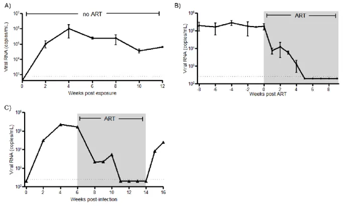
macrophages harbor latent virus. Previous attempts to characterize the “latent reservoir” in macrophages have largely depended solely on the presence of viral DNA in these cells, which could instead be the result of phagocytosis of T cells and not latent infection [131, 132]. ART effectively suppressed plasma viremia and reduced tissue viral RNA (vRNA) and viral DNA (vDNA) levels in infected MoM (Chapter 4). Removal of ART resulted in viral rebound in MoM, but this rebound was very delayed in comparison to ToM or BLT mice (Chapter 4). These results indicate that macrophages are a source of viral rebound after ART-interruption. Overall, I present herein two separate but
25
CHAPTER 2: HIV INFECTION, RESPONSE TO TREATMENT AND
ESTABLISHMENT OF VIRAL LATENCY IN A NOVEL HUMANIZED T CELL-ONLY
MOUSE (TOM)1
SUMMARY
The major targets of HIV infection in humans are CD4+ T cells. CD4+ T cell depletion is a hallmark of AIDS. Previously, the SCID-hu thy/liv model was used to study the effect of HIV on thymopoiesis in vivo. However, these mice did not develop high levels of peripheral T cell reconstitution and required invasive surgery for infection and analysis. Here, we describe a novel variant of this model in which thy/liv
implantation results in systemic reconstitution with human T cells in the absence of any other human hematopoietic lineages.
NOD/SCID-hu thy/liv and NSG-hu thy/liv mice were created by implanting human fetal thymus and liver tissues under the kidney capsule of either NOD/SCID or NSG mice. In contrast to NOD/SCID-hu thy/liv mice that show little or no human cells in
peripheral blood or tissues,substantial systemic human reconstitution occurs in NSG-hu thy/liv mice. These mice are exclusively reconstituted with human T cells (hence, T cell- only mice or ToM). Despite substantial levels of human T cells, no signs of graft-versus-host disease (GVHD) were noted in these mice over a period of 14 months. ToM are
1
26
readily infected after parenteral exposure to HIV-1. HIV replication is sustained in peripheral blood at high levels and results in modest reduction of CD4+ T cells. HIV-1 replication in ToM responds to daily administration of combination antiretroviral therapy (ART) resulting in strong suppression of virus replication as determined by undetectable viral load in plasma. Latently infected resting CD4+ T cells can be isolated from
suppressed mice which can be induced to express HIV ex vivo upon activation demonstrating the establishment of latency in vivo.
INTRODUCTION
SCID-hu thy/liv mice develop a bonafide human thymic organ and have a marginal level of systemic reconstitution with human T cells [197, 198]. The human thymic organoid present in the SCID-hu model is susceptible to HIV infection [199]. However, infection of these animals requires this tissue to be surgically exposed to virus administration via direct injection [73]. HIV injection results in infection of the human thymocytes present, but there is no detectable viremia in these mice. Thus, analysis of virus replication and its effect on thymocytes requires surgical removal of a piece of tissue [73]. Subsequent monitoring of infection over time also requires additional surgical collection of tissue for analysis. Although the use of this model is extremely labor intensive and requires large numbers of animals to make meaningful
27
Following the development of the SCID-hu thy/liv model, several other novel strains of mice have been derived with a higher degree of immunodeficiency. These include the NOD/SCID and the NOD/SCID common gamma chain null (NSG) strains of immunodeficient mice [197]. Both of these strains have been extensively and
successfully used in the derivation of a variety of humanized mouse models [202]. However, neither of these two strains has been extensively used to produce humanized thy/liv implanted mice [203].
Resting CD4+ T cells represent a well-characterized reservoir for latent HIV-1 infection, and this reservoir persists long-term despite treatment with highly active antiretroviral therapy (HAART) [87, 204, 205]. Incubating resting CD4+ T cells with CCL19, secreted by mature dendritic cells, ex vivo increases HIV-1 integration efficiency [206]. Additionally, the chemokines CXCL9 and CXCL10, secreted by monocyte-derived cells and induced by IFN-γ production, seem to mediate similar effects in resting T cells [204, 206-208]. Secretion of IL-7 by dendritic cells may be important for the survival of memory T cells, and secretion of IL-15 by macrophages and other mononuclear phagocytes is important for the low level of proliferation necessary to maintain a resting memory pool over time [209]. Thus while it is known that several myeloid-derived cell types secrete cytokines and chemokines that facilitate the
development of latency and maintain the resting CD4+ T cell pool, whether or not these cells are necessary for the establishment of latency in vivo remains unknown [210].
28
myeloid (and B) cells. To this effect, we implanted human thymus and liver into NOD/SCID and NSG mice. In this study we show that whereas NOD/SCID-hu thy/liv mice do not develop high levels of systemic reconstitution with human cells, NSG-hu thy/liv mice develop high levels of human T cells in the peripheral blood. Remarkably, flow cytometric analysis of blood and tissues demonstrate the complete absence of human B and myeloid cells in these mice. Interestingly, in contrast to mice reconstituted with human peripheral blood mononuclear cells (PBMC) and some other types of
humanized mice [211, 212], these T cell-only mice (or ToM) do not develop signs of GVHD. In addition, ToM are readily susceptible to HIV infection after parenteral exposure and can sustain high levels of HIV replication. Virus replication can be
efficiently suppressed by antiretroviral therapy, and HIV latency is established in resting T cells.
EXPERIMENTAL PROCEDURE
GENERATION OF HUMANIZED MICE
29
Laboratory Animal Medicine at the University of North Carolina at Chapel Hill (UNC-CH) according to protocols approved by the Institutional Animal Care and Use Committee. Human reconstitution of mice was monitored by flow cytometric analysis for human CD45+ cells in peripheral blood, as previously described [79, 80]. Peripheral blood samples were obtained via submandibular venipuncture and were collected in tubes containing EDTA. Whole peripheral blood was stained with antibodies, red blood cells were lysed, and the remaining cells were washed and fixed using a 1%
paraformaldehyde solution. A total of 10,000-30,000 events were collected per animal at each time point as indicated below.
TISSUE HARVESTING AND FLOW CYTOMETRIC ANALYSES OF HUMANIZED
MICE
Mononuclear cells (MNCs) were isolated from the bone marrow, spleen, lymph nodes, lung, liver, and thymic organoid as previously described [80]. Tissues were minced and/or digested and filtered through a 70 µm strainer. The liver, lung and female reproductive tract (FRT) were processed as previously described [83]. For all latency determinations, mononuclear cells, with the exception of the lymph nodes, were isolated using a Percoll gradient. Red blood cells were lysed as needed (namely for the spleen, bone marrow and liver tissues). MNCs were washed, counted via trypan blue exclusion, and flow cytometric analyses were performed for the indicated markers [79, 80, 83, 87, 213]. Live cells were distinguished by their forward and side scatter profiles as
previously described [214]. Flow cytometry data was collected on either a BD
30
HIV-1 INFECTIONS
Stocks of HIV-1JR-CSF were prepared and titered as previously described [215].
Briefly, virus supernatants were prepared via transient transfection of 293T cells, and were tittered using TZM-bl cells essentially as we have previously described [77]. Parenteral exposures were performed using HIV-1JR-CSF (90,000 TCIU) administered
either intravenously or intraperitoneally. A total of two intraperitoneal and six intravenous exposures were performed, yielding 2/2 and 6/6 systemically infected animals, respectively.
ANALYSIS OF HIV-1 INFECTION
Peripheral blood was collected via retro-orbital bleed using EDTA coated
capillary tubes (approximately 100 ul total).Infection of ToM with HIV-1 was determined with a one-step reverse transcriptase real-time PCR assay (ABI custom TaqMan
Assays-by-design) according to the manufacturer’s instructions (with primers
5’-CATGTTTTCAGCATTATCAGAAGGA-3’ and 5’-TGCTTGATGTCCCCCCACT-3’; assay sensitivity of 400 RNA copies per mL). Additionally, the percent of human CD4+ T cells in the peripheral blood of ToM pre- and post-exposure to HIV-1 were monitored by flow cytometry (using 40-60 ul of blood). Changes in the percent of CD4+ T cells present in the tissues of infected and uninfected animals were compared by two-way ANOVA, and were not significantly different. Statistical analysis was performed in Prism version 5 (GraphPad Software, Inc., San Diego, CA).
ANTIRETROVIRAL TREATMENT OF TOM
31
Specifically, infected ToM were administered daily intraperitoneal injections of emtricitabine (FTC; 140-200 mg/kg), tenofovir disoproxil fumarate (TDF; 146-208 mg/kg) and raltegravir (RAL; 56-80 mg/kg) for six to nine weeks, as previously described [87]. HIV-1 infection was monitored throughout ART as described above. RESTING CELL ISOLATION AND LATENCY DETERMINATIONS OF TOM AND
PATIENT SAMPLES
All MNCs from individual mice were pooled. Resting human CD4+ T cells were isolated from pooled tissues or from leukapheresis product of patient samples using negative magnetic selection (STEMCELL Technologies, Vancouver) as previously described [87, 163, 205]. Briefly, MNCs obtained from mouse tissues were incubated with a cocktail of antibodies composed of mouse anti CD45 and TER119, and anti-human CD8, CD14, CD16, CD19, CD56, CD41, CD25, CD31, CD105, HLA-DR, and glycophorin A. For the separation of cells from human samples, the mouse antibodies were not included in the isolation cocktail. Antibody-bound cells were removed using a column based-magnetic purification system and the purified resting cells were collected as flow through. This approach resulted in a >99% pure resting CD4+ T cell population. Resting CD4+ T cells were then cultured with 15 nM efavirenz and 1 µM raltegravir for 2 days prior to performing viral outgrowth assays to prevent any de novo infection from unintegrated virus [87]. Viral outgrowth was achieved by maximally stimulating resting cells in limiting dilution cultures containing 60 U/ml IL-2, 1ug/ml phytohemagglutinin (PHA) and irradiated allogeneic PBMC from an uninfected donor [87, 205]. The culture media was replaced every 3-4 days with fresh media containing 5U/ml IL-2. CD8
32
experiment to facilitate virus spread/amplification in cultures. Cultures were scored positive if p24 was detectable at day 15 and confirmed on day 19. The number of infected resting cells was estimated by a maximum likelihood method and was expressed as the infectious units per million resting CD4+ T cells (IUPM) [205]. RESULTS
CHARACTERIZATION OF THY/LIV IMPLANTED NSG AND NOD/SCID MICE
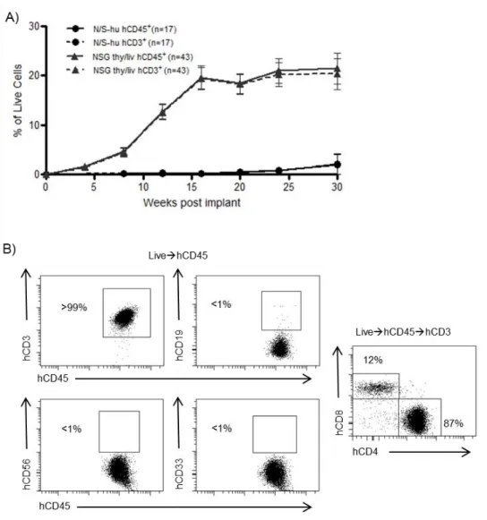
SCID-hu (thy/liv) mice have been extensively used as an in vivo model to study HIV infection of the thymus [72]. Since the original development of the SCID-hu thy/liv model, new and improved strains of immunodeficient mice like NOD/SCID and NSG have been developed [200, 201]. We implanted human thymus and liver into NOD/SCID and NSG mice to determine whether or not these strains would be an improvement over the SCID-hu model. We then monitored the peripheral blood (PB) of these mice over time by polychromatic flow cytometry for the presence of human cells (hCD45). While the NOD/SCID implanted mice, like the original SCID-hu mice, did not have significant levels of human cells in their PB, the implanted NSG mice had substantial levels of human reconstitution as determined by presence of human CD45 in their PB (Figure 2.1A). Furthermore, human cells present in the PB of these mice were identified as T cells by their cell surface expression of human CD3 (Figure 2.1B). Interestingly,
exhaustive analysis for the presence of other lymphoid or myeloid human cells did not reveal any significant levels of these cells in the PB of any animals analyzed.
33
mice (Lin-/HLA-DRhi, data not shown). Thy/liv implanted NSG mice showed sustained production of human T cells that reached approximately 20% in peripheral blood for up to 30 weeks (the last time point analyzed). Over this period, no signs of graft-versus-host disease (GVHD) were observed. Additionally, some animals were followed for up to 12 months post-implant (the last time point analyzed). These animals were found to sustain 20-30% human T cells in the PB even at these late time points (n=2, data not shown). From these results, we concluded that implantation of human thymus and liver into NSG mice results in sustained and exclusive production of human T cells in vivo.
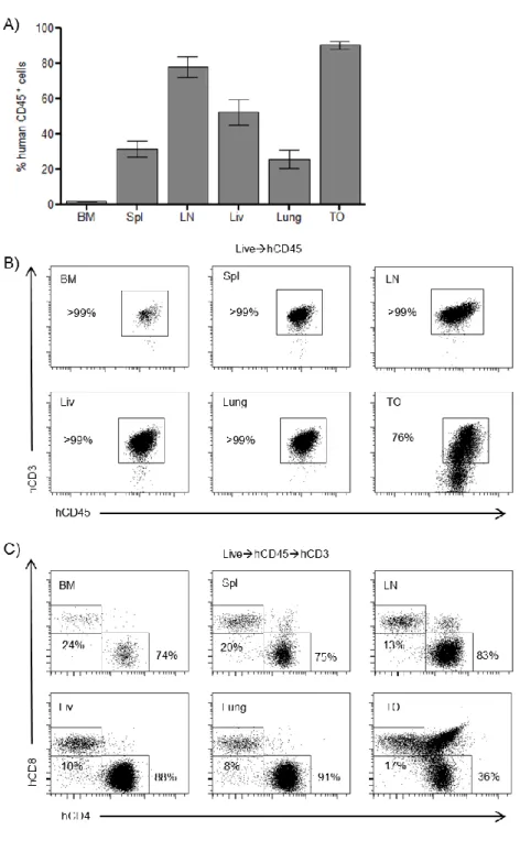
In SCID-hu mice, human cells are almost exclusively found in the thymic organoid, with little reconstitution of PB or tissues [216]. To determine the systemic distribution of the human cells present in NSG mice, mononuclear cells were isolated from the bone marrow, spleen, lymph nodes, liver, lung, and the thymic organoid. Flow cytometric analyses demonstrated that the spleen, lymph nodes, liver, lung, and thymic organoid were robustly reconstituted with human cells (CD45+) (Figure 2.2A).
34
Additionally, the small and large intestines of ToM were analyzed by flow cytometry and immunohistochemistry. In contrast to all other tissues analyzed we found no significant levels of human cells present in the gastrointestinal tract of these animals (data not shown).
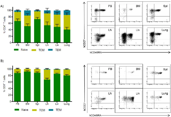
We further investigated the phenotypes of the human T cells present in PB and tissues. Both CD4+ and CD8+ T cells from PB and tissues of these mice had a
predominantly naïve phenotype, expressing both human CD45RA and CD27 (Figure 2.3). Additionally, we found CD4+ T cells with central memory (CD45RA-/CD27+) and effector memory (CD45RA-/CD27-) phenotypes (Figure 2.3A). Within the CD8+ T cell population, mainly naïve and central memory phenotypes were observed, with effector memory phenotypes being extremely rare in this population (Figure 2.3B). These results demonstrate that the human CD4+ T cells in these mice have a normal developmental phenotype.
HIV INFECTION OF TOM
Once we established the systemic reconstitution of ToM, we tested whether or not they could support HIV-1 replication. Eight ToM were infected with a single dose of cell-free HIV-1JR-CSF, a CCR5-tropic isolate administered parenterally. We then
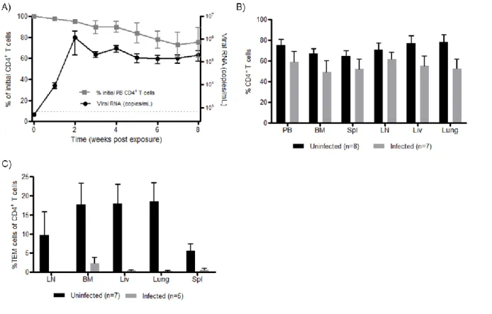
monitored the plasma of ToM for the presence of HIV-1 RNA as previously described [83, 87]. HIV-RNA was detected in the plasma of ToM one week after exposure and high levels of viremia were maintained over time (Figure 2.4A). We also monitored CD4+ T cell depletion in PB, a measure of HIV pathogenesis. The presence of HIV-RNA in PB correlated with a subsequent drop in circulating CD4+ T cells (Figure 2.4A).
35
with systemic virus spread (Fig. 2.4B). Phenotypic analysis of the remaining CD4+ T cells in the infected mice demonstrated the specific depletion of effector memory T cells (TEM) cells in PB and tissues (Figure 2.4C).These results demonstrate the susceptibility of ToM to HIV infection, their ability to sustain high levels of virus replication and the depletion of CD4+ TEM cells from PB and tissues of infected animals.
SUPPRESSION OF HIV BY ART IN TOM
Having established the capacity of ToM to support HIV infection, we proceeded to determine if virus replication could be suppressed by combination ART. For this purpose, HIV infected ToM were treated daily with a combination of FTC, TDF and raltegravir. This regimen has been shown by our laboratory to effectively suppress viral replication in the humanized BLT mouse model [87]. To assess the effectiveness of ART in infected ToM , levels of HIV-RNA were monitored in the plasma throughout treatment. ART administration resulted in a rapid reduction in the levels of plasma HIV-RNA in all treated animals (Figure 2.5B). Five weeks after initiation of treatment the levels of HIV-RNA in plasma were below the detection limit of our assay (750 RNA copies/ml). These results demonstrate the efficacy of ART in controlling HIV replication in humanized ToM. However, even long-term ART does not result in virus eradication and treatment interruption in patients leads to viral rebound [218]. To determine if this also occurs in ToM, one infected mouse was suppressed for four weeks on ART (treated for eight weeks total), after which treatment was ceased. One week after
36
QUANTIFICATION OF LATENT RESERVOIR
In humans, ART results in virus strong virus suppression, increases in CD4+ T cell levels and other significant health benefits to patients [219]. Despite this strong virus suppression seen in ART treated patients, HIV persists in resting CD4+ T cells that form a long lasting latent reservoir [220]. To determine if HIV could establish a latent
reservoir in ToM, we first confirmed the presence of resting human T cells in ToM. To assess the presence of resting human T cells in ToM, we collected cells from PB, bone marrow, spleen, lymph nodes, liver, lung, and thymic organoid from HIV+ ART
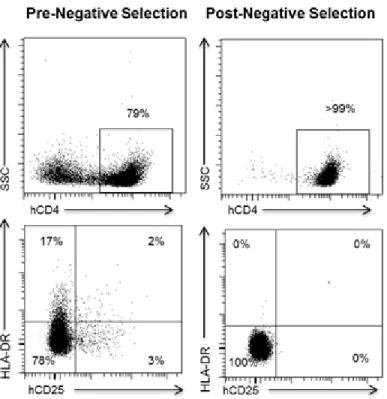
suppressed mice. Cells from all the tissues obtained from each individual mouse were pooled together and human CD4+ T cells in each pool of cells corresponding to one individual mouse were analyzed for expression of hCD25 and HLA-DR. This analysis demonstrated the presence of significant numbers of resting human CD4+ T cells in ToM (Figure 2.6). To determine if resting cells were latently infected with HIV, pooled cells from all tissues of each individual ART suppressed mouse were used to isolate resting CD4+ T cells by negative selection using magnetic beads [87, 205]. After
magnetic selection, a highly purified population of resting CD4+ T cells, shown by a lack of CD25 and HLA-DR expression was obtained from each individual mouse (Figure 2.6).
37
with PHA, IL-2 and irradiated PBMC from an uninfected donor, followed by co-culture with allogeneic PHA-activated CD8+-T-cell-depleted feeder cells. Fifteen days later, cultures were tested for the presence of HIV (Figure 2.7A). The number and density of cultures was then used in a maximum likelihood method to estimate the number of infectious units per million cells (IUPM). Latently infected cells were obtained from all four animals analyzed (Figure 2.7B). IUPM values from mice were compared with values obtained from outgrowth assays using resting CD4+ T cells of HIV infected patients treated during the acute or chronic phase of infection. The levels of latently infected cells in these mice are within those observed in the chronic patients receiving suppressive ART [159, 222]. To confirm that this indeed is induction from latency, as an added control, prior to stimulation, supernatant from resting cell cultures were assayed for P24 and were all found to be negative (data not shown) suggesting that virus recovered from outgrowth experiments originated from latent provirus. These results demonstrate the establishment of HIV latency in ToM and demonstrate that in vivo human T cells alone are sufficient for establishing latency.
DISCUSSION
Although SCID-hu thy/liv animals have been used extensively to study
38
tissue under both kidney capsules of each mouse. Using this more invasive implantation strategy combined with 20X more tissue, HIV-1 infection was achieved after IP or intra-implant injection. Using the original intra-implantation strategy described for SCID-hu mice, the use of more immunodeficient mouse strains, like the NSG strain, has overcome the limited systemic reconstitution previously seen in SCID-hu mice. Interestingly, thy/liv implantation of NOD/SCID mice did not result in systemic reconstitution with T cells suggesting that the additional immunosuppression due to the lack of a functional common gamma chain observed in NSG mice resulting in a complete lack of natural killer cells [226] is likely contributing to the increased T cell levels in these mice.
ToM were systemically reconstituted with human T cells. This reconstitution is consistent with the continued production of human T cells from the implanted thy/liv organoid as it showed a substantial and robust population of CD3+/CD4+/CD8+ thymocytes for as long as the animals were examined (1.2 years). Consistent with the lack of cryptopatches in NSG mice [227] ToM showed essentially no significant
accumulation of human T cells in the intestinal tract (data not shown). ToM show phenotypically normal CD4+ T cell development. However, we noted somewhat limited CD8+ T cell development in ToM, with few effector memory CD8+ cells. These
differences in the formation of effector phenotypes in the CD4+ and CD8+ T cell
39
since tissue from a total of 11 different donors were used to generate the mice used for these experiments.
One salient feature of ToM is the fact that despite robust levels of human T cells, they do not develop GVHD. GVHD has been observed in multiple humanized mouse models [228, 229]. Some investigators have reported a significance incidence of GVHD beginning approximately 12 weeks post-humanization and leading to death of the animals as early as 15 weeks post-humanization [228]. In contrast, we did not notice any of these effects on ToMs at these or subsequent time points. The longevity of ToM systemically reconstituted with high levels of human T cells in the absence of GVHD is an important feature of this model.
ART offers significant benefits to HIV infected patients. Our results show that combination ART is able to suppress viral replication in ToM validating this model for the evaluation of the effect of antivirals on HIV replication in vivo. As in humans, therapy interruption resulted in rapid viral rebound. Furthermore, we show that human T cells alone are sufficient for the establishment of HIV latency in resting CD4+ T cells.
40
41
Figure 2.1: Analysis of the peripheral blood (PB) of thy/liv implanted NSG mice demonstrates long-term reconstitution with human T cells. A) Flow cytometric analysis of the PB of NOD/SCID-hu (N/S-hu) (black circles) and NSG thy/liv (gray triangles) mice demonstrates systemic reconstitution of implanted NSG mice with
human cells (CD45+; solid line) and human T cells (CD3+; dashed line). Gating strategy: live cellshuman CD45human CD3. B) Flow cytometric analysis of cells from PB of a representative NSG-hu thy/liv mouse (29 weeks post-implant) demonstrates these mice are exclusively reconstituted with human T cells with greater than 99% of cells
42
Figure 2.2: Peripheral tissues of implanted thy/liv NSG mice are extensively reconstituted with human T cells. A) Flow cytometric analysis of cells harvested from the bone marrow (BM), spleen (Spl), lymph nodes (LN), liver (Liv), lung, and the thymic organoid (TO) of implanted thy/liv NSG mice (n=15) demonstrated reconstitution with human cells (CD45+). B) Flow cytometric analysis of tissues harvested from an implanted thy/liv NSG mouse (29 weeks post-implant) demonstrated that all organs were reconstituted exclusively with human T cells or thymocytes. C) Flow cytometric analysis of tissues and PB harvested from an implanted thy/liv NSG mouse
43
Figure 2.3: Naïve/memory phenotype of T cells in the PB and tissues of ToM.
44
Figure 2.4: HIV-1 replication and CD4+ T cell depletion in ToM. A) ToM (n=8) were parenterally exposed to HIV-1 and the viral load monitored in PB plasma (black circles). Changes in CD4+ T cell levels in PB were measured over time using flow cytometric analysis (gray squares). Gating scheme: live cells hCD45 hCD3 CD4. The limit of detection for viral load is indicated with a dashed line. B) The percentage of CD4+ T cells present in the tissues of infected (gray bars) and non-infected (black bars) ToM were analyzed using flow cytometric analysis. Gating scheme: live cells hCD45 hCD3 CD4. C) HIV infection results in a reduction in the levels of effector memory cells within the CD4+ T cell population of infected mice (n=5, grey bars) versus
45
Figure 2.5: There is sustained HIV replication in ToM that can be efficiently
46
Figure 2.6: Resting human CD4+ T cell isolation from ToM. (Top left)Flow
cytometric analysis of cells pooled from the different tissues of a ToM prior to magnetic negative selection showed the presence of both CD4+ and CD4- cells. (Bottom left) Prior to negative selection CD4+ T cells expressed various levels of CD25 and HLA-DR. (Top right) After isolation 99% of the cells obtained were CD4+. (Bottom right) Consistent with a resting phenotype, isolated cells were CD3+CD4+ and did not express CD25or HLA-DR. Gating strategy: (Top) livehCD45+hCD3+. (Bottom)
47
Figure 2.7: Latent HIV infection of human resting CD4+ T cells in ToM and human
48
CHAPTER 3: MACROPHAGES SUSTAIN HIV REPLICATION IN VIVO
INDEPENDENT OF T CELLS
SUMMARY
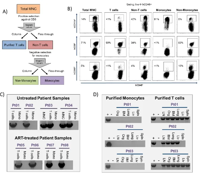
Macrophages have been long considered as contributors to HIV infection of the CNS, a sanctuary with restricted access from the periphery. However, recent studies have contradicted early work, suggesting that macrophages are not an in vivo source of virus production. To address this question, we first analyzed monocytes isolated from viremic patients and patients undergoing antiretroviral treatment, and were unable to find viral DNA or viral outgrowth in vivo. To determine if tissue macrophages are productively infected, we used three different but complementary humanized mouse models. Two models were previously described [bone marrow/liver/thymus (BLT) and T cell-only mice (ToM)] and a third [myeloid-only mice (MoM)] was used for the purpose of investigating the role of myeloid cells in HIV replication in vivo. Using MoM we
49
INTRODUCTION
HIV, the causative agent of AIDS, is severely species restricted and to date only humans and chimpanzee have been shown to be susceptible to infection [12, 230]. The limited species specificity of HIV represents a significant challenge for in vivo
experimentation, thus there is a need for the use of animal models. Human infection by HIV (and infection by its relative SIV in non-human primates) is restricted to cells
expressing the CD4 molecule [231]. In addition to CD4, productive HIV infection, meaning infection that leads to the production of viral progeny, requires one of two different G protein-coupled receptors: CCR5 or CXCR4 [232]. CD4+ T cells have been shown to harbor HIV proviruses and represent the most abundant target for HIV infection in vivo [101, 160]. Despite the prevalence of virus in CD4+ T cells, it is clear that T cells are not the only targets of HIV infection in vivo. In fact, macrophages have been shown to express CD4, CCR5 and CXCR4 and to be susceptible to HIV infection in vitro and in vivo [233-235]. Non-human primates and humanized mice have been extensively used to study HIV and SIV infection and pathogenesis in vivo. HIV/SIV infection of macrophages and microglia, the tissue-resident macrophages of the brain, is postulated to substantially contribute to the establishment and pathogenesis of HIV/SIV infection in the CNS [29, 236, 237]. The CNS is a location that has been considered to be a sanctuary for the viruses where variants of HIV can replicate and expand independently of contributions from the periphery [238, 239]. It has been
50
macrophages, such as microglia and perivascular macrophages, could then be susceptible to infection [29].
Whereas the ability of HIV to replicate in human macrophages in vitro has been extensively documented, evidence for HIV replication in human macrophages in vivo or ex vivo is limited and in some instances indirect [193, 240, 241]. Analysis of the gut has yielded somewhat conflicting results as human intestinal macrophages did not support HIV replication ex vivo and were found to be more monocyte-like in receptor expression patterns [241]; yet viral HIV-DNA was isolated from sorted CD13+ cells in rectal biopsies obtained from ART-suppressed patients, suggesting a non-T cell origin [242]. Analysis of monocytes from peripheral blood consistently shows very low levels or lack of infection in viremic or aviremic patients [117, 119, 243]. Evidence of both in vitro virus outgrowth from human monocytes obtained from patients and ex vivo virus outgrow from tissue macrophages (including the brain or CNS) is largely absent. However, the presence of infected macrophages in a variety of tissues has been clearly documented via immunohistochemistry and in situ hybridization approaches [235, 244, 245].
In vivo macrophage infection is currently a topic of intense debate. Specifically, data from Calantone et al suggests that at least in SIV infected non-human primates, macrophages are not productively infected and cannot replicate SIV [132]. Rather macrophages ingest T cells and this explains the presence of HIV nucleic acids and proteins in macrophage preparations. Further evidence in support of this postulate has also been recently presented by Baxter et al [131]. In this article the authors document that human macrophages selectively capture and engulf human T cells and that