MULTI-SCALE MODELING OF THE
STRUCTURE AND DYNAMICS OF
MACROMOLECULES
Adrian Wendil R. Serohijos
A dissertation submitted to the faculty of the University of North Carolina at Chapel Hill in partial fulfillment of the requirements for the degree of Doctor of Philosophy in the Department of Physics & Astronomy.
Chapel Hill 2009
Approved:
© 2009
ABSTRACT
ADRIAN W.R. SEROHIJOS: Multi-scale modeling of the structure and dynamics of macromolecules
(Under the mentorship of Nikolay V. Dokholyan)
Biology is defined by phenomena that are inherently complex spanning multiple length and time scales. To understand these processes, there is a need for multi-scale
approaches that provide a coherent framework for describing and interrogating these phenomena. Here, we employ multiple approaches to investigate specific biological systems.
The first system we studied was the cytoplasmic dynein motor, a protein that walks along the microtubule tracks in cells. The major objective in the dynein motors field is to understand its mechanism. Specifically, what is dynein’s structure and how does it transduce chemical
energy into mechanical work? We proposed a theoretical structural model of the motor and performed normal mode analysis and molecular dynamics on the motor unit structure. These
studies hypothesized new structural features in the dynein motor unit and proposed a potential mechanism for energy transduction [5,6,80]. The second system we studied was the CFTR channel, which regulates ion transport in the apical membrane of epithelial cells.
Mutations in the CFTR protein are the basis of the cystic fibosis disease. One of the primary question is how a single residue deletion (Phe508) lead to ~90% of cystic fibrosis cases. We
ACKNOWLEDGEMENTS
Foremost of all, I would like to thank my mentor Nikolay V. Dokholyan for the countless opportunities to work on good science. The depth of his patience and wisdom in life is unfathomable. He certainly taught me much more than science.
I would also like to thank current and former members of the Dokholyan group, with special mention to Feng Ding and Yiwen Chen. Not only did I earn esteemed colleagues but also good friends.
I would also like to thank my collaborators John Riordan, Tamas Hegedus, and the rest of the Riordan group for placing us right at the forefront of cystic fibrosis research; and to Timothy Elston and his group for introducing me to the exciting world of molecular motors. Scientific output is less a function of personal effort and talent than the people one works with; I could not be more fortunate in this regard.
Thanks to the Board of Governors Fellowship, Program in Molecular and Cellular Biophysics, and the American Heart Association Predoctoral Fellowship for funding. Thanks also to the members of my committee for supporting my efforts.
Thanks to Judith and Nanette for keeping me company and introducing to a whole new community of Filipinos and friends in Chapel Hill and the Philippines. Lastly, I would like to thank my family, friends, and loved ones for their infinite support.
CONTENTS
Page
LIST OF FIGURES………. ix
LIST OF TABLES……….. xi
LIST OF PUBLICATIONS……… xii
Chapter 1. Introduction……… 1
Chapter 2. Structure of the cytoplasmic dynein motor unit………. 5
2.1 Methods……….. 8
2.1.1 Homology modeling……… 8
2.1.2 Fitting all-atom models to EM-maps……….. 10
2.2 Results……… 12
2.2.1 Models of the individual domains……….. 12
2.2.2 Motor domain organization……… 15
Chapter 3. Conformational dynamics of the dynein motor unit……….. 18
3.1 Methods………. 18
3.1.1 Preliminary investigation of the motor dynamics using normal mode analysis……… 18
3.1.2 Molecular dynamics simulation of the motor unit……….. 20
3.2 Results……… 22
3.3 Summary……… 25
4.1 Methods and models……….. 32
4.1.1 Simplified model of a protein………. 32
4.1.2 Simplified interaction using the Go-model and discrete molecular dynamics……….. 32
4.1.3 Equilibrium simulations protocol……… 33
4.1.4 Folding simulations protocol……….. 33
4.1.5 Analysis of folding simulations……….. 34
4.2 Results……… 35
4.2.1 Equilibrium dynamics………. 35
4.2.2 Difference in wild type and mutant NBD1 folding propensities…. 37 4.2.3 Folding pathways………. 37
4.2.4 Structural modulators of folding kinetics……… 38
4.2.5 Computational rescue of NBD1-F508 ………. 45
4.3 Summary………. 46
Chapter 5. Structure of the complete CFTR channel……… 48
5.1 Methods and models……….. 48
5.1.1 Modeling the CFTR structure from Sav1866…….………. 48
5.2 Results……… 52
5.3 Summary……… 56
Chapter 6. Conclusion and outlook………. 60
LIST OF FIGURES
1.1. Time and length scales of processes involving proteins………... 2
2.1. Cellular “super highways” and dynein………. 6
2.2. Dynein stepping behavior. ………...………...………. 7
2.3. Homology modeling. ………...………...………. 9
2.4. Fitting of all-atom models to low-resolution electron miscroscopy maps………… 11
2.5. All-atom models of domains within the motor unit. ……… 13
2.6. Dynein’s putative catalytic site.……… 13
2.7. Potential coiled-coil conformation of IDR4………. 15
2.8. Complete model of the motor unit……… 17
3.1. Normal mode analysis and elastic network model……… 19
3.2. Simplified protein model……….. 20
3.3. Discrete molecular dynamics……… 22
3.4. Lowest frequency normal modes of dynein motor unit……… 24
3.5. Averaged domains fluctuations from equilibrium molecular dynamics………….. 25
3.6. Model of power stroke……….. 26
4.1. CFTR and cystic fibrosis………... 29
4.2. Arrest in the processing of the CFTR-DF508 mutant……… 30
4.3. Protein folding energy landscape……….. 31
4.4. Thermodynamics of NBD1-WT, NBD1-F508A, and NBD1-F508………... 36
4.5. NBD1 folding……… 39
4.6. Energy probability distributions averaged over all successful folding trajectories... 40
4.7. Distribution of fraction of native contacts………. 41
4.9. Comparison of the folding pathways of wild type NBD1 and its mutants………… 43
4.10. Structures of folding intermediates………. 44
4.11. Contacts in NBD1-WT that perturbed in the F508A and F508 mutants………. 46
5.1. Sequence alignment of the membrane-spanning domains of human CFTR and the Sav1866 exporter………. 50
5.2. Experimental constraints satisfied in the membrane-spanning domains of the homology model………. 52
5.3. Theoretical model of CFTR structure………... 54
5.4. Predicted cytoplasmic and nucleotide-binding domain interfaces……… 55
5.5. Cross-linking schema……… 57
5.6. Validation #1: Cross-linking of interfacial residues………. 58
LIST OF TABLES
LIST OF PUBLICATIONS
The permissions for the adaption of published figures and texts in the dissertation were granted by the publishers.
Dynein Molecular Motor
1. A.W.R. Serohijos, Y. Chen, F. Ding, T.C. Elston, and N.V. Dokholyan, “A new structural model reveals energy transduction in dynein” (2006) Proc. Natl. Acad. Sci. USA, 103: 18540-18545.
2. D. Tsygankov, A.W.R. Serohijos, N.V. Dokholyan, and T.C. Elston, “Kinetic models for the coordinated stepping of cytoplasmic dynein” (2009) J. Chem. Phys., 430: 25101. 3. A.W.R. Serohijos*, D. Tsygankov*, S. Liu, T.C. Elston, and N.V. Dokholyan,
“Multiscale approaches for studying energy transduction in dynein” Submitted. [*Equal contribution]
Cystic Fibrosis and CFTR
4. A.W.R. Serohijos, T. Hegedus, A.A. Aleksandrov, L. He, L. Cui, N.V. Dokholyan, J.R. Riordan, “Phenylalanine 508 forms interdomain contact in the CFTR structure crucial to folding and function” (2008) Proc. Natl. Acad. Sci. USA, 105: 3256-3261.
5. A.W.R. Serohijos*, T. Hegedus*, J. Riordan, and N.V. Dokholyan, “Diminished self-chaperoning activity of the F508 mutant CFTR results in protein misfolding” (2008) Public Library of Science Computational Biology, 4: e1000008. [*Equal contribution] 6. T. Hegedus, A.W.R. Serohijos, N.V. Dokholyan, L. He, J.R. Riordan, “Computational
studies reveal phosphorylation dependent changes in the unstructured R domain of CFTR” (2008) J. Mol. Biol., 378: 1052-1063.
7. L. He, A.A. Aleksandrov, A.W.R. Serohijos, T. Hegedus, L.A. Aleksandrov, L. Cui, N.V. Dokholyan J.R. Riordan, “Multiple membrane-cytoplasmic domain contacts in CFTR” (2008) J. Biol. Chem., 283: 26383-26390.
Protein Folding, Protein Engineering, and Ion Channels
9. J. Hao*, A.W.R. Serohijos*, G. Newton, G. Tassone, D.C. Sgroi, N.V. Dokholyan, and J.P. Basilion, “Identification and rational redesign of peptide ligands to CRIP1, a novel biomarker for cancers” (2008) Public Library of Science Computational Biology, 4: e1000138. [*Equal contribution]
Chapter 1
Introduction
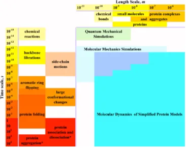
In its most elementary sense, all processes that define biology are governed by the dynamics, structure, and interactions between biomolecules such as proteins, nucleic acids such as DNA (deoxyribonucleic acid) and RNA (ribonucleic acid), and lipids. Processes involving these biomolecules are inherently multi-scale in time and length [1]. For example processes involving proteins range from 10-15 s (chemical reaction) to 104 s (aggregation) and from 10-11 m (chemical bond) to 10-6 m (protein complexes) (Fig. 1.1) [2]. Understanding biology then entails understanding the structure, dynamics, and interactions between these molecules.
Figure 1.1. Time and length scales of processes involving proteins. The left side of the figure contains examples of processes occurring at various time scales. Protein association, dissociation, and aggregation (*) are concentration dependent and may span longer times than presented here. Examples of molecular sizes are at the top. Three simulation approaches – quantum mechanical, molecular mechanics, and molecular dynamics simulations with simplified protein model – outline the time and length scales accessible to these approaches. The time-length scales area, corresponding to molecular dynamics simulations, signifies a range of simplified protein models used in simulations, i.e. to access all outlined scales one may need to use a number of mutually-consistent simplified protein models. [Diagram is taken from [2]]
This work in particular develops and uses multi-scale modeling of protein dynamics and structure to investigate two outstanding problems in biology and medicine: (1) elucidating the structure and mechanism of dynein, a motor protein that is fundamentally involved in the active transport within cells and (2) understanding the structural basis of the misfolding of the CFTR channel, which eventually leads to cystic fibrosis.
oligomeric organization of the structure, we fit the individual models of the motor unit to a low-resolution EM density derived from negatively stained electron microscopy [5]. These studies determined that the structure of the motor putatively forms an asymmetric heptamer, which may be important in generating the sequence of conformational changes during the motor’s force production.
In Chapter 3, we elucidated the conformational dynamics of the motor unit that may be associated with its force generation. Using a simple analysis of potential protein conformations, we performed normal mode analysis of the structural model constructed from Chapter 2 [5]. This analysis show that the motor contains a mobile “rough” side, which incidentally is the non-catalytic site of the motor unit, and a less mobile “smooth” side of the motor, the site containing the catalytic parts of the motor unit. Molecular dynamics simulations of the motor unit using a simplified protein model corroborate these observations [6].
either to the co-translational arrest or of the protein misassembly leading to post-translational
arrest is unknown. In Chapter 4, we explored the structural basis of the folding kinetics
defect induced in NBD1. By performing multiple folding simulations of wild type and
mutant NBD1s, we identified the metastable folding intermediates and the folding pathways
of both the wild type and mutant NBD1s [13]. We also defined a measure of the folding
propensity for each of the NBD1 constructs. These analyses showed that indeed this
difference in kinetics could be reproduced in simulations [13]. Moreover, from the structures
of the intermediate states, we found that this difference in kinetics could be attributed to the
conformation of specific loops in the nucleotide-binding domain, especially that of the
so-called S6-H6 loop [13]. In fact, when this loop in NBD1-DF508 mutant was forced to that of
the wild type conformation, we were able to partially “correct” the folding defect of the
protein [13]. Preliminary experimental results likewise validate our model.
In Chapter 5, we addressed the misassembly of the F508 CFTR mutant. We
constructed a structural model of the whole CFTR molecule to identify the location of the
Phe508 in the whole protein and to determine the specific interdomain interfaces it mediates.
This interface is presumably perturbed upon Phe508 deletion [14,15]. From the model, we
found that Phe508 in NBD1 interacts with the second membrane-spanning domain through
the fourth cytoplasmic loop (CL4). This predicted interface and other interfaces in the model
have been verified extensively using experiments [14,16].
Lastly, in Chapter 6, we synthesize the knowledge gained from the development of
multi-scale models of protein structure and dynamics and its application to specific biological
systems. We also provide an outlook of how this type of modeling may be applicable to other
Chapter 2
Structure of the cytoplasmic dynein motor unit
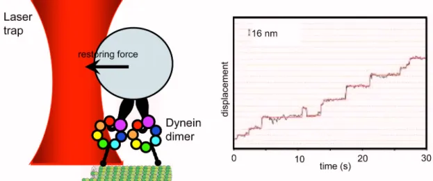
Figure 2.1. Cellular “super highways” and dynein. (Upper left) Image of a cell showing the cytoskeletal filaments actin (red) and microtubules (green). (Lower left) Simplified schema of a cell showing microtubules running from the cellular periphery towards the nucleus. Motor proteins such as dynein and kinesin walk along the microtubule transporting cellular substructures. (Right) Close-up view of a dynein dimer with a load walking towards the nucleus. A dynein monomer consists of a tail that binds to its load, a motor unit that hydrolyzes ATP and generates force, and a stalk that binds to the microtubule track.
binds and hydrolyzes ATP and putatively is the site for force production (Fig. 2, right panel).
Sequence analysis of dynein’s motor unit indicates that it consists of six concatenated AAA
subunits (denoted as A1 to A6 in the schema), an extended stalk that contains a microtubule
binding domain, and a C-terminal domain that is twice the size of an AAA subunit (Fig. 2.1,
right panel) [20,21,22]. Although experimental studies using EM revealed significant insights
into the structure of dynein, modeling the motor at the atomic level is essential in
investigating its mechanism.
The stepping mechanism of single dynein has been explored by many groups using
single molecule assays (Fig. 2.3). Studies of bead movement driven by cytoplasmic dynein in
vitro suggest that single molecule dynein molecules are processive, that is, single molecules
of dynein are capable of taking multiple steps along the microtubule track without detaching
[23,24,25].
The mechanism of other cytoskeletal motors such as myosin and kinesin, are better studied and more understood. It has been shown that nucleotide-driven conformational changes of their mechanism elements power the hand-over-hand stepping of their two identical motor domains [26,27]. In contrast, the mechanism of processivity in dynein is much less understood, and dynein’s distinct evolutionary origin and structural features of this motor suggest that its mechanism differs considerably from other molecular motors. First of all, their stepping behaviors are already different. Using dynein molecules labeled with quantum-dots, it has been shown that dynein takes both small (~8 nm) and large (12-24 nm) step sizes with occasional backward stepping, which are rarely observed in Kinesin 1 or Myosin V.
In this chapter, we first address the structure of the dynein motor unit. The dynamics of the motor unit and the stepping of the motor unit is address in the next chapter.
2.1 Methods
To build a structure of the dynein motor unit, we first performed homology modeling
of the individual domains that comprise the motor unit region. This individual domains need
to be assembled together to finally determine the quaternary structure of the protein. We used
an experimentally derived low-resolution electronic map of motor unit in determining
organization of these various domains.
2.1.1 Homology modeling
Homology modeling (or comparative modeling) is a method for deriving all-atom
theoretical models of protein tertiary structure by copying the topology of a related protein
with a known structure, usually derived from x-ray crystallography or NMR (nuclear
exhibit high sequence similarity (~30 % or greater) would most likely exhibit the same
structure. This assumption is based on the observation that protein tertiary structure is better
conserved than amino acid sequence [28]. While the structure derived from homology
modeling cannot be as definitive as those derived from X-ray crystallography or NMR, the
models are useful for generating hypothesis and directing experimental work.
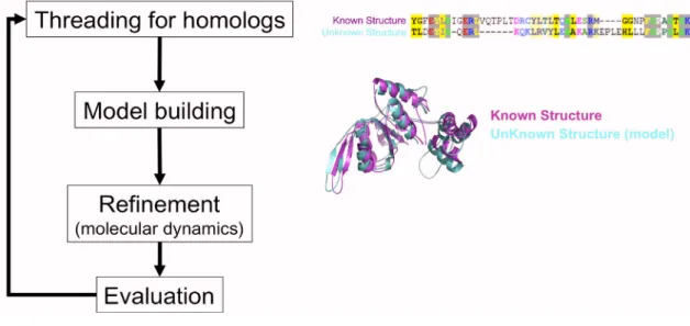
Figure 2.3. Homology modeling. To construct a model, the sequence of the protein of unknown structure is mapped to the sequence of the protein with known structure. The mapping optimizes the alignment of conserved residue regions (left panel). An all-atom model is then constructed by copying the backbone topology (and when possible, the rotameric states of the side-chains) of the known structure (left panel). The model structure is refined using molecular dynamic simulations. The quality of the model structure may be evaluated for correctness of backbone and side-chain geometry, exposure of charge residues, burriedness of hydrophobic residues, among other things. This set of procedures is performed iteratively until we arrived at a model of reasonable quality.
The sequence of the motor domain of cytosolic dynein heavy chain of slime mold D.
discodeum (GenBank accession no. P34036) was submitted for threading to 3DJury
(http://bioinfo.pl/meta)[29,30]. The templates used in building the AAA1, AAA2, and AAA4
were the Holliday junction migration motor protein RuvB from Thermus thermophilus HB8
polymerase III (PDB ID code 1JR3; [32]), and eukaryotic clamp loader (PDB ID code 1SXJ; [33]), respectively. AAA3 and AAA5 were modeled from the same model used by AAA1. Similar to AAA2, AAA6 was built from the clamp loader. We constructed the atomic models using the Homology suite of INSIGHTII (Accelrys, San Diego, CA).
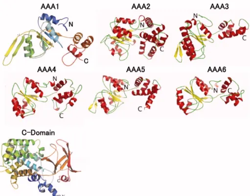
To construct a structural model for the C domain, we followed a protocol similar to the one used to construct models of the AAA subunits. We found that the C domain's first 290 residues consist entirely of -helices, whereas the remaining 128-residue stretch includes five -strands and terminates with a helix (Fig. 2.5). We then determined a family of candidate proteins that represent good structural templates for the two stretches of the C domain. Interestingly, the candidate templates for the first 290 residues were structures of the complement component C3d [Protein Data Bank (PDB) ID code 1GHQ], which attaches to foreign antigens during immune response [34]. Using the C3d fragment as template, the first 290 residues acquired a dome-shaped – toroidal fold [34]. The remaining five -strands and last helix were built from the plecktrin homology (PH) domain of the Leukemia-associated RhoGEF (PDB ID code 1TXD) [35], which folds into a flattened seven-stranded -barrel capped with a C-terminal helix. To obtain the complete structure of the C domain, the two subdomains were docked together using rigid body docking. The -helical stretch shows higher homology with its template structure than the remaining -strands suggesting that the function of the C domain is performed by the more conserved -helical stretch.
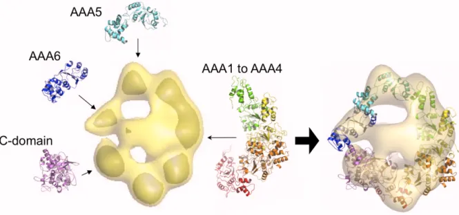
2.1.2 Fitting all-atom models to EM-maps
To preserve functionally relevant interactions between domains AAA1–AAA4 and to construct a regular tetramer for this portion of the motor, we superimposed the models of these subunits onto the 54
protein has a known homogenous heptamer structure consisting of AAA subunits with active
catalytic sites. Next, we used the vector-quantization method implemented in SITUS [37,38]
to fit the AAA1–AAA4 tetramer to the EM density (Fig. 2.4). To obtain a preliminary
orientation of the remaining domains, the atomistic models of subunits AAA5, AAA6, and
the C domain were fit separately to their corresponding electron density lobes. We also
imposed the constraint that AAA5, IDR4, and AAA6 form a continuous peptide. Thus,
AAA5 was oriented such that its C terminus faced the coiled coil. Similarly, AAA6 was
oriented such that its N terminus faced IDR4 (Fig. 2.4).
Finally, to arrive at the complete model, we docked IDR4 and the rest of the
inter-domain regions to the seven inter-domains using a rigid-body docking protocol and shape
complimentarity as criteria. When the complete atomic model was refit to the EM density of
the entire motor unit, SITUS [38] ab initio identified the correct orientation of the domains
with a correlation of 0.74 (P < 10316 ).
2.2 Results
2.2.1 Models of the individual domains
We systematically constructed a complete structural model of the motor unit of
cytoplasmic dynein from D. discoideum. The model includes the six AAA subunits, the linker regions that connect the subunits, and the C domain (Fig. 2.5). All AAA enzymes
consist of two structurally conserved units: an / Rossman fold subdomain and an -helical
globular subdomain. Despite a <20% sequence identity between proteins in the AAA
superfamily, up to 50% of equivalent C positions are within 2 Å rmsd [39]. To produce
improved folds for the six AAA subunits of the dynein motor unit, we used 3DJury (Section
2.2.1) to search for candidate homologs of these subunits and the linker regions that connect
them. 3DJury produces a consensus structure template based on the results of multiple
independent structure prediction algorithms. The structural templates obtained in this way
have consensus scores well above the confidence threshold of 50, which offers a prediction
accuracy of 90%. Using these initial alignments, we then built atomic models of the AAA
domains and their adjacent linkers using the homology modeling suite in Insight II (See
Section 2.2.1). To evaluate the accuracy of the models for each subunit, we compared the
local environments of the residues in the predicted structures to the population-averaged
residue environments determined from known structures. The profiles score show the current
model has better fold than earlier proposed models [21].
To determine the residues that form dynein's primary catalytic core, which is located
between the first and second AAA subunits, we docked an ATP molecule to the glycine-rich
P-loop (1969-GPAGTGKT-1976), which is the putative binding site for the nucleotide
Figure 2.5. All-atom models of domains within the motor unit. Six AAA domains (ATPases associated with various cellular activities) and a C-domain comprise the motor unit. [Figure adapted from [5]]
in the Walker A and Walker B motifs that bind the and NTP phosphates in all P-loop NTPases. These conserved residues found in dynein include K1975 in Walker A; D2021,
E2022, and R2025 in Walker B; and R2145 in Sensor 2 (Fig. 2.6). These results are
consistent with recent biochemical studies showing the dynein mutant K1975T trapped in a
strong-binding state and devoid of motile activity[40].
Interestingly, the interdomain region between subunits AAA5 and AAA6 (denoted as
IDR4) is 231 residues long, comparable with the size of an AAA unit (whereas the length of
the other interdomain regions IDR1, IDR2, and IDR3 are 79, 103, and 92, respectively) (Fig.
2.7). If IDR4 possesses a globular fold, then it would manifest as an additional lobe in the
reconstructed EM densities (Fig. 2.7) [41], and the motor would appear as an octamer. On the
other hand, one of the densities on the face of the motor unit forms a long arch that spans the
ring formed by the AAA subunits (Fig. 2.7), and is suggestive of a coiled coil. The IDR4 is
sufficiently long to span the 8-nm facial density of the motor unit. Moreover, coil prediction algorithms assign a coiled-coil structure in the AAA5-IDR4 sequence, although the length of
the predicted coil varies for dyneins from different species (Fig. 2.7). The search for
structural homologs also resulted in several coiled-coil structures. On the basis of these
results, we built IDR4 as a coiled coil using the cytoplasmic domain of serine chemotaxis
receptor (PDB ID code 1QU7)[42] as a template.
The smaller lump on the face opposite the arching density could be the remnant of the
dimerizing tail used in the EM studies. Another EM study [43] where the dynein tail has been
labeled with antibody-Fab tag showed that the tag is not rigid and can be found at various
positions around the planar ring. The study suggests that the tail domain docks into the center
Thus, only the point of attachment near of the tail will exhibit a density because the flexible
part will be averaged out, making the smaller facial density the more viable candidate for
docking of the N-terminal tail.
Figure 2.7. Potential coiled-coil conformation of IDR4. (A) Sequence map of dynein showing the various domains. Apart from the AAA units and the C-domains, there are also interdomain regions (IDRs) predicted to be primarily helical. There are three regions predicted to be coiled coils; the first two are already known to form the microtubule-binding stalk (panel B). We postulated that the third coiled-coil region correspond to the elongated density in the EM map (panel C).
2.2.2 Motor domain organization
The predicted structural model of the cytoplasmic dynein motor unit consists of six
AAA domains and a C-terminal domain arranged in an asymmetric heptameric ring. The
conserved AAA1-AAA4 domains form a tetramer that is organized similarly to other AAA
homomer complexes. The less well-conserved AAA5, AAA6, and C-terminal domains
consistent with the postulated evolutionary origin of the molecule in which the primordial
homodimer pairs AAA1–AAA2 and AAA3–AAA4 combined to form a tetramer, with the
subunits AAA5 and AAA6 representing later additions to the motor [44].
Another intriguing feature of the homology model is the IDR4 linker that connects
subunits AAA5 and AAA6. The model predicts that this structure accounts for the observed
density that spans the motor ring. In addition to contributing to the overall rigidity of the
motor, IDR4 provides a route for force propagation from the rigid smooth edge of the motor
where the nucleotide-binding sites are located to the flexible rough edge. Specifically, IDR4
extends from AAA5, which is at the base of the microtubule-binding stalk, to AAA3, whose
nucleotide-binding pocket regulates the motor's processivity [40,44]. The IDR4 structure
provides a clue to the important question of how distant functional sites communicate with
Chapter 3
Conformational dynamics of the dynein motor
unit
To understand the mechanism of the dynein motor, we investigated the
conformational changes that may be relevant with its force production. First, we used a
simple normal mode analysis of the protein to determine the most likely dynamic
conformations of the protein. Second, we performed molecular dynamics simulation using
simplified models of proteins and a non-traditional molecular dynamics approach called
molecular dynamics simulations. The results in this chapter have been described in two
articles [5,6].
3.1 Methods and Models
3.1.1 Preliminary investigation of the motor dynamics using normal mode
We performed normal mode analysis to establish the motor unit's dominant modes of
motion. Normal mode analysis has been shown to accurately identify structural sites that
function as pivots and, therefore, can be used to infer global motions of large molecular
complexes [45]. Normal mode analysis also can be used to explore the intrinsic flexibility of
molecular structures. In this analysis, the interactions between heavy atoms (C only) within
8 Å were approximated using a harmonic potential. Equations of motion were then computed by diagonalization of the Hessian matrix (mass-weighted second derivatives of the potential energy matrix) (Fig. 3.1). The eigenvalues of the matrix correspond to the mode frequencies and the associated vectors are the normal modes.
3.1.2 Molecular dynamics simulation of the motor unit
To make the dynamics simulation of this large molecule tractable, we used a
simplified protein model, a simplified interaction potential between atoms in the model, and
a fast sampling algorithm called discrete molecular dynamics (DMD)[2,46,47].
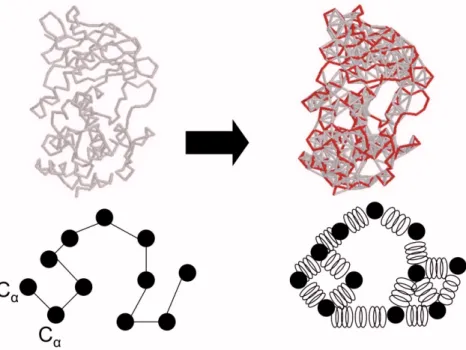
Figure 3.2. Simplified protein model.. The all-atom model of the dynein motor (Left) can be represented by by its C and C atoms. Simulation of the simplified model enables the investigation of the motor dynamics over long time scales.
The protein model consisted only of C and C atoms [48](Fig. 3.2). The interaction
potential used in the simulation is the structure-based Go-interaction [49,50], where the
residues that were proximate in the native state were assigned an attractive interaction, but
those that were not were assigned a repulsive interaction. The total potential energy of a
model protein was then U= ij=1Uij N
, where i and j denoted residues i and j, Uij was the
Ui j =
+, r ir j a0
ijij, a0<
r ir j a1
0, a1< r ir j
Here, a0 was the hard core diameter, ai was the maximum interaction distance between
residues and ij was the interaction strength between residue i and residue j, which set the
energy scale. ||ij|| was a matrix of contacts with elements
ij =1, if r i
NS r j
NS
a1 and
ij =1, if r i NS
r j NS
>a1, where
r iNS was the position of the ith residue in the native
conformation. We penalized the non-native contacts by imposing ij< 0. Temperature units were taken in terms of the typical value of interaction strength ij divided by the Boltzmann constant kB, i.e., in units of ij/kB.
The strength of the interaction between residues in contact ij defines the energy units. Physically, ij 1–2 kcal/mol, which is approximately the contribution to protein stability from a hydrogen bond. The time unit (tu) is estimated to be the shortest time between particle
collisions in the system (~0.1 ns).
The evolution of this simplified protein model with simplified atomic interactions
was calculated using DMD. In contrast to traditional molecular dynamics which employs
continuous potentials, the DMD algorithm uses discretized square well potentials [2,46,47],
thus all particles move at constant velocity until the before the soonest collision. That the
state of the system is necessarily updated only in the event of a collision enables DMD to
Figure 3.3. Discrete molecular dynamics. (A) Interaction potential between atoms are discretized (red) as opposed to being continuous in traditional molecular dynamics simulations (black). (B) Because of the discretized potential, the evolution of the system is now driven the collision between particles and entails the calculation of momentum and energy conservation equations.
3.2 Results
made quantitative in Fig. 3.4(B), which lists the RMSD of the C atoms of each subunit for
the first three normal modes.
In mode 1, the AAA5 subdomain exhibits an upward motion, whereas AAA6
partially rotates about the IDR4 linker (Fig. 3.4(C)). On the other hand, AAA1 to AAA4 and
their linkers exhibit minimal displacement. Interestingly, AAA5 is positioned at the base of
the stalk that interacts with the microtubule. The fact that the dominant motion of the lowest
frequency normal mode occurs at the base of the stalk suggests that the stalk tilts during the
motor's power stroke. Mode 2 is characterized by a “squeeze” applied to subunit AAA5 and
the C domain coupled with an outward motion by AAA6 (Fig. 3.4(C)). Similar to mode 1, in
mode 2, AAA1–AAA4 and their linkers exhibit minimal movement. EM 3D reconstructions
of the motor unit with stalks positioned at 0°, 25°, and 45° relative to vertical show greatest
variation in electron densities corresponding to subunits AAA5 and AAA6 [41] (Fig. 3.4(C)).
The direction and magnitude of the domain displacements determined for modes 1 and 2 are
consistent with these observations (Fig. 3.4(C)). For example, the motion predicted to occur
in modes 1 and 2 is consistent with the reorientation of subunit AAA6's density observed for
different stalk positions (Fig. 3.4(C)).
Using the simplified model described above, we extensively characterized the
dynamics of dynein. First, we performed discrete molecular dynamics (DMD) simulations
for 106
time units (approximately a few milliseconds) with initial temperatures from T=0.1
e/kBT to T=2.0 e/kBT (see section 2.1.2). These equilibrium simulations allowed us to
determine the thermal denaturation curve of the dynein head and the melting temperature.
temperature, we then performed molecular dynamics simulations near the identified melting
point.
Figure 3.4. Lowest frequency normal modes of dynein motor unit. (A) Superposition of
two structures displaced in opposite directions along the normal mode. The size of the backbone is proportional to fluctuations of the Ca atoms. Arrows indicate the directions of the dominant vibrations. AAA5, AAA6, and the C-domain exhibit the most prominent variation in domain architecture in the three normal modes. (B), Average rmsd of the Ca in a domain, normalized by the largest displacement and weighted by inverse frequency. (C)
To quantify the fluctuations of all the domains, we calculated the per residue root
mean square deviation (RMSD) with respect to the initial structure. The average fluctuations
of residues within a particular domain are shown in Fig. 3.5. In agreement with the normal
mode analysis, we found the “rough” side of the motor composed of AAA5, IDR4, AAA6,
and C-domain exhibits the largest fluctuations, whereas the “smooth” side, which is
composed of the AAA1 to AAA4 is a more compact structure. Interestingly, the
ATP-binding domains are located on the smooth side, suggesting that only minor conformational
changes in the catalytic binding pocket are induced upon hydrolysis or product release,
however, these conformations are then propagated to and amplified by the rough side of the
motor.
Figure 3.5. Averaged domains fluctuations from equilibrium molecular dynamics. The per residue root-mean-square deviation with respect to the starting conformation was calculated over the equilibrium simulation run.
3.3 Summary
Our analysis of the three lowest frequency normal modes indicates that large-scale
consistent with recent observations from EM reconstructed structures [41,43,44]. We
speculate that the subunits AAA1–AAA4 provide the motor with a stationary backbone
against which forces generated in the primary catalytic site can act. This generates
conformational changes that propagate sequentially through the C domain, AAA6 and AAA5
and terminate with a movement of the microtubule-binding stalk (Fig. 3.6).
Figure 3.6. Model of power stroke. Binding ATP or release of ADP_Pi in the hydrolytic sites (indicated by stars) induces conformational change that is primarily propagated through the C domain, AAA6, and AAA5. These domain reorientations cause the stalk or tail to flex about the junction that connects them to the motor unit, thus generating the power stroke.
There are three current models for dynein’s power stroke. In the first model, ATP
causes a rotation of both the stalk and the tail about the junctions that connect them
[21,27,41]. The second model assumes that a conformational change of the tail swings the
motor unit and the stalk together [44]. Lastly, the third model assumes that a flexible
structural linker between the motor unit and tail bends upon coordinated conformational
rearrangements of the AAA domains [51]. From our structural model and normal mode
analysis, model 2 is unlikely because of the large motions in AAA5 to which the stalk is
docked. We propose the possible conformational rearrangements of the domains movements
ATP or ADP induces conformational change in the catalytic domain between AAA1 and
AAA2. Because of the rigid structure formed by subunits AAA1-AAA4, the disturbance is
propagated in a clockwise direction through the C domain, AAA6, and AAA5, causing the
microtubule-binding stalk to flex. The change in the angular position of the stalk possibly
alters the microtubule binding affinity of the stalk’s globular tip. These conformational
changes may also play regulatory role, consistent with the findings in enzymatic studies of
dynein domain fragments suggesting that the stalk autoinhibits ATP or ADP release in
AAA1 and AAA3, and that the C domain also affects the ATPase activity [51].
In a recent cover article in the journal Cell [52], a new EM study of tagged and
truncated dynein constructs showed that the ring-like architecture of the motor unit only
consists of six domains. Contrary to our model and earlier experimental results, the
C-domain is not an integral part of the ring. With this revised architecture of the motor unit, the
model of energy transduction proposed in this chapter needs to be revised accordingly. The
revision of the model is an endeavor in the immediate future.
The issue of whether the proposed structure of the IDR4 is indeed coiled coil or not,
and whether it spans the motor ring or not, is still a point of contention in the field. This issue
Chapter 4
Misfolding of mutant CFTR NBD1 domains
Figure 4.1. CFTR and cystic fibrosis. (A) CFTR is a chloride channel found in apical membranes of epithelial cells. The absence of a functional CFTR channel in the epithelial cell membranes leads to hydration of the airway surface layer, eventually leading to the cystic fibrosis. (B) CFTR is an ATP-binding cassette protein consisting of membrane-spanning domains (MSD), nucleotide-binding domains (NBD), and a regulatory region (R domain). The deletion of a single residue Phe508 in NBD1 is associated with ~90% of CF cases.
Despite the extensive research was done in recent decades to find a cure, only
symptomatic treatments are currently available. Since the detailed molecular mechanism of
the CFTR function and the effect of the mutations are not known, moreover the structure of
the full length CFTR channel remains to be solved, drug discovery has been limited to high
throughput screening assays. The biggest outstanding question in cystic fibrosis is the
molecular basis of the fast degradation of the CFTR protein with the most prevalent F508
mutation. The answer could accelerate rational CF drug development.
In this chapter, we explore the structural basis of the misfolding of NBD1 mutants. In
general, when a protein proceeds from the unfolded state to the native state in the
multidimensional free energy landscape (conceptual cartoon shown in Fig. 4.3), it accesses
metastable folding intermediates along the way. The sequence of intermediate states accessed
by the protein defines its folding pathway. This folding pathway may be perturbed in the case
of mutants, which we hypothesize to be the case for CFTR NBD1.
Figure 4.3. Protein folding energy landscape. As the protein proceeds from the unfolded state to the native (folded) state, it accesses metastable folding intermediates along the way. This native folding pathway may be perturbed when the protein is mutated.
Using molecular dynamics simulation of a simplified protein model of a single
NBD1, we recapitulated the observation that there is no significant difference in the
thermodynamic stabilities of the wild type and mutant [11]. This recapitulation of
experimental observation points to the validity of the protein model. Next, by performing
mutant NBD1s, and showed that indeed these pathways are different. We also showed that
this difference could be attributed to the conformation of some loop regions in the NBD1.
4.1 Methods and Models
4.1.1 Simplified model of a protein
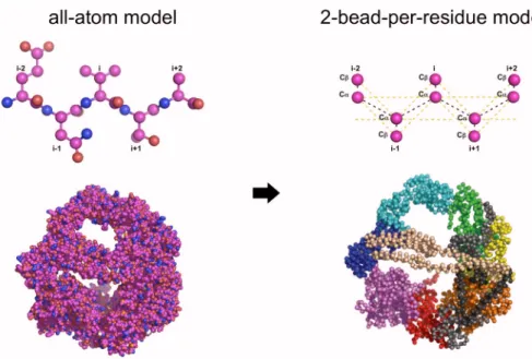
To access time scales of NBD1 folding, we used a simplified protein model that still
maintained important features of the protein such as side-chain packing. Amino acid residues
were modeled as follows: (1) glycines are represented by three beads (-N, C, C); (2)
phenylalanine, tyrosine, tryptophan, and histidine by five beads (-N, C, C, C, C), and (3)
all other residues by four beads (-N, C, C, C) [58]. This protein model has been
successfully employed in studying protein aggregation [58]. In the simulations, we used the
available crystal structures of wild type and mutant NBD1: wild type (PDB ID 2BBO),
F508 mutant (PDB ID 1XMI) and F508A mutant (PDB ID 1XMJ)[59,60]. The missing
loop between E403 and L436 in both wild type and mutant NBD1 is reconstructed using a
loop-search algorithm in SYBYL (Tripos Assoc. Inc, St. Louis, MO).
4.1.2 Simplified interaction using the Go-model and discrete molecular
dynamics
To determine the long-range interaction between the particles in the simplified
protein model, we used the model described in Section 3.1.2. In this particular
Go-model, two residues are said to be in contact if their heavy atoms are within a distance of
4.5 Å. To calculate the evolution of the system, we also used discrete molecular dynamics as
4.1.3 Equilibrium simulations protocol
Using discrete molecular dynamics (Section 3.1.2), long equilibrium simulations at
various temperatures were performed to investigate the equilibrium dynamics of the CFTR
NBD1. The primary objective was to compute the thermal denaturation plot and the heat
capacity of the protein. From these plots, we can compare the thermodynamic stabilities of
wild type and mutant NBD1 domains.
From long equilibrium simulations of 106
time units (tu), we were able to access the
long time-scale dynamics of the CFTR NBD1 in the order of 0.5 ms. Each equilibrium
simulation consumed approximately 300 CPU hours.
4.1.4 Folding simulations protocol
We performed 300 folding simulations for each NBD1-WT, NBD1-F508A, and
NBD1-F508. Starting from fully unfolded chains, the temperature of the system was
progressively reduced to allow NBD1 to fold to its native structure. Folding simulations
proceeded until max ~ 60,000 tu, which was chosen to be longer than the typical folding time
of the studied sequences [61]. A similar criterion was employed in the studies calculating the
folding probability of other proteins [62]. The NBD1 structure in a particular folding run was
considered folded it satisfied the following criteria: (1) its energy was less than or equal to
620 (the energy of the native state), (2) its structure was within 2.5 Å RMSD relative to the
native conformation, and (3) the structure possessed correct topological wiring of the
secondary structure elements.
Folding Probability=Number of successful folding trials Total number of folding trials
To estimate the error in folding probabilities, each folding trajectory was considered a
Bernoulli trial with a binary outcome, folded or unfolded. The variance of this Bernoulli
process was then 2
= p(1p)/n, where p was probability and n was the total number of trials.
4.1.5 Analysis of folding simulations
Identification of metastable folding intermediate states
For each folding trajectory that successfully folded the NBD1, we calculated energy
probability distribution, which is simply the normalized histogram of the energy over the
folding simulation time. The peaks of this normalized energy probability distribution is
indicative of metastable folding intermediate states. To identity the dominant intermediate
states for the wild type and mutants, a sum of multiple Gaussian curves
aiexp
(
xbi)
2ci 2
[
]
i
was fitted to the average energy probability distribution ofsuccessfully folded runs. The parameters ai, bi, and ci were the center, standard deviation and
height of the ith Gaussian curve, respectively.
Structural characterization of intermediate states
Because of the reduction in dimensionality of the folding process when energy was
used as a reaction coordinate, each intermediate state, as defined above, represented an
ensemble of NBD1 structures. To identify the primary structural characteristics of each
intermediate state, we clustered the structures in the corresponding state and calculated the
frequency of contacts formed between pairs of residues. For a particular structure, an nn
when residue i and j were in contact (within 4.5 Å) or 0 otherwise. Dominant contacts
between residue pairs were then determined from the average contact matrix of all the
structures within an intermediate state.
Kinetic accessibilities of the intermediate states and most likely paths
We estimated the probability of transition between states by counting the trajectories
that underwent such a transition. The sum of probabilities of the paths emanating from a
given state was normalized to 1, which physically meant that the system always exited from
its then current intermediate state.
The transition probabilities represented independent conditional probabilities, thus the
probability of a sequence of paths to be taken from the unfolded state to the native state was
estimated by multiplying the probabilities of the traced edges. The sequence of edges
connecting the unfolded and folded state with highest probability was considered the most
likely folding pathway.
4.2 Results
4.2.1 Equilibrium dynamics
To determine the equilibrium dynamics and stabilities of the wild type and mutant
NBD1, we performed equilibrium simulations (106
time units ~ 0.5 ms) of wild type and mutant NBD1 using discrete molecular dynamics (see Methods, Section 4.1.3). From the equilibrium simulations, we calculated the thermal denaturation curve of both NBD1-WT
and NBD1-F508 (Fig. 4.4) and observed two stable thermodynamic states, folded and
unfolded. In agreement with previous experimental studies by denaturation experiments
[10,11], the stabilities of wild type and F508 NBD1 were not significantly different. The slope at the transition temperature of the wild type (Tm ~ 0.68 /kB) was 9.8 103
slope at the transition temperature of the mutant (Tm ~ 0.70 /kB) was 1.6 10 3
kb ( ~ 1–2
kcal/mol and kB is the Boltzman factor; see Section 3.1.2 for further discussion on units).
This shift in slope at the transition temperature indicated a difference in folding cooperativity
of NBD1-WT and NBD1-F508 and therefore a difference in folding kinetics.
Figure 4.4. Thermodynamics of NBD1-WT, NBD1-F508A, and NBD1-F508. Average equilibrium energy was calculated from long equilibrium simulations (106
time units) of NBD1-WT, NBD1-F508A, and NBD1-F508 crystal structures. Error bars represented ±standard deviation. (Inset) The specific heat is calculated as Cv = (E
2 E2
)/T2
4.2.2 Difference in wild type and mutant NBD1 folding propensities
Folding is a stochastic process, thus to investigate in detail the difference in folding
kinetics and dynamics of NBD1-WT and NBD1-F508, we performed 300 folding
simulations on each of the structures (Section 4.1.4). Starting from fully unfolded chains of
NBD1-WTand NBD1-F508, we progressively reduced the temperature of the system to
simulate thermal folding. We found that the folding probability [61] of wild type to be
33 ± 3% while that of the mutant was 13 ± 2%. The ratio of NBD1-WT and NBD1-F508
correlated with the ratio of their folding yields derived from folding experiments. Folding
yields of NBD1-WT was approximately twice that of NBD1-F508 in the temperature range
10°C to 22°C [11]. Folding simulations of our control structure NBD1-F508A yield a folding
probability of 26 ± 4% which was intermediate to that NBD1-WTand NBD1-F508. This
folding probability value was in agreement with experimental studies showing intermediate
folding efficiencies and maturation levels of NBD1-F508A relative to NBD1-WT [10,11].
4.2.3 Folding pathways
To investigate the molecular origin of the difference in folding yields and
probabilities, we mapped the folding pathways of WT, F508A, and
NBD1-F508 by identifying their metastable folding intermediate states. The folding intermediate
states of a folding trajectory were exhibited as peaks in the energy probability distributions
(Fig. 4.5). Thus, dominant intermediate states in the folding pathways were peaks in the
average energy probability distributions (Fig. 4.6). The average energy probability
distributions of wild type and the mutant were significantly different (Kolmogorov-Smirnov
test; P-value<1.410292
), which suggested a significant difference in the folding kinetics of
an intermediate state follows a distinct distribution (Fig. 4.7), thus, an intermediate state
identified using energy as the folding reaction coordinate, forms a distinct collection of
NBD1 conformations.
To determine the difference between the sequence of folding events of the wild type,
F508, and the F508A control, we estimated the probability of transitions between
intermediate states (Fig. 4.8). The difference in transition probabilities of WT,
NBD1-F508, and NBD1-F508A is shown in Fig. 4.9. The transition probabilities showed some
states accessible only to either wild type or mutant NBD1. The difference in state
accessibilities between the two suggested a difference in contact pattern formation
(nucleation events), which could cause the observed difference in folding yields.
We also calculated the most dominant folding pathways in wild type and mutant
NBD1. The most dominant path in wild type follows a sequence of transition
UnfoldedS10S8S7S5S4S1, while the dominant path in the mutant follows the
sequence of transitions UnfoldedS9S8S7S6S4S1. Thus, NBD1-WT and
NBD1-F508 undergo different sequences of folding events.
4.2.4 Structural modulators of folding kinetics
Because of the reduction in dimensionality of the folding process when energy was
used as a reaction coordinate, each intermediate state represented an ensemble of NBD1
structures. To identify the primary structural characteristics of each intermediate state, we
clustered structures in the corresponding state and calculated the frequency of contacts
formed between pairs of residues (Fig. 4.11). In all intermediate states, we found the most
notable structural difference between NBD1-WT and NBD1-F508 occured in the S7-H6
Figure 4.9. Comparison of the folding pathways of wild type NBD1 and its mutants.
Shown are the difference transition probabilities between (A) NBD1-F508 and NBD1-WT
and between (B) NBD1-F508 and NBD1-WT. Blue edges denote transitions dominant in
interacted with F587 in the mutant but not in wild type (Fig. 4.11). This pattern of contact
formation reflected the difference in NBD1-WT and NBD1-F508 crystal structures that are
embedded in the interactions defined according to structure. Additionally, residue pairs that
had similar interactions (i.e., attractive or repulsive) in the wild type and mutant crystal
structures still exhibited different contacts in the folding intermediate states. These results
showed that the pattern of transient contact formation in the wild type was also perturbed by
Phe508 deletion. This class of residue pairs included Q525/E585 and C524/I586.
4.2.5 Computational rescue of NBD1-
F508
To verify that the identified contact pairs (Q493/P574 and F575/F587) found in the
S7-H6 loop were indeed critical in the kinetics of NBD1, we reverted their interactions in
NBD1-F508 to their interactions in NBD1-WT and performed folding simulations. In the
case of the Q493/P574 pair, the residues were in close proximity in NBD1-WT but not in
NBD1-F508, thus we changed the interaction between Q493 and P574 in NBD1-F508
from repulsive to attractive to mimic a possible rescuing mutation. Folding simulations of
“rescued” NBD1-F508 yielded a folding probability of 19 ± 2% (Table 4.1). On the other
hand, residues F575 and F508 were in close contact in NBD1-F508 but not in NBD1-WT,
thus we reverted their interaction in NBD1-F508 from attractive to repulsive. Folding
simulations of the second “rescued” NBD1-F508 yielded a folding probability of 20 ± 2%.
These folding probabilities of the two “rescued” NBD1-F508s were higher than the
13 ±2% folding probability of the original NBD1-F508, supporting our findings that the
contacts between Q493 and P574 and between F575 and F587 were indeed critical to NBD1
Figure 4.11. Contacts in NBD1-WT that perturbed in the F508A and F508 mutants.
Difference between average contact frequencies of structures within intermediate states showed malformed contacts in NBD1-F508 (green) compared to NBD1-WT (blue). These identified malformed contacts in the mutants were critical determinants of NBD1 folding kinetics. In particular, P574 interacted with Q493 in wild type but not in the mutant. Also, F575 interacted with F587 in mutant but not in wild type. Redesigning these contacts to their wild type interactions in the F508 background can potentially rescue NBD1-F508.
Table 4.1. Computational rescue of NBD1-F508. To computationally rescue the
F508, we forced the loop S7-H6 of the mutant to its wild type conformation. These
constructs (shown in gray) exhibit higher folding probability than the original NBD1-F508.
4.3 Summary
Deletion of a single residue, Phe508 in CFTR is present in approximately 90% of
cystic fibrosis (CF) patients. Experiments showed that this mutant protein exhibited
inefficient biosynthetic maturation and susceptibility to degradation probably due to
molecular dynamics simulations of NBD1-WT, NBD1-F508A, and NBD1-F508, we
showed that the deletion of Phe508 indeed altered the kinetics of NBD1 folding. We also
found that the intermediate states appearing on wild type and mutant folding pathways were
populated differently and that their kinetic accessibilities were distinct [13].
We also identified critical interactions not necessarily localized near position 508,
such as Q493/P574 and F575/F587, to be significant structural elements influencing the
kinetic difference between wild type and mutant NBD1. Forcing these locations to adopt wild
type conformations, at least from simulations, rescues the aberrant folding kinetics of the
Chapter 5
Structure of the complete CFTR channel
In this chapter, we investigate the second aspect of the defect associated to the
Phe508 deletion, which is that of the misassembly of the whole protein. To determine the
origin of the misassembly, it is essential to know where the residue Phe508 is located in the
context of the whole protein and identify the specific set of domain-domain interactions that
it mediates. We constructed a complete model of the CFTR protein, partly from homology
and partly from ab initio protein folding. The model predicted, and verified with extensive
biochemical experiments in our collaborating laboratory, that Phe508 mediates a crucial
interaction between the cytoplasmic and membrane-spanning domains of the CFTR channel.
Identification of this crucial interface is important in the targeted rational design of drugs that
can rescue the protein.
5.1 Methods and Models
5.1.1 Modeling the CFTR structure from Sav1866
CFTR consists of several domains: nucleotide-binding domains NBD1 and NBD2,
There exist crystal structures of NBD1 but none for the other domains. The NBD1-NBD2
dimer was constructed by superimposing the NBD1 crystal structure and the homology
model of NBD2 [63] on the Sav1866 (PDB ID 2HYD) structure [64]. The conformations of
the NBD1-NBD2 dimer agrees with the inter-NBD cross-links observed by Mense et al. [65].
We modeled the membrane spanning domains of CFTR using homology modeling
(Section 2.2.1). Because both CFTR and Sav1866, an ABC bacterial multidrug transporter,
contain 12 transmembrane helices that are of similar length, we opted to model the CFTR
membrane-spanning domains from that of Sav1866. The alignment of Sav1866 and CFTR
was dictated by the position of their corresponding membrane-embedded regions and the
conserved coupling helices in the intracellular loops (Fig. 4.1B). The membrane-embedded
regions of the Sav1866 helices were identified from the PDB_TM database [66], whereas the
approximate locations of CFTR TM helices were defined by using the results from earlier
glycosylation site insertions [67] and the HMMTOP transmembrane prediction server
(www.enzim.hu/hmmtop) [68]. Using the CFTR-Sav1866 alignment, the atomic model of
CFTR MSDs was constructed in the Homology suite of INSIGHTII (Accelrys, Inc.). To
eliminate clashes and refine the model, the side-chain rotamer states were optimized, and
minor backbone fluctuations were introduced by using Medusa [69].
The structural model is consistent with available experimental data on the orientation
and packing of transmembrane helices. Pairs of residues such as M348C/T1142C,
T351C/T1142C, and W356C/W1145C, which come from transmembrane helices TM6 and
TM12, could be cross-linked by molecules of different lengths [70]. Cross-linking between
I340C and S877C also exists [71]. In our model, these residue pairs were closer than
(TM8) [72], which in the model face each other directly and their C atoms are separated by
9 Å (Fig. 5.2). Likewise, Therien et al. [73]found that TM3 and TM4 form anti-parallel
helices and that Q207 (TM3) and V232 (TM4) form a hydrogen bond between them,
suggesting that this pair of residues is structurally close. In the model, Q207 and V232 side
chains directly face each other (Fig. 5.2(A)).
Aside from the experimental constraints described in the main text that were satisfied
by the structural model, the organization of the membrane-spanning helices also agreed with
studies identifying water-accessible residues along the channel pore [74,75]. Akabas et al.
[74] found that residues G91, K95, and Q98, which are located in TM1, are accessible to
water-soluble MTS reagents, which implies that these residues line the CFTR pore.
Fig. 5.2(B) shows that indeed these residues face the pore lining in the current model.
Another study by the same group [75] found that residues I331, L333, R334, K335, F337,
S341, R347, T351, R352, and Q353 (all positioned in TM6) are on the water-accessible
surface of the protein. These residues in TM6 are shown to face the CFTR pore lining, which
makes them accessible to water (Fig. 5.2(B)).
To identify the ensemble of conformations dynamically accessible to the R domain,
Dr. Tamas Hegedus (UNC-CH Department of Biochemistry and Biophysics) and I performed
ab initio folding of the R-domain and generated decoys of low-energy structures by using
discrete molecular dynamics (Section 3.1.2) with an all-atom force field called Medusa [69].
We clustered the decoy set to determine putatively dominant conformations of the R domain.
The centroid of the largest decoy set is docked to the CFTR homology model by using
desolvation energy, and electrostatics. In docking the R domain model, we imposed the
constraint that the C terminus of the R domain is close to the N terminus of MSD2.
Figure 5.2. Experimental constraints satisfied in the membrane-spanning domains of
the homology model. (A) Cross-links can be formed between M348-T1142, T351-T1142,
and W356-T1145 (red), which are pairs of residues found between TM6 and TM12 [70]. R347 (TM6) forms a salt bridge with D924 (TM8) (blue) (11). A H-bond can be formed between Q207 (TM3) and V232 (TM4) (yellow) [73]. A recent constraint from cross-linking of I340C-S877C (cyan) is also satisfied [70]. (B) Residues G91, K95, and Q98 (colored red) in TM1 are water-accessible, suggesting that they face the channel pore [74]. I331, L333, R334, K335, F337, S341, R347, T351, R352, and Q353 (colored blue), which are all found in TM6, are also water-accessible [75].
5.2. Results
We constructed a 3D structure of CFTR by molecular modeling (see above).
Full-length ABC proteins (the protein family to which CFTR belongs) can be grouped into two
classes according to the number and conformation of their transmembrane helices. Bacterial
importers have variable numbers of helices that are short, positioning their NBDs close to the
membrane plane. The exporters such as Sav1866 possess 12 transmembrane helices that are
longer than those of the importers, thus their NBDs are farther from the membrane plane.
those of Sav1866 [64,78,79], which suggested that CFTR MSDs can be modeled from those
of Sav1866. To organize the different domains of CFTR, we followed the tertiary
organization of the Sav1866 domains. The structural model is consistent with available
experimental data on the orientation and packing of CFTR transmembrane helices (Fig. 5.2).
The complete structural model of CFTR is shown in Fig. 5.3. It exhibits the
characteristic domain swapped architecture of Sav1866 whereby one MSD sits on both
NBDs. This characteristic topology also predicts that the Phe508 residue is in contact with
the cytoplasmic loop 4 (CL4) of the MSD2. The preponderance of other disease-associated
mutations in CL4 that are sensitive to CFTR misassembly suggest that indeed this interface is
crucial the assembly of the whole protein.
To verify that indeed the predicted interactions are correct, our experimental
collaborators, Dr. John R. Riordan and company (UNC-CH Department of Biochemistry and
Biophysics) performed chemical cross-linking (Fig. 5.5). Cross-linking can verify whether
two residues are spatially close as predicted by the model. This method involves mutating
the two residues in question to cysteines in a Cys-less CFTR background (see Fig 5.5). The
two cysteines are then induced to form a disulfide bond using bifunctional
methane-thiosulfonate (MTS) reagents. If the residues successfully cross-link, the cross-linked species
can be detected by a shift of a band in a western blot (see Fig 5.5). Using this methodology,
we indeed found that Phe508 plays a central role in this interface because it can be
cross-linked to cysteines introduced at many positions in CL4. These positions include Leu-1065,
We also showed that the interface mediated by the Phe508 with the CL4 of MSD2 is
crucial for channel function. Single-channel gating, which persists after the introduction of a
Cys pair at each interface, was completely inhibited by cross-linking (Fig. 5.7). In both cases,
this inhibition was completely reversed on reduction with DTT. Hence, these points of
contact are integral elements of the structure, and covalent coupling between residues on
either side restricts channel activity. This restriction is unlikely to be caused by prevention of
signal transmission per se but probably reflects the restriction of dynamics at the interfaces.
5.3. Summary
We constructed a model of the whole CFTR molecule to determine the overall
topology of the protein. More importantly, the model identified the location of the Phe508
residue, whose deletion has been known to induce misassembly of the whole CFTR complex.
We found Phe508 to mediate a crucial interaction between the NBD1 and the cytoplasmic
loop 4 (CL4) of MSD2. This architecture explains the sensitivity of Phe508 and CL4 to
mutations that also affect the maturation (presumably due to domain misassembly) of the
whole CFTR. These interactions between NBD1-CL4 and NBD2-CL2, and other
MSD-NBD interfaces, have been validated experimentally. We likewise showed that these
interfaces are crucial to the channel function since cross-linking of cysteines on either side of
the interface arrests channel gating, indicating a dynamic interface. The precise
identification of the interface perturbed upon the deletion of the Phe508 provides a focused
Figure 5.5. Cross-linking schema.(A) To verify if two residues are spatially close, they are mutated to cysteines and then cross-link using a bifunctional methane-thiosulfonate (MTS)
reagent. The disulfide bond can be removed by adding a reducing agent (DTT). (B) The