Perioperative Management of Adult Patients With External
Ventricular and Lumbar Drains: Guidelines
From the Society for Neuroscience in Anesthesiology
and Critical Care
Abhijit V. Lele, MBBS, MD, MS,
*
Amie L. Hoefnagel, MD,
w
Nina Schloemerkemper, MD, Dr. med., FRCA,z
David A. Wyler, MD,
y
Nophanan Chaikittisilpa, MD,
8
Monica S. Vavilala, MD,
z
Bhiken I. Naik, MBBCh,#
James H. Williams, MD, PhD,
**
Lakshmikumar Venkat Raghavan, MBBS, MD, FRCA, FRCPC,
ww
and
Ines P. Koerner, MD, PhD,zz
Representing SNACC Task Force for Developing Guidelines for
Perioperative Management of External Ventricular and Lumbar Drains
Abstract: External ventricular drains and lumbar drains are commonly used to divert cerebrospinal fluid and to measure cerebrospinal fluid pressure. Although commonly encountered in the perioperative setting and critical for the care of neuro-surgical patients, there are no guidelines regarding their man-agement in the perioperative period. To address this gap in the literature, The Society for Neuroscience in Anesthesiology & Critical Care tasked an expert group to generate evidence-based guidelines. The document generated targets clinicians involved in perioperative care of patients with indwelling external ven-tricular and lumbar drains.
Key Words:external ventricular drain, ventriculostomy, lumbar drain, guidelines, perioperative, management, leveling, trans-port, checklist, competency
(J Neurosurg Anesthesiol2017;00:000–000)
E
xternal ventricular drains (EVDs) and lumbar drains (LDs) are temporary devices placed into the lateral ventricles of the brain and lumbar subarachnoid space, respectively, to facilitate external cerebrospinal fluid (CSF) drainage and to monitor CSF pressure. TheirReceived for publication December 22, 2016; accepted December 27, 2016.
From the Departments of *Anesthesiology & Pain Medicine, Neurocritical Care Service;8Anesthesiology and Pain Medicine;zAnesthesiology and Pediatrics, Harborview Injury Prevention and Research Center (HIPRC), Harborview Medical Center, University of Washington, Seattle, WA;
wDepartment of Anesthesiology, University of Florida, UF Health, Jacksonville, FL;zDepartment of Anesthesiology & Pain Medicine, University of California Davis Medical Center, Sacramento, CA;yJefferson Hospital of Neuroscience, Sidney Kimmel Medical College, Thomas Jefferson University, Philadelphia, PA; #Department of Anesthesiology & Neurosurgery, University of Virginia Health System, Charlottesville, VA; **Department of Anesthesiology, University of North Carolina at Chapel Hill, N2201 UNC Hospitals, Chapel Hill, NC;wwDepartment of Anesthesia and Pain Medicine, Toronto Western Hospital, Toronto, ON, Canada; andzzDepartment of Anesthesiology & Perioperative Medicine, Neurosciences ICU, Oregon Health Sciences University, Portland, OR.
A.V.L.: manuscript design, manuscript write-up, Tables 1 to 6, Figures 1A, 1B, 2A, 2B, Sections 1 to 6, developing clinical competencies for EVD/LD among clinicians involved in perioperative care, continued medical education and following a perioperative checklist for management of EVD/LD, review and approve final manuscript. A.L.H. and N.S: manuscript write-up, Section 1: Introduction, indications and contraindications, review and final approve manuscript. D.A.W.: manuscript write-up, Section 2: Preoperative assessment of patients with EVD or LD, review and approve final manuscript. N.C. and M.S.V.: manuscript write-up, Section 3: Transporting patients with EVD or LD, review and approve final manuscript. B.I.N. and J.H.W.: manuscript write-up, Section 4: Intraoperative management of external ventricular and lumbar drains, review and approve final manuscript. L.V.R. and I.P.K.: manuscript write-up, Section 5: Management of external ventricular drains (EVD)/lumbar drains (LD) in special clinical scenarios, review and approve final manuscript.
A.L.H. has received honoraria from Design Science Consulting Inc.; N.S. has received consulting fee from Mizuho OSI. The other authors have no conflicts of interest to disclose.
Address correspondence to: Abhijit V. Lele, MBBS, MD, MS, Department of Anesthesiology & Pain Medicine, Neurocritical Care Service, Har-borview Medical Center, University of Washington, 325, 9th Avenue, Seattle, WA 98104 (e-mail: [email protected]).
Supplemental Digital Content is available for this article. Direct URL citations appear in the printed text and are provided in the HTML and PDF versions of this article on the journal’s Website, www.jnsa.com.
Copyrightr2017 Wolters Kluwer Health, Inc. All rights reserved.
placement is considered one of the most frequently per-formed procedures in neurologically critically ill pa-tients,1–3 with the majority placed in patients with subarachnoid hemorrhage (SAH), intracerebral hemor-rhage, and obstructive hydrocephalus.4 Although the worldwide incidence of placement of EVD and LD is largely unknown, it is estimated that 500,000 ven-triculostomies were placed in the United States alone between 1988 and 2010.4Since its first placement in 1744, EVDs have undergone numerous changes in materials, techniques, and indications.5–7
EVDs are commonly encountered in perioperative care by clinicians, specifically the anesthesia providers who might have limited experience in their management. Furthermore, mismanagement of EVDs can have cata-strophic consequences. Despite their importance, there are currently no guidelines for the perioperative man-agement of EVD and LD.
METHODOLOGY
Purpose of the Guidelines
These evidence-based guidelines aim to provide recommendations related to EVD and LD regarding (1) common indications, contraindications, complications, and patient preparation for placement and maintenance; (2) preoperative assessment of patients; (3) transporting patients; (4) intraoperative management including mon-itoring and CSF drainage; (5) management of these drains under special circumstances; and (6) creating a perioper-ative checklist, clinical competency, and continued med-ical education.
Application
These guidelines are intended for the use by clini-cians involved in perioperative care of adult patients with EVDs and LDs.
Task Force Members
The initial concept and design of “Society for Neuroscience in Anesthesiology & Critical Care (SNACC) EVD/LD project” began in November 2015, and an EVD/LD task force was finalized in December 2015. This task force comprised of 10 neuro-anesthesiologists and neurointensivists practicing at aca-demic medical centers across the United States and Canada. These 10 individuals were chosen after an e-mail invitation was sent to all active SNACC members seeking project membership. Applicants were required to have published peer-reviewed neuroscience research or have documented experience in the care of patients with EVD and LD. The task force members agreed on criteria for evidence and then evaluated peer-reviewed studies per-taining to EVD and LD (search strategies described in next section). The document was compiled of 6 sections, with each section equally coauthored by 2 project mem-bers, and was subsequently reviewed and approved by all members of the task force. The completed draft was submitted to the SNACC Board of Directors. After
incorporating the inputs and suggestions from the SNACC Board of Directors, the approved version was placed on the SNACC Web site (http://www.snacc.org) for member review and comments for a period of 1 month, with the final version of the guideline confirmed after incorporating member input.
AVAILABILITY AND STRENGTH OF EVIDENCE
The task force worked with a medical librarian to create a systematic strategy to search PubMed: (external ventricular drain*[tw] OR external ventricle drain*[tw] OR extraventricular drain*[tw] OR extra ventricular drain*[tw] OR ventricular catheter*[tw] OR ventricular access device*[tw] OR lumbar drain*[tw] OR cere-brospinal fluid drain*[tw] OR csf drain*[tw]) OR (ven-triculostom*[tw] AND (drain*[tw] OR catheter*[tw])). The format of this search was adapted for Embase through Elsevier. Search results were limited to journal articles and conference papers published in English and last updated on December 26, 2016. The total search re-sults (7936 references) were downloaded to Endnote. After 2729 duplicates were removed, 5207 articles were imported to a Covidence database for team review. Au-thors had previously identified 119 additional articles, and 91 articles were found by the authors including checks of reference lists as the review progressed.
Several rounds of screening were conducted, with review of each of the titles and abstracts from the original search by 2 reviewers. Inclusion criteria were all study types in adults, and exclusion criteria were nonhuman studies, laboratory investigations, and pediatric literature. References addressing infectious complications and pre-vention strategies published before the 2015 Neurocritical Care Society (NCS) guidelines8and those addressing CSF drainage for spinal cord protection published before the 2010 ACC guidelines9were eliminated. Ultimately a pool of 646 references were identified and organized by topic for the individual section authors to draw upon in con-struction of the guidelines.
We used the American Heart Association methodo-logy for the level of evidence for each recommendations proposed10(Table 1).
SECTION 1: INTRODUCTION TO EVDs AND LDs
Common Indications for Placement of EVDs
and LDs
EVDs function as intracranial pressure (ICP) monitors and as conduits for external CSF diversion. LDs, in contrast, function primarily as a conduit for ex-ternal CSF drainage, and are not used for ICP monitor-ing. Although both parenchymal ICP monitors and EVDs provide reliable and accurate ICP data, EVDs are preferred in patients with hydrocephalus.11
Placement of EVDs and LDs
EVDs are frequently inserted emergently at bedside rather than in the operating room (OR).47,48 The frontal horn of the right lateral ventricle is the preferred destination as this is assumed to be in the nondominant side for speech and language in most patients.47,49 Although different techniques for EVD placement have been described in the literature,3,50–52 anatomic landmarks are frequently used and the drain is placed free handed.49,51,53–57
LDs are placed in a manner similar to a lumbar puncture, epidural, or intrathecal catheter placement.58 Placement of an LD may be done preoperatively in an awake patient, or after the induction of anesthesia. Co-agulation profile and anticoCo-agulation medications should be reviewed and anticoagulants held per current Ameri-can Society of Regional Anesthesia and Pain Medicine (ASRA) guidelines (refer to Supplement 1, Supplemental Digital Content 1, http://links.lww.com/JNA/A46). The patient is placed in the lateral decubitus or sitting posi-tion. Using strict aseptic technique, the lumbar catheter is typically inserted at the L3-L4 or L4-L5 level through a 14-G Tuohy needle. A special, kink-resistant, flexible catheter is passed through the Tuohy needle into the in-trathecal space.58 The catheter with the guidewire is threaded approximately 5 to 8 cm past the needle into the intrathecal space and secured with a clear dressing. It should be stressed that the catheter should thread easily, like an epidural catheter. If resistance is felt, or the catheter is unable to be placed, both the needle and catheter should be removed as a single unit to prevent
inadvertent shearing of the catheter. The catheter should be flushed with 10 mL sterile saline flush and connected to a sterile pressure monitoring kit and a closed collection system. The transducer should not be connected to any pressured flushing system. When connecting the trans-ducer to a collection system ensure a continuous fluid column. This can be achieved by allowing CSF to drain back to the stopcock at the transducer. Bloody CSF or aspiration of blood during placement requires avoidance of anticoagulation for 24 hours.
Optimizing Patients Before Placement of EVDs
or LDs
Contraindications to placement of EVD or LD in-clude coagulopathy and infection at the entry site. LD also needs careful screening to rule out non-communicating hydrocephalus and large intracranial mass lesions.24The ASRA guidelines on anticoagulation should be consulted to determine appropriateness of placement and removal of the LD.59 It is generally ad-vised to diagnose and promptly correct coagulopathies before placement of either monitor.8,60
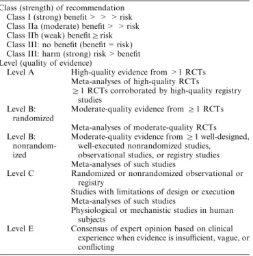
TABLE 1. Summary of Class (Strength) of Recommendation
and Level (Quality) of Evidence
Class (strength) of recommendation Class I (strong) benefit > > > risk Class IIa (moderate) benefit > > risk Class IIb (weak) benefitZrisk Class III: no benefit (benefit = risk) Class III: harm (strong) risk > benefit Level (quality of evidence)
Level A High-quality evidence from >1 RCTs Meta-analyses of high-quality RCTs
Z1 RCTs corroborated by high-quality registry studies
Level B: randomized
Moderate-quality evidence fromZ1 RCTs
Meta-analyses of moderate-quality RCTs Level B:
nonrandom-ized
Moderate-quality evidence fromZ1 well-designed,
well-executed nonrandomized studies, observational studies, or registry studies Meta-analyses of such studies
Level C Randomized or nonrandomized observational or registry
Studies with limitations of design or execution Meta-analyses of such studies
Physiological or mechanistic studies in human subjects
Level E Consensus of expert opinion based on clinical experience when evidence is insufficient, vague, or conflicting
RCT indicates randomized controlled trial.
TABLE 2. Major Indications for Placement of External
Ventricular and Lumbar Drains
External ventricular drains
Acute symptomatic hydrocephalus Aneurysmal SAH12–14
ICH and IVH with decreased level of consciousness15
Acute ischemic cerebellar stroke in concurrence with decompressive craniectomy16,17
ICP monitoring in TBI
TBI with postresuscitation GCS of 3-8, and abnormal CT scan defined as one with hematomas, contusions, swelling, herniation, or compressed basal cisterns18–20
Severe TBI with a normal CT scan ifZ2 of the following features are noted on admission (age over 40 y, unilateral or bilateral motor posturing, or SBP < 90 mm Hg18,19
Management of patients with intracranial hypertension after TBI21,22
Malfunctioning or infected ventriculoperitoneal shunts, and other neurological emergencies occurring due to infective and neoplastic diseases23–26
Facilitation of intraoperative brain relaxation27,28
Targeted therapeutic interventions
rTPA in patients with IVH29,30(efficacy and safety uncertain) and in
patients with SAH31,32
Treatment of vasospasm after aneurysmal SAH33–35
Antibiotics in management of central nervous system infections36,37 Lumbar drains
Acute symptomatic hydrocephalus in SAH12
Spinal cord–protective strategy in open and endovascular thoracic aortic repair for patients at high risk of spinal cord injury9,24,38–40
Active CSF leak (due to craniofacial trauma)41or those at risk for
CSF leak during skull base procedures42–44; however, lumbar drains
do not prevent postoperative CSF leaks44,45
Facilitate intraoperative brain relaxation27and intraoperative
exposure46
Complications Associated With EVDs and LDs
Complications associated with EVD placement are common61 requiring revision in 10% to 22% of cases.47,62 Table 3 highlights complications associated with EVDs and LDs. Although hemorrhage, infections, and overdrainage of CSF are the most recognized com-plications, clinicians involved in perioperative care must be familiar with other possible complications.
Hemorrhagic Complications Associated With EVD
and LD
There are 2 main risk factors for hemorrhagic complications: (1) coagulopathy and (2) overdrainage of CSF. Factors such as cerebrovascular disease, size of catheter,107 use of antiplatelet agents,108 and INR > 1.6,109 place patients at risk for ventriculostomy-associated hemorrhage. Although most of these bleeds are clinically insignificant,2,110,111 they can be potentially devastating.8,66 Removal of EVD also poses risk for hemorrhage. In a retrospective study of 482 EVDs by Miller and Tummala,66hemorrhage was seen in 22.5% of those patients who underwent post-EVD neuroimaging. Factors associated with hemorrhage included bedside placement of EVD. Interestingly, the investigators were unable to demonstrate impact of INR value, platelet count, and antiplatelet agents on incidence of hemor-rhage. Proposed mechanisms of hemorrhage associated with removal of EVD include: injury to and release of any tamponade effect on a small vessel, tracking of scalp bleeding along EVD track, and possible adherence of EVD to choroid plexus that may contribute to bleeding on removal. Majority of the hemorrhages in this series were small and asymptomatic.66
Hemorrhagic Complications Associated With
Chem-ical Prophylaxis Against Venous Thromboembolism.
Contrary to popular concern, chemical prophylaxis against venous thromboembolism started within 24 hours of admission and therapeutic heparinization initiated within 24 hours of placement of the EVD does not in-crease bleeding risk.112,113 The incidence of hemorrhage during removal of EVD can be higher than during placement,66and similar indices of coagulopathy must be maintained before removal of EVD.
Placement of LD in Patients Requiring Systemic
Anticoagulation. Placing a LD in an anesthetized patient
is safe, even in patients requiring subsequent heparin-ization and cardiopulmonary bypass.74,114 The risk of hematoma and neurological injury is rare, and can be minimized by following certain guidelines, that is, delay-ing surgery 24 hours in the event of a traumatic tap (blood freely aspirated), delaying heparinization for >60 minutes after catheter insertion, and maintaining tight perioperative control of anticoagulation.115 Neu-raxial hematoma associated with placement or removal of a LD is a rare but potentially serious complication. The reported incidence of surgical decompression required after epidural catheterization varied between 1/22,189 and 1/4330 in a cohort of 62,450 nonaortic surgery pa-tients.116 Of note, 4 of the 7 patients who developed a neuraxial hematoma had perioperative anticoagulation management deviated from current ASRA guide-lines.116,117
There are limited data on the incidence of neuraxial hematoma associated with LDs during aortic surgery re-quiring systemic anticoagulation, but has been reported as between 0% and 4%.67,72,73
A summary of the ASRA guidelines pertinent to LDs117and NCS consensus statement8on management of EVD can be found in the supplement document attached to the guidelines (refer to Supplement 1, Supplemental Digital Content 1, http://links.lww.com/JNA/A46).
Infectious Complications
Infections associated with EVD (0% to 28%) and LD (0% to 50%) are among the most serious of com-plications.8,69–75 Factors associated with increased in-fection risk include nontunneled catheters, nonsterile conditions, intraventricular hemorrhage, frequent sam-pling, irrigation of catheters, and longer in situ dura-tion.70,118 Adherence to an aseptic technique, that is, cleaning the insertion site using an antimicrobial agent per local antibiogram, and using a dressing as a part of a management bundle, institution of pre-EVD-insertion antibiotics, using antimicrobial-impregnated catheters whenever possible,8,62,119–125 avoiding routine CSF sam-pling, and limited manipulation of the CSF collecting system, all feature in the recommendations provided in the recently published NCS consensus statement on EVD insertion and management.8To limit potential for
Clos-tridium difficile diarrhea and antimicrobial-resistant
or-ganisms, as well as lack of efficacy, antibiotics are not routinely recommended for the duration of the EVD.8
Although there are no guidelines or consensus statements regarding intraoperative periprocedural
TABLE 3. Complications Associated With EVD and LD
Hemorrhage
Intracerebral hemorrhage, tract hematoma, or tract hemorrhages (0%-41%)1,8,63–66
Neuraxial hematoma (0%-3.2%)67 Neural injury68
Infection (0%-28% EVD, 0%-50% LD)8,69–75
Malposition2,76
Occlusion and malfunction77–79
Overdrainage of CSF
Subdural or epidural hematoma80–83
Rebleeding from a ruptured cerebral aneurysm84
Intracranial hypotension85–87
Cerebellar tonsillar herniation79,88–91 Paradoxical herniation92
Pneumocephalus79,93
Iatrogenic vascular injury (arteriovenous fistula, cerebral pseudoaneurysm)94
Fracture of catheters,95with retained fragment of catheter96,97
Inadvertent injections of drugs into EVDs98–105
Postdural puncture headache106
administration of antibiotics before LD placement for aortic and nonaortic surgery, this task force recommends following standards such as those used for EVD insertion and management.
Summary
Although there are many indications for the place-ment of EVD or LD, they are critical to monitor intra-cerebral or intraspinal pressure (ISP). Thorough knowledge of indications, contraindications, complica-tions, and cautions associated with the monitoring mo-dality, along with strict adherence to local, national, and international standards will likely enhance patient safety. Prevention of Hemorrhagic and Infectious Com-plications Associated with EVDs and LDs (Class of Recommendation and Level of Evidence):
(1) Before insertion of EVD and LD, prompt diagnosis and correction of coagulopathy utilizing institutional practice guidelines is recommended (Class I Recom-mendation; Level of Evidence E).
(2) Perioperative anticoagulation management with LD placement and removal during aortic or nonaortic surgery should be performed within the framework of the current ASRA guidelines (Class I Recommenda-tion; Level of Evidence E).
(3) Antibiotics should only be administered before place-ment of an EVD and LD with the choice based on institutional practice (Class I Recommendation; Level of Evidence E).
(4) It is recommended to practice strict aseptic technique based on national and institutional guidelines (Class I Recommendation; Level of Evidence E).
SECTION 2: PREOPERATIVE EVALUATION OF
PATIENT WITH EVD AND LD
Background
A thorough preanesthetic evaluation of patients with EVD and LD is critical for optimal perioperative care. Table 4 describes components of history, physical examination, laboratory, and imaging data that should be incorporated into the preanesthetic assessment.
Parts of evaluation that are unique to EVDs include reviewing indication for placement, relevant history, medications given, ICP trends, qualitative evaluation of components of ICP waveforms, and any data available from EVD clamp trials. P1:P2 waveform evaluation would be significant to understand ICP compliance curve, and EVD clamp trial data would be significant to un-derstand impact of EVD clamping on ICP during patient transport.
Inspection of the EVD or LD System
Inspection of EVD and LD system must be per-formed to provide information regarding (1) integrity of the system, (2) color and consistency of CSF, and (3) leveling and zeroing of the transducer system.
Baseline color and consistency of CSF, as well as presence of air bubbles or debris should be noted in the
catheter and the burette (rather than the collecting bag), and sudden change in color of CSF at any given time deserves attention (see Section 5 for further details).
EVDs are leveled at the external auditory meatus using either a Carpenter bubble or a laser level. LDs, in contrast, are leveled at the right atrium (phlebostatic axis) or at the lumbar catheter insertion site.
Understanding
“
Drain Dynamics
”
or
“
Setting of
EVD and LD
”
CSF drainage through EVD and LD is performed under controlled conditions to prevent overdrainage. Establishing CSF drain volume goals is an important part of this with a goal of 10 to 20 mL/h as this is the typical hourly CSF production and volume that resides in the ventricular system.126 To avoid overdrainage, bridging vein tear, and ultimately subdural bleed, drainage of EVD/LD >15 to 20 mL in any hour should accompany consultation of a neurosurgeon. The bedside notes should clearly indicate whether the goals are hourly drainage of
TABLE 4. Preoperative Assessment of Patients With External
Ventricular and Lumbar Drain
History Comments
Diagnosis SAH, ICH, IVH, AIS, TBI, skull base surgery, CSF leak
Medications Anticoagulant drugs, antiplatelet drugs Liver or renal disease Associated with coagulopathy Cancer or
hematological disorders
Associated with coagulopathy
Physical examination
Vital signs SpO2, PO2, EtCO2, PCO2, MAP, CPP
ICP data ICP range, ICP waveform, P1:P2 ratio EVD clamp trial results
CSF data Hourly CSF output, output over 24 h Color of CSF (clear, xanthochromia, bloody,
etc.) Multimodal
monitoring data
PbtO2, microdialysis, and autoregulation
studies Focused neurological
examination
GCS, FOUR score, cranial nerve paresis, brainstem reflexes, presence or absence of focal neurodeficits
Inspection of EVD or LD system
Tunneled catheter system
Setting of EVD with reference to zero Leveling of EVD at EAM
Leveling of LD at phlebostatic axis/EAM Laboratory data
Complete blood count
Correct thrombocytopenia
PT, INR, PTT Prompt reversal of coagulopathy Imaging data
CT or MRI findings Site and location of EVD Midline shift, cerebral edema
AIS indicates acute ischemic stroke; CPP, cerebral perfusion pressure; CSF, cerebrospinal fluid; CT, computed tomography; EAM, external auditory meatus; EtCO2, end-tidal carbon dioxide; FOUR, Full Outline of Responsiveness Score;
GCS, Glasgow Coma Score; ICH, intracerebral hemorrhage; ICP, intracranial pressure; INR, international normalized ratio; IVH, intraventricular hemorrhage; MAP, mean arterial pressure; MRI, magnetic resonance imaging; Pbto2, brain tissue oxygenation; PCO2, partial pressure of carbon dioxide; PO2, partial pressure
certain predetermined CSF volume or whether ICP data can drive the drainage volume.
Setting of EVD depends upon indication for placing catheters. In patients with aneurysmal SAH, EVDs are typically set at +20 cm H2O, before clipping or coiling of
ruptured cerebral aneurysm, and EVD setting is lowered to +10 cm H2O after aneurysm repair has been
com-pleted. Although these are arbitrary numbers, clinicians involved in perioperative care must familiarize themselves to their respective institutional practices. Sudden over-drainage of CSF in a patient with unsecured ruptured cerebral aneurysm predisposes to rebleeding due to sud-den wisud-dening of transmural pressure gradient (MAP-ICP) across the aneurysm wall.84A recent survey of US-based neurointensivists and neurosurgeons revealed that most institutions utilize a strategy involving continuously open EVD to enhance CSF drainage in a patient with secured cerebral aneurysm along with a gradual weaning strat-egy.127
In patients with ICH, EVDs are set to provide drainage so that an intraventricular clot does not develop by stasis, and thus avoids blocking ventricular system passages and egresses that lead to noncommunicating hydrocephalus and impending herniation.128
Results of Clamping Trials
EVDs and LDs are routinely clamped during change in patient positioning, such as occurring during turning patients in the intensive care unit (ICU) and getting patients out of bed to chair or during ambulation, and remains the recommended standard of practice.129
Tolerance of patients to any period of clamping depends upon the primary reason for placement of EVD or LD, ICP trends, and dependency on external CSF di-version. Patients at risk for clamp failure include those dependent on external CSF diversion, such as occurring with acute hydrocephalus, and in situations of elevated ICP.8
The preoperative evaluation of all patients with an indwelling EVD/LD should also include clinical (wor-sening headache, depressed level of consciousness, cranial nerve deficits, ICP elevation) and radiographic findings (worsening hydrocephalus) that confirm clamping trial intolerance.
Although clamping trials are performed periodi-cally, they are not standardized, and initiation and frequency of such trials vary in different institutions. It is imperative that any clamping trial data be sought for and documented in the preoperative evaluation to ensure that clamping can be safely done during patient transport to and from the OR, and in the OR.
Importance of ICP and Multimodality
Monitoring Data
Often, ICP monitoring and CSF drainage is part of a multimodal monitoring plan, which includes brain tis-sue oxygenation, brain temperature, and continuous electroencephalography.
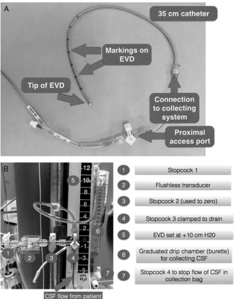
The analysis of the continuous ICP waveform can give indications of cerebral dysfunction.130 Normal ICP waveform has 3 components: P1 (percussion wave), P2 (tidal wave), and P3 (dicrotic wave). The P1 wave is the tallest and the sharpest wave, and results from arterial pressure being transmitted from the choroid plexus. The P2 wave follows P1, and is usually 80% as tall as the P1 wave, and correlates to brain compliance. The P3 wave is caused by closure of the aortic valve (Fig. 1A).
Certain patients may have an abnormally high P2 waveform (Fig. 1B) demonstrating poor compliance. In this case, the compensatory mechanism defined by the compliance curve in the Monro-Kellie doctrine may be exhausted at a lower ICP than expected. Certain patients may have an abnormally high P2 waveform demonstrat-ing poor compliance requirdemonstrat-ing therapy. In this case, the compensatory mechanism defined by the compliance curve in the Monro-Kellie doctrine may be exhausted at a lower ICP than expected. In the noncompliant brain, this reflection is stronger than the initial energy pulse P1, whereas a normally compliant brain will absorb the en-ergy generating a less intense P2 response. Dampened waveform can be observed in patients with cerebral vas-ospasm, postcraniectomy, and other skull-fusion defects. The preoperative evaluation of all patients with an indwelling EVD/LD should include recent ICP values, trends, indices, and relationship with CPP and other multimodality values made available.
Summary
To safely conduct an anesthetic on a patient with CNS injury or risk thereof, a thorough preoperative evaluation should include all important details of the EVD and LD management. Communication with the intensive care/ward staff and a thorough investigation of
FIGURE 1. A, Normal ICP waveforms. P1 percussion wave,
the ICP and other multimodal monitoring data and EVD/ LD drain settings is paramount.
Preoperative Assessment of Patients with EVDs and LDs (Class of Recommendation and Level of Evidence): (1) A thorough preoperative evaluation should be per-formed in all patients with an indwelling EVD and LD that includes a focused history and physical exam (Class I Recommendation; Level of Evidence E). (2) Recommended preoperative evaluation of all patients
with an indwelling EVD and LD should include all of the following (Class I Recommendation; Level of Evidence E).
(a) CSF color and consistency.
(b) ICP values, ICP trends, autoregulation indices, and relationship with CPP and other multimodal monitoring data.
(c) Clinical (worsening headache, depressed level of consciousness, cranial nerve deficits, ICP elevation) and radiographic evidence of clamp trial intoler-ance (worsening hydrocephalus).
(3) Incorporate all information pertinent to the EVD and LD into a standardized preoperative handoff between ICU/ward providers and anesthesia providers (Class I Recommendation; Level of Evidence E.)
SECTION 3: TRANSPORTING PATIENTS WITH
EVDs
Introduction
Neurocritically ill patients with indwelling EVDs frequently require transport from the ICU to other sites for diagnostic and/or therapeutic procedures. These pa-tients may be at risk of intracranial complications such as high ICP during intrahospital transport (IHT) because of direct patient movement and stimulation and/or discontinuation of ICP treatment. However, change in patient position can also lead to CSF overdrainage and result in complications such as rebleeding of intracranial aneurysm,131–133subdural hemorrhage from disruption of bridging veins,80–82 and reverse brain herniation.92 Anesthesiology providers are often involved in the transport of these patients to and from the ICU and to angiography and/or to and from the OR. There are no guidelines regarding EVD management during IHT.
Background
The majority of guidelines for the transport of critically ill patients, including those with ICP monitoring lack recommendations specific to EVD manage-ment.134–138 The American Association of Neuroscience Nurses Guideline recommends routine clamping of EVD before and during IHT to prevent CSF overdrainage but does not address ICP monitoring or documentation during IHT.129
The lack of recommendations regarding EVD management is problematic because published studies document several complications associated with IHT of neurocritically ill patients, including unwanted alteration
in systemic blood pressure, respiration, and neurological conditions.139–142 Andrews and colleagues prospectively observed 50 IHTs of patients with traumatic brain injury (TBI) who underwent computed tomography (CT) scan-ning, magnetic resonance imaging, and transport to the OR. Investigators reported high ICP as the most common secondary insult (16%) during IHT.143 Picetti et al144 conducted a prospective observational study of 160 neu-rocritically ill patients undergoing 288 CT transports; 32% of these IHTs were associated with EVDs. Although ICP was monitored in only 32 of the 127 patients with pretransport ICP monitoring, the incidence of ICP > 20 mm Hg was high (66%). However, neither of these 2 studies distinguished between ICP monitoring types. Moreover, in a case series of 7 patients with indwelling EVDs who were transported for CT, there was a 27% increase in average ICP from the initial value, and the highest ICP noted during CT exam was 35 mm Hg. Kleffmann et al145 recently published a prospective ob-servational study of 56 IHTs to CT of 43 patients with ICP monitoring (50% were EVD). The authors reported an 85% increase in average ICP from baseline during CT scan and ICP therapy was required in 26% of IHTs. Recently, Chaikittisilpa et al146 reported on the largest series of 178 IHTs among 19 neurocritically ill cere-brovascular patients whose EVD was clamped before IHT. They reported that 12% of IHTs were associated with post-IHT high ICP.21ICP complications were only observed among IHTs of patients who had an open EVD setting in the ICU before transport. Pre-IHT ICP values 15 to 19 mm Hg (odds ratio, 3.4 [1.08-10.76]), pre-IHT ICP values Z20 mm Hg (odds ratio, 12.94 [4.08-41.01]), IHT for therapeutic procedure (odds ratio, 5.82 [1.76-19.19]), and high hourly CSF output (odds ratio for every mL/h, 1.11 [1.01-1.23]) are risk factors for ICP-related complications during IHT.
Although these guideline focuses on perioperative management, the transport recommendations should be applicable to all IHTs. Personnel accompanying patient during transport should be trained and competent in management of intracranial hemodynamic perturbations such as intracranial hypertension and cerebral hypo-perfusion.
Summary
Best evidence from observational studies suggests that neurocritically ill patients with indwelling EVDs are at risk of intracranial hypertension during IHT. Routine clamping of EVD for IHT may predispose the patients to intracranial complications, particularly in patients with open EVD status before IHT, those with pretransport ICP >15 mm Hg, and high hourly CSF output in the ICU. Patients with LDs may experience similar compli-cations but there are no data specific to IHT among pa-tients with LDs.
Transporting Patients With Indwelling EVDs (Class of Recommendation and Level of Evidence):
mount the transducer and drainage system (Class I Recommendation; Level of Evidence E).
(2) Do not routinely clamp EVD during IHT. The decision whether to open or clamp EVD for IHT should be individualized. Factors in pretransport evaluation that may influence decision to travel with EVD open or clamped to cerebrospinal drainage (CSF) include: (1) hourly and daily CSF output and setting of EVD, (2) EVD clamp status in the ICU, (3) patient’s tolerance to clamping of EVD in ICU, and (4) reason transport is undertaken (diagnostic vs. therapeutic procedure). Test tolerance to EVD clamping before making clamping decision as patients at high risk for high ICP may benefit from opening of EVD during IHT (Class I Recommendation; Level of Evidence B-NR).
(3) If the EVD is clamped during transport, clamping should be undertaken at 2 sites: (1) proximal port on the EVD and (2) distal port on collecting system of EVD (Class I Recommendation; Level of Evidence E). (4) It is recommended to continue all pretransport ICU monitoring, including intermittent clamping of EVD for accurate ICP monitoring, and documentation of ICP and other vital signs including end-tidal carbon dioxide during IHT (Class I Recommendation; Level of Evidence B-NR).
(5) Transport personnel should be prepared to treat intracranial hypertension in patients with indwelling EVDs during IHT (Class I Recommendation; Level of Evidence E).
SECTION 4: INTRAOPERATIVE MANAGEMENT
OF EVD AND LD
Introduction
EVD and LD function as diagnostic and therapeutic devices, and planning for a procedure requires knowledge of the basic goals of management. Addition of a pressure transducer allows monitoring and waveform display of the ICP or ISP.
Setting Up an EVD or LD System in the OR
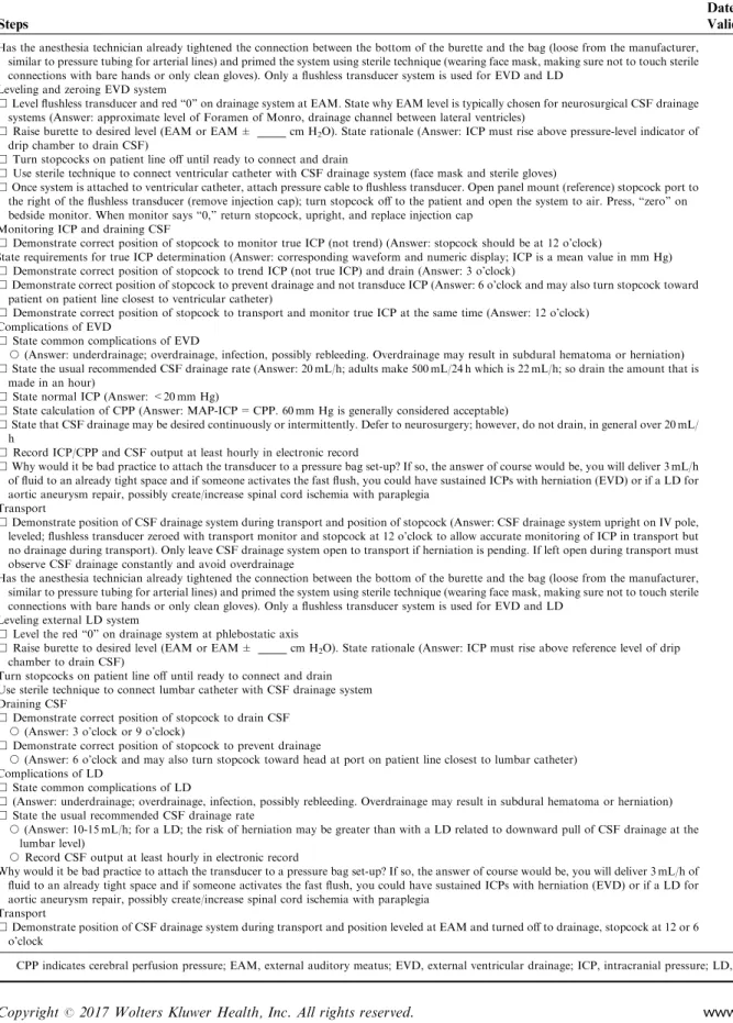
Some aspects of EVD or LD use can be idiosyn-cratic for the specific device (Figs. 2A, B). Consistency within an institution is important for choices such as the reference level and measurement scale. The reference level is most commonly the external auditory meatus for EVDs. However, this convention is not followed rigor-ously in clinical practice and published studies,94,147 which can lead to significant measurement errors if the head of the bed is elevated above zero degrees.148 For vascular surgery patients with LDs, common reference levels include the right atrium (phlebostatic axis) or lumbar catheter insertion site. The reference level is the zero-pressure point for both an attached transducer and the EVD/LD. The device is leveled by aligning the zero-pressure point on the device with the reference level on the patient; this is more accurate when a Carpenter (bubble) or laser level is utilized.149,150
A flushless pressure transducer (Fig. 2B) is used with EVD/LD that is connected to the patient through a fluid column. This is the most common configuration with the transducer located externally on the device and measurement of the ICP or ISP by the fluid column in the drainage catheter.151,152 In stark contrast to commonly used transducer-based monitoring systems such as in-vasive arterial blood pressure, central venous, or pulmo-nary artery catheter, this flushless system is not pressurized in any way. Under no circumstances should a pressure transducer system with a pressure bag be as-sembled or used in conjunction with EVD or LD.
Extra catheter systems are increasingly available and separate the pressure transducer from the drainage catheter by placing the transducer at the tip of the cath-eter.153,154ICP can also be measured separately from the EVD at a variety of anatomic sites utilizing several available technologies.11,151,152,155–157
The following steps can be followed when setting up an EVD or LD intraoperatively (refer to educational document that accompanies this article, Supplemental Digital Content 2, http://links.lww.com/JNA/A47): (1) Choose the appropriate reference level and
measure-ment scale (cm H2O). The reference level is most
commonly the external auditory meatus for EVDs or the right atrium (phlebostatic axis) for vascular surgery patients who have LDs.
(2) Mount the device upright158 by attaching to an intravenous pole or to the patient’s bed. Make appropriate changes to the drainage system when the patient’s position changes relative to the drainage system. It is good practice to clamp the EVD or LD during changes in the position of the patient until it can be releveled.156,159
(3) Level the device by aligning the zero point of the device and the reference level on the patient using a Carpenter (bubble) or laser level.149
(4) Monitor ICP by EVD or ISP by LD with an attached transducer, if possible.
(5) Adjust the collection chamber to the specified height based on the requirements of the procedure.
(6) The fluid path of the EVD/LD is a sterile system. There should be a careful assessment of the risks/ benefits before opening any of these systems to reduce the risk of infectious complications.
EVD/LD Management During Changes in
Position
releveled.156,159,163If changes in position are not accounted for, then there is the risk of both overdrainage159,163 and underdrainage of CSF as well as inaccurate pressure measurements. Nevertheless, there are clinical situations such as impending herniation where it is not feasible to clamp the EVD/LD even briefly for position changes.
Positioning the patient for an intraoperative pro-cedure is a unique phase of the case. The final patient position during the intraoperative procedure may be quite different from that in the ICU or the ward. In addition, there are very significant and dynamic changes in CSF pressure with postural changes164 as might occur as the patient is moved between beds, turned prone, and others to meet the requirements of the procedure. The EVD/LD should be closed to drainage during positioning if clin-ically feasible, and the management reassessed once in the intraoperative position.
Inaccurate Pressure Measurements With
Simultaneous Drainage
Transducers that measure pressure by the fluid column in the drainage catheter are always inaccurate if the EVD or LD is simultaneously open for drainage.153,154,157,165 For fluid-coupled systems, accurate pressure measurements re-quire a static fluid column without simultaneous drainage allowing the transducer to directly interface with the patient line.157,165–167This concern is relevant to transducers that are mounted externally and transducers placed within the tip of the drainage catheter.166 Although a value for ICP can be trended while the device is simultaneously draining,168 the EVD/LD should be closed for accurate measurement at least hourly.166,168In some situations such as an anesthetized patient with elevated ICP, it would be indicated to obtain accurate pressure measurements more frequently than hourly.
FIGURE 2. A, Components of an EVD (a representative example of antimicrobial-impregnated EVD). B, CSF collecting system.
Dramatic measurement errors are possible with transducers that measure with fluid coupling while at the same time draining.165 Notable situations reported or discussed in the literature are compressed or slit ven-tricles, catheter blockage by debris, or catheter dislodge-ment into the parenchyma.153,154,165,166 Accurately measuring the pressure by stopping drainage and con-necting the transducer directly to the patient line can sometimes overcome this limitation.165,166
Continuous Drainage Versus Continuous
Monitoring
A common management decision is whether to utilize continuous drainage with intermittent monitoring (open EVD) versus continuous monitoring with inter-mittent drainage (monitor EVD).169,170 Continuous drainage impacts the ability of the EVD as a monitor to detect trends.11 The decision to choose a specific man-agement option is dependent on the indication for drain placement and close consultation with the surgical team is important. In adult severe TBI, continuous drainage (open EVD) has been associated with better ICP con-trol,19,170 but in patients with aneurysmal SAH, open EVD management is associated with a higher rate of complications.169,171,172
Documentation on Anesthesia Record for
Patients With EVD or LD
There are 5 items that should be documented in the anesthesia record in patients with EVD or LD.
(1) Pressure = ICP/CPP or ISP/spinal cord perfusion pressure (SCPP).
(2) Amount of CSF drainage (expressed in mL).
(3) Color of CSF and any change in color of CSF observed during the procedure.
(4) Drain height relative to the reference level.
(5) EVD/LD status as set by the stopcocks in the device (ie, open, clamped).
These items should be recorded at least hourly. However, it is reasonable that frequency of ICP doc-umentation follow clinical situations as changes in ven-tilation, exposure to anesthetics, and hemodynamic changes all cause frequent perturbations in ICP and CPP. In such clinical scenarios, it may be desirable to document ICP more frequently such as every 5 to 15 minutes. CSF characteristics such as color, and any sudden change in color including the presence of blood should also be documented.129,156,173,174
With an electronic health record that automatically imports patient data, care should be exercised to prevent inaccuracies.171 This may occur due to technical issues with the monitoring system or if automatic systems con-tinue to import inaccurate data such as when the EVD system is open for continuous drainage.
Summary
Intraoperative management of EVD and LD in-cludes review of basic management goals, knowledge of the device in use, and the ability to make accurate
measurements. This section includes recommendations for preparing the EVD/LD, device management during positioning, and best practice for documentation in the anesthetic record. There is also a discussion of in-accuracies that arise with traditional fluid-coupled sys-tems if simultaneously open for drainage, and evidence that is accumulating for various management strategies.
Intraoperative Management of Patients with EVDs and LDs (Class of Recommendation and Level of Evi-dence):
(1) Anesthesia providers should be knowledgeable about the specific EVD and LD device in use locally as details vary (Class I Recommendation; Level of Evidence E).
(2) It is recommended to set up your anesthetizing location following the standards of your institution, including a consistent choice of reference level and measurement scale (Class I Recommendation; Level of Evidence E).
(3) It is recommended to level EVD or LD using a Carpenter (bubble) or laser level rather than by visual inspection (Class I Recommendation; Level of Evidence B-NR).
(4) It is recommended to close the EVD/LD to drainage during any changes in position if clinically feasible (Class I Recommendation; Level of Evidence C). (5) It is recommended to relevel the transducer after
changing patient position to ensure accurate mon-itoring of ICP and adequate drainage of CSF (Class I Recommendation; Level of Evidence C).
(6) It is recommended to monitor ICP or ISP with an attached transducer that is appropriately leveled and zeroed according to manufacturer guidelines (Class I Recommendation; Level of Evidence E).
(7) A pressure bag and pressurized flush system should not be attached to the EVD/LD (Class III; Level of Evidence E).
(8) Pressure measurements should not be made while the EVD or LD is simultaneously draining. Accurate pressure measurements require a static fluid column from the monitoring site to the externally mounted transducers without simultaneous drainage (Class III Recommendation; Level of Evidence B-NR). (9) If open for continuous drainage, it is recommended
to close the EVD or LD to measure pressure at least once per hour or more often if clinically indicated (Class I Recommendation; Level of Evidence E). (10) The decision for either continuous drainage or
continuous monitoring should be made in consulta-tion with the surgical team. Continuous monitoring with intermittent drainage may be considered in patients with aneurysmal SAH, and continuous drainage may be considered for adults with severe TBI (Class IIb Recommendation; Level of Evidence B-R).
ICP/CPP or ISP/SCPP, (2) amount of CSF drainage (expressed in mL), (3) color of CSF and any change in color of CSF observed during the procedure, (4) drain height relative to the reference level, and (5) EVD/LD status as set by the stopcocks in the device (ie, open, clamped)
SECTION 5: MANAGEMENT OF EVD AND LD IN
SPECIAL CLINICAL SCENARIOS
Background
Despite careful maintenance and vigilance, compli-cations may arise given the invasive nature of the devices. EVD and LD need monitoring with the same attention provided to other invasive monitoring devices. Safety can be enhanced using dedicated protocols and bundles that standardize the handling of EVD and LD.
Accidental Disconnection of EVD and LD
As an immune privileged organ, the brain is at high risk for infections from bacterial contamination of drainage devices. Standardized bundles for EVD place-ment have significantly reduced rates of ventriculitis/ meningitis associated with EVD placement.123,175 Rele-vant guidelines were recently published by the Society for Neurocritical Care and emphasize the importance of maintaining a closed, sterile drainage system.8 If drains become inadvertently disconnected, the most immediate threat to the patient is from uncontrolled leakage of CSF as discussed below. A clamp should immediately be put on the free end of the catheter to stop leakage. As the system becomes contaminated by disconnection, all distal parts should be replaced with new, sterile tubing.129 As replacement of the proximal catheter carries new proce-dural risk,129it will not routinely be replaced after acci-dental disconnection. There is no evidence to support empiric antibiotic treatment after disconnection of an EVD or LD system. After a new system is connected, patency must be confirmed, especially in situations where the catheter may have been displaced. In doubt, CT imaging can confirm appropriate position of an EVD catheter.
Drain Occlusion and Troubleshooting
No studies exist in the literature that compares methods to troubleshoot EVD or LD. Published recom-mendations represent expert opinion and practice sur-veys.123,129,150 Sudden reduction in the hourly volume of CSF drained can indicate an obstruction in the drainage system. Similarly, if the ICP waveform is dampened, the EVD may be occluded.176Patency can be tested by briefly lowering the drainage system. This may also be sufficient to remove small amounts of material such as air bubbles, blood clots, or tissue that may obstruct the tubing. If patency is not restored by briefly lowering the system, troubleshooting should continue by examining the drainage tubing distal to the patient. If any occluding material is present, the tubing can be flushed away from the patient to remove the debris. Alternatively, the entire
drainage system can be changed, if necessary. Occlusion of the proximal catheter can sometimes be resolved by flushing the catheter toward the patient. This may in-crease ICP, as the irrigation solution adds to the intra-cranial volume. In patients with poor intraintra-cranial compliance, irrigation with even small volumes can create disproportionately large and dangerous increases of ICP, possibly causing brain herniation. Proximal flushing should only be attempted after discussion with the neu-rosurgeon. Volumes of 0.5 to 2 mL of sterile, preservative-free isotonic sodium chloride solution can be used to flush the EVD catheter, although a variety of antibiotic sol-utions are sometimes used as well.177Maintaining aseptic conditions are essential. The technique should be guided by institutional protocol and include, as a minimum, sterile gloves, mask, and hair cover.175,178,179 The drain-age system should be releveled and rezeroed after ma-nipulation.
Overdrainage of CSF
Rapid drainage of large volumes of CSF from the ventricles (ie, more than the 15 to 20 mL produced in an hour) can collapse the ventricles (especially when com-munication to the extraventricular subarachnoid space is compromised, as in noncommunicating hydrocephalus), thus shrinking the cerebral hemispheres away from the skull and dura. This creates tension on the bridging veins and can cause acute subdural hematomas.180In patients with aneurysmal SAH and an unsecured aneurysm, rapid drainage of CSF while the dura is closed can increase the transmural pressure of the aneurysm and provoke re-bleeding.181 Overdrainage from an EVD occurs most commonly when the patient’s position is changed (eg, the head of the bed or operating table is raised) without si-multaneously adjusting the position of the EVD drainage system.163Drainage systems should be clamped whenever patient position is changed. Patients with critically ele-vated ICP may not tolerate even brief clamping of their EVD during transport, in which case extra care needs to be taken to secure the drainage system to the bed and to monitor output. Once a new position is achieved, the drain should be releveled and rezeroed.
drainage of CSF. Their skull defect exposes the brain to ambient pressure, so that relatively small decreases in CSF pressure can cause significant brain sag and her-niation (syndrome of the trephined).191,192 Tension pneumocephalus and subdural hematoma from brain sag have also been described as complications from LDs.93,193
CSF Drainage at Different Points During a Case
(eg, Before Craniotomy Bone Flap, Before Dural
Reflection, After Durotomy, or After Dural
Closure)
LDs are often placed electively for procedures that carry a risk of postoperative CSF leak, such as skull base surgery.194They are used to drain CSF and improve ex-posure during the procedure, and 10 to 20 mL of CSF is usually drained immediately before durotomy to “relax”
the brain, while the drain remains clamped for the rest of the procedure. LDs are also used to reduce CSF pressure and optimize SCPP during repair of thoracoabdominal aortic aneurysms. CSF is usually drained to a pressure goal, for example, 10 mm Hg of CSF pressure (zeroed at the right atrium). Use of CSF drainage for spinal cord protection during thoracoabdominal aortic aneurysm re-pair has recently been reviewed,195,196and is not the focus of the current document.
EVDs are not commonly placed for intraoperative CSF drainage, but rather to treat ICP elevation caused by noncommunicating hydrocephalus. Most patients will come from the ICU with an EVD in place. CSF can be drained from an EVD to reduce ICP and improve surgical conditions, like a LD, with the difference that if the pa-tient has noncommunicating hydrocephalus, CSF will be removed only from the intraventricular space. Rapid re-moval of a large volume of CSF will cause sudden de-crease in ICP and can cause subdural hematoma, as discussed above. In patients with aneurysmal SAH, sud-den CSF drainage before durotomy in patients can result in precipitous decrease in ICP and widening of transmural pressure gradient and can cause fatal rerupture of cere-bral aneurysm and should be avoided.197
Monitoring and Patient Safety
Changes in EVD output can provide important clues into changes in patient condition. Increasing output can indicate an increase in intracranial volume from edema or hemorrhage, or rising ICP. Bright red output from an EVD suggests an intraventricular hemorrhage or SAH, such as from rerupture of an unsecured aneurysm. This is a life-threatening emergency, and thus should be communicated immediately to the surgical team. Alter-natively, blood from a previous intraventricular hemor-rhage may have been mobilized and transiently color the CSF more brightly red. Although this is a much more benign scenario, suspicion should remain high for a fresh hemorrhage, especially if there are changes in vital signs, such as hypertension or bradycardia, or if ICP increases. Close communication is key to safe management.
Avoiding Accidental Injections Into EVD or LD
EVD tubing routinely has at least one 3-way stopcock that allows access to the system. Commonly used EVD systems use the Luer-lock standard and most EVD tubing is not specifically marked to differentiate it from intra-venous tubing. This creates the opportunity for providers to inadvertently inject drugs into the ventricular system that are meant for intravenous use. There are many case reports and case series that describe accidental intrathecal injection of agents as varied as anesthetic drugs,98,99 antibiotics, chemotherapeutic agents, or gadolinium contrast, fre-quently with devastating or fatal consequences.102–105Care should be taken to prevent this severe complication by carefully labeling EVD tubing and access ports, and using color-coded caps (please refer to educational document that accompanies this publication, Supplemental Digital Con-tent 2, http://links.lww.com/JNA/A47). In the future,
TABLE 5. Perioperative Checklist for Patients With External
Ventricular and Lumbar Drain
Preoperative assessment
&Obtain baseline neurological examination
&Review EVD (cm H2O) and LD setting (in mL/h of CSF drained) &Review hourly CSF output to obtain baseline
&Review baseline ICP mm Hg, ICP trends, and available multimodal monitoring data
&Review baseline CSF color and consistency
&Review clamp trials data if available
&Review coagulopathy profile
&Review antibiotic plan if anticipating new EVD/LD insertion in the operating room
&Provide EVD and LD details during preoperative handoff between intensive care/ward providers and the anesthesia providers Transporting patients with EVD and LD
&Confirm decision to travel with EVD or LD clamp vs. open
&If traveling with EVD clamp, ensure clamping at both proximal port on EVD and distal port on CSF collecting system
&Confirm HOB status during transport
&Confirm availability of dedicated intravenous pole for EVD/LD mount
&Confirm leveling EVD at external auditory meatus and LD at phlebostatic axis or at lumbar catheter insertion site
&Enable ICP monitoring during transport
&Confirm availability of medications needed to treat intracranial hypertension during transport
Intraoperative management of indwelling drains
&Prepare transducer cable
&Identify EVD/LD tubing by appropriate unique labeling
&Confirm HOB status during surgical procedure
&Confirm leveling of EVD at external auditory meatus and LD at phlebostatic axis
&Obtain ICP waveform and baseline ICP value
&Record q 1-h EVD/LD setting
&Record at least q 1-h ICP values (recorded with EVD closed to drain)
&Record at least q 1-h EVD/LD drain output (expressed in mL)
&Provide EVD and LD details during intraoperative handoffs between anesthesia providers
Inform surgeon if anyZ1 of the following
&Sudden decline in CSF drainage or no drainage from EVD or LD, or occlusion of EVD or LD
&If drain output is >15-20 mL at any time or in any given hour
&Sudden change in CSF color
&Dampening or loss of ICP waveform
cm H2O indicates centimeters of water; CSF, cerebrospinal fluid; EVD,
TABLE 6. EVD and LD Clinical Competency Checklist for Clinicians Involved in Perioperative Care
Name of Provider__________________ Date_________________ Validated by______________________
Steps
Date/Initials of Validator
Has the anesthesia technician already tightened the connection between the bottom of the burette and the bag (loose from the manufacturer, similar to pressure tubing for arterial lines) and primed the system using sterile technique (wearing face mask, making sure not to touch sterile connections with bare hands or only clean gloves). Only a flushless transducer system is used for EVD and LD
Leveling and zeroing EVD system
&Level flushless transducer and red “0” on drainage system at EAM. State why EAM level is typically chosen for neurosurgical CSF drainage systems (Answer: approximate level of Foramen of Monro, drainage channel between lateral ventricles)
&Raise burette to desired level (EAM or EAM ± _____ cm H2O). State rationale (Answer: ICP must rise above pressure-level indicator of
drip chamber to drain CSF)
&Turn stopcocks on patient line off until ready to connect and drain
&Use sterile technique to connect ventricular catheter with CSF drainage system (face mask and sterile gloves)
&Once system is attached to ventricular catheter, attach pressure cable to flushless transducer. Open panel mount (reference) stopcock port to the right of the flushless transducer (remove injection cap); turn stopcock off to the patient and open the system to air. Press, “zero” on bedside monitor. When monitor says “0,” return stopcock, upright, and replace injection cap
Monitoring ICP and draining CSF
&Demonstrate correct position of stopcock to monitor true ICP (not trend) (Answer: stopcock should be at 12 o’clock)
State requirements for true ICP determination (Answer: corresponding waveform and numeric display; ICP is a mean value in mm Hg)
&Demonstrate correct position of stopcock to trend ICP (not true ICP) and drain (Answer: 3 o’clock)
&Demonstrate correct position of stopcock to prevent drainage and not transduce ICP (Answer: 6 o’clock and may also turn stopcock toward patient on patient line closest to ventricular catheter)
&Demonstrate correct position of stopcock to transport and monitor true ICP at the same time (Answer: 12 o’clock) Complications of EVD
&State common complications of EVD
J(Answer: underdrainage; overdrainage, infection, possibly rebleeding. Overdrainage may result in subdural hematoma or herniation)
&State the usual recommended CSF drainage rate (Answer: 20 mL/h; adults make 500 mL/24 h which is 22 mL/h; so drain the amount that is made in an hour)
&State normal ICP (Answer: <20 mm Hg)
&State calculation of CPP (Answer: MAP-ICP = CPP. 60 mm Hg is generally considered acceptable)
&State that CSF drainage may be desired continuously or intermittently. Defer to neurosurgery; however, do not drain, in general over 20 mL/ h
&Record ICP/CPP and CSF output at least hourly in electronic record
&Why would it be bad practice to attach the transducer to a pressure bag set-up? If so, the answer of course would be, you will deliver 3 mL/h of fluid to an already tight space and if someone activates the fast flush, you could have sustained ICPs with herniation (EVD) or if a LD for aortic aneurysm repair, possibly create/increase spinal cord ischemia with paraplegia
Transport
&Demonstrate position of CSF drainage system during transport and position of stopcock (Answer: CSF drainage system upright on IV pole, leveled; flushless transducer zeroed with transport monitor and stopcock at 12 o’clock to allow accurate monitoring of ICP in transport but no drainage during transport). Only leave CSF drainage system open to transport if herniation is pending. If left open during transport must observe CSF drainage constantly and avoid overdrainage
Has the anesthesia technician already tightened the connection between the bottom of the burette and the bag (loose from the manufacturer, similar to pressure tubing for arterial lines) and primed the system using sterile technique (wearing face mask, making sure not to touch sterile connections with bare hands or only clean gloves). Only a flushless transducer system is used for EVD and LD
Leveling external LD system
&Level the red “0” on drainage system at phlebostatic axis
&Raise burette to desired level (EAM or EAM ± _____ cm H2O). State rationale (Answer: ICP must rise above reference level of drip
chamber to drain CSF)
Turn stopcocks on patient line off until ready to connect and drain Use sterile technique to connect lumbar catheter with CSF drainage system Draining CSF
&Demonstrate correct position of stopcock to drain CSF
J(Answer: 3 o’clock or 9 o’clock)
&Demonstrate correct position of stopcock to prevent drainage
J(Answer: 6 o’clock and may also turn stopcock toward head at port on patient line closest to lumbar catheter) Complications of LD
&State common complications of LD
&(Answer: underdrainage; overdrainage, infection, possibly rebleeding. Overdrainage may result in subdural hematoma or herniation)
&State the usual recommended CSF drainage rate
J(Answer: 10-15 mL/h; for a LD; the risk of herniation may be greater than with a LD related to downward pull of CSF drainage at the lumbar level)
JRecord CSF output at least hourly in electronic record
Why would it be bad practice to attach the transducer to a pressure bag set-up? If so, the answer of course would be, you will deliver 3 mL/h of fluid to an already tight space and if someone activates the fast flush, you could have sustained ICPs with herniation (EVD) or if a LD for aortic aneurysm repair, possibly create/increase spinal cord ischemia with paraplegia
Transport
&Demonstrate position of CSF drainage system during transport and position leveled at EAM and turned off to drainage, stopcock at 12 or 6 o’clock
manufacturers should design access ports that cannot be confused with intravenous access ports, similar to the safety pin system used for anesthetic gases.102 Treatment of acci-dental intrathecal injection is supportive. Aspiration of CSF and replacement with isotonic sodium chloride to “lavage” the intrathecal space has been suggested as an emergent intervention to reduce neurotoxicity, especially when caustic agents such as chemotherapeutic drugs have been injected inadvertently.100,101 No controlled studies exist, however, and success of this intervention is mixed in reported cases.
Intrathecal Injection of Fluorescein Dye for CSF
Leak
Intrathecal injection of fluorescein dye through the LD is sometimes indicated intraoperatively during repair of CSF leaks to identify the location of actual defect. In addition, it is useful to assist in locating additional leak sites and to confirm the watertight closure of the defect. Although it has been used worldwide for this purpose, the intrathecal fluorescein is an off-label use of the product; hence, informed consent is required in many centers.
The usual dose range used is 10 to 50 mg (0.1 to 0.5 mL of 10% fluorescein with 9.5 mL of CSF) injected over 30 minutes.198Complications are mostly related to meningeal irritation that include headache, nausea and vomiting, dizziness, nuchal pain, limb weakness, gener-alized seizures,199 and cranial nerve palsy. Most compli-cations are dose related and transient, and typically resolve within 7 to 10 days. Limited evidence suggests that fluorescein injection should be avoided in patients with history of seizures, hydrocephalus, spinal stenosis, and cerebral edema. We strongly recommend that individual institutions should develop their own protocols for in-trathecal administration of fluorescein.
Summary of Management of EVDs and LDs in
Special Clinical Scenarios
Because of invasive nature of these devices, com-plications that occur perioperatively include accidental disconnection, drain occlusion, overdrainage, and in-advertent administration of drugs into the drains. Extra vigilance, clear labeling of the drains, and the develop-ment of standardized protocol on the handling of EVD/ LDs are some of the measures that can minimize these complications.
Management of EVD and LD in Special Clinical Sce-narios (Class of Recommendation and Level of Evidence): (1) EVD or LD tubing that is accidently disconnected
should be clamped immediately to prevent over-drainage of CSF (Class I Recommendation; Level of Evidence C).
(2) If the EVD or LD systems are contaminated by disconnection, all distal parts should be replaced with new sterile tubing (Class IIa Recommendation; Level of Evidence E).
(3) Routine flushing of the EVD or LD catheter should not be performed (Class III Recommendation; Level of Evidence E).
(4) In patients with ruptured cerebral aneurysm, sudden excessive drainage of CSF before securing the aneurysm can provoke aneurysm rerupture and should be avoided (Class III Recommendation; Level of Evidence C).
(5) Identification of EVD or LD tubing by appropriate labels and use of other visual aids is recommended to prevent confusion with intravenous ports (Class I Recommendation; Level of Evidence E).
(6) Accidental intrathecal injection should be recognized and reported to the neurosurgeon (Class IIa Recom-mendation; Level of Evidence E).
(7) Lavage of the intrathecal space after accidental injection is not recommended (Class III Recommen-dation; Level of Evidence C).
(8) Establish institutional standards to ensure safe intra-thecal injection of fluorescein dye through LD in patients with suspected CSF leak (Class I Recom-mendation; Level of Evidence E).
SECTION 6: PERIOPERATIVE CHECKLIST,
DEVELOPING CLINICAL COMPETENCIES FOR
EVD/LD, AND CONTINUED MEDICAL
EDUCATION
Use of a Perioperative EVD and LD Checklist
The material presented above provides a framework from which a perioperative checklist can be constructed. Such a checklist (Table 5) incorporated into a shared mental model200–202is intended to reduce systematic errors during perioperative management of patients with EVD or LD. This perioperative checklist can be used for all patients undergoing perioperative care, and may be included in preoperative handoffs between ICU/ward providers and anesthesia providers, and during intraoperative handoffs between various anesthesia providers. Checklist can be modified and used during IHT of neurocritically ill patients.
Clinical Competence and Continued Medical
Education Related to Management of Patients
With EVD/LD
For clinicians involved in perioperative care of pa-tients, one of the core competencies is the ability to ad-minister safe and reliable care on a consistent basis. In relationship to patients with EVD and LD, this translates to acquiring knowledge about the basics of EVD and LD relevant to the perioperative period, at the same time, possessing essentially the same skills as neuroscience nursing while managing these drains within the confines of perioperative care.