Spatial and Dynamic Organization of
Molecular Structures in the Cell
Nucleus
Spatial and Dynamic Organization of
Molecular Structures in the Cell Nucleus
Proefschrift
ter verkrijging van
de graad van Doctor aan de Universiteit Leiden,
op gezag van Rector Magnificus prof. mr. P.F. van der Heijden,
volgens besluit van het College voor Promoties
te verdedigen op woensdag 8 september 2010
klokke 13.45 uur
door
Anne - Kee Brouwer
geboren te Amsterdam
Promotiecommissie
Promotor:
Prof. dr. H.J. Tanke
Co-promotor:
Dr. R.W. Dirks
Commissieleden:
Prof. dr. L.H.F. Mullenders
Dr. J.M.J.M. Zijlmans
Erasmus Universiteit Rotterdam
Prof. dr. F.C.S. Ramaekers Universiteit Maastricht
The studies described in this thesis were performed at the department of Molecular Cell
Biology, Leiden University Medical Center.
Printing of this thesis was financially supported by the J.E. Jurriaanse Stichting.
Table of contents
Chapter 1
Introduction and outline of the thesis
9
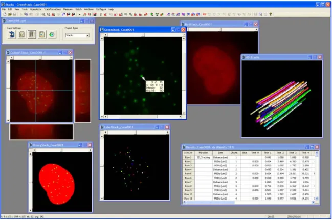
Chapter 2
STACKS: A software program for particle tracking in
living
cells
61
59
Chapter 3
Telomere movement is constrained by interactions with an
inner inner nuclear lamin structure
85
Chapter 4
Telomeric DNA mediates de novo PML body formation 105
Chapter 5
Nuclear body movement is constrained by associations
with chromatin
143
Chapter 6
Summary and discussion
165
Nederlandse
samenvatting
175
CHAPTER 1
1. The cell nucleus
1.1 A concise historical perspective
The eukaryotic cell nucleus was first named in 1831 by Robert Brown when he ob-served an opaque structure in orchid cells. Most likely, he was not the first person who observed the cell nucleus. Already in 1802, it was Franz Bauer who described a structure in plant cells that could reflect the cell nucleus. Also, it is even possible that van Leeuwenhoek was actually the first person who observed the cell nucleus back in 1682 when he studied plant cells. These scientists, however, had no clue about the content and function of this organelle at that time. Knowledge about the constituents and function of the nucleus evolved rapidly after the first isolation of the nucleus in 1869 by Friedrich Miescher (Miescher, 1871). For this isolation he used white blood cells derived from pus, which he treated with a pig-stomach extract and acid. Mi-escher discovered that the nucleus contains a substance made up of large molecules containing phosphorus and nitrogen which he named ‘nuclein’. When the substance was separated into protein and acid molecules it was in 1889 referred to as nucleic acid by a pupil of Miescher, Richard Altmann. It was only since the discovery of the chemical structure of DNA by James Watson and Francis Crick in 1953 that the pre-cise role of this molecule in life became known.
1.2 What is inside the cell nucleus?
1.2.1 Chromatin
These regions have a rather complex structure and may include several elements, e.g., topoisomerase II binding sites (for review see Razin, 1996; Vassetzky, 2000b). To-gether, these studies suggest a strict organization principle for chromatin. The reality is, however, that we know very little about the organization of chromatin in the cell nucleus. Even there is debate whether the 30 nm fibre exists in living cells (Maeshima et al., 2010).
Typically, euchromatin is referred to as a transcriptionally active open chromosome structure having ample access to the transcription and RNA processing machinery, while heterochromatin is referred to as a transcriptionally inactive, compact chromatin structure (John, 1988; Felsenfeld & Groudine, 2003). However, these morphological terms do not provoke a very clear functional distinction, as some genes show tran-scriptional activity in supposed heterochromatic regions (Bühler & Moazed, 2007) and some are silenced in supposed euchromatic regions. Despite some cell type spe-cific variation, heterochromatin is mainly positioned at the nuclear periphery and around nucleoli. It is probably also for this reason that chromatin at the nuclear pe-riphery shows a relatively low transcriptional activity and a low gene density (Boyle, 2001; Finlan, 2008). Transcriptionally competent regions preferentiallylocalize to-wards the interior of the cell nucleus and to the periphery of chromosomal territories (Verschure, 1999). The status of chromatin is characterized best by the presence or absence of specific histone and DNA modifications, rather than relying on morpho-logical features. Histone modifications associated with transcriptional repression in-clude methylation of histone H3 on Lysine 9 (Steward, 2005) and Lysine 36 (Strahl, 2002), and deacetylation (leading to hypoacetylation) of histone H3 (Grunstein, 1997; Turner, 2000). Histone H3 lysine 9 (H3-K9) methylation creates a specific binding site for heterochromatin protein 1 (HP1), which is targeted there by the methylating enzyme SUV39H1 (Steward, 2005; Krouwels et al., 2005). However, methylated H3-K9 is also able to suppress transcription in absence of HP1 by a mechanism involving histone deacetylation (Steward, 2005).
unmethylated in all cells (Bird, 1986). Both histone and DNA methylation can act as epigenetic markers providing heritable mechanisms for gene silencing (Nakayama, 2001; Grewal & Rice, 2004).
1.2.2 The interchromatin domain
The interchromatin domain is inevitably a crowded space since both proteins and RNAs travel through this compartment to reach their destination or exert their func-tion in this compartment. RNA forms together with proteins ribonucleoprotein (RNP) particles, which are thought to form a continuous nuclear network. This structure is the source of the RNP particles that are released from the nucleus by chemical or me-chanical extraction (Smetana, 1963). RNA-selective staining procedures have made a complete ultrastructural characterization of the nuclear RNP network possible (Bern-hard, 1969; Biggiogera & Fakan, 1998). Making use of EDTA regressive staining to localize RNA, Monneron & Bernhard were able to define, characterize and classify the interconnected nuclear RNP structures and distinguished interchromatin granule clusters, perichromatin fibrils, perichromatin granules and coiled bodies (Monneron & Bernhard, 1969). The discovery of these structures was important for our understand-ing of nuclear RNA metabolism (Misteli & Spector, 1998; Misteli, 2000). Perichro-matin fibrils are sites of RNA transcription (Bachellerie, 1975; Cmarko, 1999), whereas interchromatin granule clusters (or speckles) play a central role in the assem-bly and/or modification of pre-mRNA splicing factors (Mintz, 1999; Smith, 1999; Spector, 2001).
To ensure unimpeded exchange of molecules between the nucleus and the cytoplasm, the ICD has direct access to nuclear pores. Nuclear pores are multiprotein complexes embedded in the nuclear envelope, which mediate and regulate nucleocytoplasmic transport (Vasu & Forbes, 2001; Fahrenkrog & Aebi, 2003). The ‘basket’ structure at the nucleoplasmic side of the nuclear pore consists of eight filaments, which attach to a distal ‘ring’ structure. Several reports suggest that these ‘rings’ connect to filaments that extend into the nucleus and facilitate nucleocytoplasmic transport (Cordes, 1993; Parfenov, 1995; Cordes, 1997).
1.2.3
Chromatin organization is dynamic.
chromatin-chromatin interactions (Dekker, 2006). Also the positioning of specific chromatin regions at particular nuclear bodies are examples supporting the notion that the genome is not randomly organized in the cell nucleus (Smith, 1995). The chal-lenge now is to unravel the underlying mechanisms that establish and maintain this non-random organization of chromatin in the cell nucleus.
Understanding the organization principles of the nucleus is important because rear-rangements in nuclear organization have been observed in cells derived from various diseases, including cancer, and in cells with a senescent or apoptotic phenotype (Vijg & Dollé, 2002; Busuttil, 2004; Raz, 2008; Shin et al., 2010). Furthermore, a striking change in nuclear organization has been observed in embryonic stem cells at the onset of differentiation (Butler et al., 2009). Most profound rearrangements in chromatin structure have been observed when a sperm pronucleus and an egg nucleus fuse after fertilization. In many species, the size of DNA loops increases from ca. 50 kbp in early embryogenesis to 200 kbp in cells of the adult organism (Buongiorno-Nardelli, 1982). Notably, the average size of DNA loops was observed to decrease in trans-formed cells (Linskens, 1987). In several human cancer cell lines the DNA loop size was found to be about 50 kbp, i.e. significantly smaller than in normal cells where it varies between 70-700 kbp (Oberhammer, 1993). It is important to unravel the mechanisms that control these aspects of nuclear organization to understand their im-pact on the etiology, progression, and possibly treatment of human diseases. Once un-derstood, the hope is that this new knowledge might open possibilities for treatment strategies of human disease.
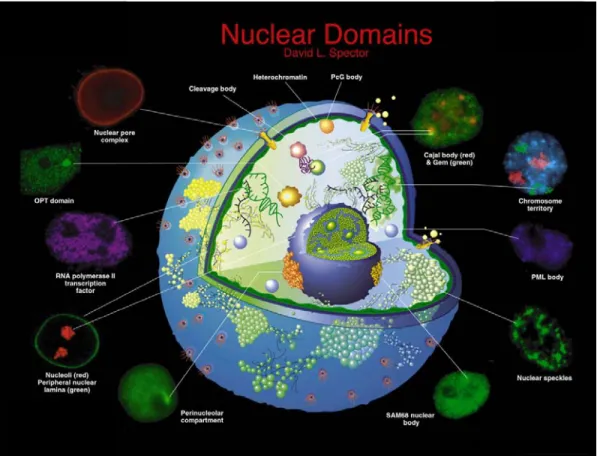
1.3 Nuclear bodies
Figure 3. Protein domains present in the mammalian cell nucleus.
OPT domains: transcriptionally active sites that contain a specific set of transcription factors and RNA pol II, appear close to nucleoli in G1. Nuclear pore complex: multiprotein complexes where the inner and outer nuclear membranes are fused and where materials can transit between the cytoplasm and the nucleus. Cleavage body: either overlap or are localized adjacent to Cajal bodies, they consist of factors involved in the cleavage and polyadenylation steps of pre-mRNA processing. Heterochromatin: inactive chromatin. PcG body: have been found to be associated with pericentric heterochromatin (Saurin, 1998) and contain polycomb group proteins (i.e. RING1, BMI1 and hPc2). Gems: Gemini of Cajal bodies, they have been found adjacent to or coinciding with Cajal bodies. Gems are characterized by the presence of the survival of motor neurons gene product (SMN) and an associated factor, Gemin2 (Matera, 1999). SAM68 nuclear bodies/ Perinucleolar compartments (PNC): have been identified as unique structures that are associated with the surface of nucleoli and are thought to play a role in RNA metabolism (Huang, 2000). Both structures are predominantly found in cancer cells and they are rarely ob-served in primary cells. Other nuclear bodies are discussed in the text. (Adapted from Spector, 2001)
1.3.1 Nucleolus
and up to 30% of these proteins are encoded by previously uncharacterized genes (Andersen, 2002;Andersen, 2005). Although it is not expected that all proteins found in nucleoli also have a function in this structure, their diversity is consistent with the idea thatthe nucleolus performs additional roles beyond generating ribosomalsubunits (Pederson, 1998; reviewed by Olson, 2002). For example, many proteins related to cell cycle regulation (about 3.5% of the identified proteome), DNA damagerepair (about 1%) and pre-mRNA processing (about 5%) have been identifiedin isolated nu-cleoli. Nucleoli have therefore been implicated in processes such as cell cycle regula-tion (Yamauchi, 2007), virus-replicaregula-tion (Jacob, 1968), regularegula-tion of tumor suppressor and oncogene activities (Itahana, 2003), DNA damage repair (van den
Boom, 2004), signal recognition particle assembly (Jacobson & Pederson, 1998), RNA modification (Sansam, 2003), tRNA processing (Paushkin, 2004), aging by modulating telomerase function (Kieffer-Kwon, 2004; Zhang, 2004), regulation of protein stability (Mekhail, 2004; Rodway, 2004), senescence (reviewed by Comai, 1999; Rosete, 2007) and apoptosis (Baran, 2003). In addition, nucleoli are thought to play a role in the maturation and transport of mRNAs (Schneiter, 1995).
A possible function of the nucleolus in mRNA export was proposed 25 years ago based on observations in interspecies heterokaryons obtained from fusing chicken erythrocytes with mouse cells. It was observed that in the dormant chicken nucleus gene expression was initiated at precisely the same time when a nucleolus became detectable (Sidebottom & Harris, 1969; Deák, 1972; Harris, 1972). Furthermore, it was observed that UV irradiation of the chicken nucleolus in these heterokaryons greatly suppressed chicken-specific gene expression (Perry, 1961; Deák, 1972). Addi-tional support for a role of nucleoli in mRNA export came by the observation that processed myc and myoD transcripts, unlike actin or lactate dehydrogenase tran-scripts, are present in the nucleolus of several cell types (Bond & Wold, 1993). Be-cause myc intron 1-containing pre-mRNA was not detected in nucleoli but instead in the nucleoplasm, it was suggested that the nucleolar localization of Pol II transcripts is a general phenomenon for transcripts that have a rapid cytoplasmic turnover only (Bond & Wold, 1993). It should be noted, however, that these observations have thus far not been confirmed by others. In cells derived from species that vary from sea ur-chins to humans, nuclear poly(A)+ RNA is found present primarily in discrete "tran-script domains", which often concentrate around nucleoli (Carter, 1991). Thus, whether nucleoli are involved in some steps of nuclear mRNA export has yet to be confirmed.
1.3.2 Cajal bodies
until 1999 that Joseph Gall suggested to link Cajal’s name to the nuclear body that was originally described by him in 1903 (Gall, 1999). The number and size of Cajal bodies varies among cell types (in mammalian cells typically 0 –10 per nucleus, rang-ing 0.1–2 μm in diameter) and they also show cell cycle variation within cell types. Cajal bodies can be discriminated in the nucleus by the presence of the protein coilin, either by immunocytochemistry or by exogenous expression of coilin-GFP (Snaar, 2000; Ogg & Lamond, 2002).
Recent studies indicated that Cajal bodies play a role in the assembly and/or modifica-tion of the transcripmodifica-tion and RNA-processing machinery (Gall, 1999; Jády, 2003). Ca-jal bodies are enriched in snRNPs (small nuclear ribonucleoproteins) and snoRNPs (small nucleolar ribonucleoproteins) spliceosome subunits. Solid evidence has been provided that the final steps in snRNP maturation including snRNA base
modifica-tion, U4/U6 snRNA annealing, and snRNA-protein assemblyof both snoRNAs and
snRNAs occur in Cajal bodies (Darzacq, 2002;Verheggen, 2002; Jady, 2003; Stanek, 2008). Despite their role in splicing factor maturation, Cajal bodies do not represent major sites of transcription per se, but they were observed frequently in association with a few specific genes coding for small nuclear snRNAs and histone genes in in-terphase cells. Because Cajal bodies do not contain either DNA (Thiry, 1994) nor non-snRNP protein splicing factors (Raska, 1991; Carmo-Fonseca, 1992) it is unlikely that these bodies are sites of transcription or pre-mRNA splicing. Thus, the current view is that Cajal bodies play a crucial role in the spliceosome cycle in which the
pro-duction of new snRNPs is promoted by the import and modification of substrates
(re-viewed by Staněk& Neugebauer, 2006). In addition, Cajal bodies may play a role in the recycling of snRNPs from splicing complexes that are released after finishing pre-mRNA splicing. Interestingly, also the RNA subunit (hTR) of the enzyme telomerase was shown to accumulate in Cajal bodies (Jady, 2004; Zhu, 2004). During the S-phase, when telomerase is likely to act, hTR has been found to associate with a subset of telomeres while Cajal bodies are present at close distance (Jady, 2006; Tomlinson, 2006 ). Mutant hTR, which fails to accumulate in Cajal bodies, was fully capable of forming catalytically active telomerase in vivo. Telomere extension, however, turned out to be strongly impaired (Cristofari, 2007). This functional deficiency was accom-panied by a decreased association of telomerase with telomeres suggesting that Cajal bodies also play an important role in telomere elongation.
1.3.3 Speckles
pre-sent throughout the nucleoplasm in regions that contain little or no DNA (Thiry, 1995). Furthermore, in situ hybridization studies revealed that speckles do not contain genes. Instead, active transcription sites were found positioned throughout the nucleoplasm and also next to speckles. Some genes have been reported to localize preferentially close to speckles (Huang, 1991; Xing, 1993; Xing, 1995; Smith, 1999; Johnson, 2000).
These observations indicate that speckles are functionally related to gene expression. Hall and coworkers proposed that speckles are hubs that spatially link the synthesis of specific pre-mRNAs to a rapid recycling of copious RNA metabolic complexes, thereby facilitating expression of many highly active genes (Hall, 2006). In addition to increasing the efficiency of each step, sequential steps in gene expression might be structurally integrated at each speckle, consistent with evidence that the biochemical machineries for transcription, splicing, and mRNA export are coupled (Hall, 2006). The observation that speckles also containpoly(A)+ RNA led to the suggestion that speckles play a role in RNA metabolism and export (Carter, 1991,1993; Molenaar, 2004). A substantial amount of mature mRNA is found to be retained in nuclear speckles until ATP is added, suggesting that speckles prevent the export of otherwise fully processed mRNAs until an energy-requiring cellular signal releases them (Schmidt, 2006).
1.3.4 PML bodies
The most mysterious of all nuclear bodies is the PML body, also known as ND10 (nu-clear domain 10) or Kremer bodies (Kr) (Dyck, 1994; Koken, 1994; Weis, 1994). Promyelocytic leukemia bodies (PML bodies) are nuclear protein bodies, ranging in size from 0.3 µm to 1.0 µm in diameter and are characterized by the presence of the PML protein. Typically there are 10-20 PML nuclear bodies (PML-NB) present in the cell nucleus and they are believed to be tightly associated with nuclear matrix proteins (Stuurman, 1992). Electron microscopy studies have shown that PML-NBs are composed of a ring-like protein structure that does not contain nucleic acids in the centre of the ring (Boisvert, 2000; Dellaire & Bazett-Jones, 2004). At the periphery of the ring, however, PML-NBs are believed to make extensive contacts with chromatin fibers through protein-based threads that extend from the core of the bodies (Eskiw, 2004). These contacts have been proposed to be essential for maintaining the integrity and positional stability of PML-NBs in the nucleus.
all-trans-retinoic acid or arsenic trioxide results in the degradation of the PML-RARα fusion protein, restoration of PML bodies and remission of the disease (Koken, 1994; Weis, 1994). Recently, it has been shown that arsenic-induced degradation of PML or PML-RARα is mediated by the ubiquitin ligase RNF4 (Lallemand-Breitenbach, 2008; Tatham, 2008).
In PML bodies, nearly eighty different proteins have been found present. Among them are Sp100, Sp140, SUMO-1, HAUSP (USP7), CBP and BLM, Daxx, pRB, and p53 (Hodges, 1998; LaMorte, 1998; Alcalay, 1998; Zhong, 1999; Zhang, 1999; Zhong, 2000; for a review see Salomoni & Pandolfi, 2002). Because of this variety of proteins, PML bodies have been implicated in many different functions, such as tran-scription regulation, protein storage, senescence and interferon-induced antiviral de-fense (Chelbi-Alix, 1995; Maul, 1998). Concerning transcription regulation, PML bodies have been suggested to be involved in both transcriptional activation (Maul, 1998; Zhong, 2000) and transcriptional repression (Everett,1999). However, whether PML bodies play indeed an essential role in transcription is not clear since PML
-/-mice show a very moderate phenotype. PML-/- mice aremorphologically normal and do not have higher rates of spontaneous cancers than littermate controls (Wang, 1998a; Wang, 1998b). Some regions of the human genome that display high transcrip-tional activity do, however, associate frequently with PML NBs, although RNAi-mediated knockdown of PML did not perturb the expression of these genes (Wang, 2004).
PML bodies have also been implicated in DNA damage repair as several repair fac-tors transit through PML bodies in a temporally regulated manner (Graham & Bazett-Jones, 2004). Furthermore, PML bodies have been shown torecruit single-stranded DNA (ssDNA) moleculesin response to exogenous DNA damage (Bøe, 2006). PML bodies are also associated with the sites of initial viral DNA transcription/ replication in virus infected cells (Maul, 1996; Maul, 1998; Guldner, 1992; Stadler,1995). PML bodies are subsequently disrupted at later stages in the infectious viral cycle (Maul, 1993). Upon treatment of cells with interferon, PML is induced and the number of nuclear bodies increases dramatically (Lavau, 1995; Gaboli, 1998). This suggests a role for PML and the nuclear bodies as part of the anti-viral defense machinery acti-vated by interferons in viral infections. DNA and RNA viruses have a variety of ef-fects on PML body morphology, where arenaviruses and the human immunodefi-ciency virus (HIV) transport PML to the cytoplasm, and herpesviruses “unwind” PML bodies (Borden, 1998; Melnick & Licht, 1999; Maul, 2000; Turelli, 2001). However, findings with HIV infected cells are somewhat controversial, since another group did not see PML NBs translocate during infection (Bell, 2001).
It has been established that PML is the primary essential component of PML NBs, and conjugation of SUMO-1 to PML is suggested to be a prerequisite for PML body formation (Ishov, 1999; Zhong, 2000). PML SUMOylation likely plays a regulatory role in the structure, composition, and function of PML bodies (Sternsdorf, 1997). Elegant studies demonstrate that the RING domain of PML directly interacts with Ubc9, an enzyme which covalently attaches the SUMO1 protein onto distal regions of PML, including one B-box and a region near the nuclear localization signal (Duprez, 1999).
It has been demonstrated that PML contains a SUMO binding motif that is independ-ent of its SUMOylation sites and is required for PML-NB formation. A model for PML-NB formation was proposed in which PML SUMOylation and noncovalent binding of PML to SUMOylated PML through the SUMO binding motif constitutes the nucleation event for subsequent recruitment of SUMOylated proteins and/or pro-teins containing SUMO binding motifs to the PML NBs (Shen, 2006).
Alternatively, PML bodies may have the ability to self-assemble. Purified RING-domains (small zinc-binding RING-domains) of PML and other proteins have been shown to self-assemble into supramolecular structures in vitro that resemble the structures they form in cells (Kentsis, 2002). Over-expression of SUMO-1 prevented the stress-mediated breakdown of PML bodies, indicating that PML body stability is partially dependent on SUMO-1 (Eskiw, 2003). Interestingly, many of the proteins found in the PML NBs have been shown to be SUMOylated (Seeler & Dejean, 2003).
Like PML bodies, also the PML protein has been implicated in different cellular func-tions including suppressing cell growth and cell transformation (Mu, 1994; Ahn 1995; Koken, 1995; review: Melnick & Licht 1999). Transduction of APL patient derived NB4 cells with a retrovirusharboring the coding sequence for PML suppressed the ability of these cells to form coloniesin soft agar. In addition, conditioned medium from these cellssuppressed colony formation of wild-type NB4 cells, suggestingthe release of negative growth control factors (Mu, 1994). Furthermore, PML-overexpressing NB4 cells, when injected into nude mice, yieldedsmaller tumors that appeared with a longer latency than vector-expressing cells (Mu, 1994). In various human tumors, PML expression was shown to be decreased (Gurrieri, 2004a) and in some cases it was shown that low levels of PML correlated with poor disease outcome (Chang, 2007).Consistent with a role as tumor suppressor, it has been reported that overexpression of PML suppresses the growth of various cancer cells (Liu, 1995; Mu, 1997; Le, 1998). Also, PML knockout mice revealed an increased susceptibility to chemical-induced carcinogenesis (Wang, 1998a) and spontaneous tumorigenesis (Trotman, 2006).
Probably one of the most important functions of PML is to control apoptosis. The physiological relevance of this is emphasized by in vivo studies demonstrating that mice and cells that lack PML are resistant to a vast variety of apoptotic stimuli (Wang, 1998a). Although the molecular mechanism remains largely unknown, PML is thought to be a pivotal factor in γ irradiation-induced apoptosis (Wang,1998a) and essential for the induction of programmed cell death by Fas, tumor necrosis factor α
(TNF), ceramide and type I and II interferons (IFNs) (Wang,1998a; Quignon, 1998). In support of these thoughts, PML−/− mice and PML−/− cells are resistant to the le-thal effects of γ-irradiation (Wang,1998a; Yang, 2002).
2. The nuclear matrix
2.1 Evidence for a nuclear matrix structure?
nuclear activities. It is the observation that nuclei withstand strong hydrodynamic shear force, compression and friction during cell or tissue homogenization as well as extreme osmotic pressure that prompted scientists to believe in a nuclear matrix struc-ture (Maggio, 1963a; Penman, 1966; Blobel & Potter, 1966; Dounce, 1995; Pederson 1997). The term ‘nuclear matrix’ was first used in 1974 to describe a filamentous structure that remained present when cell nuclei were salt extracted using 1.0- 2.0 M NaCl (Berezney & Coffey, 1974). Numerous studies followed since, using variations on extraction protocols until proteins, RNA- and DNA-sequences were all shown to be connected to the nuclear matrix (Berezny & Jeon, 1995). Interestingly, Jackson and Cook observed in 1988 an extensively anastomatized nuclear network of filaments after performing nuclear extractions of cells that were encapsulated in agarose spheres (Jackson & Cook, 1988). This network is believed to resemble the filamentous struc-ture that remains present after a high ionic strength extraction of the nucleus (Capco, 1982). Many studies found 3-5 and 10-30 nm ribonucleoprotein elements/filaments remaining present in the nucleus after extraction using depleted and RNP-containing resinless electron microscopy (resin is an embedding material that scatters electrons in a similar way as the embedded specimen does) or whole mount electron microscopy (Monneron & Bernhard, 1969; Berezney & Coffey, 1974; Comings & Okada, 1976; Capco, 1982; Small, 1985; Fey, 1986; Jackson & Cook, 1988). RNP filament domains are thought to be very important for nuclear matrix organization and for some time it was not possible to remove chromatin from the nucleus without removing the RNP filament domains as well (Fey, 1986).
These ultrastructural studies of sectioned cell nuclei did, however, not confirm the presence of a filament system that was thought to comprise the nuclear matrix in situ. In fact enormous doubt was raised concerning the procedures used to extract cell nu-clei could possibly reveal the nuclear matrix structure that may exist in vivo. All nu-clear matrix preparation procedures used thus far involved harsh treatments, including the removal of nucleic acids, heat (Mirkovitch, 1984; Martelli, 1991), Cu 2+
(Mirk-ovitch, 1984; Neri, 1997), sulfhydryl cross-linking (Kaufmann & Shaper, 1984), and highly concentrated monovalent salts such as 2 M NaCl (Berezney & Coffey, 1977). Significantly, it has been noted that such treatments themselves result in protein rear-rangements and protein aggregations (Palade & Siekevitz, 1956; Tashiro, 1958; Madi-son & Dickman, 1963; Lothstein, 1985). Also, protein-protein interactions and van der Waals forces between proteins and water change profoundly when high ionic strength is used (Kauzmann, 1959; Varshavsky & Ilyin, 1974), which is true for most standard nuclear matrix preparation procedures. Consequently, such artificially intro-duced protein filaments might easily be interpreted as a nuclear matrix structure (Finkelstein, 1997). The existence of a nuclear matrix still needs to be confirmed by other techniques, like for example live cell imaging and RNA interference.
active form of this enzyme (Dahmus, 1996). Taking all this evidence into account it can be concluded that it is very likely that there is a nuclear matrix, but still further research is necessary to precisely define its components.
Recent advances in the study of the protein composition of the nuclear matrix allowed the characterization of several proteins that are specifically associated with the nu-clear matrix in tumor cells (Konety & Getzenberg, 1999). Some of these proteins are used for the diagnosis of cancer; e.g., NMP22 is specifically present in the nuclear matrix of bladder cancer cells (Ozen, 1999). Hence, detecting changes in the nuclear matrix structure may serve as a valuable tool in cancer diagnostics.
2.2 The nuclear lamina
The nuclear envelope is a double-layered membrane that encloses the contents of the nucleus during most of the cell's lifecycle and forms a boundary between chromo-somes and the cytoplasm in eukaryotic cells. The main components of the nuclear en-velope are the inner nuclear membrane, the outer nuclear membrane, which is con-tinuous with the endoplasmatic reticulum, and the nuclear pore complexes (Stuurman, 1998; Goldman, 2002). On the inner surface of the nuclear membrane, the nuclear lamins (type-V intermediate filaments) are polymerized to form a thin fibrous struc-ture, 20-50 nm thick. The nuclear lamins form together with the inner nuclear mem-brane (INM) proteins the ‘nuclear lamina’, a stable yet dynamic network that main-tains extensive interactions with both INM-specific integral membrane proteins and chromatin (Hutchison, 2002). There are two classes of lamins, A-type lamins (lamin A/lamin C, each alternatively spliced from the same gene) and B-type lamins which bind to the lamin B receptor (LBR). Mutations in the lamin A/C and lamin B genes result in diseases ranging from cardiac and skeletal myopathies and partial lipodystro-phy to peripheral neuropathy and premature aging (Mounkes, 2003). Specifically, mu-tations in the genes encoding for A-type lamins and their binding partners have been associated with Emery-Dreifuss muscular dystrophy, dilated cardiomyopathy, Dunni-gan-type familial partial lipodystrophy and Hutchinson-Gilford progeria syndrome (Bonne, 1999; Fatkin, 1999; Cao, 2000; Shackleton, 2000; Burke & Stewart, 2002; De Sandre-Giovannoli, 2003; Eriksson, 2003). B-typelamins are constitutively ex-pressed in all somatic cells and contain a stable C-terminal farnesyl modification, which mediates tight association with the INM. Unlike B-type lamins, the A-type lamins are expressedonly in differentiated cells (Lebel, 1987; Stuurman, 1998). They are components of the peripheral lamina and of structures in the nuclear interior (Moir, 2000). The lamina may be linked to nuclear pore baskets through Nup153 (Foisner, 2001).
Mar-tins, 2003; Gruenbaum, 2003) and to support nuclear activities such as DNA replica-tion and RNA synthesis (Nili, 2001; Spann, 2002; Wilkinson, 2003; Haraguchi, 2004). It is not yet clear whether intranuclear lamins form a network and whether such a network would be required for the activities supported by the lamins.
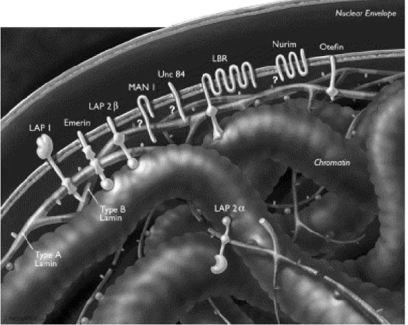
The nuclear lamins bind to severalINM proteins, including lamina-associated poly-peptides 1 and2ß (LAP1, LAP2ß), emerin and Man1, whichshare a common struc-tural motif of about 40 amino acid residues, called the LEM (LAPs, emerin and Man1) domain (Lin, 2000). Although additional LEM domain proteins such as Nesprin, Otefin, and Lem-3 have been identified (Lin, 2000). All LAP1 isoforms and LAP2 interact preferentially with A-type lamins, while the lamin B receptor and LAP2 interact with the B-type lamins and emerin interacts with both types of lamins (Foisner, 2001). Lamins can also bind to chromatin proteins (histone H2A or H2B dimers), as well as ostensibly soluble proteins including lamina-associated polypep-tide-2α (LAP2α), Kruppel-like protein (MOK2), actin, retinoblastoma protein (RB), barrier-to-autointegration factor (BAF), sterol-response-element-binding protein (SREBP) and one or more components of RNA-polymerase-II-dependent transcrip-tion complexes and DNA-replicatranscrip-tion complexes (Gruenbaum, 2003; Zastrow, 2004). In cells that lack A-type lamins, many of these proteins are not retained at the NE but instead drift throughout the NE/ER network (Sullivan, 1999; Lee, 2002; Liu, 2003; Muchir, 2003; Wagner, 2004). Lamins and their associated proteins are proposed to have roles in large-scale chromatin organization (Sullivan, 1999; Liu, 2000; Guil-lemin 2001; Liu, 2003; Raz, 2006; Raz, 2008), the spacing of nuclear pore complexes (Liu, 2000; Schirmer, 2001), the positioning of the nucleus in cells (Starr, 2001; Starr 2002) and the reassembly of the nucleus after mitosis (Lopez-Soler, 2001). Lamins have been shown to interact with chromatin at more than 1,300 sharply defined large domains, 0.1-10 megabases in size (Guelen, 2008). These lamina-associated domains are typified by low gene-expression levels, indicating that they represent a repressive chromatin environment (Guelen, 2008).
Figure 4.
Schematic view of the nuclear envelope, lamina and
chro-matin
. The inner and outer membranes of the nuclear envelope are shown with their en-closed lumen. Also lamin filaments and selected nuclear envelope proteins, including lamina-associated protein 1 (LAP1), emerin, LAP2β, MAN1, UNC-84, lamin B receptor (LBR), nurim and otefin, are shown. (Adapted from Cohen, 2001).2.3 Are lamins part of the nuclear matrix?
vivo(Goldman, 1992; Moir, 1994; Schmidt, 1994). Altogether this evidence is in fa-vour of a role for lamins constituting part of the nuclear matrix, possibly by forming a complex network with other proteins like emerin, protein 4.1, nuclear actin and nu-clear myosin (Pestic-Dragovich, 2000; Kiseleva, 2004).
2.4 Nuclear actin
Since one of the presumed functions of the nuclear matrix is to support and to facili-tate/regulate intranuclear transport, possible nuclear matrix components may be simi-lar to protein filament systems already characterized in the cytoplasm. Thus far, there is little if no evidence for the presence of tubulin or microtubules in the nucleus. Nu-clear actin, however, is present and functional in the cell nucleus of various cell types (Clark & Merriam, 1977; Fukui, 1978; Fukui & Katsumaru, 1979; Clark & Rosenbaum, 1979; Osborn & Weber, 1980; Welch & Suhan, 1985; De Boni, 1994; Yan, 1997; Rando, 2000; Pederson and Aebi, 2002; Bettinger, 2004; Castano et al., 2010). This is also true fornuclear actin binding proteins (Ankenbauer, 1989; Rimm & Pollard, 1989) and nuclear myosin (Hauser, 1975; Berrios & Fisher, 1986; Hagen, 1986; Rimm & Pollard, 1989; Nowak, 1997). Nuclear actin has initially been sug-gested to play a role in transcription (Scheer, 1984) and later also in mRNA process-ing (Sahlas, 1993), chromatin remodellprocess-ing and nuclear export (Machesky & May, 2001; Goodson & Hawse, 2002; Olave, 2002). Nuclear actin is also found present in the nucleolus (Clark & Merriam, 1977; Funaki, 1995) and TEM analysis of Xenopus
oocyte nuclei suggested that short bundles of actin extend from nucleoli towards the nuclear envelope (Parfenov, 1995). In addition to actin, nuclei also contain a specific isoform of myosin I, nuclear myosin 1 (NM1), which is an actin-dependent motor. Antibodies directed against nuclear myosin I block transcription by RNA polymerase II when injected into mammalian cells and inhibit isolated transcription complexes in vitro (Pestic-Dragovich, 2000).
The most important questions about nuclear actin revolve around its polymeric state(s). Nuclear actin does not form long actin filaments (‘F-actin’), it is proposed to assume shorter, potentially novel conformations (Pederson & Aebi, 2002; Bettinger, 2004). Nuclear actin ‘rods,’ ‘bundles,’ and ‘tubules’ have been describedby a number of investigators (Fukui & Katsumaru, 1979; Iida, 1986; Iida & Yahara, 1986; Nishida, 1987; Wada, 1998), but their supramolecular organization has remainedelusive ex-cept for one case (Sameshima, 2001). Sameshima et al. have described a new type of actin rods formedboth in the nucleus and the cytoplasm of Dictyostelium discoideum
pro-tein (Correas, 1991; Krauss, 1997; Luque & Correas, 2000). An actin network is proposed to exist at the INM and to mechanically reinforce the lamina network (Holaska, 2004). The actin-binding domain of nuclear-specific isoforms of protein 4.1 is found to be essential to reconstruct nuclei after mitosis (Krauss, 2003). In conclusion, there is ample evidence that actin is present in the cell nucleus and is involved in a variety of nuclear processes.
3. Telomeres & Nuclear organization
3.1 Telomere biology
Telomeres are structures at the ends of eukaryotic chromosomes (in greek: telo= end , mere= part). They are protein-DNA complexes that protect chromosome termini from unregulated degradation, recombination and fusion. They also serve to limit the loss of genetic material from chromosome ends that occurs during (incomplete) DNA rep-lication. After about 60-80 cell divisions, telomere repeats are shortened from a typi-cal initial length of 10-15 kb in human cells to ~5 kb and below, which triggers cell senescence or apoptosis (Harley, 1990; Martens, 2000; Blasco, 2007). The number of cell divisions that a normal cell can make before entering in to a state of senescence is also referred to as the ‘Hayflick limit’. Leonard Hayflick demonstrated in 1965 that normal human diploid cells in a cell culture divide about 50 +/- 10 times(Hayflick, 1965).
The length of telomeres is well-controlled by several different factors. If this were not the case, the chromosomes would shorten dramatically with every cell division. In most organisms, telomeres are lengthened by the enzyme telomerase (Greider & Blackburn, 1985; Greider, 1996). With the exception of a few cell types, including stem cells, human somatic cells undergo programmed telomere shortening, a process that appears to involve repression of telomerase expression (Cooke & Smith, 1986; de Lange, 1990; Harley, 1990; Hastie, 1990; Counter, 1992; Kim, 1994). This progres-sive decline of telomere length with each cell division may constitute a tumor sup-pressor mechanism that limits the replicative potential of transformed cells. In agree-ment, telomerase is frequently activated in human and mouse tumors and restoration of telomere length is correlated with immortalization of human cells in vitro (Counter, 1992, 1994a, b; Kim, 1994; Blasco, 1996; Broccoli, 1996). During the malignant pro-gression of cancer cells, the maintenanceof telomere length is a crucial prerequisite for immortalization(Bacchetti, 1996). Therefore, telomere length has emerged as a promising clinical marker to predict the risk and prognosis of patients with malignant disorders (reviewed by Svenson & Roos, 2009). Most cancer cells activate a telomere maintenance pathway and about 90% of these tumors show telomerase activity (Shay & Bachetti, 1997). A significant minority of tumors use an alternative lengthening of telomeres (ALT) mechanism (Bryan, 1995; Bryan, 1997).
Figure 6.
Schematic representation of shelterin on
telomeric
DNA. The shelterin complex consists of six subunits, TRF1,TRF2, POT1, TIN2, TPP1 and Rap1. For simplicity, POT1 is only shown as binding to the site closest to the duplex telomeric DNA although it can also bind to the 3′ end. (Adapted from de Lange, 2005)3.2 ALTernative lengthening of telomeres
requiredfor DNA recombination (Lundblad, 1993). Also, individual telomeresin hu-man ALT cells undergo steady telomere attrition upon whichsudden lengthening and shortening events are superimposed ina manner that is suggestive for recombination (Murnane, 1994). Finally, functional evidencefor the involvement of recombination in the ALT mechanism was provided by showing that DNA sequences are copied from telomere to telomere in ALT cells (Dunham, 2000). Telomere lengthening is also possible via intra-telomeric DNA copying (Muntoni, 2009). These observations are all consistentwith a recombination-mediated DNA replication mechanism.
The hallmarks of human ALT cells include a large variance in telomere length, with telomeres that range from very short ~5kb, to very long ~50 kb (Bryan, 1995), and the presence of ALT-associated promyelocytic leukemia nuclear bodies (APBs) contain-ing telomeric DNA and telomere bindcontain-ing proteins (Yeager, 1999). ABPs are a subset of PML bodies that are not found in normal cells, or in tumor cells that express telom-erase, and contain additional proteins involved in DNA replication, recombination and repair that are not found in normal PML bodies (Yeager, 1999; Yankiwski, 2000; Stavropoulos, 2002; Tarsounas, 2004). APBs are found in a minority of cells, ap-proximately 5% within asynchronously dividing ALT cell populations, from which it may be concluded that their formation is cell cycle-dependent (Yeager, 1999; Grobelny, 2000; Wu, 2000). It has been suggested that APBs may have an integral role in the ALT mechanism (Yeager, 1999; Grobelny, 2000; Wu, 2000, 2003; Mole-naar, 2003). Consistent with this suggestion, inhibition of ALT in somatic cell hy-brids, formed by fusing ALT and telomerase-positive cells, resulted in a substantial decrease in APBs (Perrem, 2001). It has been shown that inhibition of ALT is accom-panied by a reduction of APBs, providing evidence for a direct link between APBs and ALT activity (Jiang, 2005). Furthermore, it has recently been shown that the DNA recombination endonuclease MUS81 is involved in ALT specific telomerase recombination and localizes to APBs (Zeng, 2009).
Observational and clinical studies on ALT positive tumors mayhelp to fill the gaps in our understanding of the ALT mechanism.ALT is most commonly activated in tu-mors of neuroepithelialorigin (astrocytomas) or mesenchymal origin, including os-teosarcomas,and in soft tissue sarcomas (Henson, 2005). The reason for this is un-known, but it is possible that some mesenchymal and neuroepithelial cells repress telomerase more tightly than epithelial cells and thereforehave a higher probability of activating ALT duringtumorigenesis. In sarcomas, ALT is more frequently activated in subtypes that have a complex karyotype, which could be linked to chromosomal instability (Montgomery, 2004; Ulaner, 2004). It could be argued that the ALT mechanism is, in part, the cause of this instability because the critically short te-lomeres found in ALT cells areprone to end-to-end fusions, anaphase bridge forma-tion, break–fusion–breakevents and ultimately severe chromosomal rearrangements. However, notall soft tissue sarcomas showing complex karyotypesare ALT-positive (Henson, 2005), indicating that other factorscontribute to chromosomal instability as well.
re-duced tumorigenicity in vitro and in vivo compared to the telomerase positive NSCLC cell lines (Brachner, 2006). It can be concluded that there is some evidence indicating that the ALT mechanism is indicative for a better patient prognosis, although further research is needed to substantiate this conclusion.
3.3 Telomeres and the nuclear matrix
The positioning of telomeres in the cell nucleus varies among organisms (Dong & Ji-ang 1998). In yeast, telomeres are positioned at the nuclear periphery while in mam-malian cells they seem randomly distributed in the nucleoplasm (Henderson, 1996; Bilaud, 1997; Broccoli, 1997; van Steensel, 1998). Biochemical and ultrastructural data suggest that in mammalian cells telomeric DNA and telomere binding proteins colocalize in individual condensed structures at the nuclear matrix (Ludérus, 1996). The shelterin complex component TIN2 is believed to play a dual role in tethering telomeres to the nuclear matrix (Kaminker, 2009). Consistent with this association to the nuclear matrix, telomeric TTAGGG repeats were found to contain an array of nu-clear matrix attachment sites at a frequency of at least one per kb. The nunu-clear matrix association is supposed to involve large domains of up to 20-30 kb telomeric DNA, encompassing the entire length of most mammalian telomeres (Ludérus, 1996). Be-cause of their association to a nuclear matrix structure, telomeres are thought to play an important role in nuclear organization (de Lange, 2002).
In situ hybridization studies revealed that in yeast telomeres are organized in clusters at the nuclear periphery Gilson, 1993; Gotta, 1996). This organization in clusters may contribute to the repression of transcription of nearby genes, a phenomenon termed telomere position effect (TPE) (Gottschling, 1990). Telomeres in yeast have a noncleosomal chromatin structure, whereas subtelomeric DNA is assembled into nu-cleosomes (Wright, 1992). Subtelomeric chromatin in yeast has therefore many of the hallmarks of heterochromatin as present in mammalian cells: it imposes transcrip-tional repression (Gottschling, 1990) and late replication of nearby sequences (Ferguson, 1991).
3.4 Telomere and chromatin mobility in the cell nucleus
monomeric protein, and only several minutes for a large complex such as a spli-ceosome or ribosome.
If the subnuclear positioning of any particular chromosomal locus reflects a state of transcriptional activity, then genes must be able to move from a transcriptional repressive subenvironment to a transcriptional competent environment and to obtain a tissue-specific and/or developmental-stage-specific spatial organization. In the past few years, various studies addressed the dynamic properties of chromatin in general or specific sequences in particular. Using time-lapse imaging of GFP-tagged chromosomal loci, Sedat and coworkers showed in yeast and later in flies that chromatin is engaged in a continuous random-walk-like motion (Marshall, 1997). Later, slightly less constrained random movements were described for multiple yeast loci by monitoring the movements of specific chromosomal sites fused to lac repressor binding sites that were tagged with GFP-lac repressor proteins (Heun, 2001a; Heun, 2001b). Telomeres and transcriptionally active non-telomeric loci showed clear differences in movement. Active chromosomal loci displayed a random walk movement within a radius of 0.5–0.7 µm (Heun, 2001a; Gartenberg, 2004; Sage, 2005). This represents more than one-quarter of the nuclear diameter in yeast, but less than one-tenth of the nuclear diameter in mammalian cells. Because 50% of the yeast nuclear volume is contained within a peripheral shell that is <0.4 µm thick, most yeast genes have a high probability to encounter the nuclear membrane. Silent telomeres, however, moved in a highly constrained manner along the inner surface of the nuclear envelope and only rarely occupy the nuclear core (Heun, 2001a; Hediger, 2002; Gar-tenberg, 2004; Sage, 2005). The movement of a typical yeast telomere is restricted to an area at the inner-nuclear-envelope surface occupying 12% of the total nuclear volume (Rosa, 2006). Interestingly, a similar constraint movement was observed for a subset of active genes. Notably, galactose-induced loci were shown to associate with nuclear pores upon induction. In addition, a subtelomeric gene was shown to shift from a telomeric focus to a nuclear pore upon induction by low glucose (Cabal, 2006; Taddei, 2006). Given their lateral dynamics and striking radial confinement, it was suggested that a subset of active genes move from pore to pore (Cabal, 2006).
Outline of this thesis
References
Miescher F. 1871. Ueber die chemische Zusammensetzung der Eiterzellen. Hoppe-Seyler's Med. Chem.Unters. 4, 441-460
Stenoien D.L., Simeoni S., Sharp Z.D., Mancini M.A. 2000. Subnuclear dynamics and tran-scription factor function. J Cell Biochem. 3, 99 –106
Carmo-Fonseca M. 2002. The contribution of nuclear compartmentalization to gene regula-tion. Cell 108, 513–521
Rippe K. 2007. Dynamic organization of the cell nucleus. Current Opinion in Genetics & De-velopment 17, 373-380
Cremer T., Kreth G., Koester H., Fink R.H., Heintzmann R., Cremer M., Solovei I., Zink D. & Cremer C. 2002. Chromosome territories, interchromatin domain compartment, and nuclear matrix: An integrated view of the functional nuclear architecture. Crit Rev Eukaryot Gene Expr. 10, 179-212
Manuelidis L. 1985. Individual interphase chromosome domains revealed by in situ hybridi-zation. Hum. Genet.71, 288-293
Cook P.R. & Brazell I.A. 1976. Conformational constraints in nuclear DNA. J Cell Sci. 22, 287-302
Paulson J.R. & Laemmli U.K. 1977. The structure of histonedepleted metaphase chromo-somes. Cell12, 817-825
van der Velden H.M., van Willigen G., Wetzels R.H. & Wanka F.1984. Attachment of Ori-gins of Replication to the Nuclear Matrix and the Chromosomal Scaffold. FEBS Lett.
171, 13–16
Razin S.V., Kekelidze M.G., Lukanidin E.M. 1986. Replication origins are attached to the nuclear skeleton. Nucl.Acids Res. 14, 8189–8207
Buongiorno-Nardelli M., Micheli G., Carri M.T. & Marilley M. 1982. A relationship between replicon size and supercoiled loop domains in the eukaryotic genome. Nature298, 100–102 Marilley M. & Gassend-Bonnet G. 1989. Supercoiled loop organization of genomic DNA: A close relationship between loop domains, expression units, and replicon
organization in rDNA from Xenopus laevis. Exp. Cell Res.180, 475–489
Razin S.V. 1996. Functional architecture of chromosomal DNA domains. Crit. Rev. Eukaryot. Gene Expr.6, 247–269
Vassetzky Y., Lemaitre J.M. & Mechali M. 2000b. Specification of chromatin domains and regulation of replication and transcription during development. Crit. Rev. Eukaryot. Gene Expr.10, 31–38
John B. 1988. The Biology of Heterochromatin. (ed.Verma R.S.) (Cambridge Univ. Press, Cambridge).
Bühler M. & Moazed D. 2007. Transcription and RNAi in heterochromatic gene silencing.
NatureStructural & Molecular Biology14, 1041-1048
Boyle S., Gilchrist S., Bridger J.M., Mahy N.L., Ellis J.A. & Bickmore W.A. 2001. The spa-tial organization of human chromosomes within the nuclei of normal and emerin-mutant cells.
Hum. Mol. Genet.10, 211–219
Finlan L.E., Sproul D., Thomson I., Boyle S., Kerr E., Perry P., Ylstra B., Chubb J.R., Bick-more W.A. 2008. Recruitment to the Nuclear Periphery Can Alter Expression of Genes in Human Cells. PLoS Genet.4, e1000039
Verschure P.J., van Der Kraan I., Manders E.M. & van Driel R. 1999. Spatial relationship between transcription sites and chromosome territories. J. Cell Biol.147, 13– 24
Stewart M.D., Li J. & Wong J. 2005. Relationship between Histone H3 Lysine 9 Methyla-tion, Transcription Repression, and Heterochromatin Protein 1 Recruitment. Molecular and cellular biology25, 2525–2538
Strahl B.D., Grant P.A., Briggs S.D., Sun Z.W., Bone J.R., Caldwell J.A., Mollah S., Cook R.G., Shabanowitz J., Hunt D.F.& AllisCD.2002. Set2 is a nucleosomal histone H3-selective methyltransferase that mediates transcriptional repression. Mol. Cell. Biol.22, 1298 -1306
Grunstein M. 1997. Histone acetylation in chromatin structure and transcription. Nature389, 349-352
Turner B.M. 2000. Histone acetylation and an epigenetic code. BioEssays450, 1-10
Krouwels I.M., Wiesmeijer K., Abraham T.E., Molenaar C., Verwoerd N.P., Tanke H.J., and Dirks R.W. 2005. A glue for heterochromatin maintenance: stable SUV39H1 binding to het-erochromatin is reinforced by the SET domain J. Cell Biol. 170, 537-549
Goll M.G. & Bestor T.H. 2005. Eukaryotic cytosine methyltransferases. Annual Review of Biochemistry 74, 481-514
Razin A. & Cedar H. 1991. DNA methylation and gene expression. Microbiol. Rev. 55, 451-458
Bird A.P. 1986. CpG-rich islands and the function of DNA methylation. Nature 321, 209-213 Nakayama J., Rice J.C., Strahl B.D., Allis C.D., Grewal S.I. 2001. Role of histone H3 lysine 9 methylation in epigenetic control of heterochromatin assembly. Science292, 110–113
Grewal S.I. & Rice J.C. 2004. "Regulation of heterochromatin by histone methylation and small RNAs". Curr. Opin. Cell Biol.16, 230–238
Maeshima K., Hihara, S., and Eltsov, M. 2010. Chromatin structure: does the 30-nm fibre exist in vivo? Curr. Opin. Cell Biol. In press
Smetana K., Steele W. J. & Busch H. 1963. A nuclear ribonucleoprotein network. Exp. Cell Res. 31, 198-201
Biggiogera M. & Fakan S. 1998. Fine structural specific visualization of RNA on ultrathin sections. J. Histochem. Cytochem. 46, 389-395
Monneron A. & BernhardW. 1969. Fine structural organization of the interphase nucleus in some mammalian cells. J. Ultrastruct. Res. 27, 266- 288
Misteli T. & Spector D.L.1998. The cellular organization of gene expression. Curr. Opin. Cell Biol. 10, 323-331
Misteli T. 2000. Cell biology of transcription and pre-mRNA splicing: nuclear architecture meets nuclear function. J. Cell Sci. 113, 1841-1849
Bachellerie J.P., Puvion E. & Zalta J.P.1975. Ultrastructural organization and biochemical characterization of chromatin - RNA – protein complexes isolated from mammalian cell nu-clei. Eur. J. Biochem. 58, 327- 337
Cmarko D., Verschure P.J., Martin T.E., Dahmus M.E., Krause S., Fu X.D., van Driel R. & Fakan S. 1999. Ultrastructural analysis of transcription and splicing in the cell nucleus after bromo-UTP microinjection. Mol. Biol. Cell 10, 211-223
Mintz P.J., Patterson S.D., Neuwald A.F., Spahr C.S. & Spector D.L.1999. Purification and biochemical characterization of interchromatin granule clusters. EMBO J. 18, 4308-4320
Smith K.P., Moen P.T., Wydner K.L., Coleman J.R. & Lawrence J.B. 1999. Processing of endogenous pre-mRNAs in association with SC-35 domains is gene specific. J. Cell Biol.
144, 617-629
Spector D.L. 2001. Nuclear domains. J. Cell Sci.114, 2891–2893
Vasu S. & Forbes D.J. 2001. Nuclear pores and nuclear assembly. Curr. Opin. Cell Biol. 13, 363-375
Fahrenkrog B. & Aebi U. 2003. The nuclear pore complex: nucleocytoplasmic transport and beyond. Nat. Rev. Mol. Cell Biol. 4, 757- 766
Cordes V.C., Reidenbach S., Kohler A., Stuurman N., van Driel R. & Franke W.W. 1993. Intranuclear filaments containing a nuclear pore complex protein. J. Cell Biol. 123, 1333-1344
Cordes V., Reidenbach S., Rackwitz H.R. & Franke W.W. 1997. Identification of protein 270/Tpr as a constitutive component of the nuclear pore complex-attached intranuclear fila-ments. J. Cell Biol. 136, 515-529
Parfenov V.N., Davis D.S., Pochukalina G.N., Sample C.E., Bugaeva E.A. & Murti, K.G. 1995. Nuclear actin and their topological changesin frog oocytes. Exp. Cell Res. 217, 385-394
Zink D., Amaral M.D., Englmann A., Lang S., L.A. Clarke L.A., Rudolph C., Alt F., Luther K., Braz C., Sadoni N., Rosenecker J. & Schindelhauer D. 2004. Transcription-dependent spatial arrangements of CFTR and adjacent genes in human cell nuclei. J. Cell Biol. 166, 815–825
Lonard D.M. & O'Malley B.W. 2008. Gene transcription: Two worlds merged. Nature 452, 946-947
Dekker J. 2006.The three 'C' s of chromosome conformation capture: controls, controls, con-trols.Nature Methods 3, 17-21
Smith K.P., Carter K.C., Johnson C.V., Lawrence J.B. 1995. U2 and U1 snRNA gene loci associate with coiled bodies. J. Cell. Biochem.59, 473-85
Vijg J. & Dollé M.E.T. 2002. Large genome rearrangements as a primary cause of aging.
Mechanisms of ageing and development123,907-915
Busuttil R.A., Dollé M.E.T., Campisi J., Vijg J. 2004. Genomic Instability, Aging, and Cellu-lar Senescence. Ann. N. Y. Acad. Sci. 1019, 245–255
Raz V., Vermolen B.J., Garini Y., Onderwater J.J.M., Mommaas-Kienhuis M.A., Koster A.J., Young I.T., Tanke H.J. & Dirks R.W. 2008. The nuclear lamina promotes telomere aggrega-tion and centromere peripheral localizaaggrega-tion during senescence of human mesenchymal stem cells. Journal of Cell Science121, 4018-4028
Shin D-M., Kucia M., Ratajczak M.Z. 2010. Nuclear and chromatin reorganization during cell senescence and aging – a mini-review. Gerontology, in press
Butler J.T., Hall L.L., Smith K.P. & Lawrence J.B. 2009. Changing nuclear landscape and unique PML structures during early epigenetic transitions of human embryonic stem cells. J. Cell Biochem.107, 609-621
Buongiorno-Nardelli M., Micheli G., Carri M.T., Marilley M. 1982. A relationship between replicon size and supercoiled loop domains in the eukaryotic genome. Nature298, 100-102
Linskens M.H., Eijsermans A., Dijkwel P.A. 1987. Comparative analysis of DNA loop length in nontransformed and transformed hamster cells. Mutat. Res178, 245-256
Oberhammer F., Wilson J.W., Dive C., Morris I.D., Hickman J.A., Wakeling A.E., Walker P.R., Sikorska M. 1993. Apoptotic death in epithelial cells: cleavage of DNA to 300 and/or 50 kb fragments prior to or in the absence of internucleosomal fragmentation. EMBO J12, 3679-3684
Tsutsui K.M., Sano K. & Tsutsui K. 2005. Dynamic View of the Nuclear Matrix. Acta Med.
Okayama 59, 113-120
Misteli T. 2001.Protein dynamics: implications for nuclear architecture and gene expression.
Science291, 843 -847
Snaar S., Wiesmeijer K., Jochemsen A.G., Tanke H.J., Dirks R.W. 2000. Mutational analysis of fibrillarin and its mobility in living human cells. J. Cell. Biol.151, 653-662
Olson M.O.J. 2004. Nucleolus. BookISBN-10: 0306478730 ISBN-13: 978-0306478734
Spector D.L. 2001. Nuclear domains. J. Cell Sci.114, 2891–2893
Matera A.G. 1999. Nuclear bodies: multifaceted subdomains of the interchromatin space.
Trends Cell Biol.9, 302 -309
Huang, S. 2000. Review: perinucleolar structures. J. Struct. Biol.129, 233 -240
Montgomery T.H. 1898. Comparative cytological studies, with especial regard to the mor-phology of the nucleolus. J. Morphol.15, 265-565
Olson M.O., Hingorani K. & Szebeni A. 2002. Conventional and nonconventional roles of the nucleolus. Int. Rev. Cytol.219, 199-266
Andersen J.S., Lyon C.E., Fox A.H., Leung A.K., Lam Y.W., Steen H., Mann M. & Lamond A.I. 2002. Directed proteomic analysis of the human nucleolus. Curr. Biol.12, 1-11
Andersen J.S., Lam Y.W., Leung A.K.L., Ong S.E., Lyon C.E., Lamond A.I. & Mann M. 2005. Nucleolar proteome dynamics. Nature433, 77-78
Pederson T. 1998. The plurifunctional nucleolus. Nucleic Acids Res.26, 3871-3876
Yamauchi K., Yang M., Hayashi K., Jiang P., Yamamoto N., Tsuchiya H., Tomita K., Moossa A.R., Bouvet M., Hoffman R.M. Imaging of nucleolar dynamics during the cell cycle of cancer cells in live mice. 2007. Cell cycle6, 2706-2708
Jacob J. 1968. Involvement of the nucleolus in viral synthesis in the cells of primary renal tumors of leopard frogs. Cancer Research28, 2126-2136
Itahana K., Krishna P., Jin B.A., Itahana Y., Hawke D., Kobayashi R. & Zhang Y. 2003. Tu-mor suppressor ARF degrades B23, a nucleolar protein involved in ribosome biogenesis and cell proliferation. Molecular Cell12, 1151–1164
van den Boom, V., Citterio, E., Hoogstraten, D., Zotter, A., Egly, J.M., van Cappellen, W.A., Hoeijmakers, J.H., Houtsmuller, A.B. and Vermeulen, W. 2004. DNA damage stabilizes in-teraction of CSB with the transcription elongation machinery. J. Cell Biol.166, 27-36
Jacobson M.R. & Pederson T. 1998. Localization of signal recognition particle RNA in the nucleolus of mammalian cells. PNAS 95, 7981-7986
Sansam C.L., Wells K S. & Emeson R.B. 2003. Modulation of RNA editing by functional nucleolar sequestration of ADAR2. Proc. Natl. Acad. Sci. USA100, 14018-14023
Paushkin S.V., Patel M., Furia B.S., Peltz S.W. & Trotta C.R. 2004. Identification of a human endonuclease complex reveals a link between tRNA splicing and pre-mRNA 3' end forma-tion. Cell117, 311-321
Kieffer-Kwon P., Martianov I. & Davidson I. 2004. Cell-specific nucleolar localization of TBP-related factor 2. Mol. Biol. Cell15, 4356-4368
Zhang S., Hemmerich P. & Grosse F. 2004. Nucleolar localization of the human telomeric repeat binding factor 2 (TRF2). J. Cell Sci.117, 3935-3945
Mekhail K., Gunaratnam L., Bonicalzi M.E. & Lee S. 2004. HIF activation by pH-dependent nucleolar sequestration of VHL. Nat. Cell Biol.6, 642-647
Comai L. 1999. The nucleolus: a paradigm for cell proliferation and aging. Brazilian Journal of Medical and Biological Research32, 1473-1478
Rosete M., Padros M.R., Vindrola O. 2007. The nucleolus as a regulator of cellular senes-cence. 67, 183-194
Baran V., Fabian D., Rehak P., Koppel J. 2003. Nucleolus in apoptosis-induced mouse pre-implantation embryos. Zygote11, 271-283
Schneiter R., Kadowaki T. & Tartakoff A.M. 1995. mRNA transport in yeast: time to reinves-tigate the functions of the nucleus. MCB 6, 357-370
Sidebottom E. & Harris H. 1969. The role of the nucleolus in the transfer of RNA from nu-cleus to cytoplasm. J. Cell Sci. 5, 351-364
Deák I., Sidebottom E. & Harris H. 1972. Further experiments on the role of the nucleolus in the expression of structural genes. J. Cell Sci. 11, 379-391
Harris H. 1972. A new function for the nucleolus. Aust. J. Exp. Biol. Med. Sci. 50, 827-832
Perry R.P., Hell A. & Errera M. 1961. The role of the nucleolus in ribonucleic acid and pro-tein synthesis. Biochem. Biophys. Acta 49, 47-57
Bond V.C. & Wold B. 1993. Nucleolar localization of myc transcripts. Mol. Cell. Biol. 13, 3221-3230
Carter K.C., Taneja K.L. & Lawrence J.B. 1991. Discrete nuclear domains of poly(A) RNA and their relationship to the functional organization of the nucleus. J. Cell Biol. 115, 1191-1202
Cajal S.R. 1903. Un sencillo método de coloración selectiva del retículo protoplásmico y sus efectos en los diversos órganos nerviosos de vertebrados e invertebrados. Tra. Lab. Invest. Biol.2, 129–221
Ogg S.C. & Lamond A.I. 2002. Cajal bodies and coilin - moving towards function. J Cell Biol.159, 17–21
Gall J.G. 2000. Cajal bodies: the first 100 years. Annu. Rev. Cell Dev. Biol. 16, 273-300 Gall J.G., Bellini M., Wu Z. & Murphy C. 1999. Assembly of the nuclear transcription and processing machinery: Cajal bodies (coiled bodies) and transcriptosomes. Mol. Biol.Cell 10, 4385–4402
Snaar S. Wiesmeijer K., Jochemsen A.G., Tanke H.J., Dirks R.W. 2000. Mutational analysis of fibrillarin and its mobility in living human cells. J.Cell Biol.151, 653-662
Jády B.E., Darzacq X., Tucker K.E., Matera, A.G., Bertrand E. & Kiss T. 2003. Modification of Sm small nuclear RNAs occurs in the nucleoplasmic Cajal body following import from the cytoplasm. EMBO J.22, 1878–1888
Verheggen C., Lafontaine D.L., Samarsky D., Mouaikel J., Blanchard J.M., Bordonne R., & Bertrand E. 2002. Mammalian and yeast U3 snoRNPs are matured in specific and related nu-clear compartments. EMBO J.21, 2736–2745
Staněk D., P idalová-Hnilicová J., Novotny I., Huranová M., Bla íková M., Wen X., Sapra A.K. & NeugebauerK.M. 2008. Spliceosomal snRNPs repeatedly cycle through Cajal Bod-ies. Mol Biol Cell 19, 2534-2543
Thiry M. 1994. Cytochemical and immunocytochemical study of coiled bodies in different cultured cell lines. Chromosoma103, 268–276
Raska I., Andrade L.E., Ochs R.L., Chan E.K., Chang C.M., Roos G. & Tan E.M. 1991. Im-munological and ultrastructural studies of the nuclear coiled body with autoimmune antibod-ies. Exp. Cell Res.195, 27–37
Carmo-Fonseca M., Pepperkok R., Carvalho M.T. & Lamond A.I. 1992.
Transcription-dependent colocalization of the U1, U2, U4/U6, and U5 sn-RNPs in coiled bod-ies. J. Cell Biol.117, 1–14
Staněk D. & Neugebauer K.M. 2006. The Cajal body: a meeting place for spliceosomal snRNPs in the nuclear maze. Chromosoma115, 343–354
Jady B.E., Bertrand E. & Kiss T. 2004. Human telomerase RNA and box H/ACA scaRNAs share a common Cajal body-specific localization signal, J. Cell Biol.164, 647–652
Zhu Y., Tomlinson R.L., Lukowiak A.A., Terns R.M. & Terns M.P. 2004. Telomerase RNA accumulates in Cajal bodies in human cancer cells. Mol. Biol. Cell15, 81–90
Jady E., Richard P., Bertrand E. & Kiss T. 2006. Cell cycle-dependent recruitment of telom-erase RNA and Cajal bodies to human telomeres. Mol. Biol. Cell 17, 944–954
Tomlinson R.L., Ziegler T.D., Supakorndej T., Terns R.M. & Terns M.P. 2006. Cell cycle-regulated trafficking of human telomerase to telomeres, Mol. Biol. Cell 17, 955–965
Christofari G., Adolf E., Reichenbach P., Sikora K., Terns R.M., Terns M.P. & Lingner J. 2007. Human telomerase RNA accumulation in Cajal bodies facilitates telomerase recruit-ment to telomeres and telomere elongation. Molecular Cell 27, 882–889
Dirks R.W., Hattinger C.M., Molenaar C. & Snaar S.P. 1999. Synthesis, processing, and transport of RNA within the three-dimensional context of the cell nucleus. Critical reviews in eukaryotic gene expression. 9, 191-201
Lamond A.I. & Spector D.L. 2003. Nuclear speckles: a model for nuclear organelles. Nat. Rev. Mol. Cell. Biol. 4, 605-612
Dirks R.W., de Pauw E.S. & Raap A.K. 1997. Splicing factors associate with nuclear HCMV-IE transcripts after transcriptional activation of the gene, but dissociate upon transcription in-hibition: evidence for a dynamic organization of splicing factors. J. Cell Sci.110, 515–522.
Misteli T., Caceres J.F. & Spector D.L. 1997. The dynamics of a pre-mRNA splicing factor in living cells. Nature 387, 523-527
Visa N., Puvion-Dutilleul F., Bachellerie J. P. & Puvion E. 1993. Intranuclear distribution of U1 and U2 snRNAs visualized by high resolution in situ hybridization: revelation of a novel compartment containing U1 but not U2 snRNA in HeLa cells. Eur. J. Cell Biol.60, 308–321
Huang S. & Spector D.L. 1991. Nascent pre-mRNA transcripts are associated with nuclear regions enriched in splicing factors. Genes Dev.5, 2288–2302
Xing Y., Johnson C.V., Dobner P.R. & Lawrence J.B. 1993. Higher level organization of individual gene transcription and RNA splicing. Science259, 1326–1330
Xing, Y., Johnson, C. V., Moen, P. T., McNeil, J. A. & Lawrence, J. B. 1995. Nonrandom gene organization: structural arrangements of specific pre-mRNA transcription and splicing with SC35 domains. J. Cell Biol.131, 1635–1647
Smith K.P., Moen P.T., Wydner K.L., Coleman J.R. & Lawrence J.B. 1999. Processing of endogenous pre-mRNAs in association with SC-35 domains is gene specific. J. Cell Biol.
144, 617–629
Johnson C., D. Primorac, M. McKinstry, J. McNeil, D. Rowe & J. Bentley Lawrence. 2000. Tracking COL1A1 RNA in osteogenesis imperfecta. Splice-defective transcripts initiate transport from the gene but are retained within the SC35 domain. J. Cell Biol.150, 417–432
Hall L.L., Smith K.P., Byron M., Lawrence J.B.2006. Molecular anatomy of a speckle. The Anatomical record part A 288A, 664–675
Carter K.C., Taneja K.L., Lawrence J.B. 1991. Discrete nuclear domains of poly(A) RNA and their relationship to the functional organization of the nucleus. J.Cell Biol. 115, 1191-1202
Carter K.C., Bowman D., Carrington W, Fogarty K., McNeil J.A., Fay F.S., Lawrence J.B. 1993. A three-dimensional view of precursor messenger RNA metabolism within the mam-malian nucleus. Science 259, 1330–1335
Molenaar C., Abdulle A., Gena A., Tanke H.J. & Dirks R.W. 2004. Poly(A)+ RNAs roam the
cell nucleus and pass through speckle domains in transcriptionally active and inactive cells. J. Cell Biol. 165, 191-202
Schmidt U., Richter K., Berger A. B. & Lichter P. 2006. In vivo BiFC analysis of Y14 and NXF1 mRNA export complexes: preferential localization within and around SC35 domains.
J. Cell Biol.172, 373-381
Dyck, J.A., Maul, G.G., Miller, W.H., Jr., Chen, J.D., Kakizuka, A. & Evans R.M. 1994. A novel macromolecular structure is a target of the promyelocyte- retinoic acid receptor onco-protein. Cell 76, 333-343
Koken M.H.M., Puvion-Dutilleul F., Guillemin M. C., Viron A., Linares- Cruz G., Stuurman N., de Jong L., Szostecki C., Calvo F., Chomienne C. 1994. The t(15;17) translocation alters a nuclear body in a retinoic acid- reversible fashion. EMBO J. 13, 1073-1083