LYSYL OXIDASE REGULATES TRANSFORMING GROWTH FACTOR-ß1
FUNCTION IN BONE
Phimon Atsawasuwan
A dissertation submitted to the faculty of the University of North Carolina at
Chapel Hill in partial fulfillment of the requirements for the degree of Doctor of
Philosophy in the curriculum of School of Dentistry (Oral Biology)
Chapel Hill
2008
Approved by:
Professor
Mitsuo Yamauchi
Professor
Phillip Trackman
Professor
Timothy Wright
Assistant Professor Yuji Mishina
2008
ABSTRACT
PHIMON ATSAWASUWAN:
Lysyl Oxidase Regulates Transforming Growth Factor-ß1 Function in Bone(under the direction of Mitsuo Yamauchi)
ACKNOWLEDGEMENTS
I would like to express my gratitude to all those who gave me the opportunity to complete this dissertation. To graduate PhD study at UNC-Chapel hill is the most prestigious experience. I am so grateful and thankful for every help I received throughout my study.
I am deeply indebted to my mentor Prof. Dr. Mitsuo Yamauchi whose help, stimulating suggestions and encouragement helped me in all the time of research and writing of this dissertation. He is a major key person for my doctoral student life from the beginning to the end. He has given me his precious time, guidance and constructive comments. His mentorship was paramount in providing a well-rounded experience consistent with my long-term career goals. He encouraged me to grow not only as a researcher but also as an independent thinker. I am not sure that many graduate students are given the opportunity to develop their own individuality and self-sufficiency by being allowed to work with such independence. He also provided me a financial support throughout my study. I would like to express my sincere gratitude and appreciation to him for giving me this honorable privilege to work in his laboratory and finish my research project.
I would like to thank all members of my doctoral dissertation committee, Professor Timothy Wright, Professor Phillip Trackman, Assistant Professor Yuji Mishina and Assistant Professor Yoshiyuki Mochida for their input, valuable discussion and accessibility. In particular, Dr. Mochida who always gives me invaluable advices, intriguing analogy, and edutainment quizzes.
I would like to thank my colleagues in collagen biochemistry laboratory in the past and present, Drs. Parisuthiman, Katafuchi, Pornprasertsuk, Sricholpech, Kaku, Nagaoka, Tokutomi, Kitamura, and Shiiba for their encouragement and friendship. In addition, I would like to express my appreciation to Mrs. Chandlers who passed away many years ago but her friendship is always around.
I would like to gratefully and sincerely thank Dr. Si-urai for his guidance, suggestion and encouragement during the down-time in my graduate study at University of North Carolina at Chapel Hill, Dr. and Mrs. Maixner who always offer helps when I need and a group of Thai students at UNC-Chapel hill who accompany me to stay healthy in the badminton courts.
TABLE OF CONTENTS
Page
LIST OF TABLES……… vii
LIST OF FIGURES………. viii
LIST OF ABBREVIATIONS AND SYMBOLS………. x
Chapter
I.
Introduction……….. 1
II.
Hypothesis ………..……… 36
III.
Study I: Lysyl oxidase regulates collagen quality and
quantity in osteoblasts………... 37
Abstract………. 38
Introduction……….. 39
Experimental procedures………... 41
Results……….. 46
Discussion……… 55
IV.
Study II: Lysyl oxidase regulates transforming growth
factor-β1 function in bone via its amine oxidase activity…….. 58
Abstract………. 59
Introduction……….. 60
Experimental procedures………... 63
Results……….. 74
Discussion……… 92
V.
Concluding remarks……… 96
LIST OF TABLES
Table
Table 1.1
The various regulators and effects on LOX mRNA and
LIST OF FIGURES
Figure
Figure 1.1
Schematic for the biosynthesis of type I collagen. ……… 6
Figure 1.2
The sites and reaction of LOX on collagen molecules……….. 7
Figure 1.3
Major cross-linking pathways in type I collagen………….…… 9
Figure 1.4
Pathway for LOX biosynthesis………...…….. 14
Figure 2.1
The level of LOX protein expression in stable clones and
controls... 50
Figure 2.2
Cell proliferation rate of S and AS clones………...…… 50
Figure 2.3
Amounts of collagen cross-links and their precursor
expressed in moles/ mole of collagen at 2 weeks of cultures….………. 51
Figure 2.4
Total collagen content from culture matrix and medium
at day 3 and 7 of the cultures……… 51
Figure 2.5
Cross-section of the collagen fibrils in the ECM at 3 weeks
of cultures observed under TEM and their diameter…………...……….. 52
Figure 2.6
Distribution of the collagen fibril diameter in the ECM at ……
3 weeks of cultures observed under TEM and their diameter distribution…
based on total numbers of 500 fibrils……….……… 53
Figure 2.7
In vitr
o mineralization assay. ………....54
Figure 3.1
Binding of LOX to TGF-ß1/BMPs………. 80
Figure 3.2
LOX constructs and their binding to TGF-ß1 by IP-WB…….…81
Figure 3.3
Purity and activity of LOX-V5/His protein……… 82
Figure 3.4
Direct binding of LOX to TGF-ß1……….. 83
Figure 3.5
Co-localization of LOX and TGF-ß1 in a MC cell culture
system……….. 84
Figure 3.6
Binding of LOX and TGF-ß1 in bone extracellular matrix……. 85
Figure 3.7
Effect of LOX overexpression on TGF-ß signaling in
Figure 3.9
Effect of LOX overexpression on BMP signaling in
osteoblasts …….………. 88
Figure 3.10
Effect of exogenous LOX protein on TGF-ß signaling in
osteoblasts …….………. 89
Figure 3.11
Effect of LOX on TGF-ß induced type I and type V collagen
expression in osteoblasts………..…… 90
Figure 3.12
Effects of LOX suppression on TGF-ß signaling by RNA
LIST OF ABBREVIATIONS AND SYMBOLS
ACP aldol condensation product ALP alkaline phosphatase
AS clone MC3T3-E1 cells derived clones expressing lower level of LOX Asp aspartic acid
Asn asparagine
ATTC American type culture collection bFGF basic fibroblast growth factor Bip binding proteins
BMP bone morphogenetic protein
bp basepair(s)
C- carboxy-
cDNA complimentary deoxyribonucleic acid COLI type I collagen
COL3A1 type III collagen alpha I
Cu copper
DDW distilled deionized water DEAE Diethylaminoethyl
deH- dehydro-
DHLNL dihydroxylysinonorleucine DHNL dihydroxynorleucine DTT dithiothreitol
ECM extracellular matrix ER endoplasmic reticulum
EV empty vector
FACIT fibril-associated collagen with interrupted triple helices FBS fetal bovine serum
FN fibronectin
Glu glutamine
GRP glucose-regulated protein
GGT galactosylhydroxylysyl glucosyltransferase GT hydroxylysyl galactosyltransferase
H2O2 hydrogen peroxide HEK human embryonic kidney HHL histidinohydroxylysinonorleucine HHMD histidinohydroxymerodesmosine His histidine
HLNL hydroxylysinonorleucine HNL hydroxynorleucine
HPLC high performance liquid chromatography HRP horseradish peroxidase
HSP heat shock protein Hyl hydroxylysine
Hylald hydroxylysine aldehyde Hyp hydroxyproline
IGF insulin-like growth factor IFN-γ interferon-gamma
kDa kilodalton
LAP latency-associated peptide LH lysyl hydroxylase
LLC large latent complex LOPP lysyl oxidase propeptide LOX lysyl oxidase
LOXdm Lysyl oxidase with Lys314 and Tyr349 mutated LOXL lysyl oxidase-like
LTBP latent TGF-β binding proteins LTQ lysyl tyrosylquinone
Lysald lysine aldehyde
M molar
Mann high-manose N-linked oligosaccharide MC, MC3T3-E1 osteoblastic cell line from mouse calvaria mRNA messenger ribonucleic acid
mTLD mammalian tolloid mTLL mammalian tolloid-like
N- amino-
NaB3H4 tritiated sodium borohydride
NF-B nuclear factor-kappa B
NH3 ammonia
OPG osteoprotegerin
PAGE polyacrylamide gel electrophoresis PBS phosphate buffered saline
PCP procollagen C-proteinase PCR polymerase chain reaction PDI protein disulfide isomerase PH prolyl hydroxylase
pI Isoelectric point
PNP procollagen N-proteinase
PPI peptidyl-prolyl cis-trans isomerase
Prl pyrrole
Pro proline
Pyr pyridinoline
RANK receptor/activator of NF-B RANKL receptor/activator of NF-B ligand
res residue
rrg ras recision gene
S clone MC3T3-E1 cells derived clones expressing higher level of LOX
SLC small latent complex
Smad small mothers against decapentaplegic protein SRCR scavenger receptor cysteine-rich region TGF-ß1 transforming growth factor-beta 1 TNF-α tumor necrosis factor-alpha
-MEM alpha minimum essential medium
ßAPN ß-aminopropionitrile
α alpha
ß beta
delta
γ gamma
epsilon
micro
m milli
n nano
+/+ homozygous wildtype
+/- heterozygous
-/- homozygous deficiency
C cysteine
D aspartic acid
E glutamic acid
G glycine
L leucine
K lysine
P proline
Q glutamine
R arginine
S serine
W tryptophan
CHAPTER I
Introduction
Biology of bone
Bone is a specialized form of connective tissue in vertebrates. It serves both mechanical and metabolic functions and is composed of two components, cellular and matrix components. The cellular components include bone lining cells, osteoblasts, osteocytes and osteoclasts while its matrix components contain organic and inorganic components (1, 2). Morphologically, bone is characterized either as cortical (compact) or as cancellous (spongy, trabecular) bone. Functionally, cortical bone provides mechanical resistance and strength while cancellous bone serves for mineral homeostasis and mechanical strength. The bone homeostatic events include bone formation, resorption and remodeling.
Cellular components of bone
Extracellular components of bone
The extracellular matrix (ECM) in bone is composed of organic and inorganic components. The organic matrix accounts for approximately 35% of the total weight of bone tissue compared with 65% for the inorganic part (3).
The inorganic component is generally referred to as hydroxyapatite (Ca10(PO4)6(OH)2), a plate-like crystal 20-80 nm in length and 2-5 nm thick. Because
bone apatite is four times smaller than naturally occurring apatites and less perfect in structure, it is more reactive and soluble and facilitates chemical turnover (10).
The freshly synthesized matrix prior to its mineralization, osteoid, consists primarily (approximately 94%) of collagen type I and is secreted by osteoblasts. Major non-collagenous proteins in bone consist of proteoglycans. In addition to their role in defining the spatial organization of the ECM, type I collagen interacts with growth factors during the development (11). Osteocalcin, osteopontin, osteonectin and matrix-gla protein play roles during the mineralization process (12, 13). Other proteins such as bone morphogenic proteins, growth factors, cytokines, and adhesion molecules also play roles in bone homeostasis (11).
Collagens in bone
Collagens are a large family of structurally related proteins that assemble in the ECM and contain one or more domain(s) of unique triple helices (collagenous domain). This collagenous domain, the hallmark of these proteins, is a coiled-coil right-handed triple helix composed of three polypeptide chains, called chains. The non-triple helical
domains are called non-collagenous domains. Each chain in the molecule is coiled into
chains are intertwined to one another and folded into a ropelike right-handed triple helix structure (14). The triple helical structure is stabilized by the high content of imino acids, i.e. proline (Pro) and hydroxyproline (Hyp) and the presence of Hyp is essential for interchain hydrogen bonds that further stabilize the triple helical structure. Collagen molecules consist of the repetitive sequences of amino acids [Gly(glycine)-XY]n (X and Y
can be any amino acid but often X is Pro and Y Hyp) are required. Every third amino acid is situated in the center of the triple helix in a very restricted space where only Gly, the smallest amino acid, can fit (15). Collagen is the most abundant protein in vertebrates accounting for about 30% of the body’s total proteins and is present in
essentially all tissues and organs of the body. The collagen superfamily consists of 27 different genetic types and these distinct types of collagen show marked diversity and complexity in the structure, their biological function and tissue distribution. The collagen superfamily can be divided roughly into 3 groups: fibril forming (type I, II, III, V, XI, XXIV and XXVII), fibril-associated collagen with interrupted triple helices (FACIT) (type IX, XII, XIV, XVI, XIX, XX, XXI, XXII and XXVI), and non-fibril forming (type IV, VI, VII, VIII, X, XIII, XV, XVII, XVIII, XXIII, XXV and XXVI)(15-19). In bone, type I collagen is the most predominant type of collagen and type V collagen is present as a minor type. Type I collagen is a heterotrimeric molecule composed of two 1 chains and one 2 chain,
[1(I)]22(I), although a homotrimeric form of 1 chains [1(I)]3, does exist as a minor
form. The precursor molecule called tropocollagen consists of three domains: the NH2
Figure 1.1 Schematic for the biosynthesis of type I collagen. The upper part shows the
intracellular events and the lower part shows the extracellular events. The intracellular events
include extensive post-translational modifications such as Pro- or Lys- hydroxylation,
glycosylation, association of pro chains and folding into a triple helical molecule from the C- to
N-terminus. The extracellular events involve the removal of both N- and C-propeptides,
self-assembly of collagen molecules into fibril, enzymatic oxidative deamination of lysine and
hydroxylysine residues by lysyl oxidase (LOX) and subsequent intra- and intermolecular covalent
cross-linking (15).
Cross-linking pathway
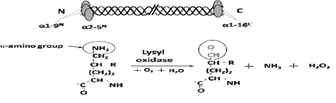
The process of cross-linking is initiated by the oxidative deamination of ε-amino
2-hydroxy-5-amino-5-carboxypentanal [(δ-hydroxy, α-aminoadipic acid-δ-semialdehyde) (Hylald)] and ammonia
(NH3) and hydrogen peroxide (H2O2)(Figure 1.2), and initiates a series of condensation
reactions forming covalent intra- and intermolecular cross-links (15, 38, 39). Covalent intermolecular cross-linking is essential to provide the tissues with mechanical properties to perform their structural functions. In collagen, once the aldehydes (either Lysald or Hylald) are formed by the action of LOX in the C- and the N- telopeptide domains, they undergo a series of condensation reactions involving another aldehyde in the same molecule and/or the juxtaposed Lys, Hyl and histidine (His) residues on the neighboring molecules. The results are the formation of covalent intra- and intermolecular cross-links as shown in figure 1.3 (15, 38-40). The cross-linking chemistry/pattern varies from tissue to tissue rather than particular collagen types since a number of tissue specific factors govern the chemistries. Depending on the state of hydroxylation of Lys residues at telopeptides, two major cross-linking pathways evolved Lysald and Hylald pathways. Besides the enzymatic cross-linking mentioned above, the non-enzymatic cross-linking of collagen has also been taken place (see review in (41-44)).
Figure 1.2 The sites and reaction of LOX on collagen molecules. The specific Lys or Hyl at
telopeptidyl domain of a collagen molecule which can be oxidized by LOX and the reaction of
LOX oxidation in the presence of oxygen (O2) and water (H2O). The end products are Lys ald
or
The Lysald pathway, a major cross-link pathway in non-mineralized tissues, leads to the formation of a tetravalent cross-link, dehydrohistidinohydroxymerodesmosine (deH-HHMD, Lysald x Lysald x His x Hyl) via intramolecular aldol condensation product (ACP), and a trivalent stable cross-link, histidinohydroxylysinonorleucine (HHL, Lysald x Hyl x His) via the iminium cross-link, dehydro-hydroxylsinonorleucine (deH-HLNL, Lysald x Hyl). ACP (α,ß-unsaturated aldol) occurs as an intramolecular cross-link located in the
N-telopeptide domain of the molecule (45, 46). Then Michael addition of N3 imidazole of His to the ß-carbon of ACP resulting as aldolhistidine and further condenses with
ε-amino group of Hyl forming an iminium bond to produce HHMD (46). The deH-HHMD is abundant in skin. Lysald located in the C- and N-telopeptide domains can also cross-link to the juxtaposed ε-amino group of helical Hyl or Lys on the neighboring
molecules to form iminium intermolecular cross-links, deH-HLNL. Then deH-HLNL further condenses with a helical His residue and forms the HHL cross-link (47). This HHL cross-link is abundant in skin and cornea (48) but minimal in skeletal tissues such as dentin, bone, ligament and tendon (49).
described above, the final post-translational modification of collagen is cross-linking formation and this process is initiated by LOX enzymes.
Figure 1.3 Major cross-linking pathways in type I collagen. L.H.: Lysyl hydroxylase, LOX: Lysyl
oxidase, ACP: Aldol condensation product (Intramolecular cross-link), deH: dehydro HLNL:
hydroxylysinonorleucine, DHLNL: dihydroxylysinonorleucine, HHMD:
histidinohydroxymerodesmosine, HHL: histidinohydroxylysinonorleucine, Pyr: pyridinoline, d-:
deoxy, Prl: pyrrole. (15)
Lysyl oxidases
(57). This suggested the existence of alternative spliced or the presence of isozymes with similar but not identical properties. The human LOX gene is located in chromosome 5q23.3-31.2 encoding a 417 amino acid polypeptide, of which the first 21 residues correspond to the signal peptide (58, 59) and the mRNA for human LOX is found as multiple species with sizes of 5.5, 4.3, 2.4 and 2.0 kb due to the use of alternate polyadenylation sites, and partially to the existence of multiple transcription initiation sites (58-60). The mouse Lox gene has been mapped to chromosome 18 (61-63).
Biosynthesis and processing of the LOX precursor
according to the human sequence) (81, 82), that may occur at cell surface in a bound complex with cellular fibronectin (83). It was also found that other extracellular proteases, including mammalian tolloid (mTLD) and tolloid-like-1 and 2 proteases (mTLL-1 and -2) cleaved proLOX at the correct physiological site but at lower efficiency (84). The N-terminal propeptide of LOX has been implicated in regulating the localization of the enzyme within the specific matrix (85) and reversion of ras-transformed cells to the non-oncogenic phenotype (86).
The C-terminal of human LOX is homologous to the N-terminal extracellular domain of the growth factor and cytokine receptor superfamily and this domain overlaps the catalytic site. The consensus sequence found in the N-terminal modules of Class 1 receptors, C-x9-C-x-W-x26-32-C-x10-13-C (where C is cysteine, W is tryptophan, and Xn is a
defined number of any amino acid), is conserved in human LOX (87). The cytokine receptor module is known to play an adhesion role in several proteins of the growth factor and cytokine receptor superfamily. Based on structure prediction, the cytokine receptor-like domain in the LOX protein forms a partial receptor site and it is questionable if it binds cytokines in the same way (88).
Catalytic mechanism
Cofactor
the resting enzyme is in the Cu(II) state, and is bound tetragonally distorted, octahedrally coordinated ligand field (93). The sequence WEWHSCHQHYH in human has been suggested to be the actual copper-binding region, which provides four histidine residues that are involved in the copper-binding coordination complex (94). In addition to the tightly bound Cu ion, purified LOX preparations have been shown to contain 5-9 atoms of loosely bound copper per enzyme molecule (93). Experiments in a cell free transcription/translation system in vitro have shown that the unprocessed 50 kDa LOX precursor binds copper (95). A study showed that protein synthesis was important for the incorporation of copper into enzyme but N-linked glycosylation had no effect on secretion of the copper that was bound to LOX. In addition, an inhibition of processing of the 50 kDa LOX precursor into the 32kDa active form with a PCP inhibitor had minimal effect on the amount of copper that was bound to LOX in the cell media (95). Kagan et. al. in 1995 (96) has shown that the propeptide region is not essential to the folding and secretion of the functional enzyme. A truncated rat LOX cDNA lacking sequences which encode for the bulk of the propeptide region was transfected into Chinese hamster ovary cells. The 29 kDa form of mature LOX which catalyzed the deamination of human recombinant tropoelastin and alkylamines was secreted and this secreted enzyme could be inhibited by ß-aminopropionitrile (ßAPN). However, the expression analysis of recombinant LOX in myofibroblast-like cells showed that propeptide region was important for the secretion (97). These controversial data might be due to the difference of type of promoter of the vectors used in the study. These results indicated that copper is incorporated into proLOX in the endoplasmic reticulum or during protein trafficking through the Golgi elements and independent to glycosylation (95, 98).
reaction involving Cu2+-mediated/assisted oxidation of a specific peptidyl tyrosine (res 345 in ratLOX) followed by covalent cross-linking with the ε-amino group of a conserved
peptidyl Lys (res 314 in rat LOX)(105, 106). This unique intramolecular cross-link, LTQ, together with several disulfide linkages within the LOX molecules, most likely contributes to the remarkable physicochemical stability of LOX (34, 104). The covalently linked lysyl component of LTQ of LOX might play an important role in the preference of LOX for peptidyl lysine substrates in cationic protein microenvironments both as a result of its donation of anionic charge to the active site and possibly by restricting the rotation of the LTQ ring in a manner that properly orients the carbonyl cofactor with respect to peptidyl lysine substrates (104). LTQ could be formed in the endoplasmic reticulum or during protein trafficking through the Golgi apparatus (98)(Figure 1.4)
The mechanism of action of LOX has been described in many studies (93, 100, 107-109). The ε-amino group of the substrate lysine residue condenses with one of the
Figure 1.3 Pathway for LOX biosynthesis. The LOX precursor is trafficked into the rough
endoplasmic reticulum where its signal peptide is cleaved. The precursor is glycosylated within
the endoplasmic reticulum at two possible Asn residues located in the propeptide region. The
addition of copper (Cu2+) and the formation of LTQ cofactor consequently occur. After secretion of
the LOX precursor into the ECM, the propeptide region is cleaved by procollagen C-proteinase
(PCP) between Gly-168 and Asp-169 to obtain and an active 32 kDa enzyme. Lysyl oxidase
propeptide (LOPP) and mature LOX has been reported to enter the cell and nucleus after
secretion. Molecular mass of LOX polypeptide is indicated according to human LOX enzyme. (69,
70, 112, 113). Cu: Copper, LTQ: Lysyl tyrosylquinone.
Inhibitors
(50). ßAPN was found to be a potent irreversible inhibitor and its inhibition was temperature- and time-dependent (116). ßAPN binds to the LTQ, the active site of LOX, and forms a dead-end complex. A free aldehyde product was not generated and the copper content of LOX was not altered upon incubation with ßAPN (117, 118). ßAPN has been used to specifically as a lysyl oxidase inhibitor. haloamines and ß-nitroethylamine are also reported as mechanism-based irreversible inhibitors of LOX similar to ßAPN (119). Benzylamine derivatives containing para substituents of increased electronegativity and isomers of aminomethypyridine are also identified as reversible ground state inhibitors by forming LOX-bound intermediates that are not completely processed into aldehydes (107). Vicinal diamines like cis-1,2-diaminocyclohexane and ethylenediamine are potent irreversible inhibitors (120). LOX is inhibited by heparin (121), N-(5-aminopentyl) azidine (122), and trans-2-phenylcyclopropylamine (123). LOX is inhibited by homocysteine thiolactone and its selenium and oxygen analogs in an active-site-directed and irreversible manner (124). LOX is inhibited by ascorbic, and esthoric acids and 3,4-dihydroxybenzoate by possibly the ascorbic acid structure (125).
Substrate specificity
development of fluorescence-based assay for LOX-dependent H2O2 production (127,
128).
The sequence region within the LTQ domain between Lys314 and Tyr349 are enriched in anionic residues. Once these two regions of LOX become covalently cross-linked to each other as the LTQ cofactor is generated, both of these regions would cooperatively provide an abundance of negatively charged sites in the microenvironment of the active site. It is likely that such an arrangement underlies the strong preference of LOX for cationic protein substrates (104). Interestingly, the sequences surrounding the susceptible lysines in collagen are in hydrophilic sequences containing anionic residues. For example, the lysine residue within the Asp-Glu-Lys-Ser sequence which occurs at the N-terminal region of the α1(I) collagen chain within the mature type I collagen
functional catalyst (83). Histone H1 and H2 have been demonstrated to have an interaction with LOX in vitro (132) and incubation of Histone H1 with LOX results in the catalytic formation of hydrogen peroxide implicating that histone H1 is a substrate of LOX (133). Basic fibroblast growth factor was reported to be a substrate of LOX as the oxidation of lysine residues in bFGF by LOX resulted in the covalent cross-linking of bFGF monomers to form dimers and higher order oligomers and dramatically altered its biological properties (134). Recently, LOX has been reported to be essential for hypoxia-induced metastasis because the administration of ßAPN, specific anti LOX antibody or short hair-pin RNA could inhibit the metastasis in animal model. It is still unclear whether LOX might oxidize an unknown key protein and inhibit its function in the cancer metastasis, or whether the by-product H2O2 from the oxidation plays a role in the
inhibition of cancer metastasis (135).
Regulation of LOX
The factors that were reported to play roles in the regulation of LOX gene expression are transcriptional factors and growth factors as shown in Table 1.1.
Table 1.1 The various regulators and effects on LOX mRNA and enzymatic activity.
Effector (reference) Cell or tissue Effect
IFN- (136) Rat aortic smooth muscle cells Down regulation of mRNA; decreased mRNA half-life
bFGF (75) Mouse osteoblastic cells Decreased mRNA level (1-10nM) upregulated of mRNA (0.01-0.2 nM)
FGF-2 and IGF-1 (137) Inflamed oral tissue, rat fibroblastic mesenchymal cells
Increased of mRNA
PGE2 (138-140) Rat lung fibroblasts
Human embryonic lung fibroblasts
TGF-ß1 (75, 76, 138, 139, 141-146)
Rat aortic smooth muscle cells human gingival fibroblast, flexor reticulum cells, renal cell lines, rat lung fibroblasts, kidney tubular epithelial cells, mouse osteoblasts
Increased mRNA level and enzyme activity
BMP-2 (147) Murine pre-myoblast cells Increased mRNA level
TNF- (79, 148) Mouse osteoblastic cells Aortic endothelial cells
Decreased mRNA, protein level and enzymatic activity
Decreased mRNA and level and enzymatic activity
Cadmium (149) Mouse fibroblasts
Mouse cadmium-resistant fibroblasts
Decreased mRNA level Increased mRNA level
Testosterone (150, 151) Calf aortic smooth muscle cells, rat granulosa cells
Increased enzyme activity and increase mRNA expression Follicle stimulating
hormone (151)
Rat granulosa cells Decrease mRNA expression and enzyme activity
Growth differentiation factor-9 (151)
Rat granulosa cells Increase mRNA expression and enzyme activity
Activin A (151) Rat granulosa cells Increase mRNA expression and enzyme activity
Bleomycin (152) Human lung fibroblasts Human dermal fibroblasts
Increased mRNA level Decreased mRNA level Hydralazine (152) Human dermal fibroblasts Increased mRNA level Minoxidil (152) Human dermal fibroblasts Increased mRNA level Adriamycin (153) Rat kidney glomeruli, medulla Increased mRNA level cAMP (140, 154) Rat and human vascular smooth
muscle cells
Upregulated of transcription
PDGF (155) Rat vascular smooth muscle cells Upregulation of mRNA Dexamethasone (156) Cultured fetal murine lungs Upregulation of mRNA
Retinoic acid (157) Adipocytes in early adipogenesis Prevents downregulation of mRNA and enzyme activity. Hypoxia inducible
factor-1 (factor-158)
Breast cancer cells Increased mRNA and protein level but decreased enzymatic activity
(158) and enzyme activity Low density lipoprotein
(77)
Vascular endothelial cells Decrease mRNA expression and enzyme activity
Homocysteine (159) Vascular endothelial cells Decrease LOX activity (35 M)
Decrease LOX mRNA
expression and LOX promoter activity (250M)
Pulsed ultrasound (160) Mouse osteoblastic cells Increase LOX mRNA expression and enzyme activity (30 mW/cm2) Metavanadate (161) Rat fibroblast Decrease LOX activity
Modified from (88, 162)
Animal models
respectively compared with those of wild-type littermates. There were significant decreases in DHLNL and HLNL of 43 and 39% respectively in the Lox-/- animals as compared with wild type total body collagen cross-links. Interestingly in Lox+/- mice, DHLNL was 100% of wild type, while HLNL was only 64% of wild type content. This could represent a greater role for LOX in HLNL cross-links. Moreover, DHLNL in lung of Lox-/- animals was not different among genotypes while decreased in HLNL in Lox+/- and Lox-/- was 14 and 32% respectively (164).
Lysyl oxidase isoenzymes
Table 1.2 The comparison of LOX family member. Family member Human Chr. Mouse Chr. mRNA and protein size Highest mRNA tissue distribution Protein domains %similarity to LOX domain %similarity to LOXL2 domain
LOX 5 18 6.8, 4.8 kb 417 res. Lung, skeletal muscle, kidney, heart Amine oxidase
100 63
LOXL1 15 9 2.4 kb
574 res. Lung, heart, spleen skeletal muscle, pancreas Amine oxidase
85 63
LOXL2 8 14 4.0 kb
774 res. Lung, thymus, skin, testis, ovary 4 SRCR, Amine oxidase
58 100
LOXL3 2 6 3.3 kb 753
res. Heart, uterus, testis, ovary 4 SRCR, Amine oxidase
65 78
LOXL4 10 19 3.5 kb 756 res. Skeletal muscle, testis, pancreas 4 SRCR, Amine oxidase
62 79
Lysyl oxidase-like protein (LOXL1)
(180, 181). LOXL1 protein is a secretory protein that is expressed in active fibrotic diseases and in the early stromal reaction of breast cancer (182). Coincident appearance of increased steady-state levels of LOXL1 and COL3A1 mRNAs was detected in the early development of liver fibrosis, suggesting that LOXL1 protein is involved in the development of lysine-derived cross-links in collagenous substrates. In contrast, steady-state levels of LOX mRNA were increased throughout the onset of hepatic fibrosis and appeared in parallel with increased steady-state level of COL1A1 mRNA (183). A specific antibody against LOXL1 has been used to identify proteins immunochemically distinct from LOX in various cells and in bovine aorta. The species of the protein are approximately 68, 52, 42 and 30 kDa (182). A 56 kDa LOXL1 protein was isolated from bovine aorta and the precursor needs to be cleaved by BMP-1 to be active (176). The mice lacking LOXL1 do not deposit normal elastic fibers in the uterine tract post partum and develop pelvic organ prolapse, enlarged airspaces of the lung, loose skin and vascular abnormalities with concomitant tropoelastin accumulation. Distinct from the prototypic LOX, LOXL1 localizes specifically to sites of elastogenesis and interacts with fibulin-5. Thus elastin polymer deposition is a crucial aspect of elastic fiber maintenance and is dependent on LOXL1, which serves both as a cross-linking enzyme and an element of the scaffold to ensure spatially defined deposition of elastin (184).
Lysyl oxidase-like protein 2 (LOXL2)
LOXL2 was originally cloned, characterized, and named as WS9-14 for its possible association with Werner syndrome (167). Later it was found that WS9-14 mRNA corresponds to LOXL2 mRNA but WS9-14 transcript encodes one additional SRCR domain in its 5’ end region (88, 168). LOXL2 mRNA encodes an 87 kDa
polypeptide in its amino acid sequence from residue 546 to 751 in its C-terminal region. This region contains all conserved amino acid sequences needed for the proper function of the mature 32 kDa form of LOX enzyme (167, 168). LOXL2 gene has been mapped to chromosome 8p21 in humans (185) and chromosome14 in mice (186), and it consists of at least 11 exons (168). LOXL2 is abundantly expressed in senescent fibroblasts and several adherent tumor cell lines, but is down-regulated in several non-adherent tumor cells. This suggests that it may be involved in cell adhesion and that a loss of this protein may be associated with the loss of tumor cell adhesion leading to tumor metastasis. LOXL2 shows similar spatial expression with LOX and LOXL in human placenta and fetal tissues in early pregnancy. However, this pattern diverges during gestation (187). In full-term human placenta, LOX is expressed predominantly in the amniotic epithelium, with little expression in the placenta, while LOXL shows the highest expression in the placenta and lowest expression in the amnion. LOXL2 expression differs in that it is detected predominantly in chorionic cytotrophoblasts of the membranes with only low expression levels in the amnion and placenta (168, 187, 188).
Lysyl oxidase-like protein 3 (LOXL3)
shown to be due in part to the LOX polypeptide (112) or LOXL (189). LOXL3 gene has been mapped to chromosome 2p13 in humans (170) and chromosome 6 in mice (171, 172). LOXL3 is expressed highly in placenta, heart, ovary, testis, small intestine and spleen (162, 171).
Lysyl oxidase-like protein 4 (LOXL4)
LOXL4 was cloned from placental, kidney, fetal tissues and chondroblast and osteoblast cDNA (173-175). The deduced 756-amino acid protein contains an N-terminal signal sequence, 4 scavenger receptor cysteine-rich (SRCR) domains, lysyl and tyrosyl residues that form the carbonyl cofactor within the catalytic site, and a cytokine receptor-like domain at the C terminus. LOXL4 shares 51% and 54% amino acid identity with LOXL2 and LOXL3, respectively. LOXL4 gene has been mapped to chromosome 10q24 in humans (175) and chromosome 19 in mice (162, 173). LOXL4 is highly expressed in skeletal muscle, testis and pancreas and not expressed in leukocytes (173).
Novel biological functions of LOX
LOX in tumor suppression
carcinomas, small cell carcinomas and neuro-endocrine carcinoma (200). It has been demonstrated that a loss or reduction of LOX function during tumor development may be a direct consequence of somatic mutations and may be associated with the pathogenesis of colon cancer (201). The LOX gene is located in chromosome region 5q23, which is known to be deleted in a high frequency in many different types of cancer (202). LOX was reported to inhibit ras-mediated transformation by preventing the activation of NF-Kappa B (NF-B) (203). Lysyl oxidase propeptide (LOPP) was also
reported to have tumor suppression ability by inhibiting Erk1/2 Map kinase activation (86, 204) and recently by inhibiting Akt activity and BCL2, a tissue-specific NF-B target gene (205).
LOX dependent chemotaxis
ßAPN has been reported to inhibit fibroblast migration in a dose-dependent fashion without inhibiting proliferation (206). Purified 32 kDa mature LOX was shown to play a role as a potent chemoattractant for human peripheral blood mononuclear cells, with a 237% increase in migration over the enzyme-free or catalytically inhibited LOX controls seen at 10-9 M LOX (207). The result showed that H2O2 product of the
LOX-catalytic reaction appeared to mediate the chemotactic response since the observed result was not detected when the LOX was inactivated or absent from the reaction (208). The result also suggested that the chemotactic effect was not due to the reaction of LOX with secreted protein in the media but more likely due to the direct access of LOX to cell-associated substrates. The cellular responses after addition of LOX showed elevated level of intracellular H2O2, enhanced stress fiber formation, and increased focal adhesion
its isoform mRNA levels are upregulated in invasive type compared to those of non-invasive type and the invasion phenotype was facilitated by active but not inactive LOX. In addition, the invasion was inhibited when the antisense mRNA of LOX was transfected or ßAPN was added into the cultured (209). A further report from the same group demonstrated that this LOX-dependent chemotactic response of breast cancer cells was elicited by the H2O2 product of the LOX-catalytic reaction on unidentified
substrates (210). Recently it has been shown that actin stress fiber formation and Rho activity in breast cancer cell line are increased through the p130 (Cas)/Crk/DOCK180 signaling complex by inhibition of LOX in these cells (211). It suggests that the down-regulation of LOX activity could limit the invasiveness of breast cancer. Other isozymes have been reported that their expression are high in metastatic breast cancer cells and correlate with increased tumor malignancy and increased fibrotic foci (209, 212). The elevation of LOX under hypoxic condition in head and neck tumor cells appeared to be essential for the hypoxia-induced metastatic response of these cells and the increased invasiveness was prevented by treatment of ßAPN, LOX antisense oligonucleotides, LOX antibody or short hairpin RNA expression but not with LOX sense oligonucleotides (135). This study did not show the role of H2O2 product of the LOX-catalytic reaction.
Another study showed that LOX interacts with hormone placental lactogen and synergistically promotes breast epithelial cell proliferation and migration. However, the study showed that lactogen was neither a substrate nor an inhibitor so the H2O2 from the
Intracellular and intranuclear activities
LOX was reported to be associated with cytoskeleton protein in cytoplasm in cultured fibroblasts (215). It was not conclusive that the observed LOX represented the proenzyme and/or mature LOX since the molecular weight of the LOX was not determined. It has been reported that an intracellularly expressed recombinant mature 32 kDa LOX might regulate the activity of COL3A1 gene promoter (195). The coinjection of LOX with oncogenic p21-rasval12 into Xenopus laevis oocytes suggests one possible intracellular role for LOX in antagonizing a Ras-induced meiotic maturation of these cells. It has been suggested that a LOX-dependent block in oocyte maturation may be downstream of Erk2, a member of the mitogen-activated protein kinases (216).
LOPP 18kDa in osteoblasts. In proliferating cells, mature LOX located in nucleus and perinuclear region while LOPP associated with Golgi and endoplasmic reticulum. In differentiating cells, mature LOX and LOPP colocalized with the microtubule network (113). However, the mechanism of how LOX and LOPP can be uptaken intracellularly or intranuclearly remains unelucidated (112). Recently LOX has been reported to regulate elastin promoter via intracellular effects of transforming growth factor-ß1 (TGF-ß1). The author showed the reduction of Smad 3 and 4 in the addition of TGF-ß1 when the coding sequence of mature LOX without signal peptides was transfected in 293T cells. The cross control mechanism remains unclear (219).
Growth factor and cellular modulation
In addition to collagen and elastin, purified LOX oxidized a number of basic globular proteins in vitro with pI values > 8.0, but did not oxidize neutral or acidic globular proteins with pI value < 8.0 (126). The study demonstrated Histone H1 as a substrate of LOX by detecting the increase of lysinonorleucine cross-link and ACP in histone H1 sample after incubation with purified LOX. Histones are involved in chromatin packing in nucleus and can be oxidized by LOX. The histone oxidation might modulate the packing state of nuclear chromatin (220). Basic fibroblast growth factor (bFGF) was reported to be a substrate of LOX in vitro. The oxidation of lysine residues in bFGF by LOX resulted in the covalent cross-linking of bFGF monomers to form dimers and higher order oligomers. The fluorescence LOX assay showed temperature dependent of amine
oxidase activity of LOX in which LOX oxidized 14 lysine residues at 55C, and 5-6
Lysyl oxidases in an osteoblast cell culture systems and in mineralized tissues
LOX was first identified in bone from saline extract in 1968 (50). Several reports have shown that the activity of purified bone LOX was inhibited by ßAPN but it was not well characterized (50, 90). LOX was also identified in dentin (221). In chick calvarial
diminished collagen processing and LOX activity; however, the collagen cross-links, Pyr and d-Pyr were not altered. Moreover, a slight increase (10%) of collagen fibril diameter was reported (225). LOX has been also shown to be regulated by bFGF (226), TGF-ß1 (75) and TNF-α (79) in MC3T3-E1 (MC) cells. The expression of LOX and its isoforms in
MC cells have recently been investigated and the result showed that LOX and all LOXLs except LOXL2 were expressed in this cell line and the expression during the cell differentiation and matrix mineralization was distinct from one another (74). The stage-dependent intracellular distribution of mature LOX 32kDa and LOPP 18kDa was investigated in MC cells. In proliferating cells, mature LOX located in nucleus and perinuclear region while LOPP associated with Golgi and endoplasmic reticulum. In differentiating cells, mature LOX and LOPP colocalized with the microtubule network (113).
Osteolathyrism
kyphoscoliosis, bone deformities, weakening of tendons and ligament attachments, dislocation of joints, weakening of skin and cartilage, hernias and dissecting or saccular aneurysm of the aorta (231). In a rabbit model, osteolathyrism does not become manifest until practically all mature cross-linking that can be affected has been inhibited (232). In experimental osteolathyritic rat model, osteolathyrogens cause a spectrum of tissue alterations in skeleton, soft connective tissue and arteries (233). Ectopic exostoses and aneurysmal-like bone cysts in mandible and long bones were observed (234, 235) due to significantly impaired turnover and remodeling rates of periosteal and endosteal bone (236). Fracture healing in osteolathyritic animals results in excessive callus formation. The vast fracture callus is very fragile and consists of irregular cartilage and premature woven bone (237, 238). Fractured tibias in the animal developed excessive amounts of mechanically weak callus tissue with irregular cartilage and showed reduced glycosaminoglycan accumulation. In lathyritic calluses, the maximal mRNA of type II and IX collagens and aggrecan core protein was peaked 4 days earlier than in controls reflecting the less well control of endochondral ossification in lathyritic calluses. Interestingly the expression of TGF-ß1 mRNA in lathyritic calluses was already peaked at the early time-points while that of control was relatively low and increased gradually at later time-points of study (239).
TGF-ß in bone and its potential regulation by LOX
these members share a cluster of conserved cysteine residues forming a characteristic cysteine knot. They are synthesized as precursors with a large N-terminal propeptides that is proteolytically cleaved from a mature C-terminal domain peptide.
TGF-ß1 is the prototypic member of the TGF-ß superfamily, and bone ECM is a major storage site in the body for this growth factor (247). Though TGF-ß2 and 3 are also present in bone, TGF-ß1 is the predominant isoform (>90%) at the level of 200-700 μg/Kg in this tissue (248, 249). In bone, TGF-ß1 plays pivotal roles in many, if not all,
aspects of the tissue development, remodeling, mechanical properties and aging (250-252). Numerous studies, though not always consistent, have shown that TGF-ß1 stimulates recruitment and proliferation of osteoblast progenitors (253, 254), stimulates matrix production such as collagen, fibronectin, osteopontin, osteonectin, proteoglycans, but inhibits osteocalcin (250, 255), inhibits late stage of osteoblast differentiation and matrix mineralization (256, 257), and modulates osteoclast differentiation (258). The abundance of this growth factor with such potent effects on cells predicts the need for tight regulation of its biological activities. Indeed, several studies have shown that deregulation/overactivation of TGF-ß could be detrimental to osteoblastogenesis and bone formation causing bone defects (259-261).
TGF-ß1 is secreted as a latent complex that needs to be activated before being capable of eliciting biological effects (262, 263), and it appears that it is the only growth factor known to be produced in a latent/inactive form. The latency is achieved by noncovalent association between TGF-ß and its propeptide, called latency-associated peptide (LAP), forming a ~100 kDa small latent complex (SLC). For this association, thus, to confer “latency” to TGF-ß, res 50-85 near the N-terminus of LAP is important
the authors also identified the RKPK sequence near the C-terminus of mature TGF-ß as a potential recognition sequence for LSKL. Interestingly, the C-terminus of the mature TGF-ß (res 83-112) including the RKPK sequence has also been demonstrated to be the critical site for the binding to its type II receptor (TßRII) that initiates the signaling cascade of TGF-ß (241, 266, 267). Thus, the association of LSKL of LAP and RKPK of TGF-ß may sterically prevent receptor binding (265). It is of interest to note that the vicinity of the critical regions of LAP and TGF-ß for their association and the region of TGF-ß to bind TßRII for signaling are enriched in basic amino acids, including several Lys residues, yielding high pI values (e.g. estimated pI for res 50-85 of LAP is 9.98, and for res 83-112 of mature TGF-ß1 is 9.24). In many cell types, LAP is further disulfide-bonded by latent TGF-ß binding proteins (LTBPs) to form a ~290kDa large latent complex (LLC) prior to secretion, and in the ECM, LLC is stored possibly by covalently linked to matrix components (263, 268). LTBP is thought to be important for efficient secretion, storage of TGF-ß in ECM and TGF-ß activation (268). Although almost all nonmalignant cells secrete TGF-ß as a part of LLC, bone cells form an exception as they efficiently secrete SLC, thus, lacking LTBP (269-271). At least 50% of the latent TGF-ß forms produced by bone cells was found to be SLC (272) indicating its specific role in bone biology. Bone cells also produce LLC like other cell types (273) where LTBP may play dual roles in matrix storage and structural element for bone formation (272). The latent TGF-ß can be activated by plasmin, thrombospondin-1, αvß 6 integrin, heat, low
binding molecules including small leucine-rich proteoglycans, decorin and biglycan (260, 275, 276) that may sequester TGF-ß in the matrix and/or diminish TGF-ß binding to its cell receptor (277). However, the significance of these interactions in bone ECM is not known. Furthermore, potential extracellular modification of TGF-ß that may change and/or stabilize the potency is unknown.
During the course of my study on the biological significance of the post-translational modifications of collagen in bone, I discovered that in an osteoblastic cell culture system, higher LOX expression resulted in less collagen production and suppressed TGF-ß1 signaling while lower LOX expression exhibited more collagen production and enhanced TGF-ß1 signaling. This intriguing finding prompted me to examine the potential interaction between LOX and this growth factor. When these two proteins were co-expressed in 293 cells, the binding was detected in a dose-dependent manner. No binding was identified between LOX and BMPs tested or LAP. Furthermore, TGF-ß1 was found to be bound LOX in the mineralized bone matrix. Considering the facts that LOX possesses a strong preference towards basic proteins as its substrates and TGF-ß1 (and LAP) is a basic protein, especially its critical domain for “latency” and “signal transduction” is enriched in basic amino acids including Lys residues, it is then
CHAPTER II
Hypothesis
CHAPTER III
STUDY I
Lysyl oxidase regulates collagen quality and quantity in osteoblasts
Phimon Atsawasuwan, Yoshiyuki Mochida, Michitsuna Katafuchi, Mitsuo Yamauchi
Specific aims
1. To establish several MC cell clones that express higher or lower levels of LOX by overexpression or antisense approaches.
ABSTRACT
INTRODUCTION
EXPERIMENTAL PROCEDURES
Cell Lines and Culture Conditions– MC cells subclone 4, (280) were purchased
from American Type Culture Collection (CRL-2593) and maintained in -minimum essential medium (Gibco) containing 10% FBS (Atlanta Biologicals) and supplemented with 100 U/ml penicillin G sodium (Gibco), 100 g/ml streptomycin sulfate (Gibco) in a
5% CO2 atmosphere at 37 C. The medium was changed twice a week.
Isolation of LOX cDNA and generation of constructs– Total RNA was isolated from MC cells using TRIzol reagent (Invitrogen) according to the manufacturer’s
protocol. Two g of total RNA was used for reverse transcription and the cDNA was synthesized using the Omniscript Reverse Transcriptase kit (Qiagen). Specific primers for the coding region of the mouse LOX (Genbank accession NM010728) was as follows; forward primer, 5’-CCCGGTCTTCCTTTTTCTCCTAGCC-3’ and reverse primer, 5’-ATACGGTGAAATTGTGCAGCCTGA-3’. PCR amplification was performed by
ProofStart DNA polymerase (Qiagen) with an annealing temperature of 62 C for 35 cycles. After adding 3’A-overhangs, the PCR products were then ligated into the
Transfection and generation of stable cell clones– MC cells were transfected with
pcDNA3.1/V5-His/LOX constructs (S and AS orientation) or pcDNA 3.1/V5-His A vector (empty vector [EV]; Invitrogen) using Fugene 6 transfection reagent (Roche). After 48 h, cells were trypsinized and plated at a low density. Single cell-derived clones (S and AS clones) were isolated and maintained in the presence of 400 g/ml G418 (Invitrogen) for
up to 4 weeks.
Immunoprecipitation and Western blot analysis of the S and AS clones– MC, EV,
S and AS clones were plated onto 10-cm dishes (Falcon) at a density of 2.0 x10⁵/dish and cultured in -minimum essential medium (-MEM, Gibco containing 10% FBS) with the same supplements described above. After 7 days of cell culture, culture media were collected and incubated with anti-LOX antibody (Imgenex) at 4 C overnight. After the addition of rec-Protein G-Sepharose conjugate beads (Zymed laboratories) and incubation at 4C for 30 min, the samples were washed with lysis buffer containing
150mM NaCl, 20 mM Tris-HCl pH 7.5, 10mM EDTA, 1% Triton X-100, 1% deoxycholate, 1.5% aprotinin, and 1mM phenymethylsulfonyl fluoride three times. Protein bound to the beads were dissolved in SDS sample buffer (100mM Tris HCl, pH 8.8, 0.01% bromophenol blue, 36% glycerol, and 4% SDS) in the presence of 10mM dithiothreitol (DTT), applied to 4-12% gradient SDS-PAGE (Invitrogen), transferred onto a polyvinylidene fluoride membrane (Immobilon-P, Millipore Corp.), and subjected to Western blot analysis with anti LOX antibody and a secondary antibody, an anti-rabbit IgG conjugated to alkaline phosphatase [(ALP), Pierce Biotechnology]. The immunoreactivity was visualized by an ALP conjugate substrate kit (Bio-Rad Laboratories).
Proliferation assay– MC, EV, S and AS clones were plated in triplicate at a
day 2, 4 and 6 days of cell culture, the cells were subjected to MTS cell proliferation assay (CellTier 96®, Promega) according to the manufacturer’s protocol. Briefly, 100 l
of MTS solution was added into each well and incubated at 37 C for 4 hrs. The amounts of formazan compound, 3-(4,5-dimethylthiazol-2-yl)-5-(3-carboxymethoxyphenyl)-2-(4-sulfophenyl)-2H-tetrazolium), MTS, produced by metabolically active cells were measured by absorbance at 490 nm.
Quantitative Collagen Cross-link Analysis– MC, EV, S and AS clones were cultured in -MEM, 10% FBS and 50g/ml ascorbic acid. After 2 weeks of culture, cells/matrices were washed with phosphate buffered saline (PBS), scraped into a 1.8 ml eppendorf tube and washed with PBS, distilled deionized water (DDW) twice then lyophilized. Two milligrams of the lyophilized samples were suspended in 0.15M N-trismethyl-2-aminoethanesulfonic acid and 0.05M Tris-HCl buffer (pH 7.4) and reduced with standardized NaB3H4 (281), hydrolyzed with 6 N HCl in vacuo, after flushing with N2
at 105C for 22 h. The hydrolysates were dried by a speed vacuum concentrator (Savant
were done in triplicate and the values are means+SD. To confirm the reproducibility of the results, three independent experiments were performed.
Collagen content determination– MC, EV, S and AS clones were cultured as
described above. At day 3 and 7, cell culture medium were collected and cell/matrix layers were collected and lyophilized as described above. The dry cells/matrices and lyophilized medium were hydrolyzed with 300 µl of 6 N HCl (Pierce) in vacuo, after flushing with N2, for 22 h at 105ºC, dried, dissolved in 300 l of distilled water, filtered
with 0.22 m filter unit and kept in 4C. An aliquot of each hydrolysate was subjected to amino acid analysis on a Varian high-performance liquid chromatography (HPLC) system (9050/9012; Varian Associates Inc) using ninhydrin (Pickering laboratories) for color development at 135oC to determine hydroxyproline residue (282). The collagen content in the cells/matrices was calculated using a value of 300 residues of Hyp per collagen molecule. The total collagen content in the medium and matrices was calculated and normalized with the number of the cells in each dish. To confirm the reproducibility of the results, three independent experiments were performed.
Transmission electron microscopy– The cell/matrix layers of MC, EV, S and AS
Micrograph software (Gatan, Inc). For each sample, a total of 500 collagen fibril
diameters and the number of fibrils per m2 were measured randomly using Scion Image software (Scion Corporation).
In vitro mineralization assay– MC, EV, S and AS clones were plated on 35 mm
plastic dishes at the density of 2x10⁵ cells/dish (Falcon) and cultured until confluence. The medium was then replaced with the one supplemented with 50 g/ml of ascorbic acid and 2 mM of ß-glycerophosphate (mineralization medium), and maintained for up to 4 weeks. In vitro mineralization assay was performed at the end of week 2 and 4. At each time point, cell/matrix layers were washed with 1X PBS twice, fixed with 100% cold methanol, stained with 1% Alizarin Red S (Sigma) for 15 min and washed with DDW and dried at room temperature(74). At week 4, the calcium contents were quantified by measuring the amount of Alizarin red S bound to mineralization nodules in the cultures. Briefly, after staining with Alizarin red S, the cultures were washed with 10% (w/v) cetylpyridinum chloride in 10 mM sodium phosphate, pH 7.0 for 15 min. The dye concentration in the extracts were subjected to spectrophotometer at absorbance 562 nm (284, 285) to compare the amount of alizarin and normalized with cell numbers. To confirm the reproducibility of the results, three independent experiments were performed.
Statistical analyses– All statistical analyses were performed using Sigma stat
RESULTS
Generation of the S and AS clones– The immunoprecipitation/Western blot analysis of three S, three AS clones and the controls are shown in Figure 2.1. S3 showed the highest level of LOX (S3: 2.34, S2: 2.25 and S1: 1.47 fold) while AS1 showed the lowest level of LOX (AS1: 0.49, AS2: 0.73, AS3: 0.85 fold) compared to the average expression level of MC and EV. Using MTS assay, the proliferation rate of S clones were comparable with controls (MC and EV) while those of AS clones were slightly lower compared to controls at the same timepoint of study but there is no statistically difference among the clones and when compared to controls (P>0.05) (Figure 2.2).
Quantitative collagen cross-link analysis–To confirm if the generated S and AS
clones secreted functional recombinant LOX, the collagen cross-links in each clones were quantified. The collagen cross-links produced in MC, EV, S, and AS clones at 2 weeks of culture were bifunctional reducible cross-links, i.e. DHLNL and HLNL and a trivalent nonreducible cross-link, Pyr. The cross-link analysis of S clone exhibited higher levels of total aldehydes (DHLNL+HLNL+2xPyr), DHLNL and Pyr whereas that of AS exhibited lower levels of total aldehydes, DHLNL and Pyr (Figure 2.3). The Pyr contents in S clones were constantly higher (157, 206 and 260 % increase) whereas those in AS clones lower (64, 57and 42% decrease) than those of controls (P<0.05) (Table 2.1). The data from cross-link analysis is consistent with the level of LOX in S and AS clones (Figure 2.1).
Collagen content determination– The total collagen content in each clone at day
all S clones was significantly lower than those of controls (MC and EV) at both time-point of study (P<0.05) while that in all AS clones was comparable to those of the controls at day 3 but significantly higher than those of controls at day 7 in AS1 and AS2 clones (P<0.05).
Ultrastructural analysis of collagen fibrils– Cross-sectional views of collagen
fibrils obtained from cultures of S (S1, S2, S3) and AS (AS1, AS2, AS3) clones and the controls (MC, EV) are shown in Figure 2.5. As shown in histogram, MC cells had a mean diameter of 46.2+9.4 nm with a range of 29.2-87.5 nm, which is similar to that of the EV clone (mean 42.3+6.8 nm; range 29.2-62.5 nm). In all S clones, the mean fibril diameters and their range were significantly smaller than those of controls (P<0.05). Among the S clones, S3 that exhibited the highest level of LOX overexpression (Fig 2.1) showed the smallest fibril diameter (mean 21.1+7.9 nm; range 3.7-46.6 nm) followed by S2 clones (mean 24.2+9.97 nm; range 3.7-44.1 nm) and S1 clones that exhibited the lowest level of LOX overexpression (mean 30.1+4.4 nm; range 3.7-44.1 nm). In all AS clones, the mean fibril diameters and their range were significantly larger than those of controls (P<0.05). Among AS clones, AS1 that exhibited the lowest level of LOX showed the largest fibril diameter (mean 99.1+43.2 nm; range 31.3-226.7 nm) followed by AS2 clones (mean 66.9+21.6 nm; range 22.2-170.8 nm) and AS3 clones that exhibited LOX only slightly lower than controls (mean 58.4+19.6 nm; range 16.7-129.2 nm) (Figure 2.6).Therefore, the fibril diameter and distribution range were inversely correlated with the level of LOX expression in the clones. The number of collagen fibrils were counted per square micron randomly from 6 areas and presented in Table 2.2. The number of fibrils in all S clones were significantly lower than those of the controls (P<0.05).
In vitro mineralization assay– The result of in vitro mineralization assay is shown
Table 2.1 The amount (moles/mole collagen) of total aldehydes and reducible and,
non-reducible cross-links in each clone are expressed as mean+S.D. ( - : decrease, * P<0.05,
ANOVA)
DHLNL HLNL PYR Total
Aldehydes
% change of PYR
AS1 0.354+0.064* 0.123+0.007 0.012+0.005* 0.500 -64.61 AS2 0.448+0.023 0.138+0.025 0.014+0.003* 0.614 -56.92 AS3 0.463+0.013 0.079+0.015* 0.019+0.010 0.580 -41.54
MC 0.475+0.010 0.168+0.030 0.029+0.007 0.701 0 EV 0.476+0.032 0.172+0.024 0.036+0.015 0.719 0 S1 0.487+0.052 0.130+0.001 0.084+0.008* 0.783 156.92 S2 0.514+0.021 0.140+0.022 0.100+0.013* 0.853 206.15 S3 0.609+0.126* 0.127+0.003 0.117+0.010* 0.970 260.0
Table 2.2 The fibril density (number fibrils per square micrometer) from each clone is expressed
as mean+S.D. (* P<0.05, ANOVA)
Clones Mean + S.D.
AS1 93.2+48.2
AS2 122.2+26.9
AS3 132.7+27.4
MC 129.8+49.2
EV 130.5+42.0
S1 59.0+24.0*
S2 70.0+25.6*
Figure 2.1 The level of LOX protein expression per dish in stable clones and controls. Cultured
medium pooled during the first week of culture were analyzed by IP-WB analysis using anti LOX
antibody. The immunoreactive bands at ~35 kDa can be detected in all stable clones and
controls. S clones, S1, S2 and S3, possessed higher levels of LOX (S3>S2>S1) compared to
controls, MC cells or EV clone. AS clones, AS1, AS2 and AS3, synthesized lower levels of LOX
(AS3>AS2>AS1) compared to that of controls.
MTS assay
0 0.1 0.2 0.3 0.4 0.5 0.6 0.7 0.82 4 6
Days of Cultures
O D (4 9 0 nm ) MC EV S1 S2 S3 AS1 AS2 AS3
Figure 2.2 Cell proliferation rate of S and AS clones. Cell proliferation of each clones and
controls were assessed by MTS cell proliferation assay. Note that cell proliferation was
Figure 2.3 Amounts of collagen cross-links and their precursor expressed in moles/mole of
collagen at 2 weeks of cultures. Total aldehydes and DHLNL in all AS clones were lower and in
all S clones were higher than those of controls. Non reducible crosslink represented as Pyr in
clones and controls was shown as bar graph. S1, S2 and S3 clones exhibited significantly higher
level while AS 1 and 2 exhibited significantly lower level than that of controls (* P<0.05, ANOVA).
Figure 2.4 Total collagen content from culture matrix and medium at day 3 and 7 of the cultures
expressed as microgram of collagen per 104 cells in each dish. All S clones showed significantly
lower collagen production compared to those of controls at each timepoint of study (*,# P<0.05,
Figure 2.5 Cross-section of the collagen fibrils in the ECM at 3 weeks of cultures observed under
TEM and their diameter. All S clones produced smaller collagen fibrils while all AS clones
produced larger collagen fibrils than those of the controls. The S3 clone, highest LOX level,
produced the smallest collagen fibrils while the AS1 clone, lowest LOX level, produced the largest
Figure 2.6 Distribution of the collagen fibril diameter in the ECM at 3 weeks of cultures observed
under TEM and their diameter distribution, based on total numbers of 500 fibrils. The fibril
diameter in all S clones were significantly smaller while those of all AS clones were significantly
larger than those of the controls (* P<0.05, ANOVA). The S3 clone, highest LOX level, produced
the smallest collagen fibrils while the AS1 clone, lowest LOX level, produced the largest collagen