THE INFLUENCE OF OVERWEIGHT- AND OBESITY-ASSOCIATED MAMMARY ADIPOSE INFLAMMATION ON PROGRESSION OF TRIPLE-NEGATIVE BREAST CANCER
Alyssa Joy Cozzo
A dissertation submitted to the faculty at the University of North Carolina at Chapel Hill in partial fulfillment of the requirements for the degree of Doctor of Philosophy in the Department of
Nutrition (Nutritional Biochemistry) in the Gillings School of Global Public Health.
Chapel Hill 2018
Approved by:
Liza Makowski Hayes Melinda Beck
iii ABSTRACT
Alyssa Joy Cozzo: The Influence of Overweight- and Obesity-associated Mammary Adipose Inflammation on Progression of Triple-Negative Breast Cancer
(Under the direction of Liza Makowski Hayes)
Triple-negative breast cancers (TNBCs) are a collection of highly proliferative and invasive breast cancers primarily comprised of the basal-like (BBC) and claudin-low (CLBCs) molecular subtypes. We previously reported that weight gain and weight loss regulated pre-neoplastic lesion formation, tumor latency, and tumor progression in C3(1)-TAg mice, a
transgenic model of spontaneous BBC. These findings coincided with elevated concentration of hepatocyte growth factor (HGF), ligand for the proto-oncogene cMET, in the mammary
microenvironment of high-fat diet (HFD)-fed mice. Thus, herein we conducted a two-phase study investigating whether crizotinib, an inhibitor of cMET, would delay onset of BBC in low-fat (LFD) and HFD-fed C3(1)-TAg mice. When administered prophylactically – before tumor onset –
crizotinib did not significantly affect tumor progression or tumor burden. However, with therapeutic treatment following tumor development,crizotinib significantly reduced tumor
multiplicity and vascularity in LFD- and HFD-fed C3(1)-TAg mice. These findings emphasize the importance of clarifying “windows of susceptibility” during which HGF/cMET signaling may play
disproportionately greater roles in BBC onset and progression.
We next hypothesized that adult weight gain and an inflammatory overweight mammary microenvironment would augment CLBC progression, while weight loss before tumor
iv
induced pro-inflammatory changes in the mammary gland, including increased growth factor expression and crown-like structure formation. Weight loss abrogated overweight-induced tumor growth and reduced expression of mitogenic and metastasis-associated signaling pathways in tumors, significantly attenuating inflammation-induced CLBC progression. Interestingly,
overweight also resulted in enhanced expression of a mast cell transcriptional signature in whole mammary tissue. Increased mast cell scores were also observed in cancer-adjacent breast tissue of overweight and obese relative to normal weight breast cancer patients. Conversely, lower intratumoral mast cell score was significantly associated with both triple-negative subtype and elevated risk-of-recurrence score in human breast cancers. Taken together, our results support that excess adiposity facilitates TNBC aggression. Further
v
ACKNOWLEDGEMENTS
This dissertation represents the culmination of a four-year period of intensive training and development as a scientist as well as self-reflection and personal growth. I owe a tremendous debt to my advisor, Liza Makowski, Ph.D., my husband, Brad Martin, and my family, whose patience and steadfast belief in me during this time have made completion of the studies presented herein possible. I am also grateful for my undergraduate research assistant, Ottavia Zattra and my colleague Ashley Fuller, for their help with everything from brainstorming to data acquisition to formatting and editing during manuscript preparation. A particularly special thanks to our lab manager, Alex Freemerman Ph.D., who on a daily basis provided extensive expertise for troubleshooting experiments, a great sense of humor, and unwavering faith in me.
I owe many thanks to Melissa Troester, Ph.D. for providing human breast histology and gene expression data obtained through the UNC Normal Breast Study and to Laura Bowers, Ph.D., and Stephen Hursting, Ph.D., for sharing mouse mammary histology. I would also like to acknowledge the tremendous amount of support I received from the Animal Studies Core Facility within the Lineberger Comprehensive Cancer Center (LCCC), and particularly facility director Charlene Santos. Moreover, the training and guidance provided by the UNC Flow Cytometry Core Facility over the last three years has been pivotal in completion of the studies contained within this dissertation and in other collaborations not included. Finally, this
dissertation would not have been possible without funding provided through the Royster Society of Fellows (Chancellor’s Fellowship), the Integrative Vascular Biology Fellowship (T32), the
vi
TABLE OF CONTENTS
LIST OF TABLES ... ix
LIST OF FIGURES ...x
LIST OF ABBREVIATIONS AND SYMBOLS ... xiii
CHAPTER 1: CONTRIBUTION OF ADIPOSE TISSUE TO DEVELOPMENT OF CANCER ... 1
Didactic Synopsis ... 1
Introduction ... 2
The Adipose Organ ... 4
Obesity and Cancer ... 6
Current status of the obesity epidemic, globally and in the United States ... 6
The obesity-cancer link ... 8
Anatomy of the Breast and Prostate ... 11
Mammary gland anatomy and adipose-cancer interaction in humans vs. mice ... 11
Prostate gland anatomy and adipose-cancer interaction in humans vs. mice ... 14
Microenvironmental Links between Adipose Tissue and Cancer ... 16
Context matters: extracellular matrix in adipose tissue and cancer ... 16
Adipocytes and adipocyte-cancer interactions ... 25
Adipose-derived Stem Cells ... 36
Adipose and Endothelial/Lymphendothelial cells ... 39
Adipose Tissue Immune Populations in Cancer Development and Progression ... 47
T cells in Adipose and Cancer ... 50
Macrophages and myeloid-derived suppressor cells ... 56
Myeloid-derived suppressor cells ... 66
Neutrophils ... 68
vii
Eosinophils ... 78
Conclusion ... 80
CHAPTER 2: CMET INHIBITOR CRIZOTINIB IMPAIRS ANGIOGENESIS AND REDUCES TUMOR BURDEN IN THE C3(1)-TAG MODEL OF BASAL-LIKE BREAST CANCER ... 82
Background ... 82
Methods ... 84
Results ... 89
Diet-induced adiposity accelerated tumor latency in C3(1)-TAg mice ... 89
Crizotinib treatment inhibited secondary tumor development, reduced overall tumor burden ... 90
Crizotinib treatment disrupted tumor vascularization ... 91
Prophylactic crizotinib administration did not affect body weight or adiposity... 93
Prophylactic crizotinib administration increased primary tumor progression without significantly altering tumor burden or precursor lesion development ... 94
Discussion ... 96
CHAPTER 3: WEIGHT LOSS NORMALIZES OVERWEIGHT-ASSOCIATED CLAUDIN-LOW BREAST CANCER PROGRESSION: ROLE OF MAMMARY FAT PAD INFLAMMATION AND MYELOID CELL INFILTRATES ... 99
Background ... 99
Methods ... 102
Results ... 113
High-fat feeding and diet switch-induced weight loss modulated mammary adiposity and CLBC tumor growth ... 113
Accelerated tumor growth rate in Overweight mice was associated with macrophage and neutrophil influx into mammary adipose ... 116
Weight loss reversed overweight-associated increases in growth factor and inflammatory cytokine production ... 119
Weight loss reduced expression of mitogenic and metastasis-associated gene pathways... 122
Adiposity status regulated mast cell density and activation in mouse mammary fat pad ... 124
viii
Lower intratumoral mast cell score is associated with triple-negative breast
cancer and elevated risk of recurrence ... 128
Discussion ... 129
CHAPTER 4: SYNTHESIS, SIGNIFICANCE, AND FUTURE DIRECTIONS ... 135
Triple-negative breast cancers: a heterogeneous disease ... 135
Diet, overweight, obesity, and weight loss in breast cancer outcomes... 137
Directions for future research ... 140
APPENDIX: SUPPLEMENTAL FIGURES ... 144
ix
LIST OF TABLES
Table 1. Antibodies and dilutions used in analysis. ... 108
Table 2. Demographic and clinicopathologic features of Normal Breast Study participants. ... 126
x
LIST OF FIGURES
Figure 1. Tumors as communities. ... 2
Figure 2. The adipose organ is comprised of several distinct adipose depots ... 5
Figure 3. Approximate composition of human white adipose tissue stromal-vascular fraction (percent cellularity) ... 6
Figure 4. Rising global and US obesity rates. ... 8
Figure 5. Comparison of mouse and human mammary gland anatomical structure ... 12
Figure 6. Comparison of mouse and human mammary gland histology ... 13
Figure 7. Adipose-breast cancer interactions in mice and humans ... 14
Figure 8. Anatomical comparison of mouse (left) and human (right) prostate glands ... 15
Figure 9. Desmoplasia and cancer-associated adipocytes ... 18
Figure 10. Obesity-associated modifications in the adipose tissue microenvironment. ... 23
Figure 11. HGF/cMET: an oncogenic signaling cascade ... 24
Figure 12. Adipocyte subtypes and secreted factors ... 26
Figure 13. Adipocytes promote tumor progression and metastasis. ... 34
Figure 14. Obesity, cancer increase circulating ASCs ... 39
Figure 15. Hypoxia & the Angiogenic Switch. ... 41
Figure 16. Mammary HGF/cMET signaling in the in C3(1)-TAg mouse model of basal-like breast cancer ... 45
Figure 17. Summary of changes in immune cell profile during progression to obesity ... 49
Figure 18. Macrophage activation as a spectrum. ... 57
Figure 19. Adipose tissue macrophage ontogeny. ... 59
Figure 20. Tumor-associated neutrophils have N1 and N2-like phenotypes ... 70
Figure 21. Mast cells: Unappreciated players in adipose and tumor biology. ... 74
Figure 22. Model of Treatment Study design. ... 89
Figure 23. High-fat diet exposure accelerated basal-like tumor latency in C3(1)-TAg mice ... 90
xi
Figure 25. Crizotinib impaired tumor vascularization ... 92
Figure 26. High fat diet exposure increased active cMET in tumors ... 92
Figure 27. Model of Prevention Study design ... 93
Figure 28. Crizotinib prophylactic treatment did not affect body weight or adiposity ... 94
Figure 29. Preventive administration of crizotinib prior to tumor onset did not alter tumor latency or overall tumor burden ... 95
Figure 30. Phenotyping of C3-Tag-luc cell line ... 105
Figure 31. Study design with orthotopic transplant model and tissue collection diagram ... 114
Figure 32. High fat diet induced adiposity in female FVB/NJ mice was normalized by weight loss... 115
Figure 33. Increased mammary fat pad mass in Overweight mice was associated with increased tumor growth rate ... 116
Figure 34. Overweight and weight loss regulated mammary adipocyte diameter ... 117
Figure 35. Accelerated tumor growth in Overweight mice occurred in association with increased mammary adipose myeloid cell content ... 118
Figure 36. Overweight increased mammary adipose CLS density and expression of macrophage markers. ... 119
Figure 37. Weight loss restored mammary fat pad expression of mixed inflammatory cytokines, growth factors, and markers of neutrophil infiltration to lean levels ... 120
Figure 38. Accelerated early tumor growth in Overweight was not explained by differences in vascular density or leukocyte infiltration. ... 121
Figure 39. Weight loss altered intratumoral expression of pathways associated with growth, ECM remodeling, immune response, and metastasis ... 123
Figure 40. Overweight and weight loss regulated mast cell density and activation in normal and tumor-adjacent mammary adipose ... 125
Figure 41. Overweight and obesity increased expression of mast-cell associated genes within human breast tissue samples. ... 127
Figure 42. Intratumoral mast cell score was subtype-specific and associated with risk-of-recurrence score ... 129
xii
Supplemental Figure 44. Complete gating scheme for analysis of
xiii
LIST OF ABBREVIATIONS AND SYMBOLS
ADH/AH AMPK APC ASC ASCO α-SMA ATGL ATM ATP BAI BBC BI-RADS BMI CAA CAFs CAR CCK CIDEA CLBC CLS COX-2 CPT1 DAMPs DCIS ECM
Atypical ductal hyperplasia/atypical hyperplasia AMP-activated protein kinase
Antigen-presenting cell
Adipose stromal cell, Adipose-derived stem cell American Society of Clinical Oncology
Alpha smooth muscle actin
Adipocyte triglyceride lipase Adipose tissue macrophages Adenosine triphosphate Body Adiposity Index Basal-like breast cancer
Breast Imaging Reporting and Data System Body mass index
Cancer-associated adipocytes Cancer-associated fibroblasts Chimeric antigen receptors Cholecystokinin
Cell death activator Claudin-low breast cancer Crown like structure Cyclooxygenase-2
Carnitine palmitoyltransferase 1
Damage-associated molecular patterns Ductal carcinoma in situ
xiv EMT ER FACS FGF FOXP3 GEMM HER2 HFD HGF HIF-1, HIF-1α IDC IGF-1 IHC IL-6 ILCs ILC2s iNOS LFD M1, M2 MCP-1/CCL2 MDSCs MMP MMTV-PyMT MVD N1, N2 NF-κB Epithelial-to-mesenchymal transition Estrogen receptor
Fluorescence activated cell sorting Fibroblast growth factor
Forkhead box P3 transcription factor Genetically engineered mouse model Human epidermal growth factor receptor 2 High-fat diet
Hepatocyte growth factor
Hypoxia-inducible factor, 1α subunit
Invasive ductal carcinoma Insulin-like growth factor-1 Immunohistochemistry
Interleukin-6
Innate lymphoid cells Innate lymphoid type 2 cells Inducible nitric oxide synthase Low-fat diet
Macrophage phenotypes
Monocyte-chemoattractant protein, or CC chemokine ligand 2 Myeloid-derived suppressor cells
Matrix metalloprotease
Mouse mammary tumor virus, Polyoma middle T antigen Microvessel density
Subtypes of tumor-associated neutrophils (see TAN)
xv NHANES NK cells PAI-1 PD-1 PDGF PD-L1 PGE2 PIN PPARγ PR PTHrP TAM TAN Tc TCR TDLU TEB TGF-β
Th1, Th2, Th17 TNBC TNF-α Tregs UCP-1 VEGF WHO
(United States) National Health and Nutrition Examination Survey Natural killer cells
Plasminogen activator inhibitor-1 Programmed Death-1
Platelet-derived growth factor Programmed death-1 ligand Prostaglandin E2
Prostatic intraepithelial neoplasia
Peroxisome proliferator-activated receptor gamma Progesterone receptor
Parathyroid hormone-related protein Tumor-associated macrophage Tumor-associated neutrophil Cytotoxic T cell
T cell receptor
Terminal ductal lobular unit Terminal end bud
Transforming growth factor beta T helper cell subtypes
Triple-negative breast cancer Tumor necrosis factor alpha Regulatory T cells
Uncoupling protein 1
1
CHAPTER 1: CONTRIBUTION OF ADIPOSE TISSUE TO DEVELOPMENT OF CANCER1
Didactic Synopsis
Major teaching points:
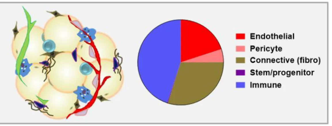
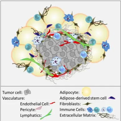
1. Solid tumor growth requires the interaction of tumor cells with the surrounding tissue, leading to a view of tumors as communities rather than exclusively tumor cells.
2. Adipose tissue, or fat, plays important roles in cancer risk and outcome because many tumors grow close to or in direct contact with adipose.
3. The adipose community – or microenvironment - includes adipocytes and
adipose-associated stromal and vascular components, such as fibroblasts and other connective tissue cells, stem cells, endothelial cells, innate and adaptive immune cells, and
extracellular signaling and matrix components.
4. Herein, we review the cellular and non-cellular parts of the adipose “organ” and the
mechanisms by which varied microenvironmental components contribute to tumor development, with emphasis on obesity.
5. Obesity dramatically modifies the adipose tissue microenvironment in numerous ways, which intriguingly resemble shifts observed within the tumor microenvironment.
6. Understanding neighboring adipose is critical in tumorigenesis.
2 Introduction
Cancer is characterized by fundamental aberrations in cellular behavior, including the ability to multiply indefinitely in the absence of growth-promoting factors and a resistance to signals that normally result in programmed cell death (apoptosis) [1]. In the case of solid tumors, carcinogenic transformation and cell proliferation are followed by establishment of a vascular supply, called tumor angiogenesis,
which facilitates the delivery of oxygen and nutrients to the growing tumor [1]. Subsequent invasion into and migration through surrounding tissues allows for the establishment of nearby satellite tumors or entry into the lymphatic or vascular systems for dissemination and secondary tumor formation
(metastases) [1]. Solid tumor growth and tissue invasion require the interaction of tumor cells with the surrounding tissue, and it is well
established that communication between cancer cells and the tissue-level context in which they reside, collectively referred to as the tumor “microenvironment”, is pivotal in determining
whether a given tumor will exist in dormancy or progress to malignancy [2]. The tumor microenvironment includes, but is not limited to, the tumor cells themselves, blood vessels (endothelial cells and pericytes), lymphatic vessels (lymphendothelial cells), adipocytes, fibroblasts, and various stem and progenitor cells [3] (Figure 1). Also present is a wide variety of innate and adaptive immune cells, which can act as critical anti-tumor defenses or,
3
alternatively, play central roles in tumor promotion. The tumor “stroma” is the connective,
functionally supportive framework of the tumor, and by definition refers to a complex mixture of signaling molecules and extracellular matrix (ECM) components, as well as the stromal cells (e.g., fibroblasts and pericytes) that produce and are embedded within them [4]. However, the term “stroma” may also be used to collectively refer to all of the aforementioned cell types and
secreted factors, as all are present within the cancer cell-adjacent tissue. Thus, considerable heterogeneity, both within the cancer cells themselves and among the interacting stromal cells, leads to a view of tumors as communities, and the process of tumorigenesis as a tissue-level
phenomenon occurring in conjunction with intrinsic genetic deviations within individual cancer cells [5].
Due to the ubiquitous nature of adipose tissue, many types of solid tumors grow in proximate or direct contact with adipocytes and other adipose-associated cell populations. Although the specific nature of the reciprocal communication occurring between a developing tumor and adjacent adipose tissue is an area of active study, a growing body of literature indicates that these interactions with the local adipose milieu are important drivers of malignancy. Many of these studies have focused on dysregulated adipose and associated systemic metabolic dysfunction in the context of obesity, as there is now adequate evidence establishing a link between obesity/adiposity and elevated risk for, or accelerated progression of, several cancers. Following an overview of the adipose organ, we will briefly address
epidemiologic links between obesity and cancer. Subsequently, we have chosen to emphasize the local physical and paracrine roles of adipose tissue in solid tumor development and
4 The Adipose Organ
Adipose tissue is a type of loose connective tissue that was long considered to be largely physiologically inert, primarily storing energy in the form of lipids while cushioning and insulating the body. However, adipose tissue is also a substantial contributor to whole body endocrine signaling, modulating feeding behavior and total body energy expenditure, as well as hematopoiesis and lymphopoiesis, overall immune function, and reproduction [6, 7].
Additionally, adipose tissue is now understood to contribute to the pathogenesis of a variety of regional and systemic diseases. The adipose tissue “organ” is in fact comprised of several
distinct adipose depots (Figure 2), each of which differentially exerts systemic and regional control on overall energy metabolism and signaling based on location and adipose tissue subtype. Broadly, adipose depots can be divided according to anatomic location into
subcutaneous and visceral subtypes. Whole adipose depots, or specific regions within depots, may be further subclassified as white, brown, or beige depending on, among other factors, adipocyte mitochondrial content, with a higher relative number of mitochondria corresponding to a darker adipocyte hue. In humans, subcutaneous adipose tissue comprises ~80% of total body fat, and is contained primarily in the abdominal, gluteal, and femoral depots [8] (Figure 2A). The breast fat pad is also a nontrivial contributor to total subcutaneous fat content in women. On the other hand, visceral depots represent approximately 5-20% of total body fat in normal weight (i.e., not overweight or obese) individuals [8]. Visceral adipose tissue surrounds vital organs, and includes omental, mesenteric, and epiploic adipose, as well as the gonadal, epicardial, and retroperitoneal fat pads. Finally, numerous smaller depots, such as intramuscular, intraorbital, and bone marrow adipose, nourish and protect tissues throughout the body. While the majority of these depots are comprised of white adipose tissue – discussed further in the Adipocytes section below – smaller brown and beige adipose tissue caches are also found in adults [9, 10].
5
function relative to humans, the laboratory mouse (Mus musculus)is a commonly used model for investigation of adipose tissue anatomy and physiology (Figure 2B).
Figure 2. The adipose organ is comprised of several distinct adipose depots. Adipose depot locations and subtypes in A) humans and B) mice (panel B adapted from [11] with permission).
Although adipocytes constitute approximately 90% of adipose tissue volume, the adipose tissue microenvironment is a rich ecosystem of additional stromal and vascular components (often referred to collectively as stromal-vascular cells). The stromal-vascular compartment of human white adipose tissue includes endothelial cells (10-20% of cells), pericytes (3-5%), fibroblasts and other connective tissue cells (15-30%), and stem and
6
Figure 3. Approximate composition of human white adipose tissue stromal-vascular fraction (percent cellularity).
Obesity and Cancer
Adipose tissue exhibits an almost unlimited capacity to expand, a unique property that has received increased attention in recent years as obesity has moved to the forefront of global public health concerns. Overweight and obesity, defined by the World Health Organization (WHO) as abnormal or excessive adiposity that presents a risk to health, are frequently measured at the population level using the body mass index (BMI), an individual’s weight in
kilograms divided by the square of his or her height in meters. However, it must be
acknowledged that, at an individual level, the BMI formula can vary considerably by sex and race and says little about body composition, often underestimating adiposity [13, 14]. For this reason, additional measures specifically of adiposity, such as waist circumference or the Body Adiposity Index (BAI; [hip circumference (cm)/height (m)1.5-18]) developed by Bergman et al. [15], are sometimes used to correlate adiposity with disease risk.
Current status of the obesity epidemic, globally and in the United States
7
published in The Lancet analyzed 1,698 population-based data sources, encompassing 186 countries and more than 19.2 million adult participants (9.9 million men and 9.3 million women), to evaluate trends in mean BMI over the last four decades [17]. The authors reported a global increase in overall age-adjusted prevalence of obesity in men from 3.2% to 10.8%, and in women from 6.4% to 14.9%, between 1975 and 2014 [17] (Figure 4A). An additional cross-sectional analysis of the United States National Health and Nutrition Examination Survey (NHANES) for the years 2013-2014 reports that the overall age-adjusted prevalence of obesity (again by BMI) among men and women in the US has now reached a staggering 35% and
40.4%, respectively [18]. Furthermore, extreme obesity (or class 3 obesity, defined as BMI >40) in the US is currently 9.9% for women and 5.5% for men [18], considerably higher than the global prevalence of 1.6% and 0.64%, respectively [17] (Figure 4B). Importantly, a
8
Figure 4. Rising global and US obesity rates. A) Global age-adjusted prevalence of obesity in men and women, 1975 and 2014; B) Class III obesity (BMI >40), globally and US; C) US obesity prevalence by race, ethnicity [17].
The obesity-cancer link
Cancer is currently the second leading cause of death in the United States and is expected to surpass heart disease as the leading cause of death within the next few years [20]. Approximately 40-60% of cancer patients are classified as overweight or obese [21, 22], and in 2004 it was estimated that overweight and obesity accounted for one in seven cancer deaths in men and one in five in women [23]. Importantly, obesity is differentially associated with
increased risk of cancer development and increased risk of poorer cancer prognosis. Indeed, there is adequate evidence to support an association between obesity and increased risk of developing colorectal, post-menopausal breast, endometrial, kidney, esophageal, liver,
gallbladder, pancreatic, and thyroid cancers, as well as non-Hodgkin’s lymphoma and myeloma
9
Interestingly, there is also a body of literature that supports a protective effect of obesity in overall survival for some cancer types, a finding known as the “obesity paradox”. Potential
explanations for the obesity paradox emphasize methodological issues, such as unmeasured confounders and/or a reliance on BMI as a metric for obesity [30, 31]. As mentioned previously, BMI is a rather crude mathematical estimate that does not capture important considerations such as percent adiposity, regional distribution of adiposity (e.g., android vs gynoid obesity), or differences in lean mass. Gonzalez et al. reported that the use of body composition indices resulted in a disappearance of the obesity paradox in 175 cancer patients in which BMI was
previously associated with a protective effect, emphasizing the importance of considering body composition in epidemiologic analyses of cancer outcomes [32]. In fact, when body composition was included, loss of lean mass (sarcopenia) was a more important prognostic indicator than BMI for patients exhibiting cancer-associated cachexia, a systemic wasting syndrome frequently observed in end-stage cancer patients that is characterized by a rapid loss of both skeletal muscle and adipose tissue [32, 33]. Thus, additional evidence is needed to determine whether isolated reports of the obesity paradox are simply artefactual or in fact clinically relevant.
Nevertheless, leading hypotheses seeking to explain observed connections between obesity and increased cancer morbidity and mortality emphasize factors such as metabolic disruption-induced growth factor dysregulation; higher levels of circulating adipokines and cytokines secreted by inflamed obese adipose tissue; and elevated production of estrogens by adipose tissue [34, 35]. These hypotheses emphasize the role of adipose as an endocrine organ and obesity as a potential state of adipose endocrine dysfunction. However, the mechanisms whereby adipose accumulation increases risk of tumor onset and/or mediates tumor progression in adipose-adjacent cancers are multifactorial, complex, and likely tissue/organ-specific, in part due to unique paracrine and physical interactions occurring
10
an increase in lymph node metastasis in patients with invasive breast carcinoma [36]. Thus, whether select adipose-mediated mechanisms of tumor promotion are merely exacerbated by obesity or are unique to a dysregulated obese adipose microenvironment in many cases remains to be determined. In this review, we have especially highlighted the role of adipose tissue in the development and progression of breast and prostate cancers due to the prevalence of these cancer types in the US population and their significant contributions to cancer-related mortality (see below).
Breast and prostate cancers are the most frequently diagnosed cancers and the second
leading causes of cancer-related death among US men and women, respectively [20]. Due to their now recognized genetic and molecular heterogeneity, these cancer types have been shown to exhibit complex associations with obesity. For example, although the association between obesity and risk of postmenopausal breast cancer is now well established, the
relationship between obesity and premenopausal breast cancer risk remains controversial and appears to be dependent upon breast cancer subtype. Specifically, recent work has clarified an association between obesity and premenopausal onset of triple-negative breast cancers
(TNBCs), with differential risk according to race [37-41]. Studies from our lab and others have also demonstrated that diet‑induced obesity is associated with accelerated TNBC latency (time
11
cancer-associated mortality [65, 66]. Thus, rising obesity rates present an oncological crisis, both globally and within the US.
Following a brief consideration of the anatomy of breast and prostate in humans and laboratory mice - a frequently used model in basic science and translational/pre-clinical cancer studies - potential mechanistic links between adipose tissue and breast and prostate cancer development or progression will be discussed in detail below through a comprehensive examination of the available literature regarding adipose-cancer interactions in each organ.
Anatomy of the Breast and Prostate
The laboratory mouse remains the most widely used animal model for the study of cancer pathophysiology. Consequently, integration of experimental findings with studies of human disease requires an understanding of human and veterinary pathology and anatomy, as well as developmental, molecular, and cellular biology. While this level of detail is beyond the scope of this review, this section will provide a brief comparative biology overview of the breast and prostate in humans and mice as a backdrop for the studies reviewed in subsequent
sections.
Mammary gland anatomy and adipose-cancer interaction in humans vs. mice
In both mice and humans, the mammary gland is a unique, dynamic organ that
continuously undergoes anatomic and functional changes over the life course [67]. In mice, the nascent mammary gland (“mammary tree”) consists of a network of epithelial ducts, each of
12
stages such as pregnancy, lactation, and post-partum involution, or epithelial regression [70, 71]. Development of the mammary tree and pregnancy/lactation-associated expansion and involution require remodeling of the surrounding stroma. In mice, mammary ductal-adjacent stroma is primarily comprised of adipose tissue, without a significant collagenous matrix layer (Figure 6).
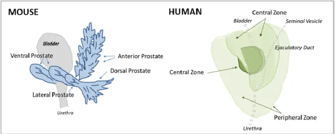
Figure 5. Comparison of mouse and human mammary gland anatomical structure. A) Murine ductal elongation and branching occur at the Terminal End Buds (TEBs). B) The human mammary gland is extensively branched, culminating in the functional terminal ductal lobular unit (TDLU).
In comparison to mouse, the human mammary gland is a more extensively branching structure. Beginning at the nipple, the lactiferous sinus branches into segmental, or interlobular, ducts (Figure 5B). Segmental ducts branch further into terminal ducts and lobules, which together comprise the functional unit of the human mammary gland, the terminal ductal lobular unit (TDLU). Immediately surrounding the TDLU is a loose intra-lobular stroma, referred to as “specialized stroma”, which contains abundant fibroblasts (Figure 6) [71]. Dense, collagenous
13
stroma is a large depot of subcutaneous adipose, comprising 7 to 56% of the volume of the adult breast [74].
Figure 6. Comparison of mouse and human mammary gland histology. Left: Adult mouse mammary fat pad from nulliparous C57BL/6 mouse (4x and 10x, H&E staining). Right: H&E-stained normal human breast tissue. Arrowhead and asterisks in right panel refer to loose intra- and dense inter-lobular stroma, respectively. Human histology images courtesy of Melissa Troester and the UNC Normal Breast Study (unpublished).
The most extreme example of tumor infiltration into adipose tissue is seen in breast cancer. Breast cancer most frequently begins in ductal epithelial cells, which proliferate to fill the ductal lumen and generate a pre-cancerous lesion called ductal carcinoma in situ (DCIS). Subsequently, invasive ductal carcinoma (IDC) cells invade the mammary stromal
14
adipocytes and other adipose cell populations (Figure 7A), whereas human invasive breast carcinoma must invade through both intra- and interlobular stroma before direct interaction with an area rich in adipose tissue (Figure 7B).
Figure 7. Adipose-breast cancer interactions in mice and humans. A) Early invasive lesions in H&E-stained mammary gland tissue from the C3(1)-TAg genetically-engineered mouse model of spontaneous basal-like breast cancer (unpublished images). B) Human breast cancer - Female, 50 years, lobular carcinoma, grade 1, Elston-Ellis score 5. Image credit: The Human Protein Atlas [75, 76].
Prostate gland anatomy and adipose-cancer interaction in humans vs. mice
Like the mammary gland, the prostate exhibits important inter-species differences
between mice and humans. However, before progressing to a comparison of mouse and human prostate anatomy, it should be acknowledged that rat and canine models have generated
important mechanistic knowledge in prostate cancer research, particularly in the context of the spontaneous development of prostate lesions [77]. With that said, genetically engineered or xenografted mice remain the most commonly used model in prostate cancer research. For an overview and critique of currently available mouse models of human prostate cancer, the reader is directed to [77, 78].
15
see diagram in Figure 8) and exhibit distinctive histology [77, 79]. The glandular acini of the prostatic lobes are surrounded by a thin fibromuscular tunica, and are embedded in a loose connective tissue stroma with minimal smooth muscle cells and sparse collagen fibers [79]. Individual mouse prostate lobes are surrounded by a delicate mesothelium-lined capsule, and are separated from each other by fibrous and adipose connective tissue [79].
In contrast to mice, the human male prostate does not have exterior lobation, but instead contains distinct glandular regions (a peripheral zone, a central zone, and a transition zone; see diagram in Figure 8) [79], again with characteristic histology. Like the breast, a conspicuous
histological difference between mouse and human prostate lies in the stromal component. In humans, the prostate gland bears an anterior, well-developed, non-glandular fibromuscular stromal region. Abundant adipose tissue is present surrounding most of the posterolateral aspects of the prostate [80], and is used as a marker of extraprostatic tissue in biopsy samples [81]. This region of adipose is referred to in subsequent sections as periprostatic adipose. Intraprostatic adipose, when present, consists of a small focus of a few adipocytes, and is rarely observed histologically [81].
16
The most common type of prostate cancer is acinar adenocarcinoma, which originates from the glandular epithelium. Pre-neoplastic prostatic intraepithelial neoplasia (PIN)progresses to invasive adenocarcinoma, in which extension of prostatic carcinoma through the prostatic capsule (extraprostatic extension) and resulting interaction with the surrounding adipose is an indicator of malignant progression and advanced histopathological stage [82]. The periprostatic adipose depot unambiguously contributes to prostate cancer malignancy [83-85]. In fact, interaction with periprostatic adipose tissue has been suggested to be a more important
determinant of cancer recurrence than an invasive phenotype [86]. Analogous to breast cancer,
recent advances in molecular phenotyping by The Cancer Genome Atlas Research Network have identified several genomically distinct molecular subtypes of prostate cancers [87]. Whether these subtypes interact differentially with adjacent adipose remains to be determined.
Microenvironmental Links between Adipose Tissue and Cancer
Context matters: extracellular matrix in adipose tissue and cancer
Adipocytes and stromal cells are embedded in a loose, three-dimensional ECM, the non-cellular tissue component that provides both structural and biochemical support to surrounding cells, such as cell adhesion, paracrine communication, and differentiation signals. Maintenance of the adipose tissue ECM – primarily comprised of fibronectin and collagens [88] – involves a
variety of cell types, including fibroblasts, macrophages, adipocytes, and preadipocytes. Importantly, adipocyte function and survival is tightly regulated by both the molecular composition and mechanical properties of the surrounding ECM [89].
17
programs in adipocytes and other stromal cells that ultimately lead to excess deposition of fibrillar ECM components such as collagens I, III, and VI and development of tissue fibrosis [88, 90]. Indeed, adipose depots of obese subjects often exhibit greater total fibrosis, and particularly pericellular fibrosis around adipocytes, than lean individuals [91, 92]. Importantly, hypoxia-induced adipose tissue fibrosis is associated with onset of metabolic perturbations in adipocytes [88, 93], while dysregulation in visceral adipose function is linked to the pathogenesis of insulin resistance and type II diabetes mellitus [92-94]. Furthermore, as adipocytes become
encapsulated in a shell of rigid ECM, impaired cellular function also results in apoptosis and
necrosis [95]. Release of damage-associated molecular patterns (DAMPs) from dead and dying adipocytes and adjacent live adipocytes promotes recruitment of macrophages and other inflammatory cells; histologically, these macrophages can be observed within crown-like
structures (CLS), foci of macrophages and other inflammatory cells surrounding dead and dying adipocytes [96]. Macrophages are fully integrated into all stages of the fibrotic process through secretion of soluble mediators and cytokines such as transforming growth factor β1 (TGF-β1),
platelet-derived growth factor (PDGF), and chemokines that attract and activate fibroblasts and collagen-producing myofibroblasts [88, 97].
Interestingly, while adipose tissue fibrosis in the context of obesity is well described, increased adipose ECM deposition, fibrosis, and immune cell infiltration are also observed in cancer-associated cachexia [98]. Abdominal subcutaneous adipose depots of lean cachectic subjects bearing gastrointestinal cancers displayed extensive adipose ECM remodeling, including a dramatic increase in deposition of collagens I, III, and VI as well as elastin and fibronectin [99]. These changes were associated with increased myofibroblast content and elevated activation of TGF-β/SMAD signaling pathways [99]. As described later in the
18
In addition to adipocytes, epithelial tissue homeostasis and tissue organization is also heavily dependent upon a dynamic dialogue with the surrounding ECM. Disruption of ECM structure or misinterpretation of ECM-derived signals due to alterations in signaling receptor profiles is associated with development of a malignant phenotype in transformed epithelial cells [100-102]. Enhanced ECM stiffness also triggers the process known as
epithelial-to-mesenchymal transition (EMT) in cancer cells, which is characterized by the loss of epithelial polarity, de-differentiation, and local migration and invasion [103-106]. Hence, modifications in the adipose tissue ECM that provide a hospitable environment to developing tumors, such as
enhanced stiffness in obese breast tissue, may provide a link between adipose tissue and tumorigenesis.
As discussed in later sections, chronic low-grade inflammation, macrophage infiltration, hypoxia, and aberrant wound healing responses, including an increase in myofibroblast and
19
activated fibroblast content, are features of both the tumor and adipose tissue
microenvironments [4, 102, 107]. Chronic activation of the wound repair response leads to excess deposition of ECM components and accumulation of scar-like fibrotic tissue in a process known as desmoplasia, or the desmoplastic reaction (Figure 9A). In both breast and prostate cancers desmoplasia is associated with poor outcomes [108, 109], and can facilitate cancer progression by interfering with drug delivery. Thus, ECM remodeling and the resultant disturbances in cytoskeletal tension and mechanotransduction have emerged as important factors that promote neoplastic transformation, cancer malignancy, and cancer metastasis [4,
102, 110], and may provide another connection between adipose dysregulation and cancer.
Adipose extracellular matrix composition and viscoelasticity: influence on the normal
breast and breast cancer
Mammographic density denotes the radiologic appearance of the breast, and is a metric of the fibroglandular (epithelial and non-fatty stromal) content in that tissue [111]. A number of qualitative and quantitative methods have been developed to estimate mammographic density, including Breast Imaging Reporting and Data System (BI-RADS) categories, Wolfe’s
parenchymal patterns, Tabar’s classification scheme, and numerous two- and three-dimensional
image analysis techniques [112]. Within heterogeneous breast tissue, tumors most frequently arise within the most mammographically dense regions of the breast, suggesting that denser fibroglandular tissue directly influences carcinogenesis [113]. Indeed, regardless of the reporting method [111], high mammographic density is consistently and strongly associated with both elevated risk of breast cancer [114] and more aggressive tumor characteristics [115], even after adjustment for other risk factors such as age and BMI [116].
20
reactive population of so-called “cancer-associated fibroblasts” (CAFs). CAFs display
remarkable plasticity, and frequently differentiate into myofibroblasts, a cell type exhibiting properties of both fibroblasts and smooth muscle cells [120-122]. In non-malignant tissue, myofibroblasts play an important role in wound healing responses, secreting a fibronectin- and collagen type I-rich ECM characterized by fibrillary architecture and increased cross-linking and density [123]. They are also the predominant source of fibrogenic and/or inflammatory cytokines in fibrotic lesions [124]. Despite the utility of this cell type to normal wound healing programs, however, the presence of myofibroblasts in tumors contributes to pathological desmoplasia
[122], and may thus promote cancer progression [125].
In addition to fibroblasts, local (adipose-derived) mesenchymal stem cells, bone marrow-derived mesenchymal stem cells, myeloid precursors, and cells marrow-derived from EMT may also present alternative sources of myofibroblasts in tumor stroma [126-128]. Furthermore, in tumors growing in an adipose tissue-rich microenvironment, cancer cell-induced reprogramming of local adipocyte gene expression and function has been observed to promote adipocyte delipidation and atrophy/regression [129]. This process occurs concurrently with the accumulation of
fibroblast-like cells and a desmoplastic stroma; this synchronicity raises the possibility that some CAFs might be derived from dedifferentiated adipocytes [129] (Figure 9B). However, the extent to which their specific lineages determine the contribution of CAFs to tumor progression remains inconclusive.
Although obesity is associated with reduced mammographic density – in part because fat is radiolucent – several studies have unveiled close links between chronic inflammation and
the development of fibrosis and associated ECM rigidity in obese mammary adipose tissue [123, 130, 131]. Myofibroblasts are typically absent from normal, uninflamed breast tissue [132]. However, Seo et al. showed that obesity elevated matrix rigidity in non-cancerous breast tissue by enhancing myofibroblast content in mammary adipose [123]. Adipose stromal cells (ASCs, also called adipose-derived stem cells) isolated from obese mice exhibited increased
21
fibronectin and a more fibrillar, partially unfolded, and stiffer ECM [123], implicating ASCs as a source of myofibroblasts in obesity. Furthermore, obese ASCs also exhibited enhanced proliferative capacity and secreted increased quantities of matrix components [123], thereby mimicking characteristics of tumor-associated stromal cells [122, 133]. Consistent with findings in mice, histologically normal breast tissue from obese patient mastectomies exhibited
increased α-SMA staining and collagen fiber length and thickness relative to tissue from lean individuals [123]. Obesity-associated increases in α-SMA levels also correlated with formation of
CLS, further implicating macrophages in the development of mammary adipose tissue fibrosis
[123]. However, distinct from tumors [133], obesity-associated increases in myofibroblast content and matrix rigidity occurred in a TGFβ-independent manner [123], suggesting that ECM
composition and stiffness may be differentially regulated in benign obese and malignant breast tissue.
Increased matrix rigidity in breast adipose tissue may be an important mediator of cancer initiation and progression in obese individuals. To test the effects of obesity and ECM on tumor cell behavior, Seo et al. cultured pre-invasive human MCF10AT cells upon decellularized matrices produced by ASCs isolated from lean or obese mice. The authors reported that, relative to ECMs deposited by lean ASCs, obesity-associated ECMs increased MCF10AT cell motility and promoted the formation of disorganized three-dimensional acini, indicative of greater tumorigenic potential [123]. Additionally, ECM generated by obese mammary ASCs significantly enhanced the proliferation of the highly invasive MDA-MB-231 cancer cell line by altering mechanotransduction through enhanced RhoA/ROCK-mediated cell contractility and YAP/TAZ transcription factor activity [123]. Collectively, these results are suggestive of a relationship between obesity-associated mammary adipose tissue fibrosis and accelerated tumor initiation and/or proliferative capacity.
In addition to fibroblasts/myofibroblasts, adipocytes play a vital role in defining the ECM environment through secretion and processing of factors such as collagen VI, an ECM
22
134, 135]. Excess adipocyte collagen VI expression in obesity is associated with adipose tissue fibrosis and metabolic dysregulation, while the absence of collagen VI in mouse models of obesity allowed for uninhibited adipocyte expansion and an improved metabolic phenotype [93]. Increased adipocyte collagen VI expression is also associated with elevated local
concentrations of endotrophin, the collagen VI α3 chain cleavage product, which has been
identified as a driving factor in adipose tissue fibrosis, macrophage chemotaxis, and
inflammation, and appears to mediate adipose metabolic dysregulation in obesity (Figure 10) [131, 135]. Unsurprisingly, increased collagen VI production also coincides with increased
adipose tissue macrophage content [130, 135].
To further illustrate parallels in the obese adipose and tumor microenvironments,
collagen VI and its cleavage product have also been implicated in the initiation and progression of breast cancers. Collagen VI is abundantly expressed by breast cancer-associated adipocytes (discussed at greater length in the Adipocytes section below), and its increased deposition in the ECM promotes tumorigenesis and malignant progression both in vitro and in vivo by inducing alterations in cancer cell signaling programs, gene expression patterns, and post-translational modifications [136, 137]. For example, treatment of MCF-7 human invasive breast cancer cells with collagen VI significantly elevated activity of the oncogenic Akt-GSK3β–β-catenin–Tcf/Lef pathway, ultimately resulting in cyclin D1 protein stabilization and enhanced cell proliferation [136, 137]. Accordingly, expression of the proto-oncogenes GSK3β and cyclin D1 in
23
endotrophin concentration in obese adipose may influence both early tumor development and treatment outcomes.
Figure 10. Obesity-associated modifications in the adipose tissue microenvironment. Adipose tissue expansion in obesity occurs in association with extracellular matrix changes such as fibrosis. Adipocyte hypertrophy and hypoxia trigger macrophage infiltration and crown-like structure formation, which further exacerbates development of fibrosis and inflammation.
Adipose extracellular matrix-derived factors: direct effects on epithelial cells
In addition to modulating composition and viscoelasticity of the breast ECM, stromal cells within the obese breast microenvironment secrete numerous soluble signaling mediators that have direct effects on epithelial cells. In particular, HGF is an excellent candidate for stromal-mediated breast cancer promotion in the context of obesity. Although HGF is classified as an adipokine [139], it is produced by a number of breast cell types including stromal
24
[141]. HGF is also elevated in the serum of breast cancer patients and correlates with advanced disease [142-145]. However, HGF signaling impacts the phenotypes of both early- and late-stage breast cancers. With respect to early-late-stage lesions, we have reported that treatment of pre-malignant basal-like breast cells with HGF-blocking antibodies inhibited 3D morphogenesis, reflecting a reduction in epithelial malignant potential [142]. An HGF gene expression signature generated via treatment of pre-malignant breast cells with recombinant HGF was also found to correlate with both basal-like subtype and poor survival in >700 breast cancer samples from three publicly available datasets [142].
Importantly, basal-like breast cancer is a clinically intractable TNBC subtype that is more prevalent in obese individuals [37-41], while serum HGF is also elevated in obese individuals and is reduced with weight loss [146-148]. Our laboratory previously demonstrated that high fat diet-induced obesity increased HGF concentration and enhanced expression and activation of cMET in the mammary fat pad of C3(1)-T-antigen (TAg) mice, a unique genetically engineered mouse model (GEMM) of spontaneous basal-like breast cancer [43, 149, 150]. We also reported that obesity increased HGF production by primary murine fibroblasts isolated from both normal mammary glands and tumors, and that CAFs isolated from obese animals induced epithelial cell
25
migration in an HGF-dependent manner [43]. Obesity-mediated regulation of HGF secretion from other stromal cell types such as adipocytes is currently under investigation.
Adipose extracellular matrix in prostate cancer
Despite being a common feature of mouse models of prostate cancer, histologically conspicuous reactive stroma is much less prevalent in human prostate tumors compared to breast cancers [77]. However, like the breast, induction of a myofibroblastic phenotype and
degree of reactive stroma carry important prognostic value for prostate cancer malignancy [109, 151, 152]. Notably, as the literature regarding the contribution of adipose tissue to breast cancer onset and progression has greatly outpaced that of prostate cancer, obesity-associated ECM modifications are currently better characterized in the mammary, relative to the periprostatic, fat pad. Additionally, conflicting data exist regarding the association between periprostatic fat density (measured by magnetic resonance imaging or computed tomography) and tumor aggressiveness in prostate cancer patients [153-155]. Our literature search also revealed no publications reporting that periprostatic adipose tissue fibrosis occurs in obesity, but whether this is due to a lack of occurrence or a lack of examination is unknown. Furthermore, no studies investigating links between adipocyte-derived endotrophin and prostate cancer were available at the time of writing this review. Therefore, future obesity-prostate cancer studies may be
informed by the sundry findings linking breast cancer and adipocyte-associated fibrosis, modifications in ECM dynamics, and endotrophin release.
Adipocytes and adipocyte-cancer interactions
26
neutral lipids. However, brown and/or beige adipocytes (also called “brite” or “inducible”
adipocytes [9]) have also been reported in adults, and likely play important roles in
thermogenesis [156]. More recently, “pink” adipocytes have been described in murine mammary
gland, arising exclusively during pregnancy and lactation due to a process wherein white adipocytes progressively transdifferentiate to acquire secretory, epithelial-like features [9]. Adipocytes secrete a broad range of signaling molecules that exert local and/or systemic effects with the potential to influence tumor growth. Among the better studied adipocyte-derived factors are metabolic factors such as leptin, adiponectin, resistin, visfatin, and plasminogen activator
inhibitor-1 (PAI-1); hematopoietic factors such as GM-CSF; growth factors such as
angiopoietins, HGF, vascular endothelial growth factor (VEGF), insulin-like growth factor-1 (IGF-1), and TGF-β; and a variety of cytokines, including interleukin-6 (IL-6) and TNF-α and the
chemokine monocyte chemoattractant protein (MCP-1) [also referred to as chemokine (C-C motif) ligand 2 (CCL2)] (Figure 12) [157, 158].
27
Several of the aforementioned adipocyte-derived growth factors influence development of a tumor vascular supply (tumor angiogenesis), as discussed in the Endothelial
Cells/Lymphendothelial Cells section below.Whereas leptin and adiponectin are considered true adipokines, many of the other signaling molecules, including resistin, visfatin, TNF-α, IL-6,
MCP-1 and PAI-1, are not, as they are expressed by both adipocytes and immune cell populations such as macrophages, and play a variety of well-known roles in immunity [157]. Thus, select functions for several of these signaling molecules will be discussed within the section titled Adipose Tissue Immune Populations in Cancer Development and Progression.
Finally, although there are clear and important roles for leptin and adiponectin in tumorigenesis and malignancy, these roles have been reviewed extensively by others [139, 159-162] and will be addressed only briefly within this review.
Adipocytes exhibit both short- and long-range interactions with cancer cells, and may be found in close proximity to tumors, along tumor margins, and within the tumor body. These cancer-associated adipocytes (CAAs; also referred to as peritumoral, intratumoral, or tumor-infiltrating adipocytes) influence tumor biology in a number of ways, including by promoting angiogenesis and inflammation [reviewed in 163, 164, 165]. Although it is reasonable to hypothesize that proliferation and invasion of tumor cells into cancer-adjacent adipose may account for the presence of CAAs within the tumor body, the origin of CAAs in fact remains unclear. As explained in further detail in the section on Adipose-derived Stromal Cells below, several cell types may give rise to intratumoral CAAs.
In addition to indirect mechanisms of tumor growth promotion (e.g., stimulation of angiogenesis, production of proinflammatory cytokines), the proximity of CAA to growing tumors may also provide direct metabolic benefits to cancer cells. In the phenomenon known as
28
droplet size within mature white adipocytes is the net result of several processes, including fatty acid uptake or de novo fatty acid synthesis, esterification, and lipolysis. Interestingly, Nieman et al. showed that co-culture of primary omental adipocytes with ovarian cancer cells, which frequently metastasize to the omentum, induced lipolysis in adipocytes, upregulation of
β-oxidation in cancer cells, and direct transfer of lipids between the two cell types [170]. Notably, the transfer of lipids from adipocytes to cancer cells has also been observed in prostate cancer [171] and breast cancer [172]. These findings indicate that active heterotypic cellular
interactions between cancer cells and adipocytes induce metabolic symbiosis.
CAAs may also influence cancer cell phenotypes through the shedding of exosomes, small vesicular bodies released from cells as a form of short- or long-range communication. Lazar et al. [173] reported that exosome shedding by mature human adipocytes induced increased migratory and invasive behavior in melanoma cells, which grow in proximity to the hypodermal adipose layer. Proteomic analysis of adipocyte-derived exosome composition revealed enrichment for proteins involved in mitochondrial lipid metabolism, particularly fatty acid oxidation. Remarkably, their results suggested that these enzymes were incorporated and utilized by melanoma cells. Melanoma cells pre-treated with exosomes exhibited an increased ability to form lung metastases in mice and an increase in fatty acid oxidation without a
29 Adipocytes in the normal breast and breast cancer
Mouse models have revealed that adipocytes act as local regulators of normal mammary epithelial cell growth and function. In fact, mammary epithelial cells require adjacent adipocytes during embryonic and postnatal development, as well as throughout later life stages such as pregnancy, lactation, and involution [174]. Indeed, using the novel FAT-ATTAC mouse, a model of inducible and reversible adipocyte loss developed by Scherer and colleagues, Landskroner-Eiger et al. showed that adipocytes play crucial roles in normal growth and development of mammary ductal epithelium [175, 176], contributing both to ductal branching morphogenesis
during puberty and to maintenance of normal alveolar structures in adulthood [176].
Due to the proximity of the adipose pad to the mammary glandular organ, ductal tumor invasion results in interaction of breast cancer cells with adipocytes (Figures 6 & 7), with dramatic implications for tumor cell biology. Carter and Church reported that mature breast adipocytes, but not preadipocytes, increased motility of both normal and malignant breast epithelial cell lines through secretion of PAI-1 [177]. Similarly, higher levels of CAA-specific IL-6 expression in human breast tumors were associated with larger tumor size and more extensive lymph node involvement [178]. Co-culture with adipocytes also induced mesenchymal features in human breast cancer cells, including repolarization of vimentin and downregulation of E-cadherin, thereby promoting tumor cell invasion and metastasis [178]. Furthermore, adipocytes co-cultured with malignant breast epithelial cells exhibited the profound phenotypic changes associated with CAA, including delipidation and decreased expression of adipocyte markers [178]. Hence, bidirectional communication between adipocytes and breast tumor cells also alters adipocyte biology.
30
delipidation was not identified, although IL-6 and β-adrenergic stimulation – factors previously implicated in lipolytic induction in cancer-associated cachexia [179] – were eliminated as
potential candidates [172]. Similar to Lazar et al. [173], Wang et al. reported that co-culture with adipocytes increased both in vitro invasion toward a stimulus and formation of breast cancer lung metastases in vivo, each of which were restored to basal levels by administration of the fatty acid oxidation inhibitor etomoxir [172]. In vitro etomoxir administration also reduced the morphological hallmarks of EMT. Remarkably, the increase in fatty acid oxidation by breast cancer cells appeared to be dependent on an upregulation of both adipocyte triglyceride lipase
(ATGL) and the carnitine palmitoyltransferase 1 (CPT1) isoform CPT1A, enzymes not expressed at appreciable levels in noncancerous human breast epithelial cells. Short-hairpin (sh)RNA-mediated knockdown of CPT1A and ATGL reduced hallmarks of EMT and invasive potential, respectively.
In addition to oxidizing transferred fatty acids, breast cancer cells also esterified free fatty acid from adipocyte lipolysis [172], incorporating the newly synthesized triglyceride into lipid droplets within the cancer cells themselves. Breast cancer cell lipid droplet accumulation was supported by both in vitro co-culture experiments employing radiolabeled palmitate and the observation of lipid droplet accumulation in breast cancer cells along the tumor margin in
histological sections (i.e., in close proximity to adipocytes). Interestingly, despite increased fatty acid oxidation, breast cancer cells also showed reduced ATP content and activation of AMP-activated protein kinase (AMPK). AMPK activation following co-culture with adipocytes was associated with increased mitochondrial biogenesis and function, indicated by increased levels of PGC-1α and its associated transcription factor PPARα as well as an increase in the ratio of
31
insight into mechanisms of metabolic symbiosis between adipocytes and cancer cells in breast tumors.
Interactions between cancer cells and adjacent adipose may also increase breast cancer stem cell abundance and facilitate metastatic progression. Picon-Ruiz et al. isolated human adipocyte stem cells and used adipogenic differentiation media to generate “immature”
adipocytes. Co-culture of these “immature” adipocytes with both primary breast cancer cells and
established cancer lines conferred stem-like features to the epithelial cells, including elevated expression of the pluripotency markers Sox2, c-Myc, and Nanog [180]. Co-culture with
adipocytes also increased mammosphere-forming capacity, indicating a more stem-like phenotype due to a greater ability to grow under non-adherent conditions. Furthermore, when co-cultured breast cancer lines were orthotopically injected into mouse models, the resulting tumors exhibited reduced latency, increased abundance of tumor-initiating cells, and an enhanced capacity to form distant metastases. Taken together, this study demonstrates that interactions between immature adipocytes and breast cancer cells drive initiation of highly metastatic cancers by enhancing epithelial cell tumor-initiating potential.
32
breast cancer subtypes, as well as the reported roles for adipocytes in regulating breast epithelial tumorigenicity and metastatic potential, additional studies are needed to address concerns regarding the potential risks associated with fat grafting in breast reconstructive surgery. Stratification by BMI and/or molecular tumor subtype may be necessary to fully assess the influence of fat grafting on breast cancer recurrence rates.
Adipocytes and prostate cancer
Bidirectional communication between adipocytes and prostate epithelial cells also
influences prostate tumor biology, particularly with regard to chemokine activity. Chemokines, or chemotactic cytokines, are small secreted signaling proteins that induce directed, gradient-driven migration (chemotaxis) in nearby cells that express the appropriate chemokine receptor. The functions of chemokines in malignancy depend on both tumor characteristics and the specific chemokine in question, but are frequently associated with leukocyte infiltration as well as metastatic potential and site-specific spread of tumor cells [183]. Adipose tissue-specific expression of many CC subfamily chemokines and their receptors is upregulated in human obesity [184]. For example, Laurent et al. [185] identified a CCR3/CCL7 axis regulated by obesity, through which secretion of CCL7 by mature periprostatic adipocytes supported the directed migration of prostate cancer cells, thereby promoting cell migration toward the
periprostatic fat pad and the spread of cancer cells outside of the prostate gland. This process appeared to be augmented in obesity by both enhanced secretion of CCL7 by hypertrophic adipocytes and increased expression of the CCL7 receptor, CCR3, by prostate cancer cells [185].
33
production of CCL2 by bone marrow adipocytes and other stromal cells is also strongly
implicated in the propensity of prostate cancer cells to metastasize preferentially to bone [187, 188]. An increase in bone marrow adipocyte content with age, obesity, and obesity-associated metabolic pathologies [189, 190], suggests a potential link between obesity and elevated rates of prostate cancer metastasis [187, 188].
Interestingly, prostate cancer-adipocyte crosstalk also appears to induce tumor-promoting changes in periprostatic adipocytes. Treatment of periprostatic adipose tissue organotypic explants with PC3 prostate carcinoma cell-conditioned medium activated a cancer-promoting secretory profile, including increased secretion of osteopontin, TNF-α, and IL-6, and
reduced production of adiponectin [191]. These changes were not observed upon treatment of cells comprising the periprostatic adipose stromal vascular fraction (i.e., all stromal populations except adipocytes) with PC3 cell-conditioned medium, suggesting that the observed increase in pro-tumorigenic factor production by explanted tissue was due specifically to tumor-mediated education of adipocytes [191]. Indeed, adipocytes appear to be a major source of
microenvironmental IL-6 in prostate cancer. Periprostatic adipose tissue harvested from patients undergoing radical prostatectomy secreted IL-6 at concentrations 375 times greater than that in patient-matched serum and correlated with histological grade [192]. Additionally, Tang et al. [83] recently showed that co-culture of prostate cancer cells increased production of the cysteine protease cathepsin B by adipocytes. Further probing revealed that adipocyte co-culture induced secretion of the peptide hormone cholecystokinin (CCK) by prostate cancer cells, resulting in establishment of an autocrine/paracrine amplification loop in which CCK, acting through the CCK receptor CCKBR, induced expression of cancer stem cell markers such as CD49f and Sca-1 in prostate cancer cells and further production of cathepsin B by adipocytes. Importantly, cathepsin B has been shown to facilitate prostate cancer invasion and metastasis via
34
important mediators of tumor progression. Figure 13 briefly summarizes adipocyte-cancer cell crosstalk findings described above.
Adipocytes and adipose wasting in cancer-associated cachexia
An example of long-range adipocyte-tumor interactions can be observed in cancer-associated cachexia (referred to hereafter as cancer cachexia or simply cachexia). Cancer cachexia is a fatal energy-wasting syndrome that is estimated to be the immediate cause of death in approximately 20-40% of end-stage cancer patients [195]. A key feature of cancer cachexia is white adipocyte “browning”, characterized by greatly increased levels of brown
fat-mediated thermogenesis in white adipose depots [196, 197]. Accordingly, cachectic patients exhibit irreversible, pathologically elevated basal energy expenditure levels, adipocyte lipolysis and adipose tissue wasting, rapid weight loss, and eventually, death [196-199]. Although prolonged systemic inflammation plays a well-established role in cachexia-associated adipose tissue wasting [198, 199], tumor-derived factors have also been shown to contribute to the
Figure 13. Adipocytes promote tumor progression and metastasis. Adipocytes may provide metabolic substrates directly to cancer cells, or may indirectly influence cancer metabolism through exosome secretion. Adipocytes also secrete a variety of factors that promote tumor growth, EMT (epithelial-mesenchymal
35
pathophysiology of this syndrome. For example, in a murine model of Lewis lung carcinoma, Kir et al. [196] demonstrated that tumor-derived parathyroid hormone-related protein (PTHrP) induced the expression of thermogenesis-associated genes in adipose tissue, implying a crucial role for this hormone in energy expenditure and tissue wasting. Accordingly, administration of a PTHrP neutralizing antibody prevented cachexia-associated weight loss and ablated
thermogenic gene expression in white and brown adipose tissue. Furthermore, compared to cancer patients lacking detectable levels of blood PTHrP, patients with detectable blood PTHrP levels exhibited significantly higher resting energy expenditure levels per kilogram of lean body
mass, implying a clinically relevant association between this hormone and wasting. On the other hand, Rohm et al. [200] reported that browning and associated
thermogenesis in major white adipose depots was not the primary mechanism of adipose tissue wasting in mouse models of colon cancer-induced cachexia. Although the brown adipose-associated protein cell death activator (CIDEA) was upregulated in both brown and white adipose depots of cachectic mice relative to healthy controls, this upregulation occurred in the absence of changes in other proteins implicated in adipocyte browning and thermogenesis, such as uncoupling protein 1 (UCP-1). Furthermore, while increased free fatty acid release was observed in primary mouse adipocytes exposed to serum from cachectic mice, this increase in lipolysis was not associated with well-characterized lipolytic inducers such as increased
expression of lipases (e.g., hormone-sensitive lipase and ATGL), or increased β-adrenergic
receptor activation. Instead, CIDEA-mediated degradation of AMPK, evidenced by a reduction in AMPK protein and enzymatic activity, contributed to adipocyte metabolic dysfunction. For example, although increased lipolysis was observed, decreased AMPK activity also resulted in reduced inhibitory phosphorylation of acetyl-CoA carboxylase, suggesting the establishment of a futile cycle in adipocytes characterized by simultaneous increases in both lipolysis and
lipogenesis. Microinjection of white adipose depots with a peptide designed to interfere with the AMPK-CIDEA interaction (termed AMPK–CIDEA-interfering peptide, or ACIP), followed by
![Figure 2. The adipose organ is comprised of several distinct adipose depots. Adipose depot locations and subtypes in A) humans and B) mice (panel B adapted from [11] with permission)](https://thumb-us.123doks.com/thumbv2/123dok_us/8310695.2200999/20.918.120.822.226.527/adipose-comprised-distinct-adipose-locations-subtypes-adapted-permission.webp)