Original Article
Protective effect of ursolic acid on ischemic brain injury
by regulating hypoxia-inducible factor 1-alpha
Jianwei Li1,2*, Renjie Wang1,2*, Yingnan Zhang3, Ruichao Jia1,2, Kai Zhao1,2, Sai Zhang1,2, Haiqian Liang1,2
1Institute of Traumatic Brain Injury and Neurology, 2Department of Neurosurgery, Pingjin Hospital, Logistics
University of Chinese People’s Armed Police Forces, Tianjin, China; 3Medical Unit, Yangjiang Detachment,
Guangdong Crops of PAP, China. *Equal contributors.
Received February 16, 2017; Accepted October 13, 2018; Epub April 15, 2019; Published April 30, 2019
Abstract: Ischemic brain injury is a dynamic process involving oxidative stress, inflammation, cell death and the activation of relative endogenous transcription factors, including hypoxia-inducible factor 1-alpha. Ursolic acid, one of the main biologically active triterpenoids derived from plant, has been shown to exert various pharmacological activities. In the present study, we hypothesize that ursolic acid has neuroprotective effects in ischemic brain that could suppress neuronal apoptosis and promote neuronal survival. Middle cerebral artery occlusion (MCAO) models and TTC staining were carried out to evaluate the protective effect of UA on ischemia injury. TUNEL assay was used to detect the neuronal cell apoptosis. Primary cortical neuronal cell was cultured and subjected to oxygen glucose deprivation (OGD). Flow cytometry was used to investigate the cell apoptosis. Western blot was carried out to detect the protein expression level. The results shown UA decreased the area of the ischemic brain and the cell apoptosis of neuronal cell. In addition, UA inhibited primary cortical neuronal cell apoptosis induced by ODG, and this effect was reversed by infection of len-si-AKT and len-si-HIF-1α along with the LY294002 AND 2ME2 treatment. UA pro -moted the expression of p-AKT, p-mTOR, HIF-1α and Bcl-2 and inhibited the expression level of bad and cleaved caspase3. In conclusion, UA can inhibit ischemia brain injury and neuronal cell apoptosis through regulating the AKT/mTOR/HIF-1α pathway and the downstream protein expression such as Bcl-2, Bad and Caspase3.
Keywords: Ursolic acid, ischemic brain injury, hypoxia-inducible factor 1-alpha
Introduction
Hypoxic-ischemic encephalopathy (HIE), as one of the most common causes of neonatal death, can lead to severe long-term neurological dis-ability [1, 2]. Development of novel therapeutic therapies to treat these injuries have occurred these past years, including glutamate receptor antagonists, calcium channel blockers, radical scavengers, and anti-inflammatory and anti-ap- optotic agents. However, the low efficacy of th- ese treatments forced us to discover more novel strategies for HIE.
Hypoxia-ischemia associated brain damage, primarily due to the impaired glucose and oxy-gen supply, caused neuronal injury and the ex- haustion of cellular energy stores. This damage could lead to a multi-faceted cascade of bioch- emical events involving blood-brain-barrier dis-ruption, inflammation, oxidative stress,
gluta-mate neurotoxicity, energy depletion, and cell death [3]. In addition, other cellular reactions, su- ch as angiogenesis and apoptosis [4] in the sur-rounding tissue of the lesion, are known to con-tribute to neuronal functional disruption and ce- ll death. However, the molecular mechanism go- verning these regulatory factors is still not fully understood.
UA protects ischemic brain injury
Methods
Establishment of middle cerebral artery occlu-sion (MCAO) models
Male Sprague-Dawley rats (Charles River Bree- ding Laboratories, Beijing, China) weighing 280 to 350 g were used. All experimental procedu- res conformed to the Guide for the Care and Use of Laboratory Animals by the National Ins- titutes of Health (NIH) and were approved by the Animal Care and Use Committee of Pingjin Hospital. Rats were anesthetized with chloral hydrate (0.4 g/kg) intraperitoneally. The tem-perature was maintained between 36.5 and 37.0°C using a feedback-controlled heating sys- tem. The detailed construction of MCAO was de- scribed previously [5]. Additionally, sham oper-ation was performed by the same method with-out bipolar electrocoagulation of the MCAO.
Assessment of brain infarction area
Rats were killed 24 hours after MCA, and their brains were removed and frozen at -80°C for 30 min. Next, 1 mm coronal sections were cut on a vibratome. Brain sections (approximately 12 per brain) were incubated in a 2% solution of triphenyl tetrazolium chloride (TTC) at 37°C for 20 minutes, which stains for viable tissue. Areas of infarct on each brain section were tr- anscribed onto scale diagrams and quantified by computer-based image analysis. Infarct vol-ume of the coronal slices (2-mm thickness) fr- om each brain was quantified by capturing im- ages with a digital camera and subsequently pe- rforming computerized analysis [6].
Primary cortical neuronal cell culture
Six- to eight-day-old neonatal Sprague-Dawley rats (Charles River, Beijing, China) were sacri-ficed and the hippocampi were rapidly removed. Next, 400-500-μm slices were separated and placed into ice-cold growth medium that consis- ted of 50% minimum essential medium (MEM), 25% Hank’s balanced salt solution (HBSS), 25 % heat-inactivated horse serum, supplemented with 5 mg/mL glucose, 1 mM glutamine, and 1.5% fungizone [7]. Cultures were placed onto semiporous membranes and were grown for 10 to 14 days in an incubator at 37°C with 5% CO2.
Oxygen glucose deprivation (OGD)
In vitro ischemic injury was induced by oxygen-glucose deprivation. To initiate oxygen-oxygen-glucose
deprivation (OGD), cortical neurons cells were cultured in DMEM without serum or glucose in a humidified atmosphere containing 95% nitro-gen and 5% CO2. Neurons were fed with serum and glucose-supplemented original medium af- ter 3 h of hypoxia and returned to the incubator under normoxic conditions (95% air, 5% CO2).
TUNEL assay
Apoptosis was detected using a TUNEL assay kit (Boster, Wuhan, China) following the manu-facturer’s instructions. TUNEL-positive cells and normal cells in each group were counted using a light microscope at 200× magnification (Olympus, Tokyo, Japan).
Western blot assay
Total protein was extracted from cells or brain tissue using a radio-immune precipitation ass- ay (Beyotime, Shanghai, China). Total protein con- centrations were measured using a BCA Protein Assay Kit (Thermo Fisher Scientific, MA, USA). Next, 20 μg of total protein was separated in 10% sodium dodecyl sulfate-polyacrylamide gel and later transferred to a polyvinylidene difluoride membranes (PVDF) membrane. The membranes were blocked in skimmed milk for 2 hours. Proteins were detected by incubation with primary antibodies AKT, p-AKT, mTOR, p-mTOR, HIF-1alpha, Bcl-2, Bad, Caspase-3 or GAPDH polyclonal antibodies (1:1,000; Abcam, Cambridge, MA, USA) at 4°C overnight. The next day, membranes were incubated with horseradish peroxidase-labeled goat anti-rab-bit polyclonal antibody (1:1,000; Abcam) at room temperature for 2 hours. Immunoblots were visualized using a Millipore ECL Western Blotting Detection System.
Statistical analysis
Data are presented as the mean ± standard de- viation. Comparison between treatment groups was analyzed by one-way ANOVA followed by post hoc Student’s Newman-Keuls test (SPSS 17.0 Software). Data with p values of <0.05 were considered to be statistically significant.
Results
UA protected against middle cerebral artery occlusion-induced ischemic brain injury
extensive infarction of the cerebral cortex com-pared to the sham group. The infarct volumes of the UA-treated group were decreased com-pared to the MCAO group. Neurological score
was used to evaluate additionally the neurologi-cal function of rats in each group. Next, 50 mg/ kg and 100 mg/kg UA treatments significantly reduced the neurological score that was
[image:3.612.95.525.73.600.2]UA protects ischemic brain injury
ed notably by MCAO (Figure 1C). In addition, we investigated the neuron apoptosis in cerebral cortex. In accordance with the previous find-ings, MCAO could remarkably induce neuron ap- optosis, while 50 mg/kg and 100 mg/kg UA tr- eatments attenuated the phenotype significan- tly (Figure 1E, 1F). At the same time, we mea-sured the water content in the brain in each group and observed a notable increase in the group subjected to MCAO. Next, 50 mg/kg and 100 mg/kg UA decreased the elevation of wa- ter content by MCAO (Figure 1D).
UA regulated PI3K/AKT/mTOR signal pathway in the MCAO rat brain
To explore the molecular mechanism of the UA protection phenotype in the MCAO rat brain, re- lative PI3K/AKT/mTOR signal pathway factors
[image:4.612.96.523.72.458.2]were detected by Western blotting. We found that MCAO notably decreased the phosphoryla-tion of AKT and mTOR, while 50 mg/kg and 100 mg/kg UA treatments significantly upregulated the phosphorylation of AKT and mTOR. However, there was no influence on the total AKT and mT- OR expression (Figure 2A-C). We next investi-gated the alteration of HIF-1α expression. The results showed that 50 mg/kg and 100 mg/kg UA treatment could elevate the HIF-1α expres-sion (Figure 2A, 2D). As UA protected against MCAO-induced cortical neuronal cell apoptosis, we subsequently detected the apoptosis-relat-ed proteins. The results demonstratapoptosis-relat-ed that 50 mg/kg and 100 mg/kg UA treatments upregu-lated the anti-apoptotic protein bcl-2 but down-regulated the pro-apoptotic proteins Bad and Caspase-3 (Figure 2A, 2D and 2E).
UA protected against OGD-induced cortical
neuronal cell apoptosis in an Akt-and HIF-
1α-dependent manner
Oxygen-glucose deprivation (OGD) was used to establish an in vitro ischemic injury model. Ne-
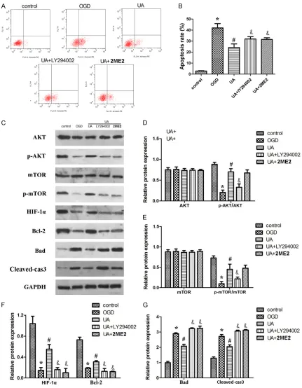
[image:5.612.91.521.71.583.2]xt, we performed the OGD assay to evaluate the UA protection against apoptosis in cortical neuronal cells. The results showed that the cortical neuronal cells subjected to OGD had a 40% apoptotic rate, while 20 μmol/L UA restored the apoptosis rate to 30%. Meanwhile,
UA protects ischemic brain injury
content that was elevated notably by MCAO (Figure 5C-E).
Discussion
Ursolic acid (UA) belongs to triterpenoid acid, which is one of the major components of cer-tain medicinal plants. Although many triterpe-noids have been used for a variety of clinical diseases with medicinal purposes in Asia, UA was identified mostly as an anticancer agent. To date, few studies have investigated the usage of UA in ischemic brain injury. A little-known mechanism was that UA protected brains from ischemic injury through the oxida-tive and inflammatory responses by suppress-ing TLR4 and upregulatsuppress-ing Nrf2 [8-10].
The pathophysiology of ischemic brain injury is a complex progress that includes cytotoxic responses, such as apoptosis, oxidative stress, proinflammation and neurological damage. Es- pecially, cell apoptosis plays a crucial role in ischemic brain injury. Previous studies have revealed that UA acts as an anticancer regent in its apoptosis-inducing function. UA can medi-ate cell apoptosis by degrading anti-apoptosis proteins and cleaving DNA repair molecules, extracellular matrix proteins, skeleton proteins and other related molecules in human cells [11-17]. In this study, we first demonstrated that UA protected the brain from ischemic inju-ry by attenuating neuron apoptosis. Our re- sults revealed that UA treatment significantly decreased the infarction area in rats subjected to MCAO. Consistently, neuron cell apoptosis was attenuated at the same time. With a neuron oxygen and glucose deprivation (OGD) model, we investigated if UA treatment sup-pressed the apoptosis of neuron cells undergo-ing OGD.
The PI3K pathway plays an important role in the regulation of cell growth, motility, survival and metabolism, as well as angiogenesis [18]. Numerous studies reported that HIF-1α activa-tion was regulated by a PI3K/Akt/mTOR-depen-dent mechanisms [19, 20]. In our study, we determined that the inhibition of HIF-1α and Bcl-2 family proteins by UA is mediated by the AKT pathway, and we observed that downgreg-ulation of p-AKT and p-mTOR was induced by UA treatment. These results demonstrate that UA inhibits the phosphorylation of AKT and mTOR.
knock down of AKT and HIF-1α could abolish the UA-induced phenotype (Figure 3A, 3B). In addition, we detected the expression level of some proteins. The results revealed that knock down of AKT reduced the expression level and the phosphorylation of AKT protein. Moreover, inhibited the phosphorylation of mTOR and the downstream bcl-2 but promoted the expression of bax and cleaved caspase3. Knock down of HIF-1α have no influence on the expression of AKT or mTOR but significantly reduced the ex- pression of HIF-1α and also of Bcl-2. Meanwhile promoted the expression of bax and cleaved caspase3 (Figure 3C, 3G). These results imply that UA protected against OGD-induced cortical neuronal cell apoptosis in an Akt- and HIF-1α-dependent manner.
UA exert its anti-apoptotic role by regulating the PI3K/AKT/mTOR signal pathway
To verify that UA exerts its anti-apoptotic effect through the PI3K/AKT/mTOR signal pathway, we also used 2ME2 and LY294002 to block HIF- 1α and AKT expression and evaluated the cell apoptosis after the block of HIF-1α and AKT. We found that block of HIF-1α and AKT significantly restored the ptotective effect of UA treatment (Figure 4A, 4B). Interestingly, the treatment of LY294002 not only inhibited the phosphoryla-tion of AKT but also the phosphorylaphosphoryla-tion of mTOR and HIF-1α expression induced by UA. Moreover, LY294002 blocked the elevation of bcl-2 expression and the downregulation of Bad along with Caspase3. Furthermore, 2ME2 significantly reduced the HIF-1α expression without affecting the phosphorylation or expression of AKT and mTOR. Similarly, si-HIF-1α also inhibited the elevation of Bcl-2 expres-sion and the downregulation of Bad along with Caspase3 (Figure 4C-G).
Knock down of AKT and HIF-1α reversed the
protective effect of UA on ischemia reperfu-sion induced injury
Hypoxia-inducible factor 1α (HIF-1α) is an
important transcriptional factor implicated in many cerebrovascular pathological disorders that targets many critical factors, such as
[image:7.612.91.522.70.624.2]UA protects ischemic brain injury
oxygenase-2, inducible nitric oxide synthase (iNOS), vascular endothelial growth factor (VE- GF) and erythropoietin [21]. 2ME2 is a known to be an effective HIF-1α inhibitor evaluated by different clinical trials [22, 23]. In our work, we used 2ME2 and LY294002 to inhibit HIF-1α and AKT expression. Additionally, the rescue assay by 2ME2 and LY294002 investigated the protective effect of UA in ischemic brain injury though an AKT and HIF-1α signal pathway. In this study, we investigated ursolic acid as a pro-tective effect and provided a potential therapy target for ischemic brain injury.
Conclusion
[image:8.612.89.515.73.488.2]In conclusion, our study could summarize as the following major founding: 1, We success-fully established middle cerebral artery occlu-sion (MCAO) models and found UA could pro-tected against MCAO-induced ischemic brain injury. 2, We found a new mechanism that UA can inhibit ischemia brain injury and neuronal cell apoptosis through regulating the AKT/ mTOR/HIF-1α pathway and the downstream protein expression such as Bcl-2, Bad and Cas- pase3.
Figure 5. Knock down of AKT and HIF-1α reversed the protective effect of UA on ischemia reperfusion induced injury. A, B. The MCAO model was established and was treated with UA or AKT nad HIF-1α knockdown in each group, then were evaluated by TTC staining. C. The neurological score was performed to evaluate the injury of the brain. D. The brain water content of the samples in each group was examined. E. The cortex neuron apoptosis in each group was detected by TUNEL assay. Sham group as control. *P<0.05 versus sham, #P<0.05 versus MCAO, &P<0.05 versus
Acknowledgements
This work was supported by the Project of the Affiliated Hospital of Logistics University of People’s Armed Police Force (WHJ2015016, WHZ201602, WHM201604 and WHM2016- 023); Tianjin Science and Technology Plan Project (16ZXHLSY00120).
Disclosure of conflict of interest
None.
Address correspondence to: Haiqian Liang, Institute of Traumatic Brain Injury and Neurology, Pingjin Hospital, Logistics University of Chinese People’s Armed Police Forces, Tianjin, China; Department of Neurosurgery, Pingjin Hospital, Logistics University of Armed Police Forces, 220 Chenglin Road, Tianjin 300162, China. Tel: 2260577125; Fax: +86-2260577125; E-mail: lianghaiqian711@163.com
References
[1] Cowan F, Rutherford M, Groenendaal F, Eken P, Mercuri E, Bydder GM, Meiners LC, Dubowitz LM and de Vries LS. Origin and timing of brain lesions in term infants with neonatal encepha-lopathy. Lancet 2003; 361: 736-42.
[2] Carli G, Reiger I and Evans N. One-year neuro-developmental outcome after moderate new-born hypoxic ischaemic encephalopathy. J Paediatr Child Health 2004; 40: 217-20. [3] Huang BY and Castillo M. Hypoxic-ischemic
brain injury: imaging findings from birth to adulthood. Radiographics 2008; 28: 417-39. [4] Bhattacharya P, Pandey AK, Paul S, Patnaik R
and Yavagal DR. Aquaporin-4 inhibition medi-ates piroxicam-induced neuroprotection again- st focal cerebral ischemia/reperfusion injury in rodents. PLoS One 2013; 8: e73481. [5] Yulug B, Kilic U, Kilic E and Bahr M. Rifampicin
attenuates brain damage in focal ischemia. Brain Res 2004; 996: 76-80.
[6] Mackay KB, Loddick SA, Naeve GS, Vana AM, Verge GM and Foster AC. Neuroprotective ef -fects of insulin-like growth factor-binding pro-tein ligand inhibitors in vitro and in vivo. J Cereb Blood Flow Metab 2003; 23: 1160-7. [7] Stoppini L, Buchs PA and Muller D. A simple
method for organotypic cultures of nervous tis-sue. J Neurosci Methods 1991; 37: 173-82. [8] Zhang T, Su J, Wang K, Zhu T and Li X. Ursolic
acid reduces oxidative stress to alleviate early brain injury following experimental subarach-noid hemorrhage. Neurosci Lett 2014; 579: 12-7.
[9] Zhang T, Su J, Guo B, Zhu T, Wang K and Li X. Ursolic acid alleviates early brain injury after experimental subarachnoid hemorrhage by suppressing TLR4-mediated inflammatory pa-thway. Int Immunopharmacol 2014; 23: 585-91.
[10] Li L, Zhang X, Cui L, Wang L, Liu H, Ji H and Du Y. Ursolic acid promotes the neuroprotection by activating Nrf2 pathway after cerebral isch-emia in mice. Brain Res 2013; 1497: 32-9. [11] Kim ES and Moon A. Ursolic acid inhibits the
invasive phenotype of SNU-484 human gastric cancer cells. Oncol Lett 2015; 9: 897-902. [12] Li R, Wang X, Zhang XH, Chen HH and Liu YD.
Ursolic acid promotes apoptosis of SGC-7901 gastric cancer cells through ROCK/PTEN medi-ated mitochondrial translocation of cofilin-1. Asian Pac J Cancer Prev 2014; 15: 9593-7. [13] Weng H, Tan ZJ, Hu YP, Shu YJ, Bao RF, Jiang L,
Wu XS, Li ML, Ding Q, Wang XA, Xiang SS, Li HF, Cao Y, Tao F and Liu YB. Ursolic acid induces cell cycle arrest and apoptosis of gallbladder carcinoma cells. Cancer Cell Int 2014; 14: 96. [14] Li Y, Lu X, Qi H, Li X, Xiao X and Gao J. Ursolic
acid induces apoptosis through mitochondrial intrinsic pathway and suppression of ERK1/2 MAPK in HeLa cells. J Pharmacol Sci 2014; 125: 202-10.
[15] Yang Y, Li C, Xiang X, Dai Z, Chang J, Zhang M, Cai H, Zhang H, Zhang M, Guo Y and Wu Z. Ursolic acid prevents endoplasmic reticulum stress-mediated apoptosis induced by heat stress in mouse cardiac myocytes. J Mol Cell Cardiol 2014; 67: 103-11.
[16] Wang J, Jiang Z, Xiang L, Li Y, Ou M, Yang X, Shao J, Lu Y, Lin L, Chen J, Dai Y and Jia L. Synergism of ursolic acid derivative US597 with 2-deoxy-D-glucose to preferentially induce tumor cell death by dual-targeting of apoptosis and glycolysis. Sci Rep 2014; 4: 5006. [17] Wang J, Liu L, Qiu H, Zhang X, Guo W, Chen W,
Tian Y, Fu L, Shi D, Cheng J, Huang W and Deng W. Ursolic acid simultaneously targets multiple signaling pathways to suppress proliferation and induce apoptosis in colon cancer cells. PLoS One 2013; 8: e63872.
[18] He Z, Chen AY, Rojanasakul Y, Rankin GO and Chen YC. Gallic acid, a phenolic compound, ex-erts anti-angiogenic effects via the PTEN/AKT/ HIF-1alpha/VEGF signaling pathway in ovarian cancer cells. Oncol Rep 2016; 35: 291-7. [19] Choi YH, Jin GY, Li LC and Yan GH. Inhibition of
protein kinase C delta attenuates allergic air-way inflammation through suppression of PI3K/Akt/mTOR/HIF-1 alpha/VEGF pathway. PLoS One 2013; 8: e81773.
re-UA protects ischemic brain injury
presses Akt/mTOR/HIF-1alpha axis and re-stores tamoxifen sensitivity in antiestrogen-re-sistant breast cancer cells. PLoS One 2015; 10: e0132285.
[21] Semenza GL. Hypoxia-inducible factor 1: con-trol of oxygen homeostasis in health and dis-ease. Pediatr Res 2001; 49: 614-7.
[22] Dahut WL, Lakhani NJ, Gulley JL, Arlen PM, Kohn EC, Kotz H, McNally D, Parr A, Nguyen D, Yang SX, Steinberg SM, Venitz J, Sparreboom A and Figg WD. Phase I clinical trial of oral 2-me-thoxyestradiol, an antiangiogenic and apoptot-ic agent, in patients with solid tumors. Cancer Biol Ther 2006; 5: 22-7.