organic papers
Acta Cryst.(2005). E61, o3435–o3436 doi:10.1107/S160053680502773X Silvaet al. C
16H12Br2N2O2S
o3435
Acta Crystallographica Section E
Structure Reports Online
ISSN 1600-5368
5,7-Dibromo-
N
-tosylquinolin-8-amine
Luiz Everson da Silva,a,b Antonio Carlos Joussef,aSabine Foroband Boris Schmidtb*
aDepartamento de Quı´mica–UFSC, 88040-900
Floriano´polis, SC, Brazil, andb
Clemens-Scho¨pf-Institut fu¨r Organische Chemie und Biochemie, Technische Universita¨t Darmstadt,
Petersenstrasse 22, D-64287 Darmstadt, Germany
Correspondence e-mail: [email protected]
Key indicators
Single-crystal X-ray study
T= 299 K
Mean(C–C) = 0.007 A˚
Rfactor = 0.041
wRfactor = 0.109
Data-to-parameter ratio = 14.3
For details of how these key indicators were automatically derived from the article, see http://journals.iucr.org/e.
#2005 International Union of Crystallography
Printed in Great Britain – all rights reserved
In the crystal structure of the title compound, C16H12Br2N2O2S, molecules are linked by two hydrogen
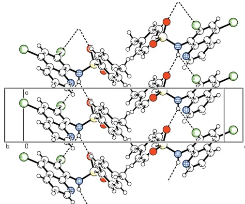
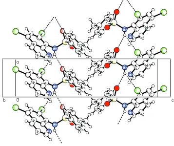
bonds to form a three-dimensional network. The H atom of the NH group has two intermolecular contacts; one to a sulfonyl O atom (H O = 2.22 A˚ ) and the other to the Br at the quinoline C-7 position (H Br = 3.11 A˚ ).
Comment
The investigation of the crystal structure of the title compound, (I), is part of our search for Zinquin Ester analogues (Kimberet al., 2000). This class of compounds has been shown to be effective in the detection of zinc (II) in a range of mammalian cells (Pearceet al., 2001).
The molecular structure of the title compound (I) is illus-trated in Fig. 1. The quinoline ring system is planar and inclined to the benzene ring of the tosyl moiety [torsion angle C1—N1—S1—C10 being 76.5 (3)].
The crystal packing of (I) is stabilized through a hydrogen-bonding network, as shown in Fig. 2. Details of the hydrogen bonding are given in Table 1.
Experimental
Compound (I) was prepared according to the literature procedure (Xueet al., 2000). Suitable crystals were obtained by recrystallization from methanol-dichloromethane (1:1).
Crystal data
C16H12Br2N2O2S Mr= 456.16
Orthorhombic,P212121 a= 4.939 (1) A˚ b= 16.595 (3) A˚ c= 20.254 (4) A˚ V= 1660.1 (6) A˚3 Z= 4
Dx= 1.825 Mg m 3
CuKradiation Cell parameters from 25
reflections
= 5.3–18.8
= 7.46 mm1 T= 299 (2) K
Long needle, light brown 0.650.080.05 mm
Data collection
Nonius CAD-4 diffractometer
!/2scans
Absorption correction: scan (Northet al., 1968) Tmin= 0.486,Tmax= 0.708 3465 measured reflections 2997 independent reflections 2758 reflections withI> 2(I)
Rint= 0.019
max= 68.0
h= 0!5 k= 0!19 l=24!24 3 standard reflections
frequency: 120 min intensity decay: 1.0%
Refinement
Refinement onF2 R[F2> 2(F2)] = 0.041 wR(F2) = 0.109 S= 1.08 2997 reflections 209 parameters
H-atom parameters constrained w= 1/[2
(Fo2) + (0.076P)2 + 0.4357P]
whereP= (Fo2+ 2Fc2)/3
(/)max< 0.001
max= 0.69 e A˚
3
min=0.92 e A˚
3
Extinction correction:SHELXL97 Extinction coefficient: 0.0012 (2) Absolute structure: Flack (1983) Flack parameter:0.01 (3)
Table 1
Hydrogen-bond geometry (A˚ ,).
D—H A D—H H A D A D—H A
N1—H1N O1i 0.86 2.22 2.884 (4) 134 N1—H1N Br1i
0.86 3.11 3.873 (3) 149
Symmetry code: (i)x1;y;z.
The H atoms were included in idealized positions and refined as riding atoms withUiso(H) = 1.2Ueq(parent atom), and N—H = 0.86 A˚
and C—H = 0.93 A˚ (0.96 A˚ for methyl groups).
Data collection:Nonius Diffractometer Control Software(Nonius, 1996); cell refinement:Nonius Diffractometer Control Software; data reduction: REDU4 (Stoe & Cie, 1987); program(s) used to solve structure:SHELXS97(Sheldrick, 1997); program(s) used to refine structure: SHELXL97 (Sheldrick, 1997); molecular graphics:
PLATON (Spek, 2003); software used to prepare material for publication:SHELXL97.
The authors thank Professor Dr Hartmut Fuess, FG Strukturforschung, FB Material- und Geowissenschaften, Technische Universita¨t Darmstadt, Petersenstrasse 23, 64287 Darmstadt, for diffractometer time.
References
Flack, H. D. (1983).Acta Cryst.A39, 876–881.
Kimber, M. C., Mahadevan, I. B., Lincoln, S. F., Ward, A. D. & Tiekink, E. R. T. (2000).J. Org. Chem.65, 8204–8209.
Nonius (1996). Nonius Diffractometer Control Software. PC Version 2.0. Nonius GmbH, Solingen, Germany.
North, A. C. T., Phillips, D. C. & Mathews, F. S. (1968).Acta Cryst.A24, 351– 359.
Pearce, D. A., Jotterand, N., Carrico, I. S. & Imperiali, B. (2001).J. Am. Chem. Soc.123, 5160–5161.
Sheldrick, G. M. (1997). SHELXS97 and SHELXL97. University of Go¨ttingen, Germany.
Spek, A. L. (2003).J. Appl. Cryst.36, 7–13.
Stoe & Cie. (1987).REDU4. Version 6.2c. Stoe & Cie GmbH, Darmstadt, Germany.
[image:2.610.310.563.70.281.2] [image:2.610.44.298.73.232.2]Xue, G., Bradshaw, Jerald S., Dalley, N. K., Savage, P. B., Izatt, R. M., Prodi, L., Montalti, M. & Zaccheroni, N. (2000).Tetrahedron,58, 4809–4815. Figure 1
Molecular structure of (I), showing the atom labeling and displacement ellipsoids drawn at the 50% probability level.
Figure 2
supporting information
sup-1
Acta Cryst. (2005). E61, o3435–o3436supporting information
Acta Cryst. (2005). E61, o3435–o3436 [doi:10.1107/S160053680502773X]
5,7-Dibromo-
N
-tosylquinolin-8-amine
Luiz Everson da Silva, Antonio Carlos Joussef, Sabine Foro and Boris Schmidt
S1. Comment
The investigation of the crystal structure of the title compound, (I), is part of our search for Zinquin Ester analogues (Kimber et al., 2000). This class of the compounds has been shown to be effective in the detection of zinc (II) in a range of mammalian cells (Pearce et al., 2001).
The molecular structure of the title compound (I) is illustrated in Fig. 1. The quinoline ring system is planar and inclined to the phenyl ring of the tosyl moiety [torsion angle C1—N1—S1—C10 being 76.5 (3)°].
The crystal packing of (I) is stabilized through a hydrogen-bonding network, as shown in Fig. 2. Details of the hydrogen bonding are given in Table 1. The H atom of the NH group has an intermolecular contact to atom O1, with an H···O distance of 2.22 Å, and is also connected to the bromine atom, indicating very weak N—H···Br hydrogen bonding, with an H···Br distance of 3.11 Å.
S2. Experimental
Compound (I) was prepared according to the literature procedure (Xue et al., 2000). Suitable crystals were obtained by recrystallization from methanol-dichloromethane (1:1).
S3. Refinement
The H atoms were included in idealized positions and refined as riding atoms with Uiso(H) = 1.2Ueq(parent atom), and N
Figure 1
Molecular structure of (I), showing the atom labeling and displacement ellipsoids drawn at the 50% probability level.
Figure 2
[image:4.610.125.482.343.642.2]supporting information
sup-3
Acta Cryst. (2005). E61, o3435–o34365,7-Dibromo-N-tosylquinolin-8-amine
Crystal data
C16H12Br2N2O2S Mr = 456.16
Orthorhombic, P212121
Hall symbol: P 2ac 2ab
a = 4.939 (1) Å
b = 16.595 (3) Å
c = 20.254 (4) Å
V = 1660.1 (6) Å3 Z = 4
F(000) = 896
Dx = 1.825 Mg m−3
Cu Kα radiation, λ = 1.54180 Å Cell parameters from 25 reflections
θ = 5.3–18.8°
µ = 7.46 mm−1 T = 299 K
Long needle, light brown 0.65 × 0.08 × 0.05 mm
Data collection
Nonius CAD-4 diffractometer
Radiation source: fine-focus sealed tube Graphite monochromator
ω/2θ scans
Absorption correction: psi-scan (North et al., 1968)
Tmin = 0.486, Tmax = 0.708
3465 measured reflections
2997 independent reflections 2758 reflections with I > 2σ(I)
Rint = 0.019
θmax = 68.0°, θmin = 3.4° h = 0→5
k = 0→19
l = −24→24
3 standard reflections every 120 min intensity decay: 1.0%
Refinement
Refinement on F2
Least-squares matrix: full
R[F2 > 2σ(F2)] = 0.041 wR(F2) = 0.109 S = 1.08 2997 reflections 209 parameters 0 restraints
Primary atom site location: structure-invariant direct methods
Secondary atom site location: difference Fourier map
Hydrogen site location: inferred from neighbouring sites
H-atom parameters constrained
w = 1/[σ2(F
o2) + (0.076P)2 + 0.4357P]
where P = (Fo2 + 2Fc2)/3
(Δ/σ)max < 0.001
Δρmax = 0.69 e Å−3
Δρmin = −0.92 e Å−3
Extinction correction: SHELXL97, Fc*=kFc[1+0.001xFc2λ3/sin(2θ)]-1/4
Extinction coefficient: 0.0012 (2) Absolute structure: Flack (1983) Absolute structure parameter: −0.01 (3)
Special details
Geometry. Bond distances, angles etc. have been calculated using the rounded fractional coordinates. All su's are estimated from the variances of the (full) variance-covariance matrix. The cell e.s.d.'s are taken into account in the estimation of distances, angles and torsion angles
Refinement. Refinement of F2 against ALL reflections. The weighted R-factor wR and goodness of fit S are based on F2,
conventional R-factors R are based on F, with F set to zero for negative F2. The threshold expression of F2 > σ(F2) is used
only for calculating R-factors(gt) etc. and is not relevant to the choice of reflections for refinement. R-factors based on F2
are statistically about twice as large as those based on F, and R- factors based on ALL data will be even larger.
Fractional atomic coordinates and isotropic or equivalent isotropic displacement parameters (Å2)
x y z Uiso*/Ueq
S1 0.45127 (18) 0.15331 (5) 0.33555 (5) 0.0360 (3) O1 0.7323 (5) 0.14564 (17) 0.31743 (15) 0.0460 (8) O2 0.3241 (7) 0.09229 (18) 0.37390 (16) 0.0534 (10) N1 0.2761 (6) 0.15936 (18) 0.26682 (15) 0.0364 (9) N2 0.0963 (8) 0.3138 (2) 0.24236 (19) 0.0459 (11) C1 0.3804 (8) 0.2041 (2) 0.21271 (19) 0.0368 (10) C2 0.5625 (10) 0.1704 (3) 0.1682 (2) 0.0470 (12) C3 0.6661 (11) 0.2144 (3) 0.1149 (2) 0.0543 (16) C4 0.5827 (10) 0.2916 (3) 0.1050 (2) 0.0510 (16) C5 0.3953 (9) 0.3298 (2) 0.1475 (2) 0.0443 (12) C6 0.3020 (13) 0.4095 (3) 0.1402 (2) 0.0600 (14) C7 0.1102 (12) 0.4385 (3) 0.1829 (3) 0.0650 (16) C8 0.0162 (12) 0.3887 (3) 0.2315 (3) 0.0603 (17) C9 0.2905 (8) 0.2838 (2) 0.20064 (19) 0.0397 (11) C10 0.4153 (8) 0.2452 (2) 0.37766 (18) 0.0369 (10) C11 0.5991 (9) 0.3069 (3) 0.3663 (2) 0.0470 (12) C12 0.5790 (11) 0.3769 (3) 0.4025 (2) 0.0537 (16) C13 0.3806 (11) 0.3870 (3) 0.4501 (2) 0.0553 (16) C14 0.1963 (11) 0.3243 (3) 0.4598 (2) 0.0581 (15) C15 0.2108 (9) 0.2538 (3) 0.4241 (2) 0.0483 (12) C16 0.3710 (16) 0.4613 (4) 0.4915 (3) 0.080 (2) H1N 0.12190 0.13560 0.26360 0.0440* H3 0.79130 0.19110 0.08650 0.0650* H6 0.37000 0.44210 0.10680 0.0720* H7 0.04530 0.49090 0.17890 0.0780* H8 −0.11600 0.40930 0.25950 0.0720* H11 0.73420 0.30100 0.33470 0.0560* H12 0.70150 0.41840 0.39470 0.0650* H14 0.06040 0.33020 0.49120 0.0700* H15 0.08570 0.21280 0.43090 0.0580* H16A 0.34950 0.50760 0.46360 0.0960* H16B 0.53630 0.46610 0.51610 0.0960* H16C 0.22090 0.45800 0.52150 0.0960*
Atomic displacement parameters (Å2)
U11 U22 U33 U12 U13 U23
supporting information
sup-5
Acta Cryst. (2005). E61, o3435–o3436C5 0.047 (2) 0.042 (2) 0.044 (2) −0.0095 (16) −0.0116 (17) 0.0056 (16) C6 0.074 (3) 0.051 (2) 0.055 (2) −0.012 (2) −0.011 (3) 0.017 (2) C7 0.075 (3) 0.043 (2) 0.077 (3) 0.004 (2) −0.019 (3) 0.009 (2) C8 0.063 (3) 0.049 (3) 0.069 (3) 0.007 (2) −0.003 (3) 0.000 (2) C9 0.0370 (19) 0.0425 (19) 0.0397 (18) −0.0062 (17) −0.0089 (16) 0.0006 (15) C10 0.0319 (18) 0.0452 (19) 0.0336 (17) 0.0004 (15) −0.0018 (14) 0.0053 (15) C11 0.041 (2) 0.048 (2) 0.052 (2) −0.0017 (18) 0.0074 (18) 0.0003 (18) C12 0.052 (3) 0.050 (2) 0.059 (3) −0.002 (2) −0.006 (2) −0.004 (2) C13 0.068 (3) 0.056 (3) 0.042 (2) 0.019 (2) −0.013 (2) −0.0046 (19) C14 0.056 (3) 0.081 (3) 0.0374 (19) 0.010 (3) 0.007 (2) −0.002 (2) C15 0.037 (2) 0.062 (2) 0.046 (2) 0.0037 (19) 0.0098 (18) 0.0038 (19) C16 0.107 (5) 0.071 (3) 0.061 (3) 0.024 (3) −0.015 (3) −0.019 (3)
Geometric parameters (Å, º)
Br1—C2 1.887 (5) C10—C15 1.388 (6) Br2—C4 1.893 (4) C10—C11 1.388 (6) S1—O1 1.441 (3) C11—C12 1.377 (7) S1—O2 1.422 (3) C12—C13 1.385 (7) S1—N1 1.642 (3) C13—C16 1.492 (8) S1—C10 1.756 (4) C13—C14 1.396 (7) N1—C1 1.421 (5) C14—C15 1.377 (7) N2—C8 1.323 (6) C3—H3 0.9300 N2—C9 1.372 (5) C6—H6 0.9300 N1—H1N 0.8600 C7—H7 0.9300 C1—C2 1.391 (6) C8—H8 0.9300 C1—C9 1.416 (5) C11—H11 0.9300 C2—C3 1.400 (6) C12—H12 0.9300 C3—C4 1.361 (7) C14—H14 0.9300 C4—C5 1.414 (6) C15—H15 0.9300 C5—C6 1.408 (6) C16—H16A 0.9600 C5—C9 1.417 (5) C16—H16B 0.9600 C6—C7 1.370 (8) C16—H16C 0.9600 C7—C8 1.367 (8)
Br1···S1 3.7413 (14) C15···C14vii 3.600 (7)
Br1···O1 3.210 (3) C8···H11v 2.9000
Br1···N1 3.137 (3) C10···H14vii 3.0200
Br1···C7i 3.686 (6) C11···H8ii 3.0900
Br1···C12i 3.662 (5) C12···H14ii 3.0800
Br1···H1Nii 3.1100 C13···H15vii 3.0900
Br1···H16Ai 2.9600 C14···H15vii 3.0000
Br2···H6 2.8100 C15···H11v 3.0700
S1···Br1 3.7413 (14) C15···H14vii 2.8000
O1···Br1 3.210 (3) C16···H6viii 3.0700
O1···N1ii 2.884 (4) H1N···Br1v 3.1100
O1···C2 3.164 (5) H1N···O1v 2.2200
O1···H11 2.6000 H6···C16ix 3.0700
O1···H7i 2.7900 H6···H16Cix 2.4400
O2···H15 2.5900 H7···O1vi 2.7900
O2···H16Biii 2.8100 H7···O2x 2.7000
O2···H7iv 2.7000 H8···C11v 3.0900
N1···Br1 3.137 (3) H8···H11v 2.4700
N1···O1v 2.884 (4) H11···O1 2.6000
N1···N2 2.757 (5) H11···N2ii 2.6000
N2···N1 2.757 (5) H11···C8ii 2.9000
N2···C10 3.360 (5) H11···C15ii 3.0700
N2···H11v 2.6000 H11···H8ii 2.4700
C2···O1 3.164 (5) H14···C12v 3.0800
C7···Br1vi 3.686 (6) H14···H16C 2.3500
C10···N2 3.360 (5) H14···C10iii 3.0200
C11···C14ii 3.517 (7) H14···C15iii 2.8000
C11···C15ii 3.358 (6) H15···O2 2.5900
C12···Br1vi 3.662 (5) H15···C13iii 3.0900
C12···C14ii 3.377 (7) H15···C14iii 3.0000
C14···C15iii 3.600 (7) H16A···Br1vi 2.9600
C14···C11v 3.517 (7) H16B···O2vii 2.8100
C14···C12v 3.377 (7) H16C···H14 2.3500
C15···C11v 3.358 (6) H16C···H6viii 2.4400
supporting information
sup-7
Acta Cryst. (2005). E61, o3435–o3436N2—C8—C7 125.9 (5) C13—C16—H16B 109.00 N2—C9—C5 121.8 (3) C13—C16—H16C 109.00 C1—C9—C5 121.3 (3) H16A—C16—H16B 109.00 N2—C9—C1 116.9 (3) H16A—C16—H16C 109.00 C11—C10—C15 120.9 (4) H16B—C16—H16C 109.00 S1—C10—C11 119.6 (3)
O1—S1—N1—C1 −38.8 (3) C2—C3—C4—Br2 −178.3 (4) O2—S1—N1—C1 −168.2 (3) C2—C3—C4—C5 0.7 (7) C10—S1—N1—C1 76.5 (3) Br2—C4—C5—C9 177.5 (3) N1—S1—C10—C11 −90.6 (3) C3—C4—C5—C9 −1.5 (7) O1—S1—C10—C15 −152.4 (3) Br2—C4—C5—C6 −1.3 (7) O2—S1—C10—C15 −21.6 (4) C3—C4—C5—C6 179.7 (5) N1—S1—C10—C15 92.4 (3) C6—C5—C9—N2 2.7 (6) O1—S1—C10—C11 24.7 (4) C6—C5—C9—C1 −177.9 (4) O2—S1—C10—C11 155.5 (3) C4—C5—C6—C7 176.9 (5) S1—N1—C1—C2 84.2 (4) C9—C5—C6—C7 −1.9 (7) S1—N1—C1—C9 −99.0 (4) C4—C5—C9—C1 3.2 (6) C8—N2—C9—C1 179.0 (4) C4—C5—C9—N2 −176.2 (4) C8—N2—C9—C5 −1.6 (6) C5—C6—C7—C8 0.2 (8) C9—N2—C8—C7 −0.3 (8) C6—C7—C8—N2 1.0 (9) N1—C1—C9—N2 −1.5 (5) S1—C10—C15—C14 175.8 (3) N1—C1—C9—C5 179.0 (4) C15—C10—C11—C12 0.9 (6) C9—C1—C2—C3 3.2 (6) S1—C10—C11—C12 −176.1 (3) C2—C1—C9—C5 −4.0 (6) C11—C10—C15—C14 −1.2 (6) N1—C1—C2—Br1 2.5 (6) C10—C11—C12—C13 0.3 (7) C2—C1—C9—N2 175.4 (4) C11—C12—C13—C14 −1.1 (7) N1—C1—C2—C3 −179.9 (4) C11—C12—C13—C16 176.5 (5) C9—C1—C2—Br1 −174.4 (3) C16—C13—C14—C15 −176.9 (5) Br1—C2—C3—C4 176.1 (4) C12—C13—C14—C15 0.8 (7) C1—C2—C3—C4 −1.6 (7) C13—C14—C15—C10 0.4 (7)
Symmetry codes: (i) −x+1, y−1/2, −z+1/2; (ii) x+1, y, z; (iii) x−1/2, −y+1/2, −z+1; (iv) −x, y−1/2, −z+1/2; (v) x−1, y, z; (vi) −x+1, y+1/2, −z+1/2; (vii)
x+1/2, −y+1/2, −z+1; (viii) −x+1/2, −y+1, z+1/2; (ix) −x+1/2, −y+1, z−1/2; (x) −x, y+1/2, −z+1/2.
Hydrogen-bond geometry (Å, º)
D—H···A D—H H···A D···A D—H···A
N1—H1N···O1v 0.86 2.22 2.884 (4) 134
N1—H1N···Br1v 0.86 3.11 3.873 (3) 149
C6—H6···Br2 0.93 2.81 3.209 (5) 107 C11—H11···N2ii 0.93 2.60 3.514 (6) 169
C15—H15···O2 0.93 2.59 2.921 (6) 101