metal-organic papers
Acta Cryst.(2005). E61, m1131–m1132 doi:10.1107/S1600536805014893 Jones, Gillon and Davey Na+C
7H3N2O6
m1131
Acta Crystallographica Section E Structure Reports Online
ISSN 1600-5368
Sodium 3,5-dinitrobenzoate
Helen P. Jones,* Amy L. Gillon‡ and Roger J. Davey
Colloids, Crystals and Interfaces Group, School of Chemical Engineering and Analytical Sciences, The University of Manchester, PO Box 88, Manchester M60 1QD, England
‡ Current address: Pharmaceutical R&D, Pfizer Global R&D (IPC 435), Ramsgate Road, Sandwich, Kent CT13 9NJ, England
Correspondence e-mail:
h.jones-2@postgrad.manchester.ac.uk
Key indicators
Single-crystal X-ray study
T= 150 K
Mean(C–C) = 0.002 A˚
Rfactor = 0.025
wRfactor = 0.066 Data-to-parameter ratio = 7.3
For details of how these key indicators were automatically derived from the article, see http://journals.iucr.org/e.
#2005 International Union of Crystallography Printed in Great Britain – all rights reserved
Sodium 3,5-dinitrobenzoate, Na+C7H3N2O6, was obtained by evaporation at room temperature of an aqueous solution of ethylenediammonium 3,5-dinitrobenzoate in sodium hydrox-ide. The structure is trigonal and the benzoate ion has twofold crystallographic symmetry.
Comment
During work on crystallization of the salt ethylenedi-ammonium 3,5-dinitrobenzoate, an aqueous solution of the salt at pH 12 was prepared and allowed to evaporate at room temperature, giving red prisms of sodium 3,5-dinitrobenzoate (NaDNB), (I). The crystal structure was not found in the Cambridge Structural Database (CSD, Version 5.25; Allen, 2002) and hence its structure was determined by single-crystal X-ray diffraction at 150 K.
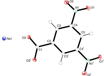
The benzoate ion is on a twofold axis of symmetry, passing through the carboxylate group (Fig. 1).
Experimental
3,5-Dinitrobenzoic acid (Aldrich, 99%) was dissolved in sodium hydroxide solution and a solution of ethylenediamine (Aldrich, 99%) was added. The solution was filtered and the pH recorded as 12.14. The solution pH was measured using an Accumet Basic AB15 pH
[image:1.610.290.371.356.427.2] [image:1.610.226.441.556.713.2]Received 5 April 2005 Accepted 10 May 2005 Online 14 May 2005
Figure 1
View of NaDNB, showing the whole benzoate anion. Displacement ellipsoids are drawn at the 50% probability level. [Symmetry code: (i)
meter with an Accumet glass calomel pH electrode. The solution was allowed to evaporate to dryness in air at room temperature. Crystals of ethylenediammonium 3,5-dinitrobenzoate, sodium hydroxide and red prisms of sodium 3,5-dinitrobenzoate formed.
Crystal data
Na+C7H3N2O6
Mr= 234.1 Trigonal,P3121
a= 10.7701 (5) A˚
c= 6.3526 (2) A˚
V= 638.15 (5) A˚3
Z= 3
Dx= 1.828 Mg m
3
MoKradiation Cell parameters from 2522
reflections
= 1.0–27.5
= 0.20 mm1
T= 150 K Prism, red
0.250.250.25 mm
Data collection
Nonius KappaCCD diffractometer Thick-slice’and!scans Absorption correction: multi-scan
(Blessing, 1995)
Tmin= 0.796,Tmax= 0.951
3498 measured reflections 554 independent reflections
537 reflections withI> 2(I)
Rint= 0.027 max= 27.5
h=12!13
k=8!13
l=8!8
Refinement
Refinement onF2
R[F2> 2(F2)] = 0.026
wR(F2) = 0.066
S= 1.09 554 reflections 76 parameters
H-atom parameters constrained
w= 1/[2(F
o2) + (0.0336P)2 + 0.1582P]
whereP= (Fo
2
+ 2Fc
2
)/3 (/)max< 0.001
max= 0.24 e A˚ 3
min=0.18 e A˚ 3
Extinction correction:SHELXL97 Extinction coefficient: 0.14 (2)
In the absence of significant anomalous dispersion effects, Friedel pairs were merged. The choice of space group P3121 rather than
P3221 is arbitrary. All H atoms were positioned geometrically and refined as riding, with C—H = 0.93–0.98 A˚ andUiso(H) = 1.2Ueq(C).
Data collection: COLLECT (Nonius, 2000); cell refinement:
SCALEPACK (Otwinowski & Minor, 1997); data reduction:
SORTAV (Blessing, 1987,1989, SCALEPACK and DENZO
(Otwinowski & Minor, 1997); program(s) used to solve structure:
SHELXS97(Sheldrick, 1997); program(s) used to refine structure:
SHELXL97 (Sheldrick, 1997); molecular graphics: ORTEP-3 for Windows (Farrugia, 1997); software used to prepare material for publication:WinGX(Farrugia, 1999).
The authors thank Sanofı–Aventis Ltd for funding.
References
Allen, F. H. (2002).Acta Cryst.B58, 380–388. Blessing, R. H. (1987).Crystallogr. Rev.1, 3–58. Blessing, R. H. (1989).J. Appl. Cryst.22, 396–397. Blessing, R. H. (1995).Acta Cryst.A51, 33–38. Farrugia, L. J. (1997).J. Appl. Cryst.30, 565. Farrugia, L. J. (1999).J. Appl. Cryst.32, 837–838.
Nonius (2000).COLLECT.Nonius BV, Delft, The Netherlands.
Otwinowski, Z. & Minor, W. (1997). Methods in Enzymology, Vol. 276,
Macromolecular Crystallography, Part A, edited by C. W. Carter Jr & R. M. Sweet, pp. 307–326. New York: Academic Press.
[image:2.610.228.540.71.336.2]Sheldrick, G. M. (1997). SHELXS97 and SHELXL97. University of Go¨ttingen, Germany.
Figure 2
[image:2.610.44.295.72.333.2]The packing of sodium 3,5-dinitrobenzoate, viewed along the c axis, showing the threefold symmetry.
Figure 3
[image:2.610.71.268.377.579.2]supporting information
sup-1 Acta Cryst. (2005). E61, m1131–m1132
supporting information
Acta Cryst. (2005). E61, m1131–m1132 [https://doi.org/10.1107/S1600536805014893]
Sodium 3,5-dinitrobenzoate
Helen P. Jones, Amy L. Gillon and Roger J. Davey
Sodium 3,5-dinitrobenzoate
Crystal data Na+·C
7H3N2O6−
Mr = 234.1 Trigonal, P3121
Hall symbol: P 31 2" a = 10.7701 (5) Å c = 6.3526 (2) Å V = 638.15 (5) Å3
Z = 3 F(000) = 354
Dx = 1.828 Mg m−3
Mo Kα radiation, λ = 0.71073 Å Cell parameters from 2522 reflections θ = 1.0–27.5°
µ = 0.20 mm−1
T = 150 K Prism, red
0.25 × 0.25 × 0.25 mm
Data collection Nonius KappaCCD
diffractometer
Radiation source: Enraf Nonius FR590 Graphite monochromator
CCD rotation images, thick slices scans Absorption correction: multi-scan
(Blessing, 1995) Tmin = 0.796, Tmax = 0.951
3498 measured reflections 554 independent reflections 537 reflections with I > 2σ(I) Rint = 0.027
θmax = 27.5°, θmin = 3.8°
h = −12→13 k = −8→13 l = −8→8
Refinement Refinement on F2
Least-squares matrix: full R[F2 > 2σ(F2)] = 0.026
wR(F2) = 0.066
S = 1.09 554 reflections 76 parameters 0 restraints
Primary atom site location: structure-invariant direct methods
Secondary atom site location: difference Fourier map
Hydrogen site location: inferred from neighbouring sites
H-atom parameters constrained w = 1/[σ2(F
o2) + (0.0336P)2 + 0.1582P]
where P = (Fo2 + 2Fc2)/3
(Δ/σ)max < 0.001
Δρmax = 0.24 e Å−3
Δρmin = −0.18 e Å−3
Extinction correction: SHELXL97, Fc*=kFc[1+0.001xFc2λ3/sin(2θ)]-1/4
Extinction coefficient: 0.14 (2)
Special details
Refinement. Refinement of F2 against ALL reflections. The weighted R-factor wR and goodness of fit S are based on F2,
conventional R-factors R are based on F, with F set to zero for negative F2. The threshold expression of F2 > σ(F2) is used
only for calculating R-factors(gt) etc. and is not relevant to the choice of reflections for refinement. R-factors based on F2
are statistically about twice as large as those based on F, and R- factors based on ALL data will be even larger.
Fractional atomic coordinates and isotropic or equivalent isotropic displacement parameters (Å2)
x y z Uiso*/Ueq
C1 0.2002 (2) 1 −0.1667 0.0113 (5)
C2 0.3418 (2) 1 −0.1667 0.0122 (5)
C3 0.3701 (2) 0.92732 (19) −0.0104 (2) 0.0131 (4)
H3 0.3033 0.8786 0.0952 0.016*
O3 0.11648 (14) 0.93745 (13) −0.01613 (17) 0.0138 (3)
C4 0.4994 (2) 0.9287 (2) −0.0143 (2) 0.0151 (4)
C5 0.6034 (2) 1 −0.1667 0.0155 (5)
H5 0.6897 1 −0.1667 0.019*
N2 0.52972 (17) 0.85204 (18) 0.1513 (2) 0.0186 (4)
Na1 0.87486 (9) 0.87486 (9) 0 0.0131 (3)
O1 0.63430 (15) 0.83610 (16) 0.1274 (2) 0.0242 (4)
O2 0.45083 (19) 0.8090 (2) 0.3043 (2) 0.0329 (4)
Atomic displacement parameters (Å2)
U11 U22 U33 U12 U13 U23
C1 0.0111 (8) 0.0108 (11) 0.0118 (10) 0.0054 (5) −0.0016 (4) −0.0031 (8)
C2 0.0115 (9) 0.0135 (11) 0.0122 (10) 0.0067 (6) −0.0012 (4) −0.0024 (9)
C3 0.0136 (9) 0.0142 (9) 0.0121 (8) 0.0073 (7) 0.0010 (6) 0.0001 (6)
O3 0.0118 (6) 0.0162 (7) 0.0133 (6) 0.0069 (5) 0.0013 (4) 0.0010 (5)
C4 0.0170 (8) 0.0185 (9) 0.0132 (8) 0.0115 (7) −0.0009 (6) 0.0006 (7)
C5 0.0130 (9) 0.0179 (13) 0.0173 (11) 0.0089 (6) 0.0002 (5) 0.0005 (9)
N2 0.0179 (8) 0.0226 (9) 0.0183 (7) 0.0125 (7) 0.0007 (6) 0.0056 (6)
Na1 0.0131 (4) 0.0131 (4) 0.0128 (4) 0.0064 (4) −0.00068 (18) 0.00068 (18)
O1 0.0170 (7) 0.0323 (9) 0.0300 (7) 0.0174 (7) 0.0027 (6) 0.0104 (6)
O2 0.0356 (9) 0.0548 (11) 0.0228 (7) 0.0335 (9) 0.0132 (6) 0.0209 (7)
Geometric parameters (Å, º)
C1—O3 1.2547 (16) O3—Na1ii 2.3416 (14)
C1—C2 1.525 (3) C4—C5 1.386 (2)
C2—C3 1.389 (2) C4—N2 1.471 (2)
C3—C4 1.386 (2) C5—H5 0.93
C3—H3 0.93 N2—O2 1.220 (2)
O3—Na1i 2.3083 (11) N2—O1 1.231 (2)
O3iii—C1—O3 126.5 (2) O3iv—Na1—O3v 167.89 (8)
O3—C1—C2 116.77 (11) O3v—Na1—O3vi 86.31 (5)
C3iii—C2—C3 119.9 (2) O3v—Na1—O3vii 102.24 (5)
supporting information
sup-3 Acta Cryst. (2005). E61, m1131–m1132
C4—C3—C2 118.90 (16) O3iv—Na1—O1 79.55 (5)
C4—C3—H3 120.5 O3v—Na1—O1 93.24 (5)
C2—C3—H3 120.5 O3vi—Na1—O1 82.64 (5)
C1—O3—Na1i 131.53 (9) O3vii—Na1—O1 162.96 (5)
C1—O3—Na1ii 125.81 (12) O1viii—Na1—O1 107.43 (8)
Na1i—O3—Na1ii 85.34 (5) O3iv—Na1—Na1ix 47.77 (3)
C3—C4—C5 123.23 (16) O3v—Na1—Na1ix 143.85 (5)
C3—C4—N2 119.02 (15) O3vi—Na1—Na1ix 77.64 (3)
C5—C4—N2 117.74 (16) O3vii—Na1—Na1ix 46.88 (4)
C4iii—C5—C4 115.8 (2) O1viii—Na1—Na1ix 108.62 (3)
C4—C5—H5 122.1 O1—Na1—Na1ix 116.10 (3)
O2—N2—O1 123.91 (16) Na1ix—Na1—Na1x 100.20 (3)
O2—N2—C4 118.34 (14) N2—O1—Na1 160.96 (12)
O1—N2—C4 117.74 (15)
Symmetry codes: (i) −y+1, x−y+1, z+1/3; (ii) x−1, y, z; (iii) x−y+1, −y+2, −z−1/3; (iv) −x+1, −x+y, −z+1/3; (v) −x+y, −x+1, z−1/3; (vi) y, x+1, −z; (vii)

![Overview of the labour market [March 2006]](data:image/gif;base64,R0lGODlhAQABAIAAAP///wAAACH5BAEAAAAALAAAAAABAAEAAAICRAEAOw==)