Original Article
Effect of lactobacillus on Toll-like receptors expression
and bacterial translocation in antibiotic diarrhea rats
Yangming Que1,2, Mengli Gu1, Feng Ji1, Xinxin Zhou1
1Department of Gastroenterology, The First Affiliated Hospital of Medical College of Zhejiang University, Hangzhou, P. R. China; 2Department of Gastroenterology, The Second Affiliated Hospital of Jiaxing University, Jiaxing, P. R. China
Received January 3, 2017; Accepted November 29, 2017; Epub February 15, 2018; Published February 28, 2018
Abstract: Diarrhea is a common digestive system disease. As a type of intestinal probiotics, lactobacillus plays a critical role in maintaining the normal function of intestinal tract. This study established a rat antibiotic diarrhea model to test Toll-like receptors expression, aiming to analyze the impact of lactobacillus on Toll-like receptors ex-pression and bacterial translocation. A total of 60 Wistar rats were equally randomly divided into three groups. The rats in experimental group received lincomycin intragastric administration together with lactobacillus intervention. The rats in model group received lincomycin intragastric administration for 7 days to establish diarrhea model. General state and intestinal tissue changes under the microscope were observed. Bacterial translocation in intes-tine, liver, spleen, and mesenteric lymph nodes was tested by bacterial culture method. TLR2 and TLR4 mRNA and protein expression in small intestine cells were detected by RT-PCR and Western blot. The amplitude of weight loss
in experimental group was lower than the model group. Bifidobacteria, lactobacillus, enterobacter, and enterococ -cus in intestine of the experimental group were obviously higher than those in the model group (P < 0.05). Bacterial
translocation in liver, spleen, and mesenteric lymph nodes were significantly lower than those in the model group (P
< 0.05). TLR2 and TLR4 mRNA and protein levels in experimental group was markedly higher than the model group and further elevated following time extension (P < 0.05). Lactobacillus intervention shows a protective effect on
intestinal mucosal inflammation in diarrhea rat through increasing intestinal beneficial bacteria, inhibiting bacterial
translocation, and enhancing TLR2 and TLR4 expression.
Keywords: Lactobacillus, diarrhea, Toll-like receptor, bacterial translocation
Introduction
Intestinal epithelial cells are important defen-sive line in the body that can timely resist the invasion of the intestinal bacteria and virus. There are numerous kinds of drugs for enteritis, but with the weakness of long treatment cycle, large side effect, and easy to relapse after stop-ping [1, 2]. Innate immunity is the first line of defense against pathogenic microorganism in- fection. It can effectively perceive the invasion of pathogenic microorganisms and induce im- mune response through specifically recognizing the conservative molecular structure of patho-gens [3]. Toll-like receptor (TLR) expresses on the cell membrane. It is an important recogni-tion receptor in the immune system that can identify a variety of pathogenic microorganisms [4, 5]. Up to now, there are about 13 kinds of TLR found by researchers. Only TLR2 and TLR4
lactobacil-lus can promote the intestinal normal flora engraftment and optimization by producing organic acids and reducing environmental pH value. Moreover, it also can produce protein similar to bacteria, thus having certain antibac-terial function. On this basis, lactobacillus may generate hydrogen peroxide in the metabolic process, thus to activate hydrogen peroxide enzyme and kill the gram-negative bacteria [11]. This study established antibiotic diarrhea rat model by adopting lincomycin intragastric administration for 1 week. The experimental group received lactobacilli intervention for 2 weeks to observe the symptoms of diarrhea and weight loss, and intestinal mucosa patho-logical changes under the microscope. Bacterial translocation in the mesenteric lymph nodes, liver, and spleen were tested and compared. TLR2 and TLR4 mRNA and protein expression in small intestine cells were detected by RT-PCR and Western blot to analyze the influence of lactobacillus on TLR expression and bacterial translocation in diarrhea rat.
Materials and methods
Experimental animals
A total of 60 healthy male Wistar rats in SPF grade were enrolled, with mean age at 8 weeks and weighted 180±20 g. The rats were provi- ded by the laboratory animal center of Zhejiang University.
Rats were used for all experimentsand all pro-cedures, which were approved by the Animal Ethics Committee of the First Affiliated Hospital of Medical College of Zhejiang University.
Experimental drugs
Lactobacillus freeze-drying powder was from XinYi pharmaceutical factory (Shanghai). TLR2 and TLR4 blocking buffer and rabbit anti mouse secondary antibody were from Keygentec. TLR2 and TLR4 monoclonal antibodies were from Biogot. TRIzol reagent was from Invitrogen. PCR kit was from Takara.
Methods
Rat antibiotic diarrhea model establishment: A
total of 60 Wistar rats were equally randomly divided into three groups as follows: The rats in the experimental group received 4 ml lincomy-cin daily via intragastric administration for 7
days, together with 4 ml lactobacillus intragas-tric administration every 4 h for 14 days. The rats in the model group received 4 ml lincomy-cin daily intragastric administration for 7 days. The rats in the blank group received equal amount of normal saline daily via intragastric administration for 7 days.
Specimen collection: The rats were killed after
successfully modeling. A total of small intestine tissue at 5-6 cm from ileocecal junction, liver, spleen, and mesenteric lymph nodes were col-lected and stored at -80°C.
Bacterial culture: About 0.5 g of intestinal
con-tent, liver, spleen, and mesenteric lymph nodes were collected and diluted for ten times. Bifidobacterium, lactobacillus, enterobacter, and enterococcus were cultured by Mile and Misra instillation method. The bacteria were anaerobic cultured at 37°C in Bs and Ls medi-um for 48 h, or aerobic cultured at 37°C in EMB and EC medium for 48 h for calculation. Bacterial isolated from liver, spleen and lymph node were resuspended in PBS followed by seeded into culture plate or gram-negative plate and subsequent culture for 24 hours at 37°C. Then the colony number of bacterial was calculated. Translocation rate was calculated as a ratio of the colony number of bacterial growth on culture plate to bacterial growth on gram-negative plate.
Western blot: A total of 40 μg protein isolated
from intestine tissue was separated by 8% SDS-PAGE and transferred to membrane. After blocked for 1 h, the membrane was incubated in diluted primary antibody for 30 min (TLR2 and TLR4, 1:200; β-actin, 1:500) and then incubated in secondary antibody for 1 h (1:2000). At last, the membrane was developed and analyzed. The protein level was quantified in relative to the control (β-actin) which was rep-resented as a ratio of β-actin to target protein.
RT-PCR: Total RNA was extracted from
Table 1. Primers sequence
Gene Sequence Product length
TLR2 370 bp
Forward 5’-AAA CGG TAA CAA TAC GGA G-3’ Reverse 5’-TGA CAA CTG TCG GGC ATA-3’
TLR4 410 bp
Forward 5’-CAG AGC CGT TGG TGT ATC-3’ Reverse 5’-CCC TGT GAG GTC GTT GA-3’
β-actin 150 bp
Forward 5’-AGT TGC GTT ACA CCC TTT C-3’ Reverse 5’-CAC CTT CAC CGT TCC AGT-3’
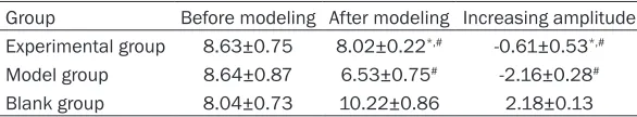
Table 2. Weight changes of rats in different groups
Group Before modeling After modeling Increasing amplitude Experimental group 8.63±0.75 8.02±0.22*,# -0.61±0.53*,#
Model group 8.64±0.87 6.53±0.75# -2.16±0.28#
Blank group 8.04±0.73 10.22±0.86 2.18±0.13
Data were shown as mean±SD. *P < 0.05, compared with model group. #P < 0.05, compared with blank group.
Statistical analysis: Data analysis was
per-formed on SPSS17.0 software. Enumeration data was compared by chi-square test, while measurement data was compared by ANOVA with Newman-Keuls multiple comparison post-hoc analysis. The data was depicted as mean±standard deviation. P < 0.05 was adopt-ed as significance level.
Results
General state
The rats in the model group appeared abdomi-nal distention, yellow watery stools, poor eat-ing, less activity, sluggish and obvious weight loss after modeling. The symptom was most severe on the fifth day. The rats in experimental group also exhibited abdominal distension and diarrhea, while the amplitude of weight loss was lower than model group. The rats in blank group presented normal eating and defecate (Table 2).
Intestinal tissue changes under microscope
Intestinal mucosa appeared hyperemia, ede- ma, erosion, bleeding in model group, to- gether with a large amount of neutrophils, lym-phocytes, and eosinophils infiltration influenc-ing the submucosa. The experimental group ex- hibited slighter intestinal mucosal hyperemia
and edema. The intestinal mucosa in the blank group was normal (Figure 1).
Intestinal flora comparison
Bifidobacteria, lactobacillus, enterobacter, and enterococ-cus in intestine of experi-mental group were lower than the blank group but obviously higher than the model group (P < 0.05). Bifi- dobacteria and lactobacillus contents reduced on the sev-enth day but lack of signifi-cant difference compared with blank group (P < 0.05) (Table 3).
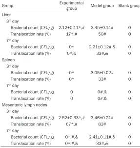
Bacterial translocation com-parison
The bacterial translocation rate of liver, spleen, and mesenteric lymph nodes in experimental group was significantly lower than the model group on the 3rd and 7th day (P < 0.05). No
bac-terial translocation in liver, spleen, and mesen-teric lymph nodes in experimental group was observed on the 7th day (P < 0.05) (Table 4).
TLR2 and TLR4 protein expression in small intestine
TLR2 and TLR4 protein expression in experi-mental group were obviously higher than the model group (P < 0.05), while their levels in experimental group on the 7th day were
signifi-cantly higher than on the 3rd day (P < 0.05)
(Figure 2; Table 5).
TLR2 and TLR4 mRNA expression in small intestine
TLR2 and TLR4 mRNA expression in experi-mental group were markedly higher than the model group (P < 0.05), while their levels in experimental group on the 7th day were
appar-ently higher than on the 3rd day (P < 0.05) (Table
6).
Discussion
[image:3.612.90.383.251.305.2]microorganisms’ invasion at this time may lead to bacterial translocation [12]. Intestinal probi-otics commonly used in clinic, including lacto-bacillus and bifidobacterium, can maintain intestinal normal function, repair the defense capability of the barrier, and regulate intestinal normal flora [13]. This study established antibi-otic diarrhoea animal model and adopted lacto-bacillus intervention to analyze its impact on TLR expression and bacterial translocation. In this study, lincomycin intragastric administra-tion was applied to establish diarrhea animal model. Lactobacillus intervene was adopted in the experimental group. The rats in the model group appeared abdominal distention, yellow watery stools, poor eating, less activity, slug-gish, and obvious weight loss after modeling. The rats in experimental group also exhibited abdominal distension and diarrhea, while the amplitude of weight loss was lower than model group. Intestinal mucosa appeared hyperemia, edema, erosion, and bleeding in model group, together with a large amount of neutrophils,
lymphocytes, and eosino-phils infiltration influencing the submucosa. The ex- perimental group exhibited slighter intestinal mucosal hyperemia and edema, con-forming to the clinical symp-toms and microscopic per- formance.
Intestinal probiotics can ma- intain the balance of intesti-nal flora. Its reduction may lead to intestinal flora disor-der, bacterial translocation, and endotoxin elevation, and intestinal mucosal barrier da- mage [14]. Bifidobacterium and lactobacillus are the rep-resentatives of dominant bacterial group in normal intestinal tract [15]. Bifido- bacteria, lactobacillus, enter- obacter, and enterococcus in intestine of experimental gr- oup were lower than the blank group but obviously higher than the model group on the 3rd day. Bifidobacteria
[image:4.612.90.382.73.175.2]and lactobacillus contents were lack of significant differ-ence compared with blank Figure 1. Intestinal tissue expression under microscope (×200). A:
experimen-tal group. B: model group. C: blank group. Arrow shows bleeding or hyperemia.
Table 3. Intestinal flora comparison
Group Experimental group Model group Blank group
Bifidobacteria
3rd day 6.67±0.25* 5.37±0.24# 8.28±0.38
7th day 8.50±0.24& 5.04±0.37# 8.59±0.25 Lactobacillus
3rd day 7.18±0.28* 6.24±0.24# 8.06±0.27
7th day 8.35±0.25& 6.51±0.25# 8.24±0.21 Enterobacter
3rd day 6.06±0.12* 5.53±0.16# 6.17±0.26
7th day 6.36±0.23& 6.42±0.25# 6.29±0.19 Enterococcus
3rd day 6.15±0.26* 5.23±0.26# 6.37±0.24
7th day 6.60±0.27& 6.45±0.38# 6.58±0.30
Data were shown as mean±SD. *P < 0.05, compared with model group. #P < 0.05, compared with blank group. &P < 0.05, compared with 3rd day.
group on the 7th day. It suggested that
bifido-bacteria and lactobacillus reduced most signifi-cantly in antibiotic diarrhea induced intestinal flora disorder. The phenomenon of dominant bacterial group reduction was more seriously following time extension, while lactobacillus can improve the structure of normal intestinal flora in diarrhea rat and maintain the home- ostasis.
[image:4.612.90.381.240.412.2]Table 4. Bacterial translocation comparison
Group Experimental group Model group Blank group
Liver 3rd day
Bacterial count (CFU/g) 2.12±0.11*,# 3.45±0.14# 0
Translocation rate (%) 17*,# 50# 0
7th day
Bacterial count (CFU/g) 0* 2.21±0.12#,& 0 Translocation rate (%) 0*,& 33#,& 0 Spleen
3rd day
Bacterial count (CFU/g) 0* 3.05±0.02# 0
Translocation rate (%) 0* 33# 0
7th day
Bacterial count (CFU/g) 0 0#,& 0
Translocation rate (%) 0 0#,& 0
Mesenteric lymph nodes 3rd day
Bacterial count (CFU/g) 2.52±0.33*,# 3.46±0.21# 0
Translocation rate (%) 67*,# 83# 0
7th day
Bacterial count (CFU/g) 0*,#,& 2.41±0.11#,& 0 Translocation rate (%) 0*,#,& 33#,& 0
*P < 0.05, compared with model group. #P < 0.05, compared with blank group. &P < 0.05, compared with 3rd day.
Figure 2. TLR2 and TLR4 protein expression in small intestine. A: experimental group. B: model group. C: blank group.
was significantly lower than the model group on the 3rd and 7th day. No bacterial translocation in
liver, spleen, and mesenteric lymph nodes in experimental group was observed on the 7th
day, indicating lactobacillus intervention can alleviate intestinal bacterial translocation in antibiotic diarrhea rat.
TLRs are important transmembrane recogni-tion receptors that expressed in intestinal epi-thelial cells. They can accurately identify the biomarker on the membrane of pathogen, trig-ger inflammation, and regulate the phagocyto-sis of phagocytes [18-20]. This experiment measured TLR2 and TLR4 protein expression in small intestine. TLR2 and TLR4 protein ex-
pression in experimental gr- oup were obviously higher than the model group, while their levels in experimental group on the 7th day were
sig-nificantly higher than on the 3rd day. Further analysis
showed that TLR2 and TLR4 mRNA expression in experi-mental group were markedly higher than the model group, while their levels in experi-mental group on the 7th day
were apparently higher than on the 3rd day. Previous
stud-ies pointed out that bifido-bacteria intervention de- clined TLR2 and TLR4 ex- pression in the terminal ileum of rats with ileitis with dose dependence [21-23]. TLR can quickly identify the corresponding ligands in a short time once the intesti-nal mucosa was attacked by toxins. They may trigger im- mune response through complicated signaling path-way, eventually inducing in- testinal mucosal inflamma-tory reaction and exogenous lactic acid bacteria engraft-ment in the intestine, inhibit-ing potential pathogenic bac-teria excessive proliferation, reducing bacterial transloca-tion, and controlling endo-toxin generation and release. They also can synthesize glutamine to promote damaged intestine recovery. Lactobacillus may have a certain protective effect on intestinal mucosal inflammation caused by TLR2 and TLR4.
[image:5.612.90.382.432.485.2]Table 6. TLR2 and TLR4 mRNA expression in small intestine
Group TLR2 mRNA TLR4 mRNA
d3 d7 d3 d7
Experimental group 0.74±0.05* 0.98±0.068*,& 0.81±0.02* 1.06±0.04*,&
Model group 0.52±0.02# 0.25±0.02#,& 0.62±0.03# 0.31±0.01#,&
Blank group 1.19±0.04 1.35±0.06 0.94±0.05 1.25±0.07
Data were shown as mean±SD. *P < 0.05, compared with model group. #P < 0.05, compared with blank group. &P < 0.05, compared with 3rd day.
Acknowledgements
This work was supported by Zhejiang Provincial Medicine Science and Technology Program Research Fund Project (NO. 2016ZA123).
Disclosure of conflict of interest
None.
Address correspondence to: Dr. Feng Ji, Department
of Gastroenterology, The First Affiliated Hospital of
Medical College of Zhejiang University, 79 Qingchun Road, Hangzhou 310006, Zhejiang, P. R. China. Tel: +86-571-87236114; Fax: +86-571-87236114; E-mail: fengjizxc@163.com
References
[1] Wang H, Moon S, Wang Y, Jiang B. Multiple vi-rus infection alters rotavivi-rus replication and expression of cytokines and toll-like receptors in intestinal epithelial cells. Virus Res 2012; 167: 48-55.
[2] Xu J, Yang Y, Wang C, Jiang B. Rotavirus and coxsackievirus infection activated different
profiles of toll-like receptors and chemokines
in intestinal epithelial cells. Inflamm Res 2009;
58: 585-592.
[3] Li GX LN. Toll like receptors on intestinal barri-er is protection or damage. Chin J Gastrointest Surg 2009; 12: 540-541.
[4] Liu Y, Zhu L, Fatheree NY, Liu X, Pacheco SE, Tatevian N, Rhoads JM. Changes in intestinal toll-like receptors and cytokines precede histo-logical injury in a rat model of necrotizing
en-terocolitis. Am J Physiol Gastrointest Liver Physiol 2009; 297: G442-450.
[5] Toiyama Y, Araki T, Yoshiyama S, Hiro J, Miki C, Kusunoki M. The expression patterns of toll-like receptors in the ileal pouch mucosa of postoperative ulcerative colitis patients. Surg Today 2006; 36: 287-290.
[6] Gribar SC, Sodhi CP, Richardson WM, Anand RJ, Gittes GK, Branca MF, Jakub A, Shi XH, Shah S, Ozolek JA, Hackam DJ. Reciprocal ex-pression and signaling of TLR4 and TLR9 in the pathogenesis and treatment of necrotizing enterocolitis. J Immunol 2009; 182: 636-646. [7] Klein Klouwenberg P, Tan L, Werkman W, van
Bleek GM, Coenjaerts F. The role of toll-like re-ceptors in regulating the immune response against respiratory syncytial virus. Crit Rev Im-munol 2009; 29: 531-550.
[8] Zhang W, Gu Y, Chen Y, Deng H, Chen L, Chen
S, Zhang G, Gao Z. Intestinal flora imbalance
results in altered bacterial translocation and liver function in rats with experimental cirrho-sis. Eur J Gastroenterol Hepatol 2010; 22: 1481-1486.
[9] Menozzi A, Ossiprandi MC. Assessment of en-teral bacteria. Curr Protoc Toxicol 2010; Chap-ter 21: Unit 21. 3.
[10] Akira S. Pathogen recognition by innate immu-nity and its signaling. Proc Jpn Acad Ser B Phys Biol Sci 2009; 85: 143-156.
[11] Sanders ME. Probiotics: definition, sources, se -lection, and uses. Clin Infect Dis 2008; 46 Suppl 2: S58-61; discussion S144-151. [12] Sharma R, Tepas JJ 3rd. Microecology,
intesti-nal epithelial barrier and necrotizing enteroco-litis. Pediatr Surg Int 2010; 26: 11-21. [13] Menard O, Gafa V, Kapel N, Rodriguez B, Butel
[image:6.612.89.524.208.276.2]MJ, Waligora-Dupriet AJ. Characterization of
Table 5. TLR2 and TLR4 protein expression in small intestine
Group TLR2 TLR4
3rd day 7th day 3rd day 7th day
Experimental group 0.56±0.05* 0.64±0.047*,& 0.044±0.07* 0.058±0.04*,&
Model group 0.054±0.04# 0.048±0.04#,& 0.042±0.05# 0.037±0.04#,&
Blank group 0.067±0.08 0.073±0.1 0.61±0.05 0.72±0.06
immunostimulatory CpG-rich sequences from
different bifidobacterium species. Appl Environ
Microbiol 2010; 76: 2846-2855.
[14] Nikitenko VI, Stadnikov AA, Kopylov VA. Bacte-rial translocation from the gastrointestinal tract in healthy and injured rats. J Wound Care 2011; 20: 114-122.
[15] Ohland CL, Macnaughton WK. Probiotic bacte-ria and intestinal epithelial barrier function. Am J Physiol Gastrointest Liver Physiol 2010; 298: G807-819.
[16] Chiva M, Soriano G, Rochat I, Peralta C, Rochat F, Llovet T, Mirelis B, Schiffrin EJ, Guarner C, Balanzo J. Effect of lactobacillus johnsonii La1
and antioxidants on intestinal flora and bacte -rial translocation in rats with experimental cir-rhosis. J Hepatol 2002; 37: 456-462.
[17] Duffy LC, Zielezny MA, Riepenhoff-Talty M,
Dry-ja D, Sayahtaheri-Altaie S, Griffiths E, Ruffin D,
Barrett H, Ogra PL. Reduction of virus
shed-ding by B. bifidum in experimentally induced
MRV infection. Statistical application for ELISA. Dig Dis Sci 1994; 39: 2334-2340.
[18] Sharma R, Young C, Neu J. Molecular modula-tion of intestinal epithelial barrier: contribumodula-tion of microbiota. J Biomed Biotechnol 2010; 2010: 305879.
[19] Meng J, Yu H, Ma J, Wang J, Banerjee S, Char-boneau R, Barke RA, Roy S. Morphine induces bacterial translocation in mice by compromis-ing intestinal barrier function in a TLR-depen-dent manner. PLos One 2013; 8: e54040. [20] Hu G, Malik AB, Minshall RD. Toll-like receptor
4 mediates neutrophil sequestration and lung
injury induced by endotoxin and hyperinflation.
Crit Care Med 2010; 38: 194-201.
[21] Yoshiya K, Lapchak PH, Thai TH, Kannan L, Rani P, Dalle Lucca JJ, Tsokos GC. Depletion of gut commensal bacteria attenuates intestinal ischemia/reperfusion injury. Am J Physiol Gas-trointest Liver Physiol 2011; 301: G1020-1030.
[22] Bauer S, Muller T, Hamm S. Pattern recogni-tion by toll-like receptors. Adv Exp Med Biol 2009; 653: 15-34.
[23] Khailova L, Mount Patrick SK, Arganbright KM,
Halpern MD, Kinouchi T, Dvorak B. Bifidobacte