Original Article
DKK1 overexpression promotes the
proliferation and cartilage differentiation
of
mesenchymal stem cells (MSCs) of antler
Chun-Mei Han1,2, Shan-Shan Wang1, Yong-Fu Zheng1, Xiao-Li Chen3, Qing-Hua Gao1,2
1Tarim University College of Animal Sciences, 3Tarim University College of Life Sciences, Alar, China; 2Xinjiang Production and Construction Corps Tarim Animal Husbandry Science and Technology Key Laboratory, Alar, China Received August 16, 2017; Accepted February 14, 2018; Epub June 15, 2018; Published June 30, 2018
Abstract: Wnt signaling plays multiple roles in mammalian mesenchymal stem cells (MSCs). This study aimed to examine the effect of Wnt signaling on the proliferation and differentiation of MSCs of antler. Antlers were harvested at 60 days of growth and MSCs were isolated. An adenovirus for DKK1 overexpression was constructed. Cartilage differentiation was evaluated using Alcian blue, alizarin red, and immunohistochemistry. MSCs were evaluated by
qRT-PCR for DKK1, collagen I, collagen II, β-catenin, cyclinD1, c-Myc, and Runx2. On days 3 and 6, Alcian blue stain
-ing in Ad-DKK1 cells was positive, but disappeared from day 9. On day 9, collagen II immunohistochemistry was weakly positive. On day 11, alizarin red staining showed positive stain. On day 11, the expression of collagen I in the
Ad-DKKI group was higher than that in the empty adenovirus group, and was higher than that on days 6 and 9 (all
P<0.05); the expression of collagen II on day 11 was significantly higher (all P<0.05). The expression of the DKK1
gene was increased from day 9 (P<0.05 vs. day 6). The expression of the β-catenin gene was decreased from day 9
(P<0.05). The expression of the CyclinD1 gene was also declined on day 11 (P<0.05). The expression of the Runx2 gene was higher from day 6 (all P<0.05). Wnt signaling is involved in the proliferation and cartilage differentiation of MSCs of antler. These results provide a theoretical basis for the growth and regeneration of antler.
Keywords: Wnt signaling, mesenchymal stem cells, antler, proliferation, differentiation, cartilage
Introduction
Tarim Wapitis is a unique animal that inhbits exclusively in Tarim Basin. The antlers of Tarim Wapitis can regenerate with an astonishing growth rate even faster than that of tumor tissues. And this regeneration is tightly con-trolled [1-3]. Therefore, the regeneration of Tarim Wapitis’ antlers represents an ideal model for deciphering the underline mecha-nism of the regeneration and remodeling of mammalian bone tissues.
Antler is currently the only mammalian organ that can regenerate and its growth, develop-ment, and regeneration is a subject of research
[4]. The growth and development of antler are accomplished through intramembranous ossi-
fication and endochondral ossification, which
are both based on stem cells [5]. Cartilage dif-ferentiation of antler is used as a study model
of the development of human cartilage. Studies have found that mesenchymal stem cells (MSCs) of antler are from the periosteal stem cells of deer spine [5, 6]. The growth of these
MSCs form fibroblasts colonies that differenti -ate into cartilage cells to form osteoblasts [5, 7]. Therefore, MSCs are one of the sources that maintains the growth and development of ant-ler [5, 7], but the exact regulation of MSCs dur-ing antler development remains unclear. The thickness of the mesenchymal cell layer is changing at different grow stages of MSCs of antler, which experiences hyperblastosis and then gradually thins from the early growth stage to the peak growth stage as well as the late
growth stage (the ossification stage). Through
in vitro experiments, MSCs of antler have been proved to have the abilities of differentiating into osteoblasts, adipocytes, and chondrocytes
separat-ed from their microenvironment (niche) for the growth and development of antler, and many signaling pathway play important role in the proliferation and differentiation of the stem cells, including Wnt signaling [4, 8]. It has been found that the Wnt signaling pathway can regu-late the growth and differentiation of MSCs in mammalians through self-renewal and differen-tiation [9].
Specific inhibitors of Wnt signaling was used to
inhibit Wnt signaling in human bone marrow MSCs and found that downstream proliferation-related gene expression can be increased and cell proliferation can be promoted [10].
Over-expression of the Wnt3a gene in pancreatic stem cells showed that the expression of cell proliferation-related genes such as c-Myc can
be significantly upregulated, which promoted
the proliferation of pancreatic stem cells and delayed cell senescence [11]. In addition, a
number of studies confirmed that the
Wnt/β-catenin classical pathway was involved in the regulation of the bone differentiation of pluri- potent stem cells and human MSCs [12-15]. It
was observed that proteins of the β-catenin/
Wnt signaling pathway were expressed in MSCs of antler, while it was lowly expressed in cartilage and osteoblasts by Immunohistoche- mistry [8]. They used epigallocatechin-3-gallate (EGCG) to block Wnt signaling and found that the number of cells was decreased while the activity of alkaline phosphatase (ALP) was
increased. On the other hand, lithium chloride
(LiCl) was used to activate Wnt signaling and showed that the number of cells was increased but ALP activity was decreased [8]. It has been
preliminarily considered that the β-catenin
gene plays a key role in the growth and bone formation of antler [8].
The Dickkopf-related protein 1 (DKK1) was
first isolated in 1999 and it was found to be located upstream of the Wnt/β-catenin signal -ing pathway [16]. DKK1 can inhibit Wnt signal-ing through combinsignal-ing Wnt and the low- density lipoprotein receptor-related protein 5/6 (LRP5/6) [16, 17]. Nevertheless, the role of DKK1 in antler development is unknown. Therefore, the aim of the present study was to examine the proliferation and differentiation of MSCs of antler after inhibiting Wnt signaling, as well as the regulatory mechanisms of the Wnt signaling pathway on the proliferation and carti-lage differentiation of MSCs of antler.
Materials and methods Animals
Two Tarim Wapitis aged 3-4 years were selected at the Experimental Station of Tarim University (Xinjiang, China). At 60 days, of the growth period of the antler, the Wapitis were anesthetized using an air gun dart loaded with
xylazine and dihydroetrphine at 6-7 O’clock in
the morning, when the weather was cool. The stag was placed under general anesthesia (the recommend dose for anesthesia of Tarim Wapitis is 0.01-0.015 ml per kilogram of body weight) with an intramuscular injection of an aqueous mixture of 2 mL of xylazine and dihy-droetrphine (0.1 g/ml xylazine and 40 ug/ml dihydroetrphine. Quartermaster University of PLA, Changchun, China). After the animals were anesthetized, the base of the antler was tied tightly with a grass rope to avoid excessive bleeding. Antlers were cut off using a hand saw. Then the styptic powder (Quartermaster University of PLA, Changchun, China) is applied immediately on the wound to stop bleeding and promote the healing [18, 19]. Contamination on the tissue surface was quickly cleaned. Tissues that were 5 cm from the top of the antler were sampled using a scalpel, and longitudinally dis-sected; then, mesenchymal cells were separat-ed under a dissecting microscope, according to the literature [20, 21]. The transition part between the antler cortex and the chondrocyte layer was cut off as much as possible. The experiments were approved by the Ethics Committee of Tarim University (Xinjiang, China).
Culture of primary cells from the antlers
Antler MSCs were isolated as previously described [6]. MSCs from antler were washed with PBS thrice and placed in a sterile 90-cm2 culture dish. The MSCs were washed with D- Hanks containing 400 IU/mL penicillin-strepto-mycin. The sample was cut into 1-mm2 pieces
and transferred into a 10-mL culture flask. Trypsin (0.25%, 7 mL; Sigma, St Louis, MO,
USA) was added and the cells were placed in a
CO2 incubator at 37°C for 1 h. Then, 5 mL of
0.1% type II collagenase (Sigma, St Louis, MO,
USA) was added and incubated for 5 h. The supernatant was transferred to a 10-mL centri-fuge tube and 3 mL of DMEM medium (Sigma,
St Louis, MO, USA) containing 10% FBS (Sigma, St Louis, MO, USA) was used to wash the tissue
for 5 min; the supernatant was discarded. Complete culture medium (5 mL) was added
and incubated at 37°C under 5% CO2. After the tissue blocks were attached to the wall, the
flask was gently flipped over, and the tissue
blocks were soaked in the complete culture medium. The medium culture was replaced every 2-3 days. When a large number of cells were observed under an inverted microscope to have migrated from the edge of the tissue block and proliferated, the tissue blocks were gently removed.
Cell culture
Passage was performed at 70% confluence.
D-Hanks solution was used to wash thrice the cells that were attached to the wall. Trypsin (0.25%) was added to detach the cells. The cells were collected in a 10-mL centrifuge tube and centrifuged at 800 rpm for 5 min. The supernatant was discarded. The cell density was adjusted to 1×10⁵ cells/mL with complete culture medium. The cells (5 mL) were
inocu-lated in a flask, and incubated. The medium
was completely replaced after 1 day, and then every 3 days.
DKK1 overexpressing adenovirus
The bovine DKK1 gene sequence was obtained from Genbank (accession number NM_00120-
5544.1). PCR primers in the ORF region were
designed using the Primer 5.0 software. The restriction endonucleases BamHI and EcoRase
(Takara Bio, Otsu, Japan) were used to intro -duce restriction sites at the two ends of the primers. cDNA from antler was used as the template to amplify the DKK1 gene. The reac-tion condireac-tions are shown in Table 1. The
puri-fied DKK1 PCR products were ligated to the pMD18-T vector (Takara Bio, Otsu, Japan). The
vectors were transformed into DH5a bacteria. After the plasmids were extracted, they were sent to Sangon Biotech (Shanghai, China) to be sequenced. After analyzing the sequencing results, the recombinant plasmid pMD18-T-DKK1 and the vector pHBAd-MCMV-GFP (Han- bio Biotechnology Co., Ltd., Shanghai, China) were digested. The recovered digested DKK1
segments were purified to be ligated to the
digested vector pHBAd-MCMV-GFP. The DKK1 recombinant plasmids were sent to Sangon Biotech (Shanghai, China) to be sequenced. After analyzing and determining the sequenc-ing results, the DKK1 recombinant shuttle plas-mid pHBAd-BHG (Hanbio Biotechnology Co., Ltd,. Shanghai, China) was used to transfect the 293A cell line no containing the E1A region of adenovirus, and was routinely cultured in DMEM 90% medium with 10% FBS. All the above-mentioned cell lines were kept at 37°C
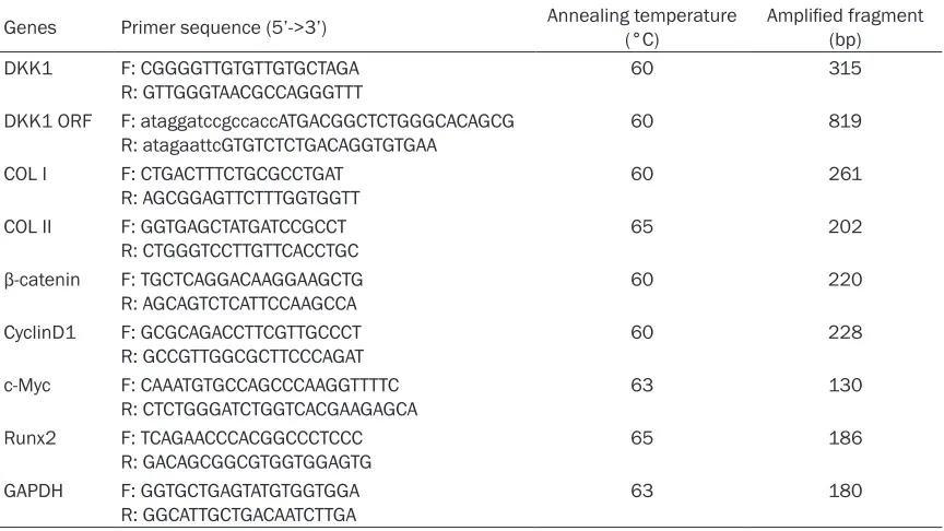
[image:3.612.91.523.85.328.2]in a humidified atmosphere of 5% CO2. Finally, DKK1 overexpressing adenovirus was obtained. Table 1. PCR primers and reaction conditions for quantitative detection of gene expression
Genes Primer sequence (5’->3’) Annealing temperature (°C) Amplified fragment (bp) DKK1 F: CGGGGTTGTGTTGTGCTAGA
R: GTTGGGTAACGCCAGGGTTT 60 315
DKK1 ORF F: ataggatccgccaccATGACGGCTCTGGGCACAGCG
R: atagaattcGTGTCTCTGACAGGTGTGAA 60 819
COL I F: CTGACTTTCTGCGCCTGAT
R: AGCGGAGTTCTTTGGTGGTT 60 261
COL II F: GGTGAGCTATGATCCGCCT
R: CTGGGTCCTTGTTCACCTGC 65 202
β-catenin F: TGCTCAGGACAAGGAAGCTG
R: AGCAGTCTCATTCCAAGCCA 60 220
CyclinD1 F: GCGCAGACCTTCGTTGCCCT
R: GCCGTTGGCGCTTCCCAGAT 60 228
c-Myc F: CAAATGTGCCAGCCCAAGGTTTTC
R: CTCTGGGATCTGGTCACGAAGAGCA 63 130
Runx2 F: TCAGAACCCACGGCCCTCCC
R: GACAGCGGCGTGGTGGAGTG 65 186
GAPDH F: GGTGCTGAGTATGTGGTGGA
R: GGCATTGCTGACAATCTTGA 63 180
Self-proliferating characteristics
Four 24-well plates were used; 84 wells were divided into four groups (n=21/group): the ade-novirus transduction group with multiplicity of
infection (MOI) of 200 (2.0×10² PFU/cell);
ade-novirus transduction group with MOI of 300
(3.0×10² PFU/cell); empty adenovirus trans- duction group (2.0×10² PFU/cell); and the blank group. Each well was seeded with about 5.0×10⁴ cells, and 0.5 mL of complete culture medium was added and incubated at 37°C
under 5% CO2. Half of the medium was replaced
every 2 days. When cells were 50-60% conflu -ent, the experimental groups were infected with DKK1 overexpressing adenoviruses and empty adenoviruses. Three wells were random-ly selected from each group every day. MTT solution (20 µL) was added and cultured for 4
h. DMSO (80 µL) was added and the absor -bance was read at 490 nm. The cell growth curve was plotted.
Cartilage differentiation
Four 6-well plates were used and the 24 wells were divided into the transduction group and the empty adenovirus transduction group (n= 12/group). Each well was seeded with 1.0×10⁵ cells, 1 mL of complete culture medium was
DAB Chromatography Kit (Boster Bioengineering Co., Wuhan, China) were used according to the manufacturer’s instructions.
Quantitative PCR
Nine 6-well plates were used and the 54 wells were divided into the transduction group, the empty adenovirus transduction group, and the blank group (n=18/group). Each well was seed-ed with 1.0×10⁵ cells; 1 mL of complete culture medium was added and incubated at 37°C
under 5% CO2. Half of the culture medium was replaced every 2 days. When the cells were
50-60% confluent, each well in the transduc -tion group was added with DKK1
overexpress-ing adenovirus at MOI of 200. After 3, 6, 9, and
11 days, the cells from three wells were harvested from the three groups. RNA was
extracted using TRIZOL (1 mL; Invitrogen Inc.,
Carlsbad, CA, USA), according to the manufac-turer’s instructions. cDNA was synthesized using a reverse transcription kit (Thermo Fisher
Scientific, Waltham, MA, USA). cDNA was dilut -ed at 1:10. Bas-ed on the Genebank database, Primer 5.0 software was used to design PCR primers (Table 1). The reaction conditions of qPCR included 95°C for 5 min; and 40 cycles at 95°C for 10 s and 60°C for 30 s. The melting curve was 55-95°C. Semi-quantitative PCR
added, and the plates were incubated at 37°C under 5%
CO2. Half of the medium was replaced every 2 days. When
the cells were 50-60% conflu -ent, each well of the transdu- ction group was added with DKK1 overexpressing
adeno-virus at MOI of 200. At days 3,
6, 9, and 11, cells from three wells were collected from each group and the culture medium was removed. After washing with 500 µL of PBS,
the cells were fixed with 4%
paraformaldehyde phosphate buffer (PBS, 0.1 M, pH 7.4) overnight at 4°C, followed by Alcian blue staining, alizarin red staining, and immunohis- tochemistry.
For immunohistochemistry,
the fixed cells were washed
[image:4.612.92.372.76.285.2]three times with PBS. The Eradication Staining Kit and Figure 1. Cell proliferation curve (MTT assay) of antler mesenchymal stem
cells (MSCs) infected with a DKK1 overexpressing adenovirus. Cells were treated with no adenovirus (control), empty adenovirus (Ad vector), with
DKK1 adenovirus at multiplicity of infection (MOI) of 200, and with DKK1 ad
-enovirus at MOI 300. *p<0.05 vs. control at the same time point. **P<0.01
reaction conditions included 95°C for 5 min; and 28 cycles at 95°C for 10 s; 65°C for 30 s, and 72°C for 2 min. The relative expression of proliferation- and differentiation-related genes of MSCs of antler (Table 1) was examined. Relative expression was evaluated using the 2-ΔΔCt method.
Statistical analysis
Differences among groups were evaluated
using ANOVA with the LSD post hoc test. SPSS
17.0 (IBM, Armonk, NY, USA) was used for data analysis. Two-sided P-values <0.05 were
con-sidered statistically significant.
Results
Effect of Ad-DKKl adenovirus on the prolifera-tion of MSCs from antler
MSCs of antler were transfected with Ad-GFP
adenovirus with MOI values of 200 and 300.
The cells grew continuously from day 1 of trans-duction in the blank, empty adenovirus, and treatment groups. Cell proliferation reached a peak on day 5 and gradually decreased
there-after. From days 0 to 7, there were no signifi -cant differences in MTT value between the blank and empty adenovirus groups, while the
MTT values in the group 200 and 300 MOI were significantly higher than that in the blank and
empty adenovirus groups from day 2 of trans-duction (P<0.05) (Figure 1). When the cell pro-liferation started to decrease (on day 7), the MTT values in the two transduction groups
were still significantly higher than in the blank
and empty adenovirus groups (P<0.001) (Figure 1).
Effects of transfected Ad-DKK1 adenovirus on cartilage differentiation
On days 3, 6, 9, and 11 after antler MSCs were
[image:5.612.92.525.69.376.2]transfected with Ad-DKK1, Alcian blue staining, Figure 2. The effect of DKK1 overexpression on the self-differentiation of antler mesenchymal stem cells (MSCs)
(magnification 100×). A-D. Alcian blue staining of the empty adenovirus group at 3, 6, 9, and 11 days. There was no obvious change in cell morphology over time. E-H. Alcian blue staining in the Ad-DKK1 transduction group. Cell morphology on day 3 began to change from a spindle to a triangle, and there was light blue positive staining.
Stain-ing was significant on day 6, but gradually disappeared from day 9. I-L. Collagen II immunohistochemistry in the
alizarin red staining, and collagen II immunohis-tochemistry were used to detect the
differentia-tion of MSCs. On day 3, Alcian blue staining in
Ad-DKK1 cells was weakly positive (Figure 2E); the staining was more obvious on day 6 (Figure 2F), but disappeared from day 9, indicating that proteoglycans were no longer secreted (Figure 2G and 2H). On day 9, collagen II immunohisto -chemistry was weakly positive (Figure 2K and 2L). On day 11, alizarin red staining showed that there was significant Ca2+ accumulation (Figure 2P).
Collagen I and collagen II expression after DKK1 overexpression in antler MSCs
The expression of collagen I and collagen II genes detected by qRT-PCR showed that the expression of the collagen I gene was high on
day 3, but without significant difference
be-tween the Ad-DKK1 and the empty adenovirus
groups. The expression was decreased signifi
-cantly on days 6 and 9. On day 11, the expres
-sion in the Ad-DKK1 group was significantly
higher than in the empty adenovirus group, and
was also significantly higher than the expres -sion on days 6 and 9 (all P<0.05) (Figure 3A). The expression of the cartilage surface marker
collagen II on day 11 was significantly higher
than the expression in each group at all time points (all P<0.05) (Figure 3B).
Self-proliferation of antler MSCs and expres-sion of downstream genes of the Wnt pathway after transduction with Ad-DKK1
Quantitative PCR and western blot were used
to detect the expressions of DKK1, β-catenin,
c-Myc, CyclinD1, and Runx2 on days 6, 9 and 11 after transduction. The expression of the DKK1 gene was sharply increased from day 9 (P<0.05 vs. day 6) (Figure 3C). The expression
of the β-catenin gene was significantly
de-creased from day 9 (P<0.05) (Figure 3C). The expression of the c-Myc gene downstream of Wnt signaling showed no difference after transduction. The expression of the CyclinD1
gene was significantly decreased on day 11
(P<0.05). The expression of the Runx2 gene was increased from day 6 (all P<0.05).
Figure 3. The expression of collagen I (A) and collagen II (B) genes after antler mesenchymal stem cells (MSCs) were
transfected with DKK1 adenovirus. There was no significant difference in collagen I gene on day 3 in the empty adenovirus and transduction groups, but the expression in the Ad-DKK1 group on day 11 was significantly higher
than in the empty vector group and at other time points (all P<0.05). The expression of the collagen II gene on day
Discussion
Wnt signaling plays multiple roles in mammali-an MSCs. This study aimed to examine the proliferation and differentiation of MSCs of ant-ler. The results showed that Wnt signaling is involved in the proliferation and cartilage differ-entiation of MSCs of antler. These results pro-vide a theoretical basis for the growth and regeneration of antler.
Two days after antler MSCs was transduced with the DKK1 adenovirus, the cells began to differentiate into chondrocytes and cell mor-phology changed. Alcian blue staining (which indicates glycisaminoglycans in cartilage) be- came positive, consistent with cartilage differ-entiation. Alcian blue staining then became negative while alizarin staining (which indicate Ca2+ accumulation) became positive. Consi- stently, collagen II immunohistochemistry be- came positive on day 11. DKK1 overexpression was used to analyze the effect of Wnt signal on human MSCs and cartilage differentiation. Their results showed that overexpression of
DKK1 and sFRP-1 genes significantly increased
the secretion of proteoglycans and upregulated
the expression of SOX-9 and collagen II [15], which was consistent with the results of the present study. Itshowed that the controlled
activation of the Wnt/β-catenin signaling
en-hanced the stem and progenitor activities when regeneration is needed [22], supporting the present study.
Antlers and tumors share similar growth char-acteristics in that antlers can regenerate after being cut off and the growth rate is extremely fast [20]. The growth and development of ant-lers is dependent upon MSCs. Mount et al. [8]
has preliminarily analyzed the effect of Wnt sig-naling on the proliferation of antler stem cells at the protein level, but the mechanisms are still unclear. In the present study, Ad-DKK1 overexpression was used to inhibit Wnt signal. DKK1 overexpression did not affect the prolif-eration of antler MSCs, which was different from the study by Mount et al. in 2006 [8]. Here, DKK1 overexpression downregulated the
expression of β-catenin, but it had no inhibitory
effect on c-Myc and CyclinD1 genes. The c-Myc and CyclinD1 genes are located downstream of Wnt, and the c-Myc gene plays roles in cell cycle regulation [23]. Increased expression of Myc genes increase the activity of Cyclins, thereby
promoting cell cycle progression of pluripotent stem cells (PSCs) and accelerating cell division
[24]. Satoh et al. [25] found that the number of hematopoietic stem cells was reduced when c-Myc was silenced. Laurenti et al. [26] and Wilson et al. [27] observed that the number of stem cells was increased when c-Myc and N-Myc were co-expressed. These studies dem-onstrated that c-Myc plays a positive role in cell division and proliferation.
In cancer, the c-Myc and CyclinD1 genes are upregulated to promote cell proliferation. CyclinD1 binds to cyclin-dependent kinase (CDK) to form a CyclinD/CDK complex that
acti-vates specific CDK and phosphorylates various
proteins to regulate the procession of the cell cycle [28, 29]. In the present study, after block-ing Wnt signalblock-ing by overexpressblock-ing DKK1, the expression of c-Myc and CyclinD1 genes was unaffected. It could be hypothesized that the expression of these two genes is not only affected by Wnt signaling, but also by other regulatory factors. The FBS in the culture medi-um contains a small amount of growth factors such as IGF and EGF, and these growth factors could promote the cell cycle through regulating the expression of c-Myc by the MAPK signaling pathway [30, 25]. Using IGF1 at different con-centrations to stimulate antler MSCs, it was demonstrated that concentrations of IGF1 as low as 10 nmol could stimulate the rapid divi-sion of antler MSCs [31]. However, the relation-ship between the proliferation of antler MSCs and the signal pathway of MAPK needs to be further investigated.
Runx2 gene is an osteogenic-specific transcrip -tion factor that plays an important role in osteo-genesis of human bone marrow MSCs and promotes cartilage and osteoclast functions
over-expression was studies. Additional studies are necessary to comprehensively determine the mechanisms involved in the regeneration of antlers, which is the only regeneration encoun-tered in mammals. Among others, microRNAs should be further studied [4], as well as Hedgehog and NELL1 signaling [33].
In conclusion, Wnt signaling is involved in the proliferation and cartilage differentiation of MSCs of antler. These results provide a theo-retical basis for the growth and regeneration of antler.
Acknowledgements
The authors appreciated the Tarim Red Deer Breeding Farm of the Second Agricultural Division of Xinjiang Production and Construction Corps for providing the antler samples. They are also thankful to postgraduates Dong Qin, Luo Xinrong, Meng Hao, and Ma Mengting for their help. This work was supported by the Project National Natural Science Foundation of
China (NO. 31260541).
Disclosure of conflict of interest
None.
Address correspondence to: Dr. Qing-Hua Gao, Tarim University College of Animal Sciences, China, Xinjiang Production and Construction Corps Tarim Animal Husbandry Science and Technology Key Laboratory, Alar 843300, China. Tel: +86-132899- 74573; Fax: +86-21-57643271; E-mail: gsy1997@ 126.com
References
[1] Price JS, Oyajobi BO, Nalin AM, Frazer A,
Russell RG and Sandell LJ. Chondrogenesis in the regenerating antler tip in red deer: expres-sion of collagen types I, IIA, IIB, and X demon-strated by in situ nucleic acid hybridization and immunocytochemistry. Dev Dyn 1996; 205: 332-347.
[2] Li C and Suttie JM. Pedicle and antler regener-ation following antlerogenic tissue removal in red deer (Cervus elaphus). J Exp Zool 1994; 269: 37-44.
[3] Li C and Suttie JM, Clark DE. Morphological ob-servation of antler regeneration in red deer (Cervus elaphus). J Morphol 2004; 262: 731-740.
[4] Ba H, Wang D and Li C. MicroRNA profiling of
antler stem cells in potentiated and dormant
states and their potential roles in antler regen-eration. Mol Genet Genomics 2016; 291: 943-955.
[5] Li C. Deer antler regeneration: a stem cell-based epimorphic process. Birth Defects Res C Embryo Today 2012; 96: 51-62.
[6] Seo MS, Park SB, Choi SW, Kim JJ, Kim HS and Kang KS. Isolation and characterization of antler-derived multipotent stem cells. Cell Transplant 2014; 23: 831-843.
[7] Nieto-Diaz M, Pita-Thomas DW, Munoz-Galdeano T, Martinez-Maza C, Navarro-Ruiz R, Reigada D, Yunta M, Caballero-Lopez MJ and Nieto-Sampedro M, Martinez-Maza R. Deer antler innervation and regeneration. Front Biosci (Landmark Ed) 2012; 17: 1389-1401. [8] Mount JG, Muzylak M, Allen S, Althnaian T,
McGonnell IM and Price JS. Evidence that the canonical Wnt signalling pathway regulates deer antler regeneration. Dev Dyn 2006; 235: 1390-1399.
[9] Serio RN. Wnt of the two horizons: putting stem cell self-renewal and cell fate determina-tion into context. Stem Cells Dev 2014; 23: 1975-1990.
[10] Hoffman MD and Benoit DS. Agonism of Wnt-beta-catenin signalling promotes mesenchy-mal stem cell (MSC) expansion. J Tissue Eng Regen Med 2015; 9: E13-E26.
[11] He X, Han W, Hu SX, Zhang MZ, Hua JL and Peng S. Canonical Wnt signaling pathway con-tributes to the proliferation and survival in por-cine pancreatic stem cells (PSCs). Cell Tissue Res 2015; 362: 379-388.
[12] Jussila M and Thesleff I. Signaling networks regulating tooth organogenesis and
regenera-tion, and the specification of dental mesenchy -mal and epithelial cell lineages. Cold Spring Harb Perspect Biol 2012; 4: a8425.
[13] Zhang F, Ren T, Wu J and Niu J. Small concen-trations of TGF-beta1 promote proliferation of bone marrow-derived mesenchymal stem cells via activation of Wnt/beta-catenin pathway. Indian J Exp Biol 2015; 53: 508-513.
[14] Visweswaran M, Pohl S, Arfuso F, Newsholme P, Dilley R, Pervaiz S and Dharmarajan A. Multi-lineage differentiation of mesenchymal stem cells - to Wnt, or not Wnt. Int J Biochem Cell Biol 2015; 68: 139-147.
[15] Im GI and Quan Z. The effects of Wnt inhibitors on the chondrogenesis of human mesenchy-mal stem cells. Tissue Eng Part A 2010; 16: 2405-2413.
[16] Fedi P, Bafico A, Nieto SA, Burgess WH, Miki T,
[17] Chen L, Wang K, Shao Y, Huang J, Li X, Shan J, Wu D and Zheng JJ. Structural insight into the mechanisms of Wnt signaling antagonism by Dkk. J Biol Chem 2008; 283: 23364-23370. [18] Gao QH, Wei HJ, Han CM, Du HZ, Zhang ZG,
Zhao WG, Zhang Y and Li S. Successful low
dose insemination of flow cytometrically sorted
Sika (Cervus nippon) sperm in Wapiti (Cervus elaphus). Anim Reprod Sci 2010; 118: 89-93. [19] Gao QH, Wang HE, Zeng WB, Wei HJ, Han CM,
Du HZ, Zhang ZG and Li XM. Embryo transfer and sex determination following superovulated hinds inseminated with frozen-thawed sex-sorted Y sperm or unsex-sorted semen in Wapiti (Cervus elaphus songaricus). Anim Reprod Sci 2011; 126: 245-250.
[20] Molnar A, Gyurjan I, Korpos E, Borsy A, Steger V, Buzas Z, Kiss I, Zomborszky Z, Papp P, Deak
F and Orosz L. Identification of differentially ex -pressed genes in the developing antler of red deer Cervus elaphus. Mol Genet Genomics 2007; 277: 237-248.
[21] Li C and Suttie JM. Tissue collection methods for antler research. Eur J Morphol 2003; 41: 23-30.
[22] Ahmadzadeh A, Norozi F, Shahrabi S, Shahja- hani M and Saki N. Wnt/beta-catenin signaling in bone marrow niche. Cell Tissue Res 2016; 363: 321-335.
[23] Schmidt EV. The role of c-myc in cellular growth
control. Oncogene 1999; 18: 2988-2996.
[24] Hindley C and Philpott A. The cell cycle and plu-ripotency. Biochem J 2013; 451: 135-143. [25] Satoh Y, Matsumura I, Tanaka H, Ezoe S,
Sugahara H, Mizuki M, Shibayama H, Ishiko E, Ishiko J, Nakajima K and Kanakura Y. Roles for c-Myc in self-renewal of hematopoietic stem cells. J Biol Chem 2004; 279: 24986-24993. [26] Laurenti E, Varnum-Finney B, Wilson A, Ferrero
I, Blanco-Bose WE, Ehninger A, Knoepfler PS,
Cheng PF, MacDonald HR, Eisenman RN, Bernstein ID and Trumpp A. Hematopoietic stem cell function and survival depend on c-Myc and N-c-Myc activity. Cell Stem Cell 2008; 3: 611-624.
[27] Wilson A, Murphy MJ, Oskarsson T, Kaloulis K, Bettess MD, Oser GM, Pasche AC, Knabenhans
C, Macdonald HR and Trumpp A. C-Myc con-trols the balance between hematopoietic stem cell self-renewal and differentiation. Genes Dev 2004; 18: 2747-2763.
[28] Li YJ, Wei ZM, Meng YX and Ji XR. Beta-catenin up-regulates the expression of cyclinD1, c-myc and MMP-7 in human pancreatic cancer: Relationships with carcinogenesis and metas-tasis. World J Gastroenterol 2005; 11: 2117-2123.
[29] Glockner S, Buurman H, Kleeberger W, Lehmann U and Kreipe H. Marked intratumor-al heterogeneity of c-myc and cyclinD1 but not
of c-erbB2 amplification in breast cancer. Lab
Invest 2002; 82: 1419-1426.
[30] Xu W, Gu J, Ren Q, Shi Y, Xia Q, Wang J, Wang S, Wang Y and Wang J. NFATC1 promotes cell growth and tumorigenesis in ovarian cancer up-regulating c-Myc through ERK1/2/p38 MAPK signal pathway. Tumour Biol 2016; 37: 4493-4500.
[31] Yang ZQ, Zhang HL, Duan CC, Geng S, Wang K, Yu HF, Yue ZP and Guo B. IGF1 regulates RUNX1 expression via IRS1/2: implications for antler chondrocyte differentiation. Cell Cycle 2017; 16: 522-532.
[32] Xu J, Li Z, Hou Y and Fang W. Potential mecha-nisms underlying the Runx2 induced osteogen-esis of bone marrow mesenchymal stem cells. Am J Transl Res 2015; 7: 2527-2535.
[33] James AW. Review of signaling pathways gov-erning MSC osteogenic and adipogenic
differ-entiation. Scientifica (Cairo) 2013; 2013: