Case Report
Fixation of unstable pelvic fractures with
minimally invasive adjustable plate
Tao Wu, Wei Chen, Qi Zhang, Yunwei Cui, Xiaodong Cheng, Yanjiang Yang, Yingze Zhang
Emergency Center of Trauma, Key Laboratory of Orthopaedic Biomechanics of Hebei Province, The Third Hospital of Hebei Medical University, Orthopaedic Research Institution of Hebei Province, Shijiazhuang, PR China
Received September 22, 2016; Accepted October 30, 2016; Epub January 15, 2017; Published January 30, 2017
Abstract:Objective: Unstable pelvic fractures are relatively rare injuries usually requiring reduction and internal fixation. Selecting appropriate methods for unstable pelvic fractures remains a challenging problem for orthopaedic surgeons. The aim of this study was to present the techniques and outcomes of MIAP for unstable pelvic fractures. Methods: We performed a retrospective analysis of patients with unstable pelvic ring fractures treated with mini-mally invasive adjustable plate at a level I trauma centre. Outcome evaluation was assessed using Majeed score standard, duration of surgery, blood loss, radiation exposure and size of incision. Results: Twenty-one patients were available for follow-up after at least 12 months. The main findings were as follows: the average duration of surgery was 67.5 min, the intraoperative blood loss was 204 ml on average, the average radiation exposure was 8 s, and the size of incision was 8.8 cm on average. The mean Majeed functional evaluation score was 85.3 points. Conclusion: Minimally invasive adjustable plate may be a good alternative for treating unstable pelvic fractures. It has the ad-vantages of technically safe, minimally invasive, less radiation exposure and time saving.
Keywords: Unstable pelvic fractures, fracture fixation, internal, minimally invasive adjustable plate
Introduction
Pelvic fractures are relatively small rare injury, accounting for 3.64% of fractures in adults [1], and 68.3% of pelvic fractures are unstable frac-tures, which are serious injuries. They usually results from high-energy trauma, such as falls and motor vehicle accidents. In contrast to the low incidence of these injuries, the high mortal-ity rate is 5-20% [2-4]. Selecting appropriate methods for unstable pelvic fractures remains a challenging problem for orthopaedic sur-geons. The stability of the pelvis is mainly relat-ed to the integrity of posterior pelvic ring [5]. Therefore, the treatment for unstable pelvic fractures needs to restore the continuity and stability of posterior pelvic ring as far as possi-ble. Greater stability of posterior pelvic ring can
be achieved by internal fixation [6]. There are
many kinds of methods available, including ilio-sacral (IS) screw, tension band plate (TBP),
tri-angular osteosynthesis and so on. IS screw fixa
-tion is a safe alternative to open fixa-tion, with
reported satisfactory results and lower rates of bleeding and infection [7-9]. However, this
tech-nique requires continuous fluoroscopic or com -puterized tomography guidance for appropriate screw insertion and remains a technically
demanding procedure. TBP fixation is a
well-recognized technique for treating the posterior pelvic ring disruption [10, 11]. But pre-bending of LCP for adapting the structure of posterior pelvic ring, which may reduce the strength of the plate or damage the threads of screw holes,
affects pelvic stability fixed with LCP [12].
Although triangular osteosynthesis can create the necessary stability for the maintenance of reducing unstable pelvic fractures, it restricts the range of L5/S1 motion segment [13, 14]. To address the above mentioned limitations, we introduced a novel minimally invasive adjust-able plate (MIAP) according to the structure characteristics of posterior pelvic ring. This arti-cle attempted to review our techniques of MIAP and provide clinical results.
Materials and methods
Subjects
Between January 2009 to July 2012, 26 patients with unstable pelvic ring fractures were included in this investigation. After at least 12-month follow-up, one patient died and 4 patients failed to be followed up. Finally, 21
patients were enrolled in this study. Of these,
10 patients had injury caused by traffic acci -dent, 7 by fall from height, and 4 by crush. The pelvic injuries were AO/OTA 61-Type B in ten cases (4 Type B1, 4 Type B2, 2 Type B3) and Type C in eleven cases (6 Type C1, 3 Type C2, 2 Type C3). The average duration from injury to operation was 7.8 days (range, 2-20 days). Of the 21, twelve were associated with multiple injuries, including head injury in one case, hae-mopneumothorax in two, L4 lumbar fracture in one, acetabular fracture in two, extremities fractures in seven, urethral disruption in three, laceration of perineum in two, bladder injury in two and renal contusion in one. The mean Injury Severity Score (ISS) [15] was 13.8 (range, 9-29).
All patients received plain X-ray films (antero -posterior, inlet, and outlet views) of the pelvis before operation and an experienced
radiolo-gist (Z.Z.K.) was assigned to read these films.
Computed tomography (CT) scanning and three-dimensional (3-D) reconstruction were performed to determine the involved fracture portion, the type of fracture, and stability of the pelvis.
Operative technique
The MIAP (Tianjin Zhengtian Medical Instrument Company Ltd., Tianjin, P.R.C.) is composed of two Z-shaped brackets and an adjustable con-nection bar (Figure 1). Each Z-shaped bracket consists of an upper wing, a web plate, and a lower wing. The connection bar is made up of two custom made eye bolts and a hexagonal tube, which can be shortened or stretched by rotating the hexagonal tube. Both Z-shaped brackets connect to the eye of the bolt to inte-grate these implants as a whole.
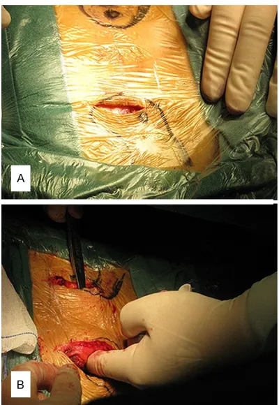
[image:2.612.90.290.72.227.2]Under general anesthesia or continuous extra-dural anesthesia, the patient lay in a prone position. Bilateral longitudinal incisions 4-6 cm long were made along the posterior superior iliac spines (PSISs) after sterilizing and draping. The bilateral PSISs were exposed after the skin and subcutaneous tissue had been incised to the periosteum without releasing the gluteal muscles from the outer side of iliac crest. The subcutaneous tunnel was created between bilateral PSISs, which were close to the dorsal surface of the sacrum (Figure 2). The displace-ment of fracture or dislocation was assessed
Figure 1. Structure of minimally invasive adjustable plate (MIAP). Both Z-shaped brackets can be com-pressed or separated by rotating the hexagonal tube.
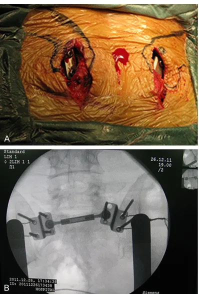
[image:2.612.89.289.287.577.2]again by using C-arm X-ray machine and re- duced as follows. The cephalad displacement of the sacral fracture or sacroiliac joint was reduced by continuous traction. For outward rotational displacement, it was reduced by compressing the affected iliac wing. When it came to ventral or dorsal displacement, a coarse thread screw was implanted into the affected PSIS in order to pull or press the pel-vis, and then the screw was pulled out after reduction. Each Z-shaped bracket was posi-tioned with the web plate close to the medial surface of the PSIS and the upper wing lying on the dorsal surface of the PSIS. Two or three long cancellous screws were inserted into the iliac crest through the holes of the upper wings in order to secure the Z-shaped bracket on the ilium. Two screws were inserted into the sacral
ala, if possible, to improve the stability of fixa -tion. The connection bar was placed through the subcutaneous tunnel and fastened to the brackets. If the median sacral crest obstructed the bar, we would make 2-3 cm longitudinal incision in the midline of sacrum or oblique
inci-sion from one side of PSIS for resecting the median sacral crest. If the sacral fracture still presented separation or compression shift in coronal plane, the hexagonal tube was rotated to shorten or elongate the bar under C-arm
X-ray fluoroscope, which in turn reduced the
separated fracture or distracted the com-pressed fractures, respectively (Figure 3).
Once fixed, the incision was irrigated and closed
appropriately with suction drains. The incision site was covered with a well-padded dressing and the patient was put in a non-weight-bearing position, postoperatively. The suction drains were taken away within 24 h usually. The patient was encouraged to take active exercis-es 3-4 days after operation. Crutch-assisted walking was allowed after two weeks. Partial weight-bearing began at six weeks postopera-tively. Progression to full weight-bearing was determined on the basis of osseous union on pelvic radiographs.
Postoperative functional recovery was asse- ssed with the Majeed score standard [16],
which includes five aspects: standing (36
points), pain (30 points), working ability (20 points), sitting (10 points), and sexual life (4 points). The maximum score is 100 points, with
excellent ≥85 points; good 70-85 points; fair 55-69 points; and bad ≤55 points.
Results
There were no iatrogenic neurovascular injuries during the operations. The average duration of surgery was 67.5 min (range, 40-90 min), the intraoperative blood loss was 204 ml on aver-age (range, 150-250 ml), the averaver-age radiation exposure was 8 s (range, 6-10 s), and the size of incision was 8.8 cm on average (range, 7-12
cm). The fractures healed without internal fixa -tion loosening or breakage. After opera-tion, the mean Majeed functional evaluation score was 85.3 points (range, 75-97 points) (Figure 4). Discussion
Sacral fractures and dislocation of sacroiliac joint are complex injuries, accompanied with vertical and rotational instability. Early
reduc-tion and fixareduc-tion can greatly reduce long-term
complications such as abnormal gait, pain and
posture. If the reduction and fixation is not
prompt, the operative outcomes are
[image:3.612.90.289.68.362.2]factory and the postoperative residual symp-tom and dysfunction are also common [17]. Therefore, early surgical treatment for posterior
pelvic ring injury is beneficial [18]. However, there is no consensus on the optimal fixation
technique for these injuries [19].
IS screw fixation is one of the most frequently
used methods with satisfactory results. How-
ever, this fixation is a highly technical proce -dure that requires extensive surgical experi-ence [20]. In addition, surgeons and patients are exposed to large amounts of radiation dur-ing operation [21, 22]. Some studies reported
that the average fluoroscopy time for IS screw fixation ranges from 26 s to 5.7 min using a
C-arm [23-25]. Improper position, length, and direction of the IS screws can lead to iatrogenic neural, vascular, or visceral injuries.
TBP technique is an optimal treatment of choice for unstable pelvic fractures. Tang treated
pos-series of 40 patients, it produced significant
rates of low-back pain (95%) and subjective restricted range of motion [14].
To address the limitations and avoid potential complications, the MIAP was introduced as an alternative to treat unstable pelvic fractures.
The MIAP can be used to fixate the posterior
pelvic ring fractures without pre-bending because it conforms to the anatomical struc-ture of posterior pelvic ring. In the current stu- dy, 21 patients were treated using MIAP. The separated posterior pelvic fractures were reduced partially by shortening the connection bar, and the compressed sacral fractures were reduced by lengthening the connection bar. Surgeons realigned the displaced bones into a suitable position by gradually rotating the bar
[image:4.612.89.379.73.361.2]under fluoroscopic guidance. There was no iat -rogenic neurovascular injury occurred. Excellent or good functional outcomes were observed in this group of patients.
Figure 4. A 57-year-old male sustained pelvic fracture (OTA 61-C1) due to fall from height. The posterior pelvic ring was fixed with MIAP. A. Preoperative pel -vic radiograph demonstrated sacral fracture associated with fractures of pu-bic rami (anteroposterior view). B. Preoperative CT image demonstrated right sacral compressed fracture (coronal view). C. Pelvic radiograph was taken at three months postoperatively (anteroposterior view). D. Pelvic CT was taken at thirty-one months postoperatively.
terior pelvic ring disruptions with TBP and obtained satis-factory functional outcomes [26]. However, pre-bending of TBP can reduce the strength of the plate or damage the threads of screw holes, which affects the stability of pelvic
fractures fixed with TBP [12].
Krappinger et al. treated 23 patients with vertically
unsta-ble pelvic injury using TBP fix -ation, and found that the aver-age residual displacement of fracture remained at 6.1 mm [27]. Ayoub et al. reported a skin infection rate of 12.5%
when TBP fixation was used
for the unstable vertical frac-tures of the sacrum [28]. Suzuki treated 19 patients with vertical fractures of the sacrum, of which two patients had skin infections [19]. Triangular osteosynthesis is a new option for the treatment of unstable pelvic fractures
Biomechanical experiments showed the MIAP provided rigid stabilization for posterior pelvic ring injuries. The Denis type I vertically sacral fracture models were fastened to the Elec- troforce 3520-AT Bose biomechanical testing
machine in sitting position and fixed with MIAP
and LCP, respectively. Under 600 N vertical load, the average displacement of the pelvis
fixed with MIAP was 1.3 mm, significantly less than the average displacement of 1.8 mm fixed
with LCP [29].
The fixation of MIAP was a minimally invasive
procedure and easy to perform. The average duration of operation is 67.5 min. During opera-tion, two or three small incisions were made to
implant this fixation with an average of 204 ml
blood loss and the size of incision was 8.8 cm on average. This procedure could avoid iatro-genic injury to the superior gluteal nerve, veins and arteries, as the muscles attached to the posterior iliac wing were not stripped off. The incision healed well and no wound complica-tion or deep infeccomplica-tion occurred. Irritative symp-toms or pressure sores were not complained since the bar lay closely to the dorsal of the sacrum. However, some patients treated with TBPs may feel uncomfortable due to the subcu-taneous implant [26]. In this study, the screws were inserted into the sacral ala through these holes in the lower wing of the MIAP in six
patients, which can provide sufficient stabiliza -tion. Molina et al. found that drilling and screw-ing into the sacral ala was a safe surgical pro-cedure [30]. There was no iatrogenic neurovas-cular or visceral complication happened. To assess the reduction quality
intraoperative-ly, the shots of fluoroscopy (anteroposterior,
inlet and outlet views) were taken and continu-ous shots were not taken. In addition, direct visualization of this operation is helpful to reduce the need of intraoperative radiation. Therefore, the average radiation exposure was 8 s, which was less than the radiation exposure in placement of IS screws [23-25].
The MIAP has the advantages of technically safe, minimally invasive, less radiation expo-sure and time saving. It can be used to reduce the separated or compressed sacral fractures and sacroiliac joint dislocations. The use of MIAP can achieve favorable clinical and radio-logical outcomes, which is a good supple- mentary option for treating unstable pelvic fractures.
Disclosure of conflict of interest
None.
Address correspondence to: Yingze Zhang, Emerg- ency Center of Trauma, Key Laboratory of Orth- opaedic Biomechanics of Hebei Province, The Third Hospital of Hebei Medical University, Orthopaedic Research Institution of Hebei Province, Shijiazhuang 050051, PR China. Tel: +86 311 88602292; Fax: +86 311 87023626; E-mail: zhangyingze66@yahoo. com
References
[1] Zhang YZ. Clinical epidemiology of orthopedic trauma: Thieme. Stuttgart 2012; 10: 548. [2] Tachibana T, Yokol H, Kirits M, Marukawa S,
Yoshiya S. Instability of the pelvic ring and in-jury severity can be predictors of death in pa-tients with pelvic ring fractures: a retrospective study. J Orthop Traumatol2009; 10: 79-82. [3] Scheyerer MJ, Zimmermann SM, Osterhoff G,
Tiziani S, Simmen HP, Wanner GA, Werner CM. Anterior subcutaneous internal fixation for treatment of unstable pelvic fractures. BMC Res Notes 2014; 7: 133.
[4] White CE, Hsu JR, Holcomb JB. Haemodyna- mically unstable pelvic fractures.Injury 2009; 40: 1023-1030.
[5] Culemann U, Seelig M, Lange U, Gansslen A, Tosounidis G, Pohlemann T. Biomechanical comparson of different stabilization devices for transforaminal sacral fracture. Is an inter-locking device advantageous? Unfallchirurg 2007; 110: 528-536.
[6] Kellam JF, McMurtry RY, Paley D, Tile M. The unstable pelvic fracture. Operative treatment. Orthop Clin North Am 1987; 18: 25-41. [7] Zheng ZL, Pan JS, Hao JD, Su YL, Yang YP. Is
the fluoroscopic view enough for accurate percutaneous sacroiliac screw insertion? An experimental study. Eur J Orthop Surg Traumatol 2012; 22: 187-192.
[8] Collinge C, Coons D, Aschenbrenner J. Risks to the superior gluteal neurovascular bundle dur-ing percutaneous iliosacral screw insertion: an anatomical cadaver study. J Orthop Trauma 2005; 19: 96-101.
[9] Hinsche AF, Giannoudis PV, Smith RM. Fluo- roscopy-based multiplanar image guidance for insertion of sacroiliac screws. Clin Orthop Relat Res 2002; 135-144.
[11] Hao T, Changwei Y, Qiulin Z. Treatment of pos-terior pelvic ring injuries with minimally inva-sive percutaneous plate osteosynthesis. Int Orthop 2009; 33: 1435-1439.
[12] Chen W, Hou ZY, Su YL, Smith WR, Liporace FA, Zhang Y. Treatment of posterior pelvic ring dis-ruptions using a minimally invasive adjustable plate.Injury 2013; 44: 975-980.
[13] Toogood P, McDonald E, Pekmezci M. A biome-chanical comparison of ipsilateral and contra-lateral pedicle screw placement for modified triangular osteosynthesis in unstable pelvic fractures. J Orthop Trauma 2013; 27: 515-520.
[14] Sagi HC, Militano U, Caron T, Lindvall E. A com-prehensive analysis with minimum 1-year fol-low-up of vertically unstable transforaminal sacral fractures treated with triangular osteo-synthesis. J Orthop Trauma 2009; 23: 313-319.
[15] Bake SP, O’Neil B, Haddon W Jr, Long WB. The injury severity score: a method for describing patients with multiple injuries and evaluating emergency care. J Trauma 1974; 14: 187-196. [16] Majeed SA. Grading the outcome of pelvic frac-tures. J Bone Joint Surg Br 1989; 71: 304-306. [17] Frevert S, Dahl B, Lonn L. Update on the roles
of angiography and embolisation in pelvic frac-ture.Injury 2008; 39: 1290-1294.
[18] Kabak S, Halici M, Tuncel M, Avsaroqullari L, Baktir A, Basturk M. Functional outcome of open reduction and internal fixation for com -pletely unstable pelvic ring fractures (type C): a report of 40 cases. J Orthop Trauma 2003; 17: 555-562.
[19] Suzuki T, Hak DJ, Ziran BH, Adams SA, Stahel PF, Morqan SJ, Smith WR. Outcome and com-plications of posterior transiliac plating for ver-tically unstable sacral fractures. Injury 2009; 40: 405-409.
[20] Hou Z, Zhang Q, Chen W, Zhang P, Jiao Z, Li Z, Smith WR, Pan J, Zhang Y. The application of the axial view projection of the S1 pedicel for sacroiliac screw. J Trauma 2010; 69: 122-127. [21] Grutzner PA, Rose E, Vock B, Holz F, Nolte LP,
Wentzensen A. Computer-assisted screw os-teosynthesis of the posterior pelvic ring. Initial experiences with an image reconstruction based optoelectronic navigation system. Unfallchirurg 2002; 105: 254-260.
[22] Sagi HC, Lindvall EM. Inadvertent intraforami-nal iliosacral screw placement despite appar-ent appropriate positioning on intraoperative fluoroscopy. J Orthop Trauma 2005; 19: 130-133.
[23] Collinge C, Coons D, Tornetta P, Aschenbrenner J. Standard multiplanar fluoroscopy versus a fluoroscopically based navigation system for the percutaneous insertion of iliosacral screws: a cadaver model. J Orthop Trauma 2005; 19: 254-258.
[24] Peng KT, Huang KC, Chen MC, Li YY, Hsu RW. Percutaneous placement of iliosacral screws for unstable pelvic ring injuries: comparison between one and two C-arm fluoroscopic tech -niques. J Trauma 2006; 60: 602-608.
[25] Zwingmann J, Konrad G, Kotter E, Sudkamp NP, Oberst M. Computer-navigated iliosacral screw insertion reduces malposition rate and radiation exposure. Clin Orthop Relat Res 2009; 467: 1833-1838.
[26] Tang H, Yang CW, Zhang QL. Treatment of pos-terior pelvic ring injuries with minimally inva-sive percutaneous plate osteosynthesis. Int Orthop 2009; 33: 1435-1439.
[27] Krappinger D, Larndorfer R, Struve P, Rosen- berqer R, Arora R, Blauth M. Minimally invasive transiliac plate osteosynthesis for type C inju-ries of the pelvic ring: a clinical and radiologi-cal follow-up. J Orthop Trauma 2007; 21: 595-602.
[28] Ayoub MA. Vertically unstable sacral fractures with neurological insult: outcomes of surgical decompression and reconstruction plate inter-nal fixation. Int Orthop 2009; 33: 261-267. [29] Chen W, Wang MY, Zhang Q, Li ZY, Wang B,
Wang J, Zhang YZ. Biomechanical comparison of the stability of posterior pelvic ring disrup-tions fixed with three kinds of internal fixator. Chin J Orthop 2011; 31: 502-507.