Gordon Sze1 Byron Simmons 1 George Krol 1 Russell Walker2 Robert D. Zimmerman1
Michael D. F. Deck1
Received October 1 0, 1987; accepted after re-vision February 10, 1988.
, Department of Radiology, Memorial Sloan-Ket-tering Cancer Center and Cornell University Medical College, 1275 York Ave., New York, NY 10021.
Address reprint requests to G. Sze, Department of Medical Imaging.
2 Department of Neurology, Memorial Sloan- Ket-tering Cancer Center, New York, NY 10021.
AJNR 9:679-686, July/August 1988 0195-6108/88/0904-0679
© American Society of Neuroradiology
Dural Sinus Thrombosis:
Verification
with Spin-Echo Techniques
Although MR imaging is being used increasingly to detect dural sinus thrombosis, accurate evaluation of images has often been hindered by the presence of artifacts,
especially flow-related enhancement, that may simulate intraluminal clot. We tried an approach with spin-echo techniques to eliminate flow-induced artifacts and, thus, facilitate the diagnosis of dural sinus thrombosis. In this investigation, a nonselective single-section spin-echo verification method was used as a prototype of this approach. Both patients and an experimental flow phantom were used to test the validity of this concept. Clinically, thrombosis was seen to persist as isointense or highly intense signal in the vascular lumina with the specialized sequence, while flow-related artifacts were replaced by hypointense signal, but not by signal void. These same changes were examined both quantitatively and qualitatively in the flow phantom by using varying velocities of flow.
Although our clinical investigation concerns suspected dural sinus thrombosis, the principles of these specialized spin-echo techniques can be applied successfully throughout the head to eliminate certain flow-related artifacts.
MR imaging is uniquely sensitive both to the evolution of hemorrhage [1] and to blood flow [2-9]. These phenomena follow orderly patterns and rules that lead to
generally predictable appearances. Blood clots initially produce hypointense signal but later become markedly hyperintense as the well-known degradation of
deoxy-hemoglobin to methemoglobin takes place.
Similarly, flowing blood can produce many different appearances, depending on a multitude of interrelated factors [2-9]. Patient factors include the velocity and pulsatile nature of the flow, the orientation of the flow with respect to the gradients, and the fluid dynamics of the flow. Operator-controlled variables are also highly important, especially in terms of the selection of pulse sequences and the position-ing and acquisition of sections.
Because of its sensitivity to both blood flow and thrombus formation, MR is used increasingly in place of CT and/or angiography for the evaluation of suspected
dural sinus thrombosis [10-15]. Additional methods have been proposed to quan -titate flow [16-19]. However, numerous artifacts can result from blood flow. In addition, if thrombus is present, its signal can vary markedly as it evolves [11, 14, 15,20]. Both phenomena can produce similar MR appearances. Therefore, careful interpretation of images is essential before committing patients to courses of
anticoagulation. Although many instances may be obvious on routine spin-echo
sequences [11, 15], in certain cases, especially those involving short segments of clot, partial thrombosis, or comparatively small vessels, specialized acquisitions may be necessary.
We have successfully used an alternative approach involving a nonselective 1800 refocusing pulse single-section spin-echo method to discriminate between true
vascular thrombosis and flow-related artifacts. This method, previously used to
and experimental techniques were used to test the validity of our approach. Clinically, patients with suspected sinus throm-bosis were examined. Experimental verification was obtained through the construction of a flow phantom.
Subjects, Materials, and Methods
Superior Sagittal Sinus Thrombosis
Five patients with suspected superior sagittal sinus thrombosis
were imaged on a superconductive magnet operating at 1 .5 T with a
25-cm-diameter head coil. There were two male and three female
patients, ranging in age from 2-42 years old. Two patients had
leukemia and were being treated with L-asparaginase, a
chemother-apeutic agent known to cause vascular thrombosis as a side effect.
Two patients with systemic tumors had metastases to the skull in
the region of the superior sagittal sinus. One patient had marked leptomeningeal deposits of tumor from spread of medulloblastoma.
Initially, T1-weighted sagittal and coronal sequences, 600/20/2
(TRITE/excitations), and spin-density and T2-weighted axial
se-quences, 2000/35, 70, were obtained in all cases. In addition, three
of the five patients had spin-density and T2-weighted coronal scans.
One patient had T1-weighted axial scans. The matrix was 256 x
256, except in one patient in whom long TR sequences were
per-formed with a 256 x 128 matrix owing to the patient's condition. In
all T1-weighted sequences, 5-mm sections with an interslice gap of
1 mm were used; in all T2-weighted sequences, 5-mm sections with
an interslice gap of 2.5 mm were obtained. In all five of our cases,
definitive evaluation of possible dural sinus thrombosis was difficult
on the basis of the routine spin-echo sequences. Therefore, all
patients had gradient-echo acquisitions (21/12,300 flip angle) as well. Very short TRs were used to maximize signal from flow. Five-mm
sections with an interslice gap of 1 mm were used. The matrix was
256 x 128.
After this examination, the single coronal T1-weighted spin-echo
section was chosen that demonstrated findings most consistent with
clot. After visualization of suspected clot on standard multislice
acquisitions, this single slice was excited by itself by using a selective 900 pulse followed by a nonselective 1800 refocusing pulse. Standard
spin-echo pulse sequences use a selective 900 pulse followed by
selective 1800 refocusing pulses, whether single-slice or multislice acquisitions are obtained. With the nonselective pulse, flow-related
enhancement is eliminated. If the suspected thrombus proves to be
an artifact, it will disappear in the single slice. However, if the
thrombus is real, its appearance will remain unchanged. All other
imaging parameters were the same as in the multislice coronal
T1-weighted sequences.
In all five cases, further evaluation was obtained with traditional
methods. This documentation included CT scans on a
fourth-gener-ation scanner with contiguous 1-cm axial sections after standard
administration of iodinated contrast material. Three of the five patients
had coronal scans as well. Four of the five patients also had
confirm-atory angiograms.
Flow Phantom
For experimental verification, a flow phantom was constructed. A
flow pump' providing essentially stable flow with a minimal pulsatile
component was used to simulate the venous system. Visually, no
perceptible pulsatile component was seen. Rubber tubing with a 3.
3-mm internal diameter was used to form a single U-shaped flow
channel. The total length of tubing was about 9 m. The portion of the
tubing to be imaged was placed with the inflow and outflow arms
parallel to the magnetic field and perpendicular to the axial plane. Full
• Laboratory Supplies Co., Inc., Hicksville, NY.
magnetization of the fluid was accomplished by coiling the remainder of the inflow portion of the tubing within the bore of the magnet. Even at the maximum velocities, the fluid was subjected to the field for a period of at least 5 x T1. Calibration of flow was performed by measuring the volume with a graduated cylinder for a given interval of time. Seven flow velocities were used and were calculated to be 0, 2.4, 3.4, 6.1, 11.6, 24.2, and 30.6 cm/sec.
The portion of the tubing to be imaged was submerged in a 28 x 36 cm rectangular container filled to a depth of 13 cm. This basin
served to hold the stationary background liquid. Both the flowing
liquid and the background liquid consisted of water to which copper sulfate was added. The approximate T1 relaxation time of the fluid was 433 msec. The basin was placed in a 25-cm-diameter head coil
in the center of the bore of the same 1.5-T imager used in the clinical
cases. Scans were obtained perpendicular to the direction of flow. Both short (500 msec) and long (2000 msec) TR multislice spin-echo sequences were performed at all seven velocities of flow. A single echo (20 msec) was used in the T1-weighted sequences; dual echoes (35 and 70 msec) were used in the spin-density and T2-weighted
acquisitions. The sections were 1 cm thick and were separated by
an interslice gap of 1 cm. The matrix was 256 x 128. After this, the
nonselective technique was used in sections displaying increased signal within the tubing. The results on the flow phantom were
compared with the clinical cases. Intensity measurements for the
selective and nonselective T1-weighted sequences on the flow
phan-tom were calculated by defining a region of interest with the operator-controlled cursor. This region of interest was not changed when
different velocities of flow were compared. The velocity of flow was plotted against the ratio of the intensity under conditions of flow to the intensity of stationary fluid.
Results
Superior Sagittal Sinus Thrombosis
On CT and/or angiography, three of the five patients were shown to have superior sagittal sinus thrombosis (Table 1). CT scans in two of the five patients showed the "empty delta" sign. These patients were considered positive. In one of the five patients, who had a clot in the anterior portion of the sinus, CT scans were not conclusive, even when coronal scans were obtained with careful windowing on the monitor (Fig. 1 A). Angiography in this patient showed probable trun-cation of the anterior portion of the sinus and was considered positive. Of the other three patients who underwent angiog-raphy, findings werd negative in two and positive in one. In the positive case the patency of the superior sagittal sinus and left transverse sinus could not be demonstrated through-out most of their course. Rather, extensive serpiginous col-lateral vessels were noted.
On the T1-weighted coronal MR sequences, all five patients showed foci of high signal in the expected course of the superior sagittal sinus (Figs. 1 and 2). In all patients, the findings were localized, often to only the anterior or middle portion of the sinus. On the T1-weighted sagittal MR se-quences, regions of thrombosis were suspected in four of the five studies. Instead of signal void seen throughout the course of the sinus, some signal appeared to be present filling certain segments of the sinus. This signal varied from isointense to hyper~ntense relative to surrounding brain parenchyma. How-ever, In no cases did signal fill the entire course of the sinus;
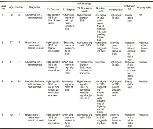
TABLE 1: Imaging Findings in Patients with Suspected Dural Sinus Thrombosis
Case
Age Gender Diagnosis
No. T1 Coronal T1 Sagittal
2 M Leukemia, on L- High signal in ?Short
seg-asparaginase SSS on ments of
five of nine mild
hy-slices
perin-tensity in SSS
2 37 F Breast carci- High signal in ?Short
seg-noma met- SSS on ments of
astatic to skull four of
isointens-nine slices ity in
SSS
3 17 F Leukemia, on L- High signal in Short
seg-asparaginase SSS on ments of three of high
sig-nine slices nal in SSS
4 4 M Medulloblastoma High signal in Isointense with subarach- SSSon signal in noid spread six of nine most of
slices; pat- SSS
ent collat-eral seen
5 42 F Breast carci- High signal in Short
seg-noma met- SSS on ments of
astatic to skull four of
isointens-nine slices ity in SSS
Note.-SSS = superior sagittal sinus; NA = not applicable.
sinus. In one case, no definite clot was detected. Signal void
appeared to be present throughout the course of the sinus as it was followed on several adjacent sections. T2-weighted axial or coronal sequences showed some signal in the sinus in all five patients. Gradient-echo acquisitions were positive in two of the five patients, negative in two, and equivocal in one.
On the nonselective examinations, unequivocal high signal was seen in the superior sagittal sinus in three patients (Fig. 1). This signal appeared identical in size and intensity to the findings on the multislice acquisitions (Fig. 2). These patients
were considered positive by the criteria of this study. In two
of the five patients, only mildly hypointense signal appeared
in the sinus, in contrast to the high signal seen on the multislice
acquisitions. These patients were considered negative. The
results of the nonselective technique correlated with those of
CT and/or angiography.
MR Findings
Enhanced T2 Coronal or Gradient
Nonselective CT Angiography
Axial Echo
Hyperintense Low signal High signal Empty NA
signal in in SSS in SSS delta
SSS with sign
some foci of
high
sig-nal,
sug-gesting throm-bus
Isointense sig- High signal Mildly hy- Negative Negative,
nal in SSS in SSS poin- in ax- slow
suggest- tense ial and flow in ing flow signal in coronal SSS
SSS for
delta
sign
?Hyperintense Equivocal High signal Negative Positive
signal in in SSS in
ax-SSS, more ial and
obvious on coronal
first echo for
delta sign
Hyperintense Low signal High signal Empty Positive
signal in in SSS in SSS; delta
SSS; hy- suggest- patent sign
pointense ing adjacent
vascular throm-
collat-signal adja- basis era I
cent to it with
small fo-cus of
high
sig-
nallat-erally
Isointense sig- High signal Mildly hy- Negative Negative
nal in SSS in SSS poin- in
ax-suggest- tense in ial for
ing flow SSS delta
sign
Not surprisingly, all patients showed high signal in the course of the superior sagittal sinus on T1-weighted coronal
images in several sections, yet only three were positive for
thrombus. Evaluation of the course of the sinus on sagittal
scans did not show large segments of signal in any of the
patients included in this study, although we have certainly
seen other patients in whom thrombus was easily imaged in
this projection. Since no question arose in these other cases,
the nonselective technique was not used and these patients
were not included in the present study. Signal was also
present in the sinus on T2-weighted scans in all cases; yet,
again, in two of these patency was proved by angiography.
The specialized sequences were very accurate in our eval-uation. Gradient-echo acquisitions easily assessed cases of
normal flow. In one patient who later proved to have throm
[image:3.614.57.558.89.513.2]B
D
E
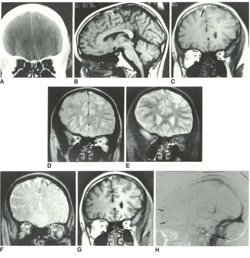
Fig. 1.-Case 3: 17-year-old girl with leukemia, on L-asparaginase, presenting with multiple seizures.
A, Coronal enhanced CT section is equivocal with respect to definitive diagnosis of superior sagittal sinus thrombosis.
B, n-weighted sagittal image, 600/20, shows clearly patent superior sagittal sinus posteriorly (arrow). However, short segments of signal appear to be present anteriorly (arrowhead).
C, T1-weighted coronal image, 600/20, shows high signal in course of sinus (arrow), consistent with both thrombosis and entry phenomenon.
D and E, Long TR coronal images, 2000/35 (D) and 2000/70 (E). Focus of high signal on first echo is not easily seen on second echo.
F, Gradient-echo image, 21/12 (300 flip angle), does not disclose unequivocal lack of flow in superior sagittal sinus. In fact, venous thrombosis was not easy to diagnose on this sequence. Sequelae of thrombosis include small, mildly hemorrhagic venous infarct on left.
G, Nonselective single-slice n-weighted image, 600/20, reveals persistent high intensity in dural sinus. Appearance is virtually identical to section from multislice acquisition (B) and suggests that thrombus, not artifact, has produced this signal.
H, Digital IV angiogram does not show patent superior sagittal sinus posterior to coronal suture. While hypoplasia of anterior portion of superior sagittal
[image:4.612.53.562.89.614.2]o
E
F
Fig. 2.-Case 2: 37-year-old woman with breast carcinoma and large skull metastasis, presenting with headache.
A, T1-weighted sagittal image, 600/20, shows patent superior sagittal sinus posteriorly (arrow), with possible signal in its mid course (arrowhead), as in Fig. 1A.
B, T1-weighted coronal scan, 600/20, shows high intensity in course of sinuses (arrow). This signal was visible on five of 12 adjacent sections anteriorly.
C and 0, Long TR coronal images, 2000/35 (C) and 2000/70 (0), disclose apparent signal in course of sinus (arrows). High intensity in brain parenchyma on left is due to edema created by protruding skull metastasis posteriorly.
E, Nonselective single-slice T1-weighted coronal scan, 600/20, shows loss of high signal, which was noted in multislice acquisition in superior sagittal sinus (arrow). This change in appearance confirms presence of flow-related artifact in original sequence. Note that hypointense signal, rather than signal void, is now present in sinus. This signal should not be mistaken for thrombus.
F, Confirmatory digital IV angiogram. Mild delay in flow is seen in superior sagittal sinus anterior to large skull metastasis, but no thrombosis is present.
high signal was noted. The nonselective method was accurate
in all five patients. It was able to depict both clot and the presence of collaterals.
Flow Phantom
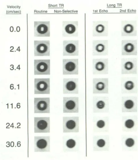
Results with the flow phantom provided experimental cor
-relation with the clinical findings (Fig. 3). Slowly moving fluid at speeds of up to about 6 cm/sec produced high signal on
short TR sequences due to flow-related enhancement.
How-ever, with the nonselective refocusing pulse, the high signal was replaced by signal that was hypointense relative to that
seen in nonmoving fluid. At speeds of about 6-11 cm/sec,
increasing peripheral signal void was noted on both the selec-tive and nonselective acquisitions. These results were also
demonstrated graphically (Fig. 4). On the long TR sequences,
Signal on the first-echo image was noted within the tubing at
speeds of up to 11.6 cm/sec. No even-echo rephasing was
seen in our flow phantom. Signal was lost after the second 1800 pulse between 6.1 and 11.6 cm/sec, earlier than after the first 1800 pulse as velocities of flow were increased.
Discussion
Traditionally, both angiography and CT have been used for
the diagnosis of dural sinus thrombosis. Although accurate, angiography is obviously invasive. In specific situations, for example, assessment of possible superior sagittal sinus thrombosis, CT has been used as a less invasive alternative [21, 22]. CT has a documented sensitivity of only 75%,
however, and careful examination at intermediate windows
[image:5.612.52.558.87.434.2]routine and may not be possible in many patients because of
difficulty in positioning. Both angiography and CT also require
substantial amounts of iodinated contrast material.
MR is very sensitive to the orderly evolution of hemoglobin
breakdown that intraluminal thrombosis undergoes [1, 15]. In
addition, MR also can accurately depict regions of flow
[2-9]. Thus, it is being used increasingly in the assessment
of possible vascular thrombosis [10-13, 16]. However, for a
number of well-described reasons, flowing blood, especially
Velocity
(cm/sec)
0.0
2.4
3.4
6.1
11.6
24.2
30.6
Shan TR Long TR Routine Non·Selective 1 st Echo 2nd Echo
Fig. 3.-Flow phantom experiment. Under Short TR are sections from
short TR multislice acquisition (left side) and results of nonselective single
slice technique (right side). Note that flow-related enhancement is elimi-nated. Under Long TR are sections from long TR multislice sequence.
1.60
1.50
1.40 1.30
1.30 1.20
1.20 1.10
§. 1.10 1.00
0 1.00
'"
~
0.90ex:
0.90 0.80
i!: 0
~
0.80 ex: '" 0.100.10 i!:
~
0.600.60 0.50
0.50 0.40
0.40 0.30
0.30 0.20
T
IT
10 20 30 Velocity (cm/sec)
A
B
when slow, can often cause artifactually high signal [2-9].
Flowing blood usually leads to signal void, explained by either
flow of spins out of the imaged volume between the 900
detection pulse and the 1800 refocusing pulse [2] or by
spin-phase changes created by the movement of spins in a
gra-dient field [3, 12]. However, visualization of signal within
vascular lumina is, of course, extremely common and can occur by several mechanisms. First, slowly moving, fully magnetized protons can enter the imaged volume and replace partially saturated spins. When this process occurs in a period
of time less than or equal to the T1 relaxation time of
sur-rounding tissues, the signal of the vessel lumen will increase
on the initial slices of a multislice acquisition [2, 4, 8]. This
effect is more prominent on short TR sequences. Second, slowly moving protons undergo more complete rephasing of
spins after even-numbered 1800 refocusing pulses, producing
increased signal on even-numbered echoes [2, 6, 8, 9]. A
similar mechanism produces signal on all echoes when
gra-dient moment nulling techniques are implemented. Third,
protons in flowing blood can be present within the imaged
volume for the first 1800 pulse but then flow out of the slice
by the time of the second 1800
pulse or subsequent pulses
[6, 12]. This phenomenon produces intravascular signal on
early echoes only. At an extreme, stagnant blood can be seen
as high signal on all echoes of a long TR sequence [23].
Finally pseudogating of pulsatile flow can also produce signal
within vessel lumina, dependent on the chance
synchroniza-tion of heart rate and TR [2]. Because of these mechanisms,
flowing blood can create signal that can appear on both short
and long TR sequences and on both first or second echoes.
This signal can be mistaken for thrombus formation.
One additional point needs emphasis. It has been stated
that flow-related enhancement can be distinguished from
thrombus because it occurs on entry slices and diminishes
progressively. However, Valk et al. [4] have pOinted out that
in multi slice imaging, enhancement in deeper sections
de-pends on the exposure of spins to 900 pulses in preceding
sections. Enhancement can follow a bimodal pattern in
con-secutive sections with two peaks, one due to "low-velocity
enhancement" and one due to "high-velocity enhancement."
I
10 20 30
Velocity (cm/sec)
Fig. 4.-Graphs of velocity of flow
plotted against 1/1. in phantom, where I
= intensity under conditions of flow and
I. = intensity of static fluid.
A, Multislice selective T1-weighted
acquisition, 600/20, shows flow-related enhancement at low velocities with
later evolution of signal void at higher
velocities.
S, Nonselective single-slice T1
-weighted acquisition, 600/20, shows
decrease in signal intensity with low
velocities, followed again by signal
void at higher velocities. In reality,
graph probably has two plateaus. The
first, at low velocity (less than 10 cm/
sec), represents loss of flow-related
enhancement. The second, at high
ve-locity (greater than 10 cm/sec), results
from dephasing of moving spins and/
[image:6.615.57.297.198.476.2] [image:6.615.60.565.547.747.2]Because of this, it can be difficult to differentiate flow-related enhancement from thrombosis on the basis of the MR signal patterns since the artifact may appear more prominent on deeper slices than on more superficial slices. Added to these effects is the fact that the acquisition of sections in most machines does not occur sequentially, thereby diminishing cross excitation.
Different MR methods have been suggested to eliminate the possibility of flow-related artifacts [11-16]. First and
sim-plest, the suspected thrombosis should be seen on at least
two different sequences, which may vary in plane of section or in TR. In superior sagittal sinus thrombosis, it may be difficult to find two projections that both demonstrate the
abnormality. Coronal or axial scans are obviously situated
perpendicular to much of the course of the superior sagittal sinus and are ideal to assess the possibility of clot in this structure. However, they are also susceptible to flow-related enhancement. If clot in the sinus is also seen on the sagittal scans, these findings together likely constitute sufficient proof
of thrombosis. Yet sagittal scans sometimes do not image
enough of the sinus in a single section to enable confident diagnosis of thrombosis. This difficulty is particularly signifi-cant when thrombosis is limited to small segments of the sinus.
Visualization of suspected clot on two separate sequences with widely varying TRs can be unreliable. The possibility of flow-related enhancement as a cause of high signal within the lumen of the vessel on T1-weighted images is well known. However, signal can also be seen in vasculature on long TR
sequences after both odd- and even-echo delays, as
men-tioned above [2, 6, 8, 9, 12]. Therefore, the appearance of
signal within a vessel on both short and long TR sequences
is not necessarily diagnostic of thrombosis. In fact,
flow-related artifacts frequently produce these findings. We have seen high signal appear in lumina of patent superior sagittal sinuses on both T1- and T2-weighted images as the result of
comparatively stagnant nonthrombosed blood (Fig. 2).
Con-versely, even if no identifiable signal arises from vessels on
T2-weighted sequences, thrombus cannot be excluded since intraluminal clot evolves in stages that parallel those of extra-vascular blood [15]. Until breakdown of deoxyhemoglobin to methemoglobin occurs, usually on the order of several days, thrombus will be hYPointense on T2-weighted images [1]. Therefore, lack of visualization of intraluminal intensity on long TR sequences does not preclude the possibility of thrombus. Other MR imaging methods that have been suggested to differentiate flow-related artifacts from thrombus include
phase-imaging techniques [12, 13, 16, 18]. The occurrence
of a parallel collateral supply, seen in one of our cases, might
prompt a falsely negative conclusion. The presence of partial
thrombosis also might not be depicted accurately.
Gradient-echo acquisitions are also extremely sensitive to flow and often sufficient. However, in their current state, they may not provide sufficient resolution, especially in cases of
partial thrombosis, substantial collateral supply, or
small-vessel involvement. In one of our angiographically proved cases, gradient-echo images appeared to show some flow on the thrombosed section (Fig. 1). No substantial collaterals were seen on the digital IV subtraction study.
Spin-echo techniques that eliminate flow-related enhance-ment have several advantages over the methods listed above. The nonselective single-section method described here
serves as a prototype for this alternative approach. Standard
spin-echo imaging uses a 900 pulse followed by 1800 pulses,
all selective in the slice-selection gradient direction. In this
flow imaging scheme, the 900 pulse is selective in the
slice-selection direction, but the 1800 pulse is, in effect,
nonselec-tive. Although this refocusing pulse is termed "nonselective,"
in reality it is nonselective in the y and z axes but selective in
the x axis to the extent of the field of view chosen. Since the
field of view is large, for practical purposes, the 1800
refocus-ing pulse is nonselective. Partially saturated spins that leave the imaged volume are replaced by partially saturated spins
from outside the slice. Therefore, augmentation of signal from
time-of-flight effects cannot occur (Fig. 3). Alternative meth
-ods of suppressing flow-related enhancement also have been described. Wehrli et al. [3] discussed a method that uses a
preliminary nonselective 900
saturation pulse followed by
selective 900 detection and 1800 refocusing pulses. They also
noted the similarity of this nonselective tagging pulse to a
1800 nonselective refocusing pulse.
Because all of these techniques eliminate flow-related en-hancement, they can be used successfully to differentiate
thrombosis from flow artifacts. True thrombosis should
ap-pear unchanged in the specialized sequence; flow-related
artifacts will disappear. In addition, because a spin-echo tech-nique, rather than a gradient-echo acquisition or phase-shift
method, is used, images can be obtained that are as detailed
and concise as in the multislice sequences. Precise anatomic comparisons are possible. Partial thrombosis and collaterals
can be demonstrated accurately. Finally, elimination of
intra-vascular artifactual signal also results in suppression of much
of the phase-shift "ghosting" that occurs along the phase
-encoding axis. One caveat must be mentioned: Very acute
and chronic clot may be mildly hYPointense rather than hy
-perintense on T1-weighted images. This appearance is
partic-ularly typical of tumor thrombus and of long-standing vascular
thrombosis. In these cases, confusion with flow-related
en-hancement is less likely, and the nonselective spin-echo
method does not have a role.
The signal in both the clinical cases and experimental model
after the suppression of flow-related enhancement was more
hYPointense than would be expected from static fluid,
al-though signal void did not result. Most likely, this decrease in
signal resulted from flow of partially saturated spins out of
the imaged section and replacement by even more saturated spins [8] or from gradient-induced phase effects arising from
spins flowing in a velocity distribution across each pixel [3].
This latter phenomenon also was responsible for the loss of
signal at the periphery of the moving liquid in all of the flow
phantom sequences as velocity increased [3] (see Appendix).
It is important to note that suppression of flow-related
enhancement did
not
create signal void. Therefore, whenusing the nonselective method to eliminate this artifact, some
signal was still seen in the vessel lumen. Clinically, this
hy-pointense signal will be present with any technique that
eliminates flow-related enhancement and should not be
Exact correlation between the appearance of liquid flowing at specific velocities in the flow phantom and of flowing blood in arteries and veins is difficult for several reasons. These reasons include differences between the flowing liquid and
blood with respect to their relaxation times, viscosity, and
flow profile. Other variables involve differences in the
diame-ter, shape, and composition of the phantom tubing compared
with actual vessels. Finally, the section thicknesses and
in-terslice gaps used in the clinical cases varied from those used
in the phantom studies. For example, the optimal speed to
achieve maximal flow-related enhancement in the initial slice
of an acquisition is dzjTR, where dz is the section thickness.
This velocity theoretically would be 0.5 cm/0.6 sec or 0.83
cm/sec for the clinical cases compared with 1 cm/0.5 sec or
2 cm/sec for the phantom studies.
In conclusion, we have applied specialized spin-echo
se-quences to eliminate certain flow-related artifacts and to study
dural sinus thrombosis. Although the technique used here
employs a single-slice nonselective 1800
refocusing pulse, the approach can also be modified to multislice sequences. Our investigation focused on dural sinus thrombosis;
how-ever, the method is applicable to intravascular clot at any site
in the head or elsewhere and seems especially useful in
thrombosis of fairly small vessels that may not be assessed
easily by other methods. We have also used it in the
assess-ment of possible internal jugular vein thrombosis.
Alterna-tively, this technique should be useful to rule out thrombus
formation in giant aneurysms. While many cases of
throm-bosis may be obvious with routine spin-echo imaging, this
technique adds another method with which to analyze
prob-lems of flow and vascular occlusion.
Appendix
Flow in a small channel, as was used in our experimental
model, has a greater tendency to be laminar than flow in
larger channels [6]; and in laminar flow, gradient-induced
phase shifts in each pixel are largest near the vessel wall
itself. In our experimental model, nearly all signal disappeared
from the lumen of the tubing by about 1 0 cm/sec on both the
selective and nonselective sequences. These results correlate
closely with those of Valk et al. [4]. Again, since the signal loss was initially primarily peripheral, it more likely was caused
by dephasing of spins than by high-velocity outflow of signs.
The latter would result in central loss of signal in a laminar
flow model. Additional proof that dephasing of spins rather
than high-velocity signal loss was operative is evident
be-cause of the velocity of the fluid at which the hYPointensity
occurred was not sufficient to produce outflow of spins
be-tween the 900
and 1800
pulses. When spins flow at a velocity
greater than dzj1hTE, where dz is the section thickness, then high-velocity signal loss will occur [2]. In our phantom, these calculations would require a velocity of approximately 100 cm/sec for the short TR sequences and of approximately 58.8 cm/sec for the first echo and 19.2 cm/sec for the second echo of the long TR sequences. Clearly, these velocities are
considerably faster than most of the velocities involved in our
experimental situation.
REFERENCES
1. Gomori JM, Grossman RI, Goldberg HI, Zimmerman RA, Bilaniuk LT.
Intracranial hematomas: imaging by high-field MR. Radiology
1985;157:87-93
2. Bradley WG Jr, Waluch v. Blood flow: magnetic resonance imaging.
Radiology 1985;154:443-450
3. Wehrli FW, Shimakawa A, MacFall JR, Axel L, Perman W. MR imaging of venous and arterial flow by a selective saturation-recovery spin echo (SSRSE) method. J Comput Assist Tomogr 1985;9(3):537-545
4. Valk PE, Hale JD, Crooks LE, et al. MRI of blood flow: correlation of image
appearance with spin-echo phase shift and signal intensity. AJR
1986;146:931-939
5. George CR, Jacobs G, Macintyre WJ, et al. Magnetic resonance signal intensity patterns obtained from continuous and pulsatile flow models.
Radiology 1984;151 :421-428
6. Bradley WG Jr, Waluch V, Lai KS, Fernandez EJ, Spa Iter C. The
appear-ance of rapidly flowing blood on magnetic resonance images. AJR
1984;143: 1167-1174
7. Dinsmore RE, Wedeen VJ, Miller SW, et al. MRI of dissection of the aorta: recognition of the intimal tear and differential flow velocities. AJR
1986;146: 1286-1288
8. Axel L. Blood flow effects in magnetic resonance imaging. AJR
1984;143: 1157-1166
9. von Schulthess GK, Higgins CB. Blood flow imaging with MR: spin-phase
phenomena. Radiology 1985;157:687-695
10. Erdman WA, Weinreb JC, Cohen JM, Maximilian Buja L, Chaney C, Peshock RM. Venous thrombosis: clinical and experimental MR imaging.
Radiology 1986;161: 233-238
11. McMurdo SK Jr, Brant-Zawadzki M, Bradley WG Jr, Chang GY, Berg BO. Dural sinus thrombosis: study using interrnediate field strength MR
imag-ing. Radiology 1986;161 :83-86
12. White EM, Edelman RR, Wedeen VJ, Brady T J. Intravascular signal in MR
imaging: use of phase display for differentiation of blood-flow signal from intraluminal disease. Radiology 1986;161: 245-249
13. Dinsmore RE, Wedeen V, Rosen B, Wismer GL, Miller SW, Brady TJ. Phase-offset technique to distinguish slow blood flow and thrombus on
MR images. AJR 1987;148:634-636
14. Glazer HS, Gutierrez FR, Levitt RG, Lee JKT, Murphy WA. The thoracic aorta studied by MR imaging. Radiology 1985;157: 149-155
15. Macchi PJ, Grossman RI, Gomori JM, Goldberg HI, Zimmerman RA,
Bilaniuk LT. High field MR imaging of cerebral venous thrombosis. J
Compul Assist Tomogr 1986;10(1):10-15
16. Moran PR, Moran RA, Karstaedt N. Verification and evaluation of internal
flow and motion. Radiology 1985;154:433-441
17. Young IR, Bydder GM, Payne JA. Flow measurement by the development
of phase differences during slice formation in MR imaging. Magn Reson
Med 1986;3:175-179
18. Bryant OJ, Payne JA, Fermin ON, Longmore DB. Measurement of flow with NMR imaging using a gradient pulse and phase difference technique.
J Comput Assist Tomogr 1984;8:588-593
19. Wehrli FW, MacFall JR, Axel L, Shutts 0, Glover GH, Herfkins RJ. Ap-proaches to in-plane and out-of-plane flow imaging. Noninvasive Med
Imaging 1984; 1 : 127-136
20. Goldberg HI, Grossman RI, Gomori JM, Asbury AK, Bilaniuk LT,
Zimmer-man RA. Cervical internal carotid artery dissecting hemorrhage: diagnosis using MR. Radiology 1986;158:157-161
21. Goldberg AL, Rosenbaum AE, Wang H, Kim WS, Lewis VL, Hanley OF.
Computed tomography of dural sinus thrombosis. J Comput Assist Tomogr
1986;10: 16-20
22. Brant-Zawadzki M, Chang GY, McCarty GE. Computed tomography in
dural sinus thrombosis. Arch Neuro/1982;39:446-447
23. Olsen W, Kucharczyk W, Brant-Zawadzki M, Norman 0, Newton TH. The
MR characterization of non-flowing intravascular blood. Acta Radiol [Suppl]