Diffusion
Canh Nguyen-minh, Lee Riley III, Khang-Cheng Ho, Roming Xu, Howard An, and Victor M. Haughton
PURPOSE: To test the hypothesis that diffusion of contrast medium into the intervertebral disk is affected by the integrity of the nucleus pulposus and annulus fibrosus. METHODS: In canine intervertebral disks, defects were made in the annulus fibrosus and nuclear material was removed from the disk with a nucleotome. MR imaging was performed with intravenous contrast medium at 15, 30, 60, and 90 days after the procedure. The diffusion of contrast medium in the intervertebral disk was studied by visual inspection and by measuring changes in signal intensity. The interver-tebral disk were classified at each MR study as normal or abnormal on the basis of the signal intensity on T2-weighted images. RESULTS: In untreated disks after intravenous injection of contrast medium, a band of increased signal intensity was observed near the endplates that became wider with time and approached the center of the disk. In six of the 12 treated disks, the band of increased signal intensity was visibly diminished or less discrete compared with that in the control disks. Weeks later, these treated disks showed diminished signal intensity on T2-weighted images and bulging of the annulus fibrosus. CONCLUSIONS: Intervertebral disks with defects in the annulus fibrosus and reduced cartilage content were characterized by abnormal diffusion of contrast medium into the disk, and changes characteristic of early disk degeneration were detected subsequently.
index terms: Spine, intervertebral disks; Spine, magnetic resonance; Animal studies
AJNR Am J Neuroradiol18:435–442, March 1997
Because the intervertebral disk is avascular, diffusion is essentially the only process by which nutrients are transported to and waste products are removed from the thousands of cells in each cubic millimeter of disk. Diffusion is required to maintain the normal function of the disk. The process of diffusion is affected by the integrity of the vertebral endplate and the annulus fibrosus; therefore, impaired diffusion across the vertebral endplates may be a marker of early disk degeneration. Impaired diffusion may have a causal role in disk degeneration or
may be the result of injury to the annulus fibro-sus or nucleus pulpofibro-sus (1– 4). Diffusion into intervertebral disks has been studied to date by counting radioactivity in disk tissue harvested after intravenous injection of a radioisotope (5). Magnetic resonance (MR) imaging has been used to detect diffusion of paramagnetic con-trast medium into intervertebral disks (6). The intervertebral disks increase in signal intensity after intravenous injection of the paramagnetic contrast medium. The increase in signal inten-sity occurs at a rate consistent with diffusion (M. A. Ibrahim, Mathematical and Pharmacokinet-ics Modelling of MRI Contrast Agent’s Transport into the Intervertebral Disks, Milwaukee, Wis: Medical College of Wisconsin; 1994, thesis). The rate of diffusion into the disk is affected by the type of contrast medium used and the state of the cartilage in the intervertebral disk (7). The charged contrast medium gadopentetate diffuses more slowly than does the uncharged paramagnetic contrast media gadoteridol and gadodiamide, probably because the charge
Received June 10, 1996; accepted after revision September 30. Supported by a grant from Bracco, Princeton, NJ.
From the Departments of Radiology (C.N-M, V.M.H.), Orthopedic Sur-gery (L.R., R.X., H.A.), and Pathology (K-C.H.), Medical College of Wis-consin, Milwaukee.
Address reprint requests to Victor M. Haughton, MD, Department of Radiology, Medical College of Wisconsin, Froedtert Memorial Lutheran Hospital, 9200 W Wisconsin Ave, Milwaukee, WI 5322.
AJNR 18:435–442, Mar 1997 0195-6108/97/1803–0435
©American Society of Neuroradiology
slows the diffusion into cartilage, which con-tains a high concentration of fixed negative charges. Diffusion is slower in mature disk car-tilage than in immature disk carcar-tilage, because of the amount of collagen in the matrix and/or the concentration of fixed negative charges (7). Diffusion of contrast medium into human inter-vertebral disks can be detected by measuring signal intensity after the injection of clinically appropriate doses of paramagnetic contrast material (8).
We tested the hypothesis that an injury to the nucleus pulposus or annulus fibrosus slows the diffusion of contrast material into the interver-tebral disks. To do this, we used percutaneous automated diskectomy to create a small perfo-ration in the annulus fibrosus and to decrease the volume of nuclear material; we then per-formed serial MR imaging to detect subsequent degenerative changes in the disk.
Materials and Methods
Six adult mongrel dogs (male and female, 26 to 35 kg) were quarantined for 30 days and tested for mycobacterial and intestinal infections. The dogs had baseline MR studies with intravenous contrast medium. Percutaneous diskec-tomy was performed (9) and then repeat MR imaging and euthanasia.
For percutaneous automated diskectomy, animals were deprived of food for 12 hours, sedated with Telazol (tiletamine hydrochloride and zolazelam hydrochloride) and atropine, intubated, and anesthetized with halothane (1% to 1.5% with oxygen). The animal was placed in the ventral decubitus position and the skin over the lumbar spine was shaved and disinfected with surgical soap and Betadine. The L4-5 and L6-7 levels were identified by C-arm fluoroscopy and a 5-mm incision was made 4 cm from the midline at each disk level. A trochar, cannula, dilator, and trephine were introduced in succession through the incision and into the intervertebral disk guided by fluoroscopic monitoring (9). Accurate positioning of the trochar and canula tip in the nucleus pulposus was confirmed by observing sufficient pressure within the disk to displace the trochar within the canula. The nucleotome probe was then inserted into the canula with its tip in the center of the disk. The cutting window in the probe and its self-contained irrigation and suction were activated for a period of 10 minutes. Nuclear material removed from the disk and collected in the vacuum’s filter was observed and measured. The nucleotome probe was removed and bleeding controlled by pressure over the incision site. Post-operatively, the dogs were confined in a humidified, warmed intensive care unit for 24 hours and then returned to their cages and allowed full activity. For analgesia, buprenorphine hydrochloride (1 ampule intramuscularly) was administered daily. Cefazolin sodium (1 g
intramus-cularly daily) was administered prophylactically for 3 days.
MR imaging was performed at 15 days (six dogs), 30 days (six dogs), 60 days (four dogs), and 90 days (one dog) exactly as in the baseline study. For the MR studies, the dogs were sedated with Telazol (4 mg/kg intramuscu-larly) and atropine (0.05 mg/kg intramuscuintramuscu-larly) and then anesthetized with phenobarbital (25 mg/kg intravenous-ly). The dogs were placed supine on a 5-in solenoid local coil in a 1.5-T imager. Images were obtained in the sagittal plane with parameters of 500/20/2 (repetition time/echo time/excitations), a 2563256 matrix, an 18-cm field of view, and a 3-mm section thickness, and with parameters of 2000/88/2 and the same matrix, field of view, and section thickness. Gadoteridol was injected intravenously in a dose of 0.3 mmol/kg. The short-repetition-time im-ages in the sagittal plane were repeated 2, 15, 30, 45, 60, 75, and 90 minutes after injection.
The animals were killed serially with phenobarbital in-travenously. The lumbar spine was removed en bloc and fixed in 10% buffered formalin for 30 days, embedded in parrafin, sectioned in the sagittal plane, stained with he-matoxylin-eosin, and examined under light microscopy for evidence of degenerative changes in the cartilage.
Baseline and postprocedure MR images were analyzed. The appearance of the intervertebral disks and endplates on the T1- and T2-weighted images was noted. On the noncontrast and contrast-enhanced T1-weighted images, signal intensity in the intervertebral disks was measured. A rectangular region of interest cursor 1 mm2was placed on each intervertebral disk in three positions: near the supe-rior endplate, in the middle of the disk, and near the infesupe-rior endplate. The signal intensity in each intervertebral disk in each of the three locations was measured. The contrast enhancement at each location was calculated as the dif-ference in signal intensity from the baseline divided by the baseline signal intensity. Contrast enhancement for the center of the disk and for the average of the two measure-ments near the endplate was plotted as a function of time after injection and time elapsed since surgery. Differences in enhancement were tested by means of Student’sttest. The sagittal sections of each spine were evaluated by the pathologist without knowledge of the MR appearance of the disk or the contrast enhancement pattern. Cellular changes in the cartilage were scored as 0 if the cells had a normal appearance and the matrix stained normally, as 1 if the matrix stained more darkly than normal and the chondrocytes were more crowded than normal, as 2 if the chondrocytes appeared enlarged and crowded and the matrix staining was darker, and as 3 if the chondrocytes appeared markedly enlarged and crowded and the staining of the matrix was markedly increased. Differences be-tween mean scores for treated and control disks were tested for significance by means of Student’sttest (two-tailed) with the significance set atP5.05. The number and size of blood vessels in the vertebral body near the disk were also characterized by the pathologist in a blinded manner. The relative amount of vascular tissue was clas-sified as small (score of 1), moderate (score of 2), or large
(score of 3). Three sections were evaluated for each disk and the average of the three taken as the score for the disk. The difference between mean scores for normal and treated disks was tested for significance by means of Stu-dent’sttest (two-tailed).
Results
No postoperative complications were ob-served in the six dogs that underwent percuta-neous partial diskectomy. Approximately 0.25 cm2of cartilaginous material and nucleus
pul-posus was removed from each disk level. Two dogs were killed at 30 days and at 60 days, and one at 90 days. One dog died during introduc-tion of anesthetic for the MR study at 90 days, probably because of an accidental overdose.
The pattern of enhancement in each interver-tebral disk in the baseline study and each con-trol disk in the subsequent studies was charac-terized by the appearance of discrete bands of increased signal intensity adjacent to each end-plate at 2 minutes (Fig 1A–C). The band per-sisted and widened through 120 minutes. The central portion of the disk increased in signal intensity to a lesser degree. This pattern was evident in the baseline MR study in all disks including two that appeared to have mild bulg-ing of the anulus fibrosus.
Abnormal patterns of increased signal were observed in six treated intervertebral disks in five animals (Table). The abnormal pattern is illustrated in images obtained at 60 days in an-imal 3 (Fig 1C and D). For about 30 minutes, the discrete enhancement near the edge of the disk was evident. Later, however (for example, in the images at 45 or 60 minutes), the band of increased signal intensity was less distinct. The abnormal pattern was observed as early as 15 days and as late as 90 days after treatment. Four of the six treated intervertebral disks lost
signal intensity on T2-weighted images after having developed an abnormal pattern of en-hancement.
In animal 1, both treated intervertebral disks showed an abnormal pattern of enhancement 15 days after surgery, which was characterized by diminished intensity and discreteness at about 45 minutes. At 15 days, the signal inten-sity of the treated intervertebral disks on the T2-weighted images was not conspicuously di-minished. At 30 days, the abnormal pattern of enhancement persisted in both disks. The end-plates of the L4-5 disk and the adjacent portions of the vertebral body at this time had contrast enhancement and diminished signal intensity on T1-weighted images. The signal intensity of the disks on the T2-weighted images was not conspicuously abnormal in either disk. At 60 days, the signal intensity of the L4-5 interverte-bral disk was appreciably diminished on T2-weighted images and the abnormal
enhance-ment of the disk, characterized by the
diminished discreteness of enhancement at 45 minutes, persisted (Fig 2). Abnormal signal in-tensity and contrast enhancement were noted in the endplates adjacent to the L4-5 disk on the 30- and 60-day studies, but not on the baseline or 15-day studies. The L6-7 intervertebral disk did not show abnormalities on the T1- or T2-weighted images. At 90 days, the diminished signal intensity of the L4-5 disk on the T2-weighted images was evident and the abnormal signal intensity on the T2-weighted images and the contrast enhancement pattern of the verte-bral endplates were less conspicuous. The ab-normally diffuse pattern of enhancement in the intervertebral disk persisted. The L6-7 interver-tebral disk had normal signal intensity on T2-weighted images and normal contrast enhance-ment.
Abnormal patterns of increased signal on T2-weighted MR images of disks after treatment
Animal Disk Level
Days Since Treatment
15 30 60 90
1 L4-5 1 2* 2* 1
1 L6-7 1 1 0 0
2 L6-7 1† 1 0 . . .
3 L4-5 0 2 2 . . .
4 L6-7 0 2 . . . .
5 L4-5 2 2 . . . .
Note.— 0 indicates normal contrast enhancement, normal appearance on T2-weighted images; 1, abnormal contrast enhancement, normal appearance on T2-weighted images; and 2, abnormal contrast enhancement, diminished signal intensity on T2-weighted images.
* Modic type I changes in vertebral endplate. † Radial tear, bulging of the anulus fibrosus.
In the other animals, one abnormal disk re-sulted from treatment. In animal 2, the nucleus pulposus and inner annulus fibrosus of the L6-7 intervertebral disk showed diminished enhance-ment relative to normal disk and less distinct-ness of the band of enhancement near the end-plate at 15 days (Fig 3). The annulus fibrosus bulged mildly and showed contrast enhance-ment, suggesting a radial tear. The signal inten-sity on T2-weighted images was not conspicu-ously diminished. At 30 days, the L6-7 disk retained the abnormally diffuse contrast en-hancement pattern. At 60 days, the abnormally diffuse contrast enhancement pattern was visi-ble, without apparent changes on the T2-weighted images. In animal 4, the abnormally diffuse pattern of enhancement was observed at 30 days but not at 15 days. In animal 5, the abnormal enhancement of the disk was ob-served at L3-4 at 15 and 30 days. The signal intensity of the intervertebral disk was visibly diminished. In the sixth animal, no abnormality was observed in the instrumented disks at 15, 30, 60, or 90 days.
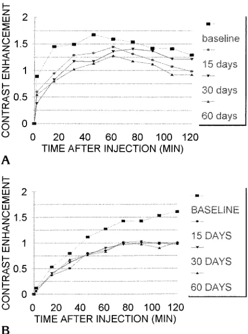
The average contrast enhancement for the edge of the treated disks that showed abnormal-ities is illustrated in Figure 4. The average en-hancement of the intervertebral disk at the edge
or in the center was greater on the baseline study than on the studies at 15, 30, 60, or 90 days after treatment. The enhancement at 60 minutes after injection was significantly less (P,.01, Student’sttest, one-tailed) in the disk studied at 30 days after treatment than in the same disk studied before treatment. Differences between baseline and treated intervertebral disks were significant (P,.01, Student’sttest) at 60 minutes.
On histologic sections, the disks with abnor-mal enhancement tended to have larger chon-drocytes that were more closely crowded to-gether, in a more deeply staining matrix. The scores for the six disks in which abnormal con-trast enhancement was observed were 2 or 3, with a mean of 2.3 (SD, 0.27). The control disks were scored as 0 or 1, with a mean of 0.8 (SD, 0.17). The difference was significant (P 5 .0003).
The osseous endplates for the six disks with abnormal enhancement tended to have more numerous and larger blood vessels. The end-plates of the six abnormal disks were scored 2.3 to 3, with a mean of 2.6 (SD, 0.03), and the endplates for the control disks had scores of 2.3 to 3, with a mean of 1.6 (SD, 0.07). The differ-ence was significant (P 5.008).
Fig 3.A and B, T1-weighted (500/20/2) MR images in animal 2 15 days after intervention in the L4-5 and L6-7 intervertebral disks. The L6-7 disk shows a less distinct band of increased signal intensity near the endplates, bulging of the annulus fibrosus, and a linear contrast enhancement pattern in the posterior annulus fibrosus, characteristic of a radial tear (arrowheadinB). The signal intensity of the disks on the T2-weighted image (2000/88/2) (C) is not conspicuously abnormal.
[image:6.587.114.485.84.337.2]Discussion
The nucleotome instrumentation produced
diffusion abnormalities and progressive
changes consistent with degeneration in half the intervertebral disks in which it was used. The model has been used previously for studying disk degeneration (9). The result of the nucle-otome procedure was variable. Some interver-tebral disks appeared to first develop abnormal-ities and then heal. In these, cartilage may have regenerated. The finding of crowded chondro-cytes in the histologic sections of treated disks suggests proliferation of cells subsequent to an injury. In some treated disks, there was no change in MR appearance or in enhancement pattern. In these, the injury may have been in-sufficient to produce progressive changes. In six disks, instrumentation produced changes in the disk that resulted in diminished diffusion and signal intensity. In these six disks, an abnormal enhancement pattern was observed 15 to 30
days after treatment. The abnormal enhance-ment pattern was associated with or was fol-lowed by other changes typical of disk degen-eration, such as diminished signal intensity on T2-weighted images. These disks also had evi-dence of proliferating chondrocytes, which may have the same significance in early degenerat-ing cartilage as in healdegenerat-ing disks. Therefore, in-tervention with the nucleotome produces a mild
injury sufficient to produce degenerative
changes in about 50% of disks.
The study has the limitations of a pilot study: a small number of animals, observer bias in the evaluation of the images, and a relatively short observation period. Whether the model simu-lated human intervertebral disk degeneration has yet to be determined. The changes in diffu-sion preceded changes in signal intensity on T2-weighted images that typify human interver-tebral disk degeneration. Therefore, the study tends to support the hypothesis that impaired diffusion in the intervertebral disk is a marker for early disk degeneration. Whether it is the cause or result of early degenerative changes is not known. The changes in vascularity in the osseous endplate and the increase in the num-ber of chondrocytes in the treated disks in these dogs are not those of advanced disk degenera-tion. Histologic studies of early human disk de-generation, which can be used for comparison with the histologic studies in this report, are not generally available. The crowded chondrocytes in the treated intervertebral disks suggest more a nonspecific response to injury than a chronic degeneration. The increased staining of carti-lage in the treated disks also is a nonspecific change suggesting increased amounts of pro-teoglycans, possibly because of some stimulus to regeneration.
The study suggests that MR imaging with an intravenous paramagnetic contrast medium may be a method to study diffusion into the intervertebral disks and to determine the role of abnormal diffusion in the development of disk degeneration. Further work is needed to deter-mine the usefulness of the method for studying intervertebral disk degeneration in humans.
References
1. Maroudas A. Physicochemical properties of cartilage in the light of ion exchange theory.Biophys J1968;8:575–595
2. Maroudas A. Transport of solutes through cartilage: permeability to large molecules.J Anat1976;122:335–347
[image:7.587.48.291.83.410.2]3. Maroudas A. Nutrition and metabolism of the intervertebral disc.
In: Ghosh P, ed.The Biology of the Intervertebral Disc. Boca Raton, Fla: CRC Press; 1988:1–38
4. Maroudas A, Bullough P, Swanson SAV, Freeman MAR. The perme-ability of articular cartilage.J Bone Joint Surg1968;50B:166 –177 5. Urban JPG, Maroudas A, Nachemson A. Nutrition of the
interver-tebral disk: an in vivo study of solute transport.Clin Orthop1977; 129:101–114
6. Ibrahim MA, Jesmanowicz A, Hyde J, Estkowski L, Haughton VM. Contrast enhancement of normal intervertebral disks: time and dose dependence.AJNR Am J Neuroradiol1994;15:419 – 424
7. Ibrahim MA, Haughton VM, Hyde JS. Effect of disk maturation on diffusion of low molecular weight gadolinium complexes: an ex-perimental study in rabbits. AJNR Am J Neuroradiol1995;16: 1307–1311
8. Akansel G, Haughton VM, Papke RA, Censky S. Diffusion into human intervertebral disks studied with MR and gadoteridol.
AJNR Am J Neuroradiol1997;18:443– 445
9. Nguyen CM, Haughton VM, Ho KC, Strandt JA. A model of study-ing intervertebral disc degeneration with MR and a nucleotome.
Invest Radiol1989;24:407– 409